1
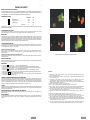
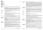
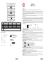
Français
English
FIGURE 1: PNEUMO CEL INDIRECT DIAGRAM FOR USE
PNEUMO CEL
INDIRECT IF TEST
Prepare specimen slide
Add 25µL of RP2 to specimen slide
INTENDED USE
Incubate for 30 minutes at 37OC
The Pneumo Cel Indirect IF Test is an invitro indirect Immunofluorescence test for the detection of Pneumocystis carinii (P.jirovecii) in human specimens.
INTRODUCTION
Wash for 1 minute in PBS and remove excess moisture
from slide
P.carinii(P.jirovecii) (PC) is an extracellular eukaryotic organism shown to be related to the fungi (1). It is an opportunistic pathogen causing interstitial pneumonia in
immunosuppressed hosts, such as premature infants, children with congenital immunodeficiencies and organ transplant recipients (2,3,4). With the advent of the acquired
immunodeficiency syndrome (AIDS), P. carini (P.jirovecii)i pneumonia (PCP) has emerged as the most common life-threatening opportunistic infection in these patients. PCP
occurs in over 75% of AIDS patients (5,6,7) and is usually associated with diminished CD4+ cells (8).
PC is generally confined to the lungs, although there have been recent reports of disseminated extrapulmonary disease (9) which may be as a result of the common use of
aerosolised pentamidine (10).
Add 25µL of RM to specimen slide
Incubate for 30 minutes at 37OC
The most common form of PC is the thick-walled cyst (5-8mm) containing up to 8 intracystic bodies (sporozoites, 1-2mm). These mature into extracystic pleomorphic forms,
the trophozoites (2-5mm). The development cycle occurs in both the alveolar epithelial cells and in the alveolar spaces.
Wash for 1 minute in PBS and remove excess moisture
from slide
PRINCIPLE OF THE TEST
PC may be identified by a variety of histochemical stains such as methenamine silver, Giemsa and toluidine blue (11). The stains can, however, be difficult to perform, they
are non-specific and they may not detect all stages of the life cycle of the organism. Experience is also required for interpretation. More recently, immunofluorescence
assays (IFA) using monoclonal antibodies to PC have been developed. These techniques are rapid, easy to perform and specimens from induced sputum and
bronchoalveolar lavage can be used, thus avoiding the need for more invasive procedures such as lung and transbronchial biopsies. IFA has been shown to be both specific
and sensitive (12,13), and also more sensitive than histochemical staining (14).
Add drop of RMF and cover
Scan at X400 – X1000
TABLE 1:
Trial
SENSITIVITY, SPECIFICITY, & OTHER DATA ON THE PNEUMO INDIRECT CEL
Sensitivity
Specificity
Repeatability
The PNEUMO-CEL INDIRECT IF TEST is an indirect immuno-fluorescence test for PC organisms in methanol-fixed specimens. The fluorescein-labelled mouse monoclonal
antibodies bind specifically to the murine raised monoclonal, initially bound to the organisms. Washing steps, following both the monoclonal and fluorescein conjugated
antibodies, removes any unbound antibody. When viewed under a fluorescence microscope, PC are seen as bright , apple green bodies which may be present singularly or
in cluster. Trophozoites, cysts and the extra cellular matrix , which binds cysts, are stained.
Reproducibility
CONTENTS OF THE KIT
A
100%
100%
-
-
B
C
100%
100%
100%
100%
-
-
D
100%
95.1%
-
-
E
-
-
100% Correlation
100% Correlation
Consult Instructions for Use
V
l
Temperature Limitation
g
Batch
8:
2:
ab
H
D
Cellabs Pty Ltd
Unit 7, 27 Dale Street (PO Box 421)
Brookvale, NSW 2100 Australia
WMDE
Bergerweg 18
6085 AT Horn
The Netherlands
Use By/Expiration Date
Insert
Version
Do Not Re-use
C
1.25mL
5mL
Mounting Fluid
2.5mL
2 x 2.5mL
50
200
Materials are supplied ready for use. Store at 2-8oC.Expiry dates are clearly marked on each kit component and on the box. Expiry dates do not change once opened.
MATERIALS REQUIRED BUT NOT PROVIDED
Positive control human PC; microscope slides with 6-8mm diameter wells; precision pipette for delivering 25mL; methanol for specimen fixation; humid chamber; wash bath;
phosphate buffered saline (PBS) for washing step; cover slips; non-fluorescing immersion oil; and fluorescence microscope with filter system for FITC (maximum excitation
wavelength 490nm, mean emission wavelength 530nm) and x400-x1000 magnification.
PREPARATION OF SLIDES
Apply 25µL to 1 well (0.8cm) slide and air dry. Fix slide for 5 minutes in acetone. If slides are not stained immediately they may be stored below 0oC for several weeks.
INSTRUCTIONS FOR USE
1. Add 25µL of RP2 monoclonal reagent to the fixed test and control slide, covering well area.
2. Incubate the slides in the dark at 37oC in a humid chamber for 30 minutes. Do no allow slides to dry out.
3. Wash the slides gently in PBS bath, drain the slides and remove excess moisture until just dry.
4. Add 25µL of the anti-mouse Ig FITC reagent RMto the specimen. Incubate slides in the dark as in step 2.
5. Wash the slides gently in PBS bath, drain the slides and remove excess moisture until just dry
6. Add a drop of RMF to the slide well. Place a coverslip on top of the drop and remove air bubbles.
7. Scan the entire specimen using a fluorescence microscope initially at X400 magnification, then at X1000 for confirmation. Read immediately or store at 2-8oC in the dark
for up to 24 hours.
READING AND INTERPRETATION OF RESULTS AND DIAGNOSIS
1. PC should exhibit bright, apple-green fluorescence of the round, thick-walled cysts and pleomorphic trophozoites in contrast to the reddish-brown colour of counterstained
material. The organisms occur either singularly or more commonly in clusters in an extracellular matrix, which is also stained. A test is considered positive if two or
more cysts are present.
3. Negative specimens should not display fluorescent organisms of characteristic morphology.
4. Fluorescent material which is not apple green and can be distinguished from typical PC forms should be disregarded.
WASTE DISPOSAL
Dispose of any unused components as biohazardous waste. Where the test reagent has been disposed of in the sink, ensure it is flushed with large quantities of water (as the
sodium azide it contains may react with copper/lead plumbing systems). For more information, please refer to the MSDS.
en fr de it es pt
1
Anti Mouse FITC Reagent
RMF
SPECIMEN COLLECTION
Clinical specimens should be processed as soon as possible after collection. Induced sputum, bronchial wash, bronchoalveolar lavage and tissue impression smears may be
used. The presence of mucous in specimen may prevent adequate staining. Work in a biohazard hood. A positive control slide should be included with each series of test
specimens.
Preparation of Induced Sputum Specimens
Prepare 0.3% dithiothreitol (DTT) in distilled water. Mix equal volumes of sputum and DTT solution. Mix vigorously (vortex) to dispense clumps. Incubate for 30 minutes at
37oC. Centrifuge for 5 minutes at 1500g. Decant supernatant. Resuspend pellet in 0.2-0.5mL of PBS and mix vigorously.
Preparation of Bronchial Wash and Bronchoalveolar Lavage Specimens
Centrifuge the specimen at 1500g for 10 minutes. Remove the supernatant leaving 0.5mL. Decant supernatant. Resuspend pellet in 0.2-0.5mL of PBS and mix vigorously
Tel: +61 2 9905 0133 Fax: +61 2 9905 6426
Web: http://www.cellabs.com.au
Email: [email protected]
Control Positive
RM
PRECAUTIONS
For in vitro diagnostic use only. Do not use after the expiry date shown on the label. If protective packaging is damaged, contact your local distributor and ask for a
replacement. Do not mix reagents from different kits. The Pneumo Cel Reagent has been optimised for use with Cellabs Mounting Fluid. Evans Blue dye contained in the test
reagent is a possible carcinogen, therefore avoid contact with the skin. Patient specimens and the positive control slide should be handled as though potentially infectious. A
Positive Control Slide must be run with each test run. Refer to Material Safety Data Sheet (MSDS) for further information.
EXPLANATION OF SYMBOLS
In Vitro Diagnostic Medical Device
Bulk
5mL
Monoclonal Antibody Reagent
Tests
Not cross reactive with Cryptococcus neoformans, Candida tropicalis, Saccharomyces cerevesiae, Aspergillus niger, Candida krusei,
Candida albicans
i
KP2 Standard
1.25mL
RP2
LP2.3
10 December 2013
0843
SENSITIVITY, SPECIFICITY, & OTHER DATA ON THE PNEUMO CEL
Refer to summary table at end of insert. All data on the Pneumo Cel can be obtained in the product information sheet. Please ask your local distributor or contact Cellabs.
INDEMNITY NOTICE
Modifications or changes made in the recommended procedure may affect the stated or implied claims. A positive or negative result does not preclude the presence of other
underlying causative agents. Cellabs and its agents and distributors shall not be liable for damages under these circumstances.
4/4
en fr de it es pt LP2.3
1/4
en fr de it es pt LP2.3
Français
PNEUMO CEL INDIRECT
PRINCIPE DU TEST ET INDICATIONS D’EMPLOI
Le coffret Pneumo Cel IF est un test in vitro par immunofluorescence directe conçu pour détecter et diagnostiquer la présence de
Pneumocystis carinii dans les échantillons humains. L’anticorps monoclonal de souris marqué à la fluorescéine se lie spécifiquement aux P.
carinii de l’échantillon. P. carinii fluorescent d’un vert intense et démontrent leur morphologie typique.
COMPOSITION DU COFFRET
RP2
Réactif Pneumo Cel
RM
Anti Mouse FITC Reagent
RMF
Liquide de Montage
KP1 Standard
1.25mL
Bulk
5mL
1.25 mL
5mL
2.5mL
2 X 2.5mL
50
200
Déterminations
o
Conserver à 2-8 C. Les dates de péremption sont clairement indiquées sur chaque composant et sur l’étiquette du coffret, et ne sont pas
affectées par l’ouverture du coffret.
MATERIELS REQUIS NON FOURNIS
Lames à spots diamètre 6-8 mm; pipette de 25 µL; acétone pour fixation des échantillons; chambre humide; bain de lavage des lames; tampon P.B.S. pour lavage;
lamelles couvre objets; huile à immersion non fluorescente; microscope à fluorescence pour FITC (490/530 nm), grossissement 400 à 1000.
PRECAUTIONS
Produit à usage uniquement in vitro. Ne pas utiliser après la date de péremption indiquée sur l’étiquette. Si l’emballage est abîmé, contactez votre fournisseur local pour
un remplacement. Ne pas mélanger les composants de coffrets différents. Pneumo Cel est optimisé pour l’emploi des lames de contrôle et du liquide de montage
fournis par Cellabs. Le Bleu d’Evans contenu dans le réactif est un carcinogène potentiel, donc évitez tout contact avec la peau. Les lames de contrôle positives ainsi
que les échantillons patients doivent être considérés comme potentiellement infectieux. Une Lame de Contrôle Positive doit être préparée pour chaque série de tests
afin d’en vérifier la qualité. Consultez la fiche de sécurité du produit (notice MSDS) pour plus amples informations.
COLLECTION DES ECHANTILLONS
Les spécimens cliniques doivent être analysés dès que possible après leur prélèvement. On peut utiliser du sputum induit, du lavage bronchial ou broncho alvéolaire ou
des impressions de tissu sur lame. La présence de mucus dans l’échantillon peut empêcher une coloration adéquate. Travaillez sous hotte à flux laminaire. Une lame
de contrôle doit être incluse avec chaque série de spécimens à analyser.
Préparation de spécimens de sputum induit
Préparer une solution à 0.3% de DTT (dithiothreitol) à l’eau distillée. Mélanger sputum et solution DTT à volume égal. Mélanger vigoureusement (vortex) pour
émulsionner la solution. Incuber 30 minutes à 37oC. Centrifuger 5 minutes à 1500g. Décanter le surnageant. Suspendre le culot dans 0.2-0.5 mL de P.B.S. et
mélanger vigoureusement.
Préparation de spécimens de lavage bronchial ou broncho alvéolaire
Images of human Pneumocystis carinii stained with Pneumo Cel Indirect IFA.
Centrifuger le spécimen 10 minutes à 1500g. Jeter le surnageant, en en laissant 0.5 mL. Décanter le surnageant restant. Suspendre le culot dans 0.2-0.5 ml de P.B.S.
et mélanger vigoureusement.
PREPARATION DES LAMES
Déposer 25 µL sur le spot (8 mm) d’une lame microscopique et laisser sécher à l’air. Fixer à l’acétone pendant 5 minutes. Si la lame n’est pas traitée immédiatement,
elle peut être conservée à 0oC pendant plusieurs semaines.
MODE D’EMPLOI
1. Déposez 25 µL de RP2 sur le spot de la lame de ab ou sur le spot de lame de l’échantillon patient fixé.
2.
3.
4.
5.
6.
7.
8.
Incubez les lames à 37oC en chambre humide pour 30 minutes. Ne laissez pas les lames sécher, car cela accroît le risque de marquages non spécifiques.
Rincez les lames délicatement dans un bain de P.B.S. pendant 1 minute. Egouttez les lames afin d’éliminer tout liquide excessif jusqu'à ce qu’elles soient sèches.
Déposez 25 µL de RM sur le spot de la lame de ab ou sur le spot de lame de l’échantillon patient fixé.
Incubez les lames à 37oC en chambre humide pour 30 minutes. Ne laissez pas les lames sécher, car cela accroît le risque de marquages non spécifiques.
Rincez les lames délicatement dans un bain de P.B.S. pendant 1 minute. Egouttez les lames afin d’éliminer tout liquide excessif jusqu'à ce qu’elles soient sèches.
Déposez une goutte de RMF sur chaque spot. Placez la lamelle en évitant les bulles.
Observez le spécimen au microscope à fluorescence sous huile à immersion, initialement à x400, puis à x1000 pour confirmation. Si l’observation est retardée,
conservez les lames à l’obscurité à 2-8oC jusqu'à 24 heures.
OBSERVATION, INTERPRETATION DES RESULTATS ET DIAGNOSTIQUE
A l’inverse de la coloration de fond brun rougeâtre, les trophozoites pleomorphes de Pneumocystis carinii (PC) présentent une intense fluorescence vert pomme, alors
que les cystes á paroi épaisse présentent une fluorescence en halo autour de leur périphérie. Les organismes peuvent être seuls ou plus fréquemment en grappes
incluses dans une membrane extracellulaire, aussi colorée. Lorsque vous observez au moins deux cystes, le résultat est positif. Utilisez la lame de contrôle à titre de
comparaison pour vérifier l’apparence et la taille des organismes. Le résultat est négatif quand aucun organisme fluorescent de morphologie caractéristique n’est
observable. Ignorez tout élément fluorescent qui n‘est pas vert pomme et qui se distingue de la forme typique de P. carinii.
DECHETS
Jetez tout composant inutilisé dans la poubelle aux déchets biologiques. Lorsque vous videz le réactif du coffret dans l’évier, assurez-vous dans le diluer avec une large
quantité d’eau courante, car l’azide de sodium qu’il contient peut être explosif en contact avec les égouts en cuivre ou en plomb. Consultez la fiche de sécurité du produit
(notice MSDS) pour plus amples informations.
SENSIBILITÉ, SPÉCIFICITÉ ET AUTRES DONNEES DU TEST PNEUMO CEL
Referez-vous au tableau récapitulatif en fin de notice. Toutes les données sur le test Pneumo Cel sont sur la fiche technique du produit. Contactez Cellabs ou votre
distributeur pour l’obtenir.
NOTICE D’INDEMNITE
Toute modification ou variation du protocole d’emploi recommandé peut affecter les performances annoncées du produit. Un résultat positif ou négatif n’exclue pas la
présence d’autres agents causatifs sous-jacents. Cellabs et ses agents et distributeurs ne sont légalement responsables d’aucun dommage dans de telles
circonstances.
2/4
en fr de it es pt LP2.3
REFERENCES
1. Edman,J.C., Kovacs, J..A, Masur, H, Santi, D.V., Elwood, H.J., Sogin, M.L. (1988). Ribosomal RNA sequence shows Pneumocystis carinii to be
a member of the fungi. Nature, 334:519-22.
2. Walzer, P.D., Schultz, M.G., Western, K..A., Robbins, J.B. (1976). Pneumocystis carinii pneumonia and primary immune deficiency diseases.
Nat,. Cancer Inst. Monogr. 43:65-72.
3. Gryzan, S., Paradis, I.L., Zeevi, A, Duquesnoy, J., Dummer, S., Griffith, B.P., Hardesty, R.L., Trento, A., Nalesnik, M.A., Dauber, J.H., (1988).
Unexpectedly high incidence of Pneumocystis carinii infection after lung heart transplantation. Am. Rev. Respir. Dis. 137:1268-1274.
4. Hardy, A.M., Wajszczuk, C.P., Suffrendini, A.F., Hakala, T.R., Ho, M., (1984). Pneumocystis carinii Pneumonia in renal transplant recipients
treated with cyclosporine and steroids. J. Infect. Dis. 149:143-147.
5. Hughes, W.T., (1987) Pneumocystis carinii pneumonia. N. Eng. J. Med. 317:1021-3.
6. Kovacs, J..A., Masur, H. (1988). Pneumocystis carinii pneumonia: therapy and prophylaxis. J.Infect. Dis. 158:254-9.
7. Murray, J.F., Garay, S.M., Hopewell, P.C., Mills, J., Snider, G.L, Stover, D.E. (1987). Pulmonary complications of the acquired immunodeficiency
syndrome: an update: report of the Second National Heart, Lung and Blood Institute Workshop. Am. Rev.Respir. Dis. 135:504-509.
8. Phair,J., Munoz, A., Detels, R., Kalslow, R., Rinaldo, C., Saarh, A. (1990). The risk of Pneumocystis carinii pneumonia among men infected
with the human immunodeficiency virus type 1. New Engl. J. Med. 322:161-165.
9. Pilon, V., Echols, R., Celo, J., Elmendorf, S., (1987). Disseminated Pneumocystis carinii pneumonia infection in AIDS. N. Eng. J. Med. 316: 14101411.
10. Raviglione, M.C. (1990). Extrapulmonary pneumocystosis: the first 50 cases. Rev. Infect. Dis. 12:1127-1138.
11. Ruskin, J. (1986). Newer developments in diagnosis and treatment of Pneumocystis infections. Curr. Clin. Top. Infect. Dis. 7:194-215.
12. Kovacs, J.A., Ng, V.L., Masur, H., Leoung, G., Hadley, W.K., Evans, G., Lane, H.C. Ognibene, F.P., Shelhamer, J., Parillo, J.E., Gill V.J. (1988).
Diagnosis of P. Carinii pneumonia: improved detection in sputum with use of monoclonal antibodies. New Eng. J. Med. 318:589-593.
13. Wolfson, J.S., Waldron, M.A, Sierra, L.S. (1990). Blinded comparison of a direct immunofluorescent monoclonal antibody staining method and
a Giemsa staining method for identification of P. Carinii in induced sputum and bronchoalveolar lavage specimens of patients infected with
human immunodeficiency virus. J. Clin. Micro. 28:2136-2138.
14. Ng. V.L., Virani, N.A., Chaisson, R.E., Yajko, C.D., Sphar, H.T., Cabrian, K.C., Rollins, N., Charache, P., Krieger, M., Haldey, W.K., Hopewell,
P.C. (1990). Rapid detection of Pneumocystis carinii using a direct fluorescent monoclonal antibody stain. J. Clin. Micro. 28:2228-2233.
4/4
en fr de it es pt LP2.3