1
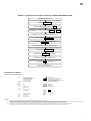
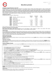
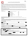
en fr LF3.06
English
Lymphatic Filariasis Bm14 Antibody CELISA
BACKGROUND
Lymphatic filariasis (LF), caused by the filarial worm, Wucheria bancroftii, and transmitted through a mosquito vector, affects more than 100 million people in more than 83
1
countries worldwide . A further billion people are at risk from the infection, which in its chronic stage (elephantiasis) has profound physical, social, and economic
consequences, and represents the world’s fourth leading cause of permanent disability. The disease has been targeted by the WHO for eradication by the year 2020 through
2
an international programme of mass drug administration (MDA) operated by affected countries in collaboration with the Global Alliance to Eliminate Lymphatic Filariasis
(GAELF).
To monitor the effectiveness of the LF eradication programme a number of diagnostic tools have been employed, including microfilaremia quantitation and parasite antigen
immunoassays, both of the rapid and ELISA format. Specific IgG4 anti – filarial antibodies have also been proposed as an effective marker for interruption of LF transmission,
3
particularly in young children .
4,5,6
The Bm14 Antibody CELISA Kit is an indirect ELISA for the detection of human IgG4 antibodies to the filarial recombinant antigen, Bm14 . Added sensitivity and specificity
result from the use of a monoclonal anti – IgG4 indicator. The test is designed to be used with serum samples as well as blood spot eluates collected on filter paper in the
field.
INTENDED USE AND PRINCIPLE OF THE TEST
This test is an indirect ELISA for the detection of Bm14 specific IgG4 antibody. Antibody is bound by the recombinant antigen Bm14 coated on the inner surface of the test
strip. Peroxidase-conjugated antibody to human IgG4 is added and reacts with bound antibody. A chromogenic substrate for peroxidase is added. If antibody to LF is present
there is a reaction which results in the development of a blue colour, which is in proportion to the serum level of the antibody. Addition of stopping solution changes the blue
colour to yellow.
CONTENTS OF THE KIT
FAMW
Celisa Plate – 1x96 wells - (single use only)
5 plates
ab
ac
Positive Control
0.05mL
Negative Control
0.05mL
FASD
Sample Diluent (10x)
60mL
FAPO
Enzyme Conjugate (100x)
0.6mL
FACD
Conjugate Diluent
60 mL
FAPT
PBS/Tween (20x)
250mL
FASC
Substrate Chromogen (TMB) (20x)
3.0mL
FASB
Substrate Buffer
60mL
FASS
Stopping Solution
30mL
O
Store all components at 2-8 C.Expiry dates are clearly marked on each kit component and on the box. Expiry dates do not change once opened.
MATERIALS REQUIRED BUT NOT PROVIDED
Micropipettes and tips; clean glassware or plastic containers for solutions; humid chamber; ELISA washer; and Spectrophotometer to read absorbances at a single
wavelength of 450nm.
PRECAUTIONS
For in vitro diagnostic use only. Reagents should not be used after the expiry date shown on the label. If protective packaging is damaged, contact your local distributor and ask
for a replacement. Do not mix reagents from different kits. Thimerosal preservative added to some components is a poison. Exercise caution when handling these
components. The stopping solution is corrosive. Avoid contact with skin, eyes and mucous membranes. Dispense all reagents with care to avoid cross contamination of wells.
Avoid exposure of the substrate to light. Treat all clinical and control material as though potentially infectious and dispose of in accordance with local operating regulations. For
further information, please refer to the Material Safety Data Sheet.
INSTRUCTIONS FOR USE
Preparation of Wash Buffer
If crystals are present, warm the concentrate to dissolve. For each microplate, add 50mL PBS-Tween concentrate FAPT to 950mL of distilled water. Label the bottle WASH
o
BUFFER. Store at 2-8 C.
Preparation of Sample Diluent
Prepare the working Sample Diluent FASD (1x) by diluting the 10x Concentrate in distilled water. Mix the buffer thoroughly before using to dilute test samples.
Preparation of Samples
Serum or Plasma
o
Collect patient blood samples by standard venipuncture procedure. The test may be used with serum or plasma. The serum or plasma should be stored below -10 C if the
analysis is delayed. Prepare a 1/100 dilution of the positive control ab, the negative control ac and the patient specimens using the prepared Sample Diluent FASD
(1x) above, ensuring proper mixing. Record the position of each diluted SAMPLE on a work sheet.
Blood Spot Eluates
The recommended filter paper for use is the TropBio filter paper (WHO format). Each filter paper circle absorbs ~10uL of blood. Upon drying at room temperature, the
o
o
spotted blood circles should be stored at 2-8 C for short term storage and transportation or -20 C in a freezer for long term storage until ready for testing. Prepare to elute the
sample by carefully removing one circle and completely immersing in a tube of 500uL of prepared working Sample Diluent FASD (1x). Leave the samples refrigerated at 2-8
o
C overnight. The following day, vortex the contents of the tube to ensure complete elution of sample. The eluate is equivalent to a 1/100 serum dilution and can be used in
the Bm14 Antibody CELISA assay without further dilution.
Assay Procedure
o
1.
Bring all reagents to room temperature (18-25 C) before use.
2.
Prepare WASH BUFFER (see Preparation of Wash Buffer).
3.
Prepare the working Sample Diluent FASD (1x) (See Preparation of Sample Diluent).
4.
Remove required number of FAMW strips. Reseal the foil bag containing unused microwell strips immediately with tape.
5.
Pipette 100µL of the SAMPLE, ab and ac, into individual microwells. Include two positive and two negatives in each assay run. Cover and incubate for one (1)
o
hour at 37 C in a humid chamber. (see Preparation of Samples using serum, plasma or blood spot from filter paper).
6.
In the last 10 minutes of the incubation period, prepare the working strength CONJUGATE. Add 10µL of Enzyme Conjugate FAPO to 990µL of FACD and mix thoroughly
(allow 1mL per strip of 8 wells).
7.
Wash the wells preferably using an automatic plate/strip washer or manually as follows:
- Empty contents from the wells. Refill with the WASH BUFFER.
- Repeat this process a further three (3) times. After the fourth wash, bang inverted wells dry on absorbent tissue.
- NB: take care when flicking out plates, hold side of frame firmly to hold strips in place.
1/4
en fr LF3.06
o
8.
Add 100µL of CONJUGATE to each well. Incubate for 45 minutes at 37 C in a humid chamber.
9.
In the last 10 minutes of the incubation period, prepare the working strength SUBSTRATE. Add 50µL of Substrate Chromogen FASC to 950µL of Substrate Buffer FASB
and mix thoroughly (allow 1mL per strip of 8 wells). The stability of the solution is 30 minutes.
10. Repeat washing as in step 7.
11. Add 100µL of fresh SUBSTRATE and incubate in the dark (covered) at room temperature for 15 minutes.
12. Add 100µL of Stopping Solution FASS. Tap the plate to mix.
13. Read the results visually or in a spectrophotometer at 450nm or 450/620 nm.
READING AND INTERPRETATION OF RESULTS AND DIAGNOSIS
Photometrically
It is optimal to use a dual beam ELISA reader. Blank machine against the well designated as the blank. Within 30 minutes of stopping the reaction read absorbance of all
wells including all the controls at 450nm on an ELISA reader.
For the test results to be accepted the controls must read as follows:
Control
O.D Value (450/620nm)
Negative Control
Positive Control
< 0.20
> 0.40
Note: A cut-off value can be determined by testing a panel of known negative samples. Cut-off value = mean + 3 Standard Deviations (+3SD). A pilot study
conducted in- house with 50 blood spot eluates from Australian residents without prior exposure to LF gave a cut-off of 0.25 OD units (mean +3SD)
Visual Reading
Results can also be read visually. Note colour of the specimen wells and compare with the colour of the negative control. If the colour, read visually, of a specimen well is
above that of the negative control, then the specimen contains antibody.
WASTE DISPOSAL
Dispose of any unused components as biohazardous waste. For more information, please refer to the MSDS.
SENSITIVITY, SPECIFICITY, & OTHER DATA ON THE BM14 ANTIBODY CELISA KIT
Data on the Bm14 Antibody CELISA test performance can be obtained from your local distributor or by contacting Cellabs.
INDEMNITY NOTICE
Modifications or changes made in the recommended procedure may affect the stated or implied claims. A positive or negative result does not preclude the presence of other
underlying causative agents. Cellabs and its agents and distributors shall not be liable for damages under these circumstances
-----------------------------------------------------------------------------------Français
Lymphatic Filariasis Bm14 Antibody CELISA
PREAMBULE
L’agent de la filariose lymphatique (FL) est la filaire Wucheria bancroftii,qui est transmise par un moustique et qui infecte plus de 100 millions de personnes dans plus de 83
1
pays du monde . Plus d’un milliard de personnes risquent cette infection qui dans son stade chronique (éléphantiasis) a de profondes conséquences physiques, sociales et
économiques et représente la quatrième cause d’handicap permanent au monde. L’éradication de cette maladie par l’OMS est prévue en l’année 2020 grâce à un programme
2
de chimiothérapie de masse (CTM) opéré par les pays affectés en collaboration avec l’Alliance Globale pour l’Elimination de la Filariose Lymphatique (AGEFL).
Afin de suivre l’efficacité du programme d’éradication FL, un nombre d’outils diagnostics ont été employés, y compris la mesure de la microfilarémie et d’antigènes
parasitaires par dosages immunologiques, en formats rapides et ELISA. Les anticorps spécifiques IgG4 anti-filariose ont également été proposés comme marqueurs
3
efficaces de l’interruption de la transmission FL, en particulier chez les jeunes enfants .
4,5,6
La trousse Bm14 Antibody CELISA est un ELISA indirecte pour détecter les anticorps IgG4 humains de l’antigène recombinant Bm14 . L’usage d’un anticorps monoclonal
de détection anti-IgG4 augmente la sensibilité et la spécificité du dosage. Le test est conçu pour usage sur échantillons de sérum ainsi que sur échantillons d’élution de
gouttes de sang sur papier filtre collectés sur le terrain.
PRINCIPE DU TEST ET INDICATIONS D’EMPLOI
Ce test est un ELISA indirecte pour détecter les anticorps IgG4specifiques Bm14. L’anticorps est lié par l’antigène recombinant Bm14 fixé sur la surface interne des puits de
dosage. Un anticorps anti-IgG4 humain conjugué à la peroxydase est ajouté et réagit avec l’anticorps immobilisé. Un substrat chromogène pour peroxydase est ajouté. Si
l’anticorps LF est présent, une réaction se produit conduisant au développement d’une couleur bleue, d’intensité proportionnelle au niveau d’anticorps dans le sérum.
L’addition d’un réactif d’arrêt change la couleur au jaune.
COMPOSITION DU COFFRET
FAMW
Plaque CELISA – 1x96 puits (usage unique)
ab
ac
Contrôle positif
0.05mL
Contrôle négatif
0.05mL
5 plaques
FASD
Réactif de dilution des échantillons (10x)
60mL
FAPO
Conjugué enzymatique (100x)
0.6mL
FACD
Diluant de conjugué enzymatique
60 mL
FAPT
PBS/Tween (20x)
250mL
FASC
Substrat chromogène (TMB) (20x)
3.0mL
FASB
Tampon du substrat
60mL
FASS
Solution d’arrêt
30mL
o
Conserver tous les composants à 2-8 C. Les dates de péremption sont clairement marquées sur chaque composant et sur le coffret de la trousse. L’ouverture n’altère pas les
dates de péremption.
MATERIELS REQUIS NON FOURNIS
Micropipettes et embouts; verrerie propre ou récipients plastiques pour solutions; eau distillée; chambre humide; laveur ELISA; spectrophotomètre capable de lire à 450 nm.
PRECAUTIONS
Produit à usage uniquement in vitro. Ne pas utiliser après la date de péremption indiquée sur l’étiquette. Si l’emballage est abîmé, contactez votre fournisseur local pour un
remplacement. Ne pas mélanger les réactifs de coffrets différents. Certains réactifs contiennent du Thimerosal comme préservatif, qui est un poison. Manipulez ces réactifs
avec soin. La solution d’arrêt est corrosive. Evitez tout contact avec la peau, les yeux ou les membranes muqueuses. Ajouter les réactifs en évitant tout risque de
contamination croisée des puits. Eviter d’exposer le substrat à la lumière. Les échantillons cliniques et les contrôles doivent être considérés comme potentiellement infectieux
2/4
en fr LF3.06
infectieux et jetés selon les procédures en vigueur. Consultez la fiche de sécurité du produit (notice MSDS) pour plus amples informations.
INSTRUCTIONS D’EMPLOI
Préparation du tampon de lavage
Si des cristaux apparaissent dans le concentré, réchauffer le réactif pour les dissoudre. Pour chaque plaque de micropuits, ajouter 50mL de concentré PBS/Tween FAPT à
o
950mL d’eau distillée. Libeller la bouteille WASH BUFFER. Conserver à 2-8 C.
Préparation du réactif de dilution des échantillons
Préparer la dilution de travail du réactif de dilution des échantillons FASD (1x) en diluant le concentré x10 avec de l’eau distillée. Mélanger le tampon vigoureusement avant
usage sur échantillons à tester.
Préparation des échantillons
Sérum ou plasma
o
Collecter les spécimens sanguins par ponction veineuse courante. Le dosage peut se faire sur plasma ou sérum. Conserver les échantillons de plasma ou sérum à -10 C si
l’analyse est retardée. Préparer une dilution au 1/100 du contrôle positif ab,du contrôle négatif ac et des échantillons patients à l’aide du réactif WASH BUFFER et
bien mélanger. Repérer la position des échantillons dilués SAMPLE sur la feuille de travail.
Elution de gouttes de sang sur papier filtre
L’emploi du papier filtre TropBio (format OMS) est recommandé. Chaque cercle de papier filtre absorbe ~10uL de sang. Apres séchage à température ambiante, les papiers
o
o
filtres utilisés doivent être conservés à 2-8 C à court terme ou au congélateur à -20 C pour un stockage de plus longue durée jusqu'à leur emploi. Eluer les échantillons en
retirant délicatement un cercle et en l’immergent dans un tube de 500uL de solution de travail de réactif de dilution des échantillons FASD (1x). Laisser les échantillons au
o
réfrigérateur à 2-8 C durant la nuit. Le lendemain, agiter au vortex le contenu du tube d’élution de l’échantillon. Cette élution est équivalente à une dilution du sérum au 1/100
et peut être utilisée dans la trousse Bm14 Antibody CELISA sans dilution supplémentaire.
Mode D’emploi
o
1.
Ramener tous les réactifs à température ambiante (18-25 C) avant l’emploi.
2.
Préparer le WASH BUFFER (voir Préparation du Tampon de Lavage).
3.
Préparer la solution de travail FASD (1x) (voir Préparation du Reactif de Dilution des Echantillons).
4.
Retirer le nombre requis de micropuits FAMW . Immédiatement refermer le sac contenant les micropuits restants avec du ruban adhésif.
5.
Ajouter 100µL de SAMPLE, de ab et de ac dans leurs micropuits individuels. Inclure deux contrôles positifs et deux contrôles négatifs dans chaque série de
o
dosage. Couvrir et incuber une (1) heure à 37 C en chambre humide. (voir Préparation des Echantillons – Elution de gouttes de sang sur papier filtre)
6.
Dans les 10 dernières minutes de l’incubation, préparer la solution de travail de CONJUGATE. Ajouter 10µL de Conjugué enzymatique FAPO à 990µL de FACD et
mélanger vigoureusement (prévoir 1mL de solution de travail par barrette de 8 micropuits).
7.
Laver les micropuits au laveur automatique ou manuellement comme suit :
- Vider le contenu des micropuits. Remplir les micropuits de WASH BUFFER.
- Répéter l’opération trois (3) fois. Vider le contenu des puits après le quatrième lavage.
- NB : opérez cette étape avec précaution, en serrant le cadre de la plaque à micropuits pour éviter qu’ils ne s’en délogent.
o
8.
Ajouter 100µL de CONJUGATE dans chaque puits. Incuber 45 minutes à 37 C en chambre humide.
9.
Dans les 10 dernières minutes de l’incubation, préparer la solution de travail de SUBSTRATE. Ajouter 50µL de Substrat chromogène FASC à 950µL de Tampon de
substrat FASB et mélanger vigoureusement (prévoir 1 mL de solution de travail par barrette de 8 micropuits). Cette solution de travail est stable pendant 30 minutes.
10. Répéter le lavage comme à l’étape 7.
11. Ajouter 100µL de SUBSTRATE frais et incuber (couvert) à l’obscurité pendant 15 minutes à température ambiante.
12. Ajouter 100µL de solution d’arrêt FASS. Taper la plaque pour mélanger.
13. Lire les résultats visuellement ou au spectrophotomètre à 450nm ou à 450/620nm.
LECTURE, INTERPRETATION DES RESULTATS ET DIAGNOSTIC
Lecture spectrophotométrique
Il est préférable d’utiliser un spectrophotomètre à double longueur d’onde. Calibrer la machine contre un blanc désigné. Lire l’absorbance de chaque puit y compris contrôles
dans un lecteur à microplaque ELISA dans les 30 minutes suivant l’addition de la solution d’arrêt. Pour être valides, les valeurs des contrôles doivent être comme suit :
Contrôle
Contrôle négatif
Contrôle positif
Valeur D.O. (450/620 nm)
< 0.20
> 0.40
Note: Un seuil discriminant peut être déterminé à l’aide d’un groupe d’échantillons négatifs confirmés. Seuil discriminant = moyenne + 3 Déviations Standard (+ 3
DS). Une étude pilote effectuée par Cellabs sur 50 échantillons sanguins d’élution sur papier filtre provenant de résidents Australiens sans exposition préalable à
FL a produit un seuil discriminant de 0.25 unités DO (moyenne + 3 DS).
Lecture visuelle
Les résultats peuvent être lus visuellement. Observer l’intensité couleur des échantillons en comparaison à celle du contrôle négatif. Si un échantillon présente une couleur
visuellement clairement supérieure à celle du contrôle négatif, cet échantillon contient des anticorps.
DECHETS
Jetez tout composant inutilisé dans la poubelle aux déchets biologiques. Consultez la fiche de sécurité du produit (notice MSDS) pour plus amples informations.
SENSIBILITE, SPECIFICITE ET AUTRES DONNEES DU TEST
Des données sur la performance du test Bm14 Antibody CELISA sont disponible auprès de votre distributeur local ou en contactant Cellabs.
NOTICE D’INDEMNITE
Toute modification ou variation du protocole d’emploi recommandé peut affecter les performances annoncées du produit. Un résultat positif ou négatif n’exclue pas la
présence d’autres agents causatifs sous-jacents. Cellabs et ses agents et distributeurs ne sont légalement responsables d’aucun dommage dans de telles circonstances.
3/4
en fr LF3.06
FIGURE 1 Lymphatic Filariasis Bm14 Antibody CELISA DIAGRAM FOR USE
Bring all components to RT
Prepare WASH BUFFER
Prepare working Sample buffer (1x) FASD
Add 100µL of ab
, ac, and SAMPLE to FAMW
o
Incubate for 1 hour in a humid chamber at 37 C
Wash 4x with WASH BUFFER
Add 100µL CONJUGATE to each well
o
Incubate 45 minutes in a humid chamber at 37 C
Wash 4x with WASH BUFFER
Add 100µL of SUBSTRATE to each well
Incubate for 15 minutes in the dark at RT
Add 100µL FASS to each well
Read at 450 or 450/620nm
EXPLANATION OF SYMBOLS
EXPLICATION DES SYMBOLES
i
Consult Instructions for Use
Consultez le mode d’emploi
In Vitro Diagnostic Medical Device
Diagnostic medical in-vitro
V
2:
l
Tel: +61 2 9905 0133 Fax: +61 2 9905 6426
Web: http://www.cellabs.com.au
Email: [email protected]
Temperature Limitation
Limites de temperature
g
ab
Batch
Lot
ac
Control Negative
Contrôle négatif
H
D
References:
1.
2.
3.
4.
5.
6.
8:
Cellabs Pty Ltd
Unit 7, 27 Dale Street (PO Box 421)
Brookvale, NSW 2100 Australia
WMDE
Bergerweg 18
6085 AT Horn
The Netherlands
Control Positive
Contrôle positif
Use By/Expiration Date
Date de péremption
Insert
Version /
Version
mode
d’emploi
C
en fr
LF3.06
6 March 2012
0843
Do Not Re-use
Ne pas ré-utiliser
Weil GJ & Ramzy RMR (2006) Diagnostic tools for filariasis elimination programs. Trends Parasitol 23: 78-82.
Ramzy RMR, Setouhy M El, Helmy H et al. (2006) Effect of yearly mass drug administration with diethylcarbamazine and albendazole on bancroftian filariasis in Egypt: a comprehensive assessment. Lancet 367: 992-999.
Ottesen EA, Skvaril F, Tripathy SP, Poindexter RW, Hussain R, (1985) Prominence of IgG4 in the IgG antibody response to human Filariasis. J.Immunol 134,2707-2712.
Chandrashekar R, Curtis KC, Ramzy RM et al.(1994) Molecular cloning of Brugia malayi antigens for diagnosis of lymphatic filariasis. Mol Biochem Parasitol 64: 261-274.
Ramzy RM, Helmy H, Faris R et al. (1995) Evaluation of a recombinant antigen-based antibody assay for diagnosis of bancroftian filariasis in Egypt. Ann Trop Med Parasitol 89: 443-446.
Lammie P, Weil G, Rahmah N et al. (2004) Recombinant antigen based assay for the diagnosis and surveillance of lymphatic filariasis – a multicenter trial. Filaria Journal 3: 9
4/4