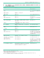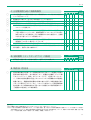
1
監訳にあたり
米国の疾病管理予防センター(Centers for Disease Control and Prevention, CDC)は、米国における感染症管理
の中心的な国家機関であり、継続的に数多くの関連のガイドラインを公表しています。これらガイドラインでは科
学的な根拠(エビデンス)に基づく感染管理の実施を医療の現場に勧めています。対象も従来の急性期の病院だけ
でなく、長期療養施設やデイケアーセンターなどのヘルスケアー全体に拡げ、感染管理体制の充実に努力を払って
います。各ガイドライン間の医療現場における運用の整合性をとる配慮の記載も、ここ数年に発行されたガイドラ
インの目立った特徴です。
そのCDCから、歯科医療における感染管理のためのCDCガイドラインが2003年12月19日発行の米国公衆衛生週報
(MMWR)に公表されました(http://www.cdc.gov/mmwr/PDF/rr/rr5217.pdf)。先のガイドライン(http://www.cdc.
gov/mmwr/PDF/rr/rr4208.pdf)が1993年であったことを考えると、10年ぶりの改訂です。今回のガイドラインで示
された勧告文数は149件にのぼり、引用文献も471件に及びます。
前版の歯科医療におけるCDCガイドラインの著述は血液媒介感染症の予防・管理に力点が置かれていましたが、
その後発表された多くの感染管理関連のCDCガイドラインの内容を受けて、今回の改訂では歯科医療の現場に対し
て、標準予防策に加え歯科医療従事者の感染予防策(ワクチンや感染症罹患時の休職措置など)、感染経路別予防策
(空気・飛沫・接触予防策)の踏襲を求めており、歯科医療の現場にとっては大きな課題が課せられたことになりま
す。廃棄や針刺し損傷対策、手袋とアレルギー対策、環境の整備、医療器具・器材の洗浄・消毒・滅菌、サージカ
ルスモーク対策、クロイツフェルト・ヤコブ病対策など細かな点についても具体的な記載がなされており、歯科診
療の現場に具体的に役立つ内容となっています。既にわが国においてもこれらの感染管理の手法論は一般医療施設
で広く病院感染管理の基本として運用されていることから、医療施設の感染管理担当者(インフェクションコント
ロールドクター[infection control doctor : ICD]やインフェクションコントロールナース[infection control nurse :
ICN])との協力体制を組むことで、歯科医療における感染管理体制の充実が期待されます。この点から、本書の監
訳を行うにあたり、歯科医療従事者にとっての日常的な用語に関しても、一般医療従事者の理解を深めるために、
あるいは一般医療従事者には日常的な用語でも歯科医療従事者の理解を深めるために、多くの用語あるいは文章に
対し監訳注釈を加筆いたしました。
本書では、勧告文を読者の歯科診療施設の実状と照らし合わせて項目毎にチェックできるよう配慮いたしました。
翻訳にあたり、原本に可能な限り忠実に和訳する努力を致しましたが、併せて巻末に英語の原文を併記いたしまし
たのでご参照戴ければ幸いです。CDCガイドラインは米国の歯科医療環境に則して策定されたものであり、その基
準の一部はわが国に適応できないものもありますのでご留意下さい。
横浜市立大学医学部附属病院臨床検査部講師 満田年宏
丸森歯科医院 丸森英史
I
目 次
日常的な清掃表面 33
要旨 2
序文
3
背景
4
血液溢出物に対する洗浄および消毒方法 33
カーペット地および布製の家具 34
規制対象でない医療廃棄物と規制対象の医療廃棄物 34
これまでに発表されている諸勧告 5
血液・その他の体液の衛生下水道または浄化槽への排出 34
用語の定義 6
デンタルユニットの給水系、バイオフィルム、および水質 34
臨床的影響 35
歯科領域における感染管理に関連する
科学的なレビュー 9
デンタルユニットの水質 35
感染管理プログラムにおける個人の健康に関する項目 9
デンタルユニットの水質を改善するための戦略 36
教育およびトレーニング 9
デンタルユニットの供給水の維持とモニタリング 36
予防接種プログラム 9
外科用滅菌洗浄液の供給 36
曝露予防および曝露後管理 10
水道水の煮沸勧告 37
健康状態、作業関連疾患および業務制限 10
特別考慮事項
記録の維持、データ管理および機密保持 11
38
歯科用ハンドピースおよび
血液媒介病原体の伝播防止 11
その他の送気/給水ラインに接続された器材 38
B型肝炎ウイルス 14
排唾管 38
D型肝炎ウイルス 15
歯科X線撮影 39
C型肝炎ウイルス 15
注射製剤投与時の無菌的操作 39
ヒト免疫不全ウイルス 15
単回使用(ディスポーザブル)の器材 40
曝露防止策 16
処置前の口腔洗浄 40
曝露後の管理と予防 17
口腔外科処置 41
生検標本の取り扱い 41
手指衛生 18
生体消毒薬の選択
手指ケア用品の保管と使用
ローション
廃棄 41
20
教育現場 41
20
指爪と付け爪
宝飾品類
抜去歯の取り扱い 41
20
歯科技工所 42
20
レーザー/電気手術器による煙柱
20
(サージカルスモーク)43
個人防護具 21
マスク、防護用の眼鏡およびフェースシールド
21
防護服 23
手袋および手袋の着用 23
手袋の種類 23
手袋の完全性 23
滅菌済み外科用手袋および
結核菌 43
クロイツフェルト・ヤコブ病(CJD)および
その他のプリオン関連疾患 45
プログラムの評価 47
感染管理の研究に関する課題 47
勧告
49
口腔外科処置中の手袋の二重着用 24
接触性皮膚炎とラテックス過敏症 24
患者診療物品の滅菌と消毒 25
汚染されたクリティカルおよびセミクリティカルな
患者診療物品の運搬および処理 26
器具の処理エリア 26
付録A:消毒薬および殺菌剤に関する規則の枠組み 67
付録B:医療従事者(HCP)に強く推奨される
予防接種の一覧 70
付録C:医療器具および環境表面の滅菌法と消毒法 71
引用文献集
72
受け取り、洗浄および汚染除去 27
滅菌準備および包装 27
滅菌 28
滅菌済みの物品および清潔な歯科用品の保管 31
【特別資料】
本ガイドラインで使用される主な略号 III
本ガイドラインに登場する器具・器材等(写真集) IV
原 文 83
環境の感染管理 32
臨床における接触表面 32
II
用語索引
120
本ガイドラインで使用される主な略号
略号
和名(原語のフルスペル)
ACIP
米国予防接種諮問委員会(Advisory Committee on Immunization Practice)
ADA
米国歯学会(American Dental Association)
AOAC
アメリカ分析科学協会(Association of Official Analytical Chemists)
AORN
米国手術室看護師協会(Association of periOperative Registered Nurses)
APHA
米国公衆衛生協会(American Public Health Association)
API
感染管理疫学専門家協会(Association for Professional in Infection Control and Epidemiology, INC)
AWWA
米国水道協会(American Water Works Association)
CDC
米国疾病管理予防センター(Centers for Disease Control and Prevention)
CFU
コロニー形成単位(colony-forming unit)
CJD
クロイツフェルト・ヤコブ病(Creutzfeld-Jakob disease)
CMV
サイトメガロウイルス(cytomegalovirus)
DHCP
歯科医療従事者(dental health-care personnel)
EPA
米国環境保護局(Environmental Protection Agency)
ETO
エチレンオキサイド(ethylene oxide)
FDA
米国食品医薬品衛生局(Food and Drug Administration)
FIFRA
連邦殺虫剤、殺菌剤、殺真菌薬、殺鼠剤法(Federal Insecticide, Fungicide, and Rodenticide Act)
HBeAg
B型肝炎e抗原(hepatitis B virus e antigen)
HBIG
B型肝炎免疫グロブリン(hepatitis B immune globulin)
HBsAg
B型肝炎表面抗原(hepatitis B virus surface antigen)
HBsAb
B型肝炎表面抗原抗体(hepatitis B virus surface antibody)
HBV
B型肝炎ウイルス(hepatitis B virus)
HCP
医療従事者(health-care personnel)
HCV
C型肝炎ウイルス(hepatitis C virus)
HDV
デルタ(D型)肝炎ウイルス(hepatitis D virus)
HICPAC
米国医療感染管理諮問委員会(the Healthcare Infection Control Practices Advisory Committee)
HIV
ヒト後天性免疫不全ウイルス(human immunodeficiency virus)
LAIV
弱毒化インフルエンザ生ワクチン(live attenulated influenza vaccine)
MSDS
製品安全性データーシート(manufacturer's Material Safety Data Sheets)
NIOSH
米国労働安全衛生研究所(National Institute for Occupational Safety and Health
OPIM
その他の潜在的な感染性物質(other potentially infectious materials)
OSHA
米国労働安全衛生局(Occupational Safety and Health Administration)
PEP
曝露後感染予防(post exposure prophylaxis)
PHS
米国公衆衛生局(Public Health Service)
PPE
個人防護具(personal protective equipment)
TB
結核(tuberculosis)
TSE
感染性海綿体状脳症(transmissible spongiofrom encephalopathies)
III
【特別資料】
「歯科医療における感染管理のためのCDCガイドライン」に登場する器具・器材等
本書の理解を深めるため、監訳者注釈として、代表的な器具・器材の写真を紹介します(p.IV∼VI)
■個人防護具を装着した状態(処置により 「飛散」・「飛沫 」の発生が予測される場合の運用例 )
(p.21)
(写真左)
①眼を保護するためのゴーグル、②サージカルマスク(透明なフィルム状のシールド付きサージカルマスクで①+②の代行
となる)、③使い捨ての手袋(蛋白量が少なく、パウダーフリーの仕様が望ましい)、④ガウン(前腕部も被覆される形状が
望ましい。スケーリングなどの処置の場合は、プラスチック製のガウンでも良い)。
■デンタルユニット設置例(p.2,32,34他)(写真右)
清掃メンテナンスが実施しやすいように、棚などが床から離れて設置されている。
←①
←
②
←④
←
③
上・左右の写真:歯科医療における感染管理のためのCDCガイドライン:2003年改訂版(“Guidelines for Infection Control in Dental Health-Care Settings 2003”, MMWR:
Recommendations and Reports, December 19, 2003/52
[RR17]
;1-61,http://www.cdc.gov/mmwr/PDF/rr/rr5217.pdf)
より
■デンタルユニット関係(p.2, 32,34他)
①デンタルユニット、
②付属のライト(両端にライトハンドル)、③患者用シンク、④コントロールスイッチ類。
床材はカーペットより、清掃・汚染除去しやすいタイル張りが望ましい
②
①
③
↓
IV
④
【特別資料】「歯科医療における感染管理のためのCDCガイドライン」に登場する器具・器材等
■X線写真撮影関係(p.39)
①X線撮影の状態(矢印:X線チューブヘッド)、②フィルム支持器(ホルダー)
、③コントローラーパネル(写真右側)、
④X線フィルム
①
②
↑
④
③
■その他の器具・器材
排唾管(p.38)
高用量吸引チップ(p.40)
スリーウェイシリンジ(p.21, 38他)
スケーラー(p.16, 38)
(次ページに続く)
V
【特別資料】「歯科医療における感染管理のためのCDCガイドライン」に登場する器具・器材等
超音波スケーラーのチップと脱着器(p.21, 38他)
探針(p.16)
バー・ポイント類と高速ハンドピース(p.2, 16, 21他)
上段:クレンザー(参考資料)[先端は鋸歯状加工]
下段:ブローチ(p.40, 46)[先端はスムースな加工]
※針状部分はホルダー(柄の部分)から脱着交換可能
ファイル(p.16, 40)
印象用トレー(p.43)
↓
シャーカステン(p.32)
スイッチ類を使用した後は患者毎にクリーニングを行う
ラバーダム関連器具・器材(p.4, 21他)
上段右より:フレーム、ナプキン、クランプ、ラバーダムシート
下段:パンチ
VI
歯科医療における感染管理のための
CDCガイドライン
2003年版
Guidelines for Infection Control
in Dental Health-Care Settings
2003
作成者
ウイリアム・G・コーン
1
エイミー・S・コリンズ
1
ジェニファー・L・クリーヴランド
ジェニファー・A・ハート
1
1
2
キャシー・J・アークランド
3
ドロレス・M・マルヴィッツ
1
米国慢性疾患予防及び健康促進センター口腔科(CDC)
2
米国空軍歯科研究所(イリノイ州グレイトレークス)
3
フォーサイス研究所(マサチューセッツ州ボストン)
Prepared by
William G. Kohn, D.D.S.1
Amy S. Collins, M.P.H.1
Jennifer L. Cleveland, D.D.S.1
Jennifer A. Harte, D.D.S.2
Kathy J. Eklund, M.H.P.3
Dolores M. Malvitz, Dr.P.H.1
1
Division of Oral Health
National Center for Chronic Disease Prevention and Health Promotion, CDC
2
United States Air Force Dental Investigation Service
Great Lakes, Illinois
3
The Forsyth Institute
Boston, Massachusetts
歯科医療における感染管理のためのCDCガイドライン―2003年版
歯科医療における感染管理のための
CDCガイドライン
2003年版
Guidelines for Infection Control
in Dental Health-Care Settings
2003
要 旨
この報告書では、歯科医療における感染管理に関する先の勧告を整理統
合し、新たな勧告を加えている。この報告書には、1)歯科医療従事者の教
育および保護、2)血液媒介病原体の伝播予防、3)手指衛生、4)個人防
護具、5)接触性皮膚炎およびラテックス過敏症、6)患者診療物品の滅菌
および消毒、7)環境の感染管理、8)デンタルユニット(dental unit*1)の
給水系、バイオフィルムおよび水質、9)特別考慮事項(ハンドピース
[handpieces*2]やその他の歯科診療器材類、X線撮影、経口治療薬、口腔
外科処置、歯科技工所など)に関する勧告を盛り込んでいる。これらの勧
告は、CDCおよびその他公的機関の感染管理当局、学術機関、ならびに民
間の専門家組織が協力して検討、策定したものである。
【監訳者注】
*1=患者さんを座らせ治療を行う診療台一式。治療に必要な機器が組み込まれている。
*2=歯牙を削るバーを装着する治療用工具。内部にマイクロモーターを組み込み、低速用、高速用がある。
2
序 文
序 文
Introduction
本報告書では、歯科医療現場における感染症を予
・歯科処置前の口腔洗浄
防・管理し、感染管理に関連した個人の健康・安全上
・口腔外科処置
の懸案事項に対処するための諸勧告を整理統合した。
・レーザー/電気焼灼術による煙柱(サージカルス
本書では、1)これまでにCDCから出されている歯科
モーク)
医療における感染管理のための勧告(1, 2)を改訂・
・結核(TB)
更新し、2)他のCDCガイドラインから関連する感染
・クロイツフェルト・ヤコブ病(CJD)およびその
管理の実施策を取り込み、3)これまでの歯科向けの
他のプリオン関連疾患
勧告では取り上げられなかった問題について考察し、
・感染管理プログラムの評価
次のように改訂・追加されている。
・研究課題
・普遍的予防策(ユニバーサル・プレコーション
本ガイドラインはCDCのスタッフと、その他の感
[universal precautions])に代わり標準予防策
染管理当局者の共同作業により策定された。ガイドラ
(スタンダード・プレコーション[standard
インの草案は、その他の連邦機関、ならびに歯科医療、
precautions]
)を適用
・感染症に罹患したか、または職業上曝露した医療
従事者(HCP)の業務制限
公衆衛生、および病院疫学・感染管理の各領域におけ
る専門家組織が査読した。政策決定過程では、連邦政
府官報(Federal Register)の通知に応えて提出され
・血液媒介病原体に対する職業上の曝露の管理(B
た民間の意見が考慮された。また、歯科領域における
型 肝 炎 ウ イ ル ス [ H B V ]、 C 型 肝 炎 ウ イ ル ス
感染管理の原理と実施策に関する既存のガイドライン
[HCV]、およびヒト後天性免疫不全ウイルス
や研究報告について検討した。勧告は、可能な限り適
[HIV]に職業上曝露した際の曝露後感染予防
切に策定された科学的研究から得られたデータに基づ
[post exposure prophylaxis:PEP]を含む)
いている。しかし、歯科診療に伴う感染のリスク因子や予
・鋭利器材による損傷を防止できるような安全設計
された器材の選定および使用
防策の有効性についての研究は限られた数しかない。
医療従事者が日常的に実践している感染管理のため
・手指衛生用品および外科用手指消毒薬
の実務の中には、倫理的な理由や流通上の理由により
・接触性皮膚炎およびラテックス過敏症
厳密な評価ができないものがある。そのような実務に
・未包装器具の滅菌
は科学的なエビデンス(evidence*4)がないことから、
・歯科医療現場における水質に関する懸案事項(歯
一部の勧告については、臨床経験、記述的研究、委員
科ユニット配管系のバイオフィルム、微生物学的
会の答申などを踏まえた強力な理論的根拠、示唆に富
に許容される治療用水の供給、配管系のフラッシ
むエビデンスあるいは権威ある専門家の意見に基づい
*3
ング[flushing ] の有用性、口腔外科処置にお
て勧告している。また、ある勧告は連邦規制から導か
ける無菌洗浄水の使用、公共水道水の煮沸勧告に
れたものである。科学的なエビデンスが十分でない実
対する対応など)
務や有効性に対する意見が分かれているような実務に
・歯科放射線
ついては勧告を与えていない。
・経静脈的治療の際の無菌的操作
【監訳者注】
*3=水を勢いよく流すこと。
*4 =根拠あるいは証拠。
3
歯科医療における感染管理のためのCDCガイドライン―2003年版
背 景
Background
米国では推定約900万人が医療従事者として就労し
ており、このうち歯科医師は16万8,000人、国家資格
を持つ歯科衛生士は11万2,000人、歯科助手は21万
8,000人(3)、歯科技工士は5万3,000人(4)とされ
ている。この報告書で使用している歯科医療従事者
(dental health-care personnel:DHCP)とは、生体物質、
汚染された物品、装置、環境表面(environmental
*5
が揃わなければ感染は起こらない。
・病原体に十分な感染性があり、その数も疾患を引
き起こすのに十分なレベルである。
・病原体が生存・増殖できるようなリザーバー(血
液など)がある。
・そのリザーバーから宿主への伝播様式がある。
・病原体が宿主に侵入できる入口がある。
surfaces )、水および空気などの感染性物質に対し
・感受性宿主である(すなわち、免疫不全者)
。
て職業上曝露する可能性のある歯科領域の有給、ある
これらの事象が揃えば感染経路が成立する( 6 )。
いは無給の労働者の全てを指している。これには歯科
感染管理の効果的な戦略は、この感染経路を1ヵ所以
医、歯科衛生士、歯科助手、歯科技工士(歯科医療施
上断ち切って疾患の伝播を防止することである。
設内または技工所の技工士)、学生・研修生、契約労
前版のCDCの歯科医療における感染管理のための
働者、ならびに、患者診療には直接関わらないが、感
勧告では、歯科医療従事者および患者間の血液媒介病
染性物質に曝露する可能性のある全ての者(管理職員、
原体の伝播リスク、およびそのリスクを低減するため
医療事務職員、清掃業者、設備管理者、ボランティア
の普遍的予防策(universal precautions)の適用に主
等)が含まれる。本報告書の勧告は、患者から歯科医
に焦点が当てられていた(1, 2, 7, 8)。それは、血液
療従事者、歯科医療従事者から患者、患者から患者へ
感染では感染者が無症候性の場合や感染した自覚がな
疾患が伝播するのを防止するか、あるいは低減するこ
い場合があるため、血液および血液混入の可能性のあ
とを目的としたものである。これらのガイドラインは
る体液は全て感染性物質として扱うべきだという考え
主に外来の歯科医療現場に焦点を合わせたものである
方が、この普遍的予防策の基本的概念であった( 9,
が、勧告した感染管理の実践方法は歯科医療を提供し
10)。血液曝露、特に経皮的曝露を減少させるための
ている全ての現場に適用される。
予防策としては、1)鋭利な器具の慎重な取り扱い、
歯科受診患者および歯科医療従事者は、サイトメガ
2)血液の飛散を最小限に抑えることを目的としたラ
ロウイルス(CMV)
、HBV、HCV、1型/2型単純ヘルペ
バーダム(rubber dams*6/写真p.VI])の使用、3)
スウイルス、HIV、ヒト型結核菌、ブドウ球菌属菌、レ
手洗い、4)防護具の着用(手袋、マスク、防護用の
ンサ球菌属菌、口腔内や呼吸器に定着または感染症を
眼鏡[protective eyewear*7]
、ガウンなど/写真p.IV]
)
引き起こすウイルスおよび細菌などの病原微生物に曝
がある。
露する可能性がある。これらの微生物は、歯科医療現
疾患伝播の他の側面に対しても普遍的予防策の妥当
場では以下の経路で伝播可能である。1)血液、口腔
性が確認され、CDCは1996年にその概念を拡大し、
内分泌液、その他の患者由来物質との直接接触、2)
用語を標準予防策(standard precautions)に変更し
汚染物(器具、装置、環境表面など)との間接接触、3)
た。標準予防策は、普遍的予防策の要素を統合し、血液や
感染者から(咳、くしゃみ、会話などで)放出され短
その他の体液、排泄物、および分泌物を介して拡散する
距離を飛散する微生物を含んだ飛沫(飛散物など)に
可能性のある病原体から、医療従事者と患者を保護す
対する結膜、鼻腔または口腔粘膜での接触、4)長時
ることを目的とした標準的ケアへと拡大させたもので
間空中を浮遊できる空気感染性の微生物の吸入(5)
。
ある(11)。標準予防策は、1)血液、2)血液混入の有
いずれの伝播経路の場合も、以下に示す全ての条件
無に関係なく全ての体液、排泄物および分泌物(汗を
【監訳者注】
*5=患者に直接接触しない表面または機器のこと。
*6=処置する歯牙にゴムのシートをかけて唾液などの汚染から守る器具。同時に器具や薬剤の誤嚥を防ぐ。
*7=眼瞼上部を覆う仕様が望ましい。結膜からの血液媒介感染症の予防と角膜の保護を行う。
4
背 景
除く)、3)損傷のある皮膚、4)粘膜、との接触に対し
そのリスクを低減するための文書化した感染管理プロ
て適用される。唾液はこれまでの歯科感染管理におい
グラムを策定すべきである。このプログラムには、歯
ても常に潜在的な感染性物質として扱われてきた。し
科医療従事者における作業関連の損傷および疾患を防
たがって、歯科医療現場における予防策として適用・
止し、患者における医療関連の感染を防止するための
実践する上で普遍的予防策と標準予防策に違いはない。
方策・手順・操作の確立と実践(技術や製品の選定・
結核、インフルエンザ、水痘などのような特定の疾
使用を含めた)に関する計画を盛り込むべきである。
患は空気媒介、飛沫あるいは接触(くしゃみ、咳、皮
また、このプログラムは感染管理と労働衛生の基本を
膚接触など)を介して伝播するが、このような疾患が
具体化し、現在の科学を反映し、連邦・州・地方の関
蔓延する可能性に対しては標準予防策に加えて別な予
連規制および法規を遵守したものでなければならな
防策(拡大予防策、感染経路別予防策など)を講じる必
い。感染管理に精通した、あるいは感染対策のトレー
要があるかもしれない(11)。通常、こうした感染症
ニングを受ける意志のある感染管理コーディネーター
のために急に具合の悪くなった患者が日常的に歯科を
(歯科医、その他の歯科医療従事者など)はプログラ
受診してくることはない。しかし、1)一部の歯科診
ム調整の責任を負う。方針・手順・操作が有用で効率
療施設は病院に付帯するか、病院施設内で限定された
的であり奏効していることを確認するために、感染管
範囲で診療が実施されること、2)こうした疾患に感
理プログラムの有効性を日常的に長期にわたって評価
染した患者が歯科医院を緊急外来受診する可能性があ
すべきである(
「プログラムの評価」を参照)
。
る、3)歯科医療従事者がこうした疾患に感染するか
感染管理コーディネーターがプログラム全体の管理
もしれない、という理由から、あらゆる経路で伝播す
について責任を負うことは当然であるが、安全な作業
る疾患の予防策を全般的に理解しておくことは重要で
環境の整備と維持には、最終的に全ての歯科医療従事
ある。必要な感染経路別予防策としては、患者の受診
者が積極的に参加して義務を果たすことが要求され
時の待合室(すなわち隔離)、適切な室内換気、歯科医
る。この報告書は、歯科医療従事者が歯科医療現場に
療従事者の呼吸器防護(N95呼吸器用微粒子ろ過マス
おける疾患の伝播を防止し、安全な作業環境の整備を
*8
ク[N95 particle respirator ]など)、緊急を要しな
推進し、歯科診療における感染対策プログラムの策
い歯科処置の延期などが挙げられるだろう。
定・実践を支援するための指針となるべく作成したも
歯科医療従事者は、各予防戦略を分類し、その優先
のである。このプログラムは、米国労働安全衛生管理
順位を決定するための感染対策の階層構造も熟知して
局(Occupational Safety and Health Administration:
おくべきである( 12 )。血液媒介病原体に対しては、
OSHA)の「血液媒介病原体に対する職業上の曝露基
危険要因を排除あるいは分離するような工学的な管理
準」によって義務付けられている労働者保護のための
方法([engineering controls]耐貫通性の鋭利物の廃
方策・手順とともに遵守すべきである( 13 )。この
棄コンテナ、自動還納機能つきの針など)が歯科医療
OSHAの基準には血液やその他の潜在的な感染性物質
従事者と患者を保護する第一の戦略である。工学的な
(other potentially infectious materials:OPIM)への曝
管理方法が適用できない場合や、その適用が適切でな
露から従業員を保護するための対策の確立が含まれて
い場合には、作業者の所作がより安全となるような作
おり、この基準によって、文書による曝露対策の計画、
業手順による管理方法([work-practice controls]針
年1回の従業員の教育、HBVワクチン接種、および曝
のリキャップを片手操作とする、鋭利な器具の使用時
露後の経過観察を実践することが義務付けられている
や縫合時に指を用いて頬粘膜の牽引をしない、など)
(13)
。このOSHAの基準を実際に適用する際に歯科医
の実施、および個人防護具(personal protective
療従事者を支援するための解説書や実施手順書が用意
equipment:PPE;すなわち目を保護する眼鏡、手袋、
されている(14)。また、有害化学物質の取り扱いや
マスクなど)の着用により曝露の防止が可能となる
これを扱う作業の正しい手順については製品安全性デ
( 13 )。さらに、ヒト型結核菌などのような空気媒介
ータシート(manufacturer's Material Safety Data
や飛沫により伝播する特定の病原体については、管理
目的でのコントロール(感染者への曝露リスクを低減
するための方策、手順、強制措置)が優先事項である。
各歯科医療施設では、疾患伝播を予防し、あるいは
Sheets:MSDS)で確認すべきである(15)
。
これまでに発表されている諸勧告
本報告書では、これまでに発表された以下のCDC
【監訳者注】
*8=結核などの空気予防策(airborne precaution)用としてN95規格のマスクを必要に応じて装着する。
5
歯科医療における感染管理のためのCDCガイドライン―2003年版
ガイドラインおよび勧告の中から関係のある感染管理
の実施策を引用した。
●CDC. Guideline for disinfection and sterilization in
health-care facilities: recommendations of CDC and the
Healthcare Infection Control Practices Advisory
Committee(HICPAC). MMWR(in press).
●CDC. Guidelines for environmental infection control in
health-care facilities: recommendations of CDC and the
Healthcare Infection Control Practices Advisory Committee(HICPAC). MMWR 2003; 52(No. RR-10).
1995; 23: 251-69.
●CDC. Guidelines for preventing the transmission of
Mycobacterium tuberculosis in health-care facilities,
1994. MMWR 1994; 43(No. RR-13).
●CDC. Recommendations for preventing transmission of
human immunodeficiency virus and hepatitis B virus to
patients during exposure-prone invasive procedures.
MMWR 1991; 40(No. RR-8).
●Garner JS. CDC guideline for prevention of surgical
wound infections, 1985. Supersedes guideline for
●CDC. Guidelines for the prevention of intravascular
prevention of surgical wound infections published in
catheter-related infections. MMWR 2002; 51(No. RR-10).
1982.(Originally published in November 1985).
●CDC. Guideline for hand hygiene in health-care
Revised. Infect Control 1986; 7: 193-200.
settings: recommendations of the Healthcare Infection
●Garner JS, Favero MS. CDC guideline for handwashing
Control Practices Advisory Committee and the HICPAC/
and hospital environmental control, 1985. Infect Control
SHEA/ APIC/IDSA Hand Hygiene Task Force. MMWR
1986; 7: 231-43.
2002; 51(No. RR-16).
用語の定義
●CDC. Updated U.S. Public Health Service guidelines for
the management of occupational exposures to HBV,
擦式アルコール製剤 alcohol-based hand rub:手指
HCV, and HIV and recommendations for postexposure
上の生存微生物数を減少させることを目的としたアル
prophylaxis. MMWR 2001; 50(No. RR-11).
コール含有製剤。
●Mangram AJ, Horan TC, Pearson ML, Silver LC, Jarvis
WR, Hospital Infection Control Practices Advisory
抗菌剤入り石けん antimicrobial soap:抗菌成分を
含有する洗剤。
Committee. Guideline for prevention of surgical site
生体消毒薬 antiseptic agent:微生物数の減少また
infection, 1999. Infect Control Hosp Epidemiol 1999;
は微生物の殺滅を目的として、皮膚および生体組織に
20:250-78.
対して使用される殺菌剤(アルコール類、グルコン酸
●Bolyard EA, Tablan OC, Williams WW, Pearson ML,
クロルヘキシジン、塩素、ヘキサクロロフェン、ヨウ
Shapiro CN, Deitchman SD, Hospital Infection Control
素、クロロキシレノール[PCMX]
、第4級アンモニウ
Practices Advisory Committee. Guideline for infection
ム塩、トリクロサンなど)
。
control in health care personnel, 1998. Am J Infect
Control 1998; 26: 289-354.
ガラスビーズ滅菌器 bead sterilizer:直径1.2∼1.5
mmのガラスビーズを用いた機器で、217∼232℃の高
●CDC. Immunization of health-care workers: recommen-
温に短時間(45秒程度)曝露することで微生物を不
dations of the Advisory Committee on Immunization
活化する。(なお、米国食品医薬品衛生局[Food and
Practices(ACIP)and the Hospital Infection Control
Drug Administration:FDA]から滅菌器としての承認
Practices Advisory Committee(HICPAC). MMWR
を受けていないため、「ガラスビーズ滅菌器」は実際
1997; 46(No. RR-18).
には誤った名称である。
)
●Rutala WA, Association for Professionals in Infection
バイオバーデン bioburden:汚染除去・滅菌前の微
Control and Epidemiology, Inc. APIC guideline for
生物の負荷量(すなわち、物体内―表面上の生存微生
selection and use of disinfectants. Am J Infect Control
物数)、または物体上・表面上の有機物。bioloadある
1996; 24: 313-42.
いはmicrobial load(微生物負荷)とも呼ばれる。
●Garner JS, Hospital Infection Control Practices Advisory
コロニー形成単位 colony-forming unit:CFU:表面
Committee. Guideline for isolation precautions in
上または半流動寒天培地上において肉眼で確認できる
hospitals. Infect Control Hosp Epidemiol 1996; 17: 53-80.
大きさのコロニーを形成する分離可能な細胞の最小数
● Larson EL, 1992, 1993, and 1994 Guidelines
(数千万単位)。CFUはペア、鎖状、集団、あるいは
Committee. APIC guideline for handwashing and hand
単独で存在するコロニーからなり、通常、ミリリット
antisepsis in health-care settings. Am J Infect Control
ル当たりのコロニー形成単位(CFU/mL)で表される。
6
背 景
汚染除去 decontamination:病原体の付着した表面
ことができる。
や器具がそれ以上感染性粒子を伝播しないよう、また
手指衛生 hand hygiene:手洗い、生体消毒薬を用
そのような表面や器具を安全に操作・使用・廃棄でき
いた手洗い、擦り込み式手指消毒、および外科処置時
るように、物理的または化学的手段によって付着した
の手指消毒に対して使用される一般的な用語。
病原体を除去、不活化または殺滅すること。
歯科治療用の水 dental treatment water:非外科処
医療関連感染 health-care-associated infection:内
科または外科的診療に伴う全ての感染。「医療関連」
置部位の洗浄、高速回転機器や超音波機器の冷却など
の代わりに「院内」を用いた場合、病院内で発生した
の歯科治療中に使用される非滅菌水。
有害転帰としての感染に限定される。
消毒薬 disinfectant:確認されているほぼ全ての病
B型 肝 炎 免 疫 グ ロ ブ リ ン hepatitis B immune
原微生物を殺滅する(必ずしも全ての微生物[細菌の
globulin : HBIG :HBV感染予防に使用される製剤。
芽胞など]を殺滅する必要はない)ことを目的として
HBIGは高力価のB型肝炎表面抗原抗体(HBsAb)を
生体以外(床、壁、シンクなど)に対して使用される
含む血漿から調製され、予防効果は3∼6ヵ月持続する。
化学薬品。米国環境保護局(Environmental Protection
B型肝炎表面抗原 hepatitis B surface antigen:
Agency:EPA)は、消毒薬の製品ラベルに表示され
HBsAg:HBV表面にある血清マーカーで、肝炎の急
ている消毒能力が限定的か、一般的か、または病院グレ
性期および慢性期に高値で検出される。体内では通常、
ードかによって消毒薬の分類を行っている。
感染に対する正常な免疫反応として表面抗原に対する
消毒 disinfection:物理的または化学的手段により
抗体が産生される。
病原性のある、あるいはその他の微生物を殺滅するこ
B型肝炎e抗原 hepatitis B e antigen:HBeAg:HBV
と。消毒とは、確認されている病原微生物種の大部分
のヌクレオカプシド遺伝子の産物で、HBV感染症の
を殺滅することであるが、必ずしも全ての微生物(細
急性期および慢性期に血清中に検出される。陽性なら
菌の芽胞など)を殺滅する必要はないため、滅菌と比
ばウイルスが増殖していることを意味し、感染価の上
較すると殺滅能力は低い。消毒は滅菌で達成されるよ
昇を示すマーカーとなる。
うな安全レベルを保障するものではない。
B型肝炎表面抗原抗体 hepatitis B surface antibody:
飛沫核 droplet nuclei:微生物を含む空中飛沫の水
HBsAb:HBsAgに対する防御抗体。血中に検出され
分が蒸発したもので、空中を長時間浮遊できる直径≦
た場合、過去のHBV感染とHBVに対する免疫性、ま
5μmの粒子。
たはB型肝炎ワクチンに対する免疫反応を意味する。
飛沫 droplets:咳やくしゃみによって発生する、あ
従属栄養細菌 heterotrophic bacteria:生育に有機炭
るいはエアレーターやシャワーヘッドで水を微細な霧
素源を必要とする(すなわち、有機化合物よりエネル
にする際に発生する水分を含んだ小さな粒子(飛散物
ギーと炭素を得る)細菌。
など)。滴と飛沫核の中間の大きさで、感染性微生物
高水準消毒 high-level disinfection:栄養型細菌、抗
を内包することができるが、空中から直ちに沈降する
酸菌、真菌およびウイルスを不活化する消毒過程であ
ため、通常、疾患が伝播するリスクは飛沫の発生源に
り、この過程で必ずしも多数の細菌の芽胞が不活化さ
極めて近接した者に限定される。
れるわけではない。FDAではさらに「高水準消毒薬」
エンドトキシン endotoxin:グラム陰性菌のリポポ
を短時間の接触に使用される滅菌剤と定義している。
リサッカライドのことで、その毒性は脂質蛋白に存在
病院消毒薬 hospital disinfectant:病院、診療所、歯
する。細菌に曝露すると、その細菌のエンドトキシン
科医院、その他の医療関連施設で生体以外に対して使
により発熱反応が引き起こされることがある。
用されるEPA承認の殺菌剤。Salmonella choleraesuis、
殺菌剤 germicide:微生物、特に病原微生物を殺滅
する薬剤。殺菌剤(germicide)と同じ接尾語を有す
黄色ブドウ球菌、緑膿菌に対して有効である。
医原性 iatrogenic:医療従事者の不注意や、(歯科)
る語(殺ウイルス剤[virucide]
、殺真菌剤[fungicide]
、
治療・診断の過誤によって生じること。特に感染症や、
殺細菌剤[bactericide]
、殺結核菌剤[tuberculocide]
、
他の治療合併症に対して使用される。
殺芽胞剤[sporicide]など)は、その接頭語が表して
予防接種 immunization:疾患に対する免疫性、ま
いる特定の微生物を殺滅する薬剤を指している。殺菌
たは疾患に対する予防効果を与える過程。「ワクチン
剤は生体組織中または生体組織上の微生物の不活化に
接種」とは、死滅または減弱した感染性微生物または
(すなわち生体消毒薬として)
、あるいは環境表面上の
トキソイドを投与する過程のことをいうが、必ずしもワ
微生物の不活化に(すなわち消毒薬として)使用する
クチン接種によって免疫性が付与されるとは限らない。
7
歯科医療における感染管理のためのCDCガイドライン―2003年版
埋め込み型器材 implantable device:外科的に形成
歯科処置時の唾液を含む)、血液の混入が肉眼で確認
した、または自然に存在する体内の空洞に留置される
される全ての体液、および体液同士の分離が困難また
器材で、予定される留置期間が≧30日のもの。
は不可能な全ての体液、2)固定していない人体(生
独立貯水槽 independent water reservoir:水やその他
体または死体)組織または臓器(健常な皮膚は除く)
、
の液を貯め、デンタルユニットに取り付けられたハンド
3)HIVを含む細胞・組織培養物または臓器培養物、
ピースおよびスリーウエイシリンジに給水するための容
HIVまたはHBVを含む培地やその他の液、HIVまたは
器。独立貯水槽の設置によりデンタルユニットが公共水
HBVに感染した実験動物の血液、臓器、その他の組織。
道から切り離される。独立貯水槽はユニットの付属のデ
非経口的 parenteral:針刺し、ヒト咬傷、切傷、擦
バイスまたは後付のデバイスとして導入することができる。
過傷などによって粘膜や皮膚を貫通する様式。
中水準消毒 intermediate-level disinfection:栄養型
持続活性 persistent activity:製品適用後、微生物の
細菌、ほとんどの真菌、抗酸菌、ほとんどのウイルス
増殖・生存を防止または抑制する効力を持続、または
(特にエンベロープウイルス)を不活化する消毒工程
拡張する活性の程度は、適用の数分ないし数時間後に
であり、この工程で芽胞は不活化されない。
中水準消毒薬 intermediate-level disinfectant:病院
消毒薬としてEPAに承認され、製品ラベルに結核菌殺
菌効果が表示されている液体化学殺菌剤(付録A)。
ラテックス latex:トウダイグサ科パラゴムより抽
出されたゴム成分cis-1,4ポリイソプレンを含む乳白色
の液状物質。
低水準消毒 low-level disinfection:ほとんどの栄養
適用部位を採取し、抗菌効果を適用前と比較すること
で確認することができる。以前はresidual activity(残
余活性)とも言われていた。
プリオン prion:核酸を欠く蛋白粒子で、一部の神
経変性疾患(スクレイピー、CJD、牛海綿状脳症
[BSE]など)の原因物質として指摘されている。
吸引 retraction:口腔内分泌液や微生物を負の水圧
で配水系へ送ること。
型細菌、一部の真菌、一部のウイルスを不活化する消
セロコンバージョン seroconversion:感染または予
毒過程であり、この工程で抗酸菌や細菌の芽胞など抵
防接種の結果、血清抗体価の検査結果が陰性から陽性
抗性の微生物の不活化は保証されない。
に転じること。
低水準消毒薬 low-level disinfectant:病院消毒薬と
滅菌状態 sterile:生存微生物が全く存在しない状態
してEPAに承認された液体化学殺菌剤。臨床における
のことで、一般的に確率として表現される(生存微生
接触表面の消毒に使用される低水準病院消毒薬には、
物の存在確率は100万分の1、など)
。
HIVおよびHBVに対する効力のラベル表示もOSHAに
より義務付けられている(付録A)
。
精密濾過フィルター(マイクロフィルター)
滅菌 sterilization:物理的または化学的手段を使用
して、耐性のある多数の細菌の芽胞を含む全ての微生
物を殺滅すること。
microfilter:水中を浮遊する微生物を捕捉するための
界面活性剤 surfactants:浮遊している汚れをより
膜フィルター。フィルターは、一般的に後付デバイス
楽に洗い流せるよう、汚れの表面張力を低下させ、解
としてデンタルユニットの給水系に設置される。孔径
離、乳化、保持することにより洗浄を助ける表面活性剤。
0.03∼10μmのフィルターであれば、一般的に精密濾
超音波洗浄器 ultrasonic cleaner:キャビテーショ
過は可能である。デンタルユニットの給水調整装置に
ンと呼ばれる工程(水溶液中に伝えられた音波エネル
は通常堆積フィルターが内蔵されているが、その孔径
ギーが物体表面に付着した粒子状物質を剥す)により
は20∼90μmであり、微生物を捕捉する機能はない。
残屑汚れを取り除く機器。
院内感染 nosocomial:医療行為の結果として病院
内で獲得される感染。
職業曝露 occupational exposure:従業員が職務を
遂行する結果として起こりうる血液・OPIMに対する
皮膚、眼、粘膜での接触、あるいは非経口的な接触で
あり、十分に予測されるもの。
その他の潜在的な感染性物質 other potentially
infectious materials:OPIM:OPIMはOSHAが使用す
ワクチン接種 vaccination:「予防接種」を参照。
ワクチン vaccine:免疫を誘導し、その結果として
疾患に対する防御を生体に与える製剤。ワクチンは、
注射、経口および噴霧吸入により投与される。
ウ ォ ッ シ ャ ー デ ィ ス イ ン フ ェ ク タ ー washer-
disinfector:薬浴ではなく高温のサイクルにより機器
を洗浄し、熱消毒する自動装置。
ウィッキング wicking:毛管現象により、液体が線維
る用語で、次のものを指している。1)体液(精液、
に沿って、あるいは物体を伝って吸引されること(液体
膣分泌液、脳脊髄液、滑液、胸水、心嚢水、腹水、羊水、
が手袋上にある検出不能の孔を通って浸透する、など)
。
8
歯科領域における感染管理に関連する科学的なレビュー
歯科領域における感染管理に関連する
科学的なレビュー
Review of Science Related to Dental Infection Control
感染管理プログラムにおける
個人の健康に関する項目
歯科医療従事者の健康保全に関する事項は、歯科診
感染管理に関するトレーニングを受けるべきである
( 13 )。教育とトレーニングは、各歯科医療従事者に
割り当てられた業務に適したものでなければならない
(交差汚染を防止するための技術、器具の滅菌など)。
療における感染管理プログラムの中で不可欠な項目で
感染媒体に対して職業上曝露する可能性の高い作業や
ある。感染管理プログラムの目的は、感染管理の原理
手順に関わる歯科医療従事者に対しては、トレーニン
について歯科医療従事者を教育すること、作業に関連
グの内容に、1)曝露のリスクがあることの説明、2)
する感染のリスクを特定すること、予防策を講じるこ
予防戦略および感染管理の実施策と手順の概要、3)
と、そして曝露に対する迅速な管理と医学的な経過観
曝露後の感染予防(PEP)を含め、作業関連の疾患お
察を保証することである。歯科医療従事者に適切なサ
よび損傷に対する対策についての議論、4)曝露また
ービスを提供するためには、各歯科医療現場の感染管
は感染が生じた場合の業務制限の概要など、を盛り込
理コーディネーターと他の有資格医療専門家との協力
むべきである。曝露リスクの低い歯科医療従事者(管
が必要である。組織的な施設(病院、医療センター、
理職員など)まで教育・トレーニングプログラムの対
教育機関など)では、職員や学生のための保健サービ
象に含めることによって、感染管理プログラムの原理
ス部門と調整しながら歯科感染管理プログラムを策定
と重要性に対する理解が、施設レベルで深まるかもし
すればよい。しかし、歯科診療の大部分は個人で開業
れない。教材の内容や教材に使用する語彙は、各個人
している外来医院で行われており、そのような歯科医
の教育レベル、識字能力および使用言語に適し、現行
院には職員に対して完全に院内で保健サービスを提供
の連邦、州および地方の規制と整合性のとれたもので
できるような有資格医療スタッフや設備が配置されて
なければならない(5,13)
。
いない。このような場合、感染管理コーディネーター
は、歯科医院向けの感染管理サービスを提供している
予防接種プログラム
外部の医療機関や提供者からサービスを受ける計画
歯科医療従事者は感染性微生物に曝露するリスクを
を、歯科医療従事者が曝露の危険に曝される前に立て
有し、場合によってはそれに感染する可能性がある。
るべきである。病院労働衛生プログラムにおける有資
予防接種を受けることによって、疾患に感染しやすい
格医療専門家の紹介や、教育機関の紹介、職員保健サ
歯科医療従事者の数は大きく減少し、他の歯科医療従
ービス部門のある医療施設の紹介を受けることも可能
事者や患者に疾患が伝播する可能性も大きく低下する
である。
(5,17)
。したがって、予防接種は歯科医療従事者の予
防プログラムおよび感染管理プログラムの中で必須の
教育およびトレーニング
項目であり、全ての歯科医療施設において包括的な予
感染管理プログラムや曝露管理計画は、その論理的
防接種戦略を実践すべきである(17,18)
。米国予防接
根拠が解れば実践率が高くなる(5,13,16)。方策や手
種諮問委員会(Advisory Committee on Immunization
順、ガイドラインを明文化しておくことは、それらを
Practices:ACIP)は、歯科医療従事者を含む医療従
一貫して効率的に実践する上で、また、それらをうま
事者の予防接種に関する国のガイドラインを出してい
く連動させる上で役立つかもしれない。職業上の曝露
る(17)
。各歯科医療現場における予防接種戦略には、
を免れない者は、初めに職場に配置される時、および
現行の州および連邦規制、ならびに米国公衆衛生局
職業上の曝露のリスクに影響するような作業や手順が
(Public Health Service:PHS)および各専門家組織か
新しく導入される時に、また最低でも年1回の頻度で
ら出されている勧告を採用すべきである(17(付録B)
)
。
9
歯科医療における感染管理のためのCDCガイドライン―2003年版
報告されている医療関連の伝播事例を踏まえると、
標準予防策、工学的な管理方法(engineering
医療従事者はB型肝炎、インフルエンザ、麻疹、流行
controls)、作業手順による管理方法(work-practice
性耳下腺炎、風疹および水痘に感染するか、またはこ
controls)
、および歯科診療管理を組み合わせて実践す
れらを伝播するリスクが高いと思われる。これらの疾
ることが、職業上の曝露を最小限に抑えるベストの方
患はいずれもワクチン接種で予防可能である。ACIP
法である。職業上の曝露の全ての事例において迅速な
は、全ての医療従事者についてこれらの疾患に対する
報告、評価、カウンセリング、治療および医学的経過
ワクチン接種を実施するか、これらの疾患に対する免
観察を円滑に実践させるための方針と手順を文書化
疫性を記録しておくことを勧告している(5, 17)。結
し、全ての歯科医療従事者が利用できるようにしてお
核に対する定期的な予防接種(すなわちBCG[bacille
くべきである。また、この文書化した方針と手順は、
Calmette-Guerin]の接種)およびA型肝炎に対する定
教育とトレーニング、曝露後の管理、および曝露の報
期的な予防接種に関しては、ACIPからの勧告はない
告に言及している連邦、州および地方の要求事項と一
(17)。HCVに対するワクチンは存在しない。ACIPの
致させておくべきである(「血液媒介病原体の伝播防
ガイドラインでは、特別な状況下にある医療従事者
(妊娠、HIV感染、糖尿病など)に対しても予防接種
の勧告を与えている(5, 17)
。
止」を参照)
。
患者との接触がある歯科医療従事者であれば感染性
の結核患者にも曝露する可能性があり、このような医
医療現場では、歯科医療従事者が曝露の危険に曝さ
療従事者は就職時にツベルクリン皮膚反応検査
れる前に予防接種することが最も効率的で有効なワク
(tuberculin skin test: TST[以下ツ反と略す];二段
チンの使用法である。一部の教育プログラムや感染管
階法が望ましい)を受けておくべきである(20)。そ
理プログラムには、学生や歯科医療従事者のための予
うしておけば、防護具を着用していない部分で職業上
防接種計画が含まれている。OSHAは雇用主に対し、
の曝露が起こった場合でも、ツ反陽転と過去の曝露に
血液やOPIMに接触する可能性のある職員全員を対象
よるツ反陽性とを区別することができる( 20, 21 )。
にB型肝炎ワクチン接種の機会を与えるよう義務付け
定期的なツ反をどの程度の頻度で実施すべきかは、各
ている。また、雇用主はワクチン接種、評価および経
施設の結核リスクレベルによって決まってくるだろう
過観察のためのCDCガイドラインに従うことも義務
(
「特別考慮事項」を参照)
。
付けられている( 13 )。患者診療に関わらない職員
(管理職員、清掃業者など)も、血液やOPIMに接触
健康状態、作業関連疾患および業務制限
する潜在的なリスクに応じて、対象に含めるべきかも
歯科医療従事者には自分自身の健康状態を監視する
しれない。さらに、雇用主はB型肝炎のワクチン接種
責任がある。日和見感染を受けやすくなるような急性
を希望しない職員には適切な辞退書に署名させなけれ
または慢性の医学的要因のある歯科医療従事者は、職
ばならない(13)。また、雇用主は要求または勧告さ
務を安全に遂行する能力がその要因に影響される可能
れているワクチン接種を受けることができない歯科医
性はないか、かかりつけの医師や資格のある専門家に
療従事者やこれを希望しない歯科医療従事者に対して
相談するべきである*9。しかし、ある状況においては、
は、自分が有する曝露リスク、自分が勤務する施設に
医療施設の管理者は感染伝播の拡大を防止するため
適用されている感染管理の実施策と手順、ならびに作
に、その歯科医療従事者を業務から外したり患者接触
業関連の疾患や、曝露・感染した歯科医療従事者の業
を控えさせたりする必要があるかもしれない( 22 )。
務制限に対する対応(適切なものであるならば)につ
業務制限は、伝播様式とその疾患が伝染力を持つ期間
いて教育を提供するべきである。
に基づいて決定する(5)(表1/p.12∼13)。業務制
限の方針は、1)文書化し、2)業務制限を決定でき
曝露予防および曝露後管理
る人物(かかりつけの医師など)を指定した権限者の
予防接種による予防とともに、血液やOPIMへの曝
陳述文を盛り込み、3)教育とトレーニングによって
露回避は職業上の感染を抑制する上で基本的な方法で
明確に伝達されるべきである。また、この業務制限の
あるが、それでも職業上の曝露は起こりうる( 19 )。
方針は、歯科医療従事者が賃金、利益、仕事の状態の
【監訳者注】
*9=歯科医療従事者本人が感染症に罹患していなくても、なんらかの基礎疾患あるは化学療法や放射線治療により医原性に免疫能の低下した状
態になっている場合は歯科医療従事者本人が職業感染を引き起こしやすい状態になりうるため、歯科医療の現場での就労について具体的に
医師による管理指導を受ける必要があることを示唆している。
10
歯科領域における感染管理に関連する科学的なレビュー
点で不利になることなく、疾患や曝露について進んで
し、従業員の医療記録に含まれる特定の情報が、1)
報告できるようなものでなければならない。
機密情報として扱われること、2)その従業員の書面
血液媒介病原体への関心が強まり、普遍的予防策が
による同意なしに職場の内外を問わずいかなる人物に
普及するにつれて、医療従事者の間でラテックス手袋
も開示・報告されないこと(OSHA基準の要求に基づ
の使用が著しく増加した(7, 23)。このような手袋の
く場合を除く)、3)少なくともその従業員の雇用期
使用が普及するにつれ、医療従事者、歯科医療従事者
間中および退職後30年間は雇用主の下で保管される
および患者の間から天然ゴムラテックスに対するアレ
ことなど、を保証するよう義務付けている。感染管理
ルギー反応の事例(24∼30)や、手指衛生用品の使
プログラムを外部の提供者と調整しながら実践してい
用、化学薬品への曝露、手袋の使用などが頻回に繰り
る歯科診療の場合、医療記録の保管場所については、
返されたことに由来する刺激性接触性皮膚炎やアレル
OSHAの血液媒介病原体基準、従業員の医療記録およ
ギー性接触性皮膚炎の事例が多く報告されるようにな
び曝露記録へのアクセスに関する基準(employee
った。
Access to Medical and Exposure Records standard)
、
歯科医療従事者はラテックス過敏症の徴候・症状を
よく把握しておく必要がある(5, 31∼33)
。ラテック
スアレルギーの症状を示した歯科医療従事者の場合、
さらにラテックスに触れることによって重篤なアレル
ギー反応を引き起こす可能性もあるため、医師の診察
その他の地方・州・連邦の適用法規を参考にして決定
することになる(13,35)
。
血液媒介病原体の伝播防止
歯科医療現場における血液媒介病原体(HBV、
を受けるべきである。診断は、病歴、診察および診断
HCV、HIVなど)の伝播は深刻な結果を招く可能性も
検査に基づいて下される。感染性物質から歯科医療従
あるが、そのような伝播は稀である。感染血液に曝露
事者や患者を保護しつつ、歯科医療従事者や患者にお
すると、患者から歯科医療従事者へ、歯科医療従事者
けるラテックス関連の健康被害を最小限に留めること
から患者へ、あるいはある患者から別の患者への伝播
ができるよう、その対策は適切なものでなければなら
につながる場合がある。歯科医療従事者は歯科処置中
ない。その対策とは、1)適切な作業方法管理によっ
に患者の血液や血液の混入した唾液に接する機会が多
てラテックス含有材料への曝露を抑制すること、2)
くなるため、患者から歯科医療従事者への伝播が最も
歯科医療従事者をトレーニング・教育すること、3)
生じやすい。
症状を監視すること、4)適切と認められれば非ラテ
1992年以降は歯科医療従事者から患者へのHIV伝播
ックス製品に変更することなど、が含まれた対策であ
事例は報告されておらず、また、歯科医療従事者から
るべきである( 32 )(「接触性皮膚炎とラテックス過
患者へのHBV伝播事例が報告されたのは1987年が最
敏症」を参照)
。
後である。歯科医療従事者から患者へのHCV伝播事
例は報告されていない。血液媒介ウイルスに感染した
記録の維持、データ管理および機密保持
歯科医療従事者の大部分は、伝播が成立するための条
歯科医療従事者の健康状態は、作業に関連する医学
件を満たすような行動をとらないため、患者に対して
的評価、スクリーニング検査、予防接種、曝露、およ
の危険要因とはならない。患者にとって血液媒介ウイ
び曝露後の管理に関する記録を残すことによってモニ
ルス伝播の危険要因となる歯科医療従事者とは、1)
タリングすることができる。こうした記録化には州お
ウイルス血症がある者(すなわち、循環血液中に感染
よび連邦の該当法規に従わなければならない。1996
性ウイルスを有する者)、2)自分の血液その他の感
年に施行された医療保険の携行性と責任に関する法律
染性体液が直接曝露するような損傷・状態(滲出皮膚
のプライバシールール(Privacy Rule of the Health
炎など)を有する者、3)自分の血液その他の感染性
Insurance Portability and Accountability Act[45 CFR
体液が患者の外傷、損傷のある組織、粘膜など体内へ
160 and 164]:HIPAA)や、OSHAより発布された
の侵入口に直接アクセスするような機会を有する者な
血液媒介病原体に対する職業上の曝露の最終規則
ど、が該当する。感染した歯科医療従事者がウイルス
(Occupational Exposure to Bloodborne Pathogens;
血症を有することはあるかもしれないが、第2、第3
Final Rule[29 CFR 1910.1030(h)(1)(i-iv)])が
該当する(34, 13)
。HIPAAのプライバシールールは、
の条件が揃わない限り、伝播は起こらない。
血液媒介ウイルスへの職業上の曝露が起こるリスク
ある特定の医療機関、医療情報センター、医療保険な
は、患者集団におけるそのウイルスの蔓延状況や、経
どの対象組織に適用されている。OSHAは雇用主に対
皮的、経粘膜的曝露を介した血液や体液との接触の性
11
歯科医療における感染管理のためのCDCガイドライン―2003年版
表1. 医療現場において主な感染性疾患に感染または曝露した医療従事者の業務制限に関する提案(州および地方
の規制がない場合)*
疾患/問題
業務制限
期間
結膜炎
患者との接触および患者環境との接触を避ける。
眼脂が止まるまで。
サイトメガロウイルス感染
業務制限なし。
下痢性疾患
急性期(下痢に他の症状を伴う)
患者との接触、患者環境との接触、および食品の取り
扱いを避ける。
症状が緩和するまで。
高リスク患者のケアを避ける。
症状が緩和するまで。便培養で陰性を
確認する必要があるか、地方および州
の保健当局に諮問すること。
エンテロウイルス感染
幼児、新生児、および免疫障害のある患者のケア、お
よびその環境との接触を避ける。
症状が緩和するまで。
A型肝炎
患者との接触、患者環境との接触、および食品の取り
扱いを避ける。
黄疸発症後7日目まで。
回復期、Salmonella属菌
B型肝炎
急性または慢性的にB型肝炎表面抗原
が陽性の者で、曝露が起こりやすい処
置を実施していない者
急性または慢性的にB型肝炎e抗原が
陽性の者で、曝露が起こりやすい処置
を実施する者
C型肝炎
単純ヘルペス
陰部
業務制限なし † 。州規制を確認すること。常に標準予
防策に従うこと。
検討委員会のカウンセリングを受けるまで曝露が起こ
りやすい侵襲的処置を行わないようにする。検討委員
会は、特定の操作、技能、技術を考慮に入れながらこ
の医療従事者が実施可能な操作を検討・推奨する。常
に標準予防策に従うこと。州および地方の規制や勧告
を確認すること。
専門業務に関しては制限なし†。HCV陽性の医療従事
者は無菌的操作を行い、標準予防策に従うこと。
業務制限なし。
手(疱疹性ひょう疽)
患者との接触および患者環境との接触を避ける。
口腔顔面
高リスク患者のケアを避ける必要があるか評価する。
ヒト免疫不全ウイルス(曝露が起こりやす
い処置を実施する者)
B型肝炎e抗原が陰性化するまで。
病変が治癒するまで。
検討委員会のカウンセリングを受けるまで曝露が起こ
りやすい侵襲的処置を行わないようにする。検討委員
会は、特定の操作、技能、技術を考慮に入れながらこ
の医療従事者が実施可能な操作を検討・推奨する。常
に標準予防策に従うこと。州および地方の規制や勧告
を確認すること。
麻疹
活動性
業務から外す。
皮疹の出現以降7日まで。
曝露後(感受性のある者)
業務から外す。
初めの曝露後7日目から最後の曝露後
21日目まで、または皮疹の出現以降
4日目まで。
髄膜炎菌感染症
業務から外す。
有効な治療を開始後24時間まで。
流行性耳下腺炎
活動性
業務から外す。
耳下腺炎の出現後9日目まで。
業務から外す。
初めの曝露後12日目から最後の曝露
後26日目まで、または耳下腺炎の出
現後9日目まで。
曝露後(感受性のある者)
出典:Bolyard EA, Hospital Infection Control Practices Advisory Committee. Guidelines for infection control in health care personnel,
1998. Am J Infect Control 1998;26:289-354より改変。
* Advisory Committee on Immunization Practices (ACIP)の勧告の修正。
† 疫学的に感染伝播との関連性がある場合を除く。
§ 水痘に対して易感染性があり、水痘の合併症を引き起こすリスクが高い者(新生児、年齢に関係なく免疫低下が認められる者、など)。
¶ インフルエンザの合併症を引き起こすリスクが高い(ACIPの定義による)患者。
12
歯科領域における感染管理に関連する科学的なレビュー
表1. (続き)医療現場において主な感染性疾患に感染または曝露した医療従事者の業務制限に関する提案(州お
よび地方の規制がない場合)*
疾患/問題
業務制限
期間
シラミ症
患者との接触を避ける。
治療し、成虫および幼虫のシラミが認
められなくなるまで。
百日咳
活動性
業務から外す。
カタル期の初めから発作発現後3週目
まで、または有効な抗生物質療法の開
始後5日目まで。
曝露後(無症候性)
業務制限なし。予防を勧告する。
曝露後(症候性)
業務から外す。
有効な抗生物質療法の開始後5日目まで。
業務から外す。
皮疹発現後5日目まで。
業務から外す。
初めの曝露後7日目から最後の曝露後
21日目まで。
患者との接触および患者環境との接触または食品の取
り扱いを避ける。
病変が消退するまで。
風疹
活動性
曝露後(感受性のある者)
黄色ブドウ球菌感染
活動性、排液性の皮膚病変
保菌状態
A群レンサ球菌感染症
結核
活動性
PPD陽転者
水痘
活動性
疫学的にこの微生物の伝播との関連性がある場合を除
き、業務制限なし。
患者のケア、患者環境との接触、および食品の取り扱
いを避ける。
適切な治療の開始後24時間まで。
業務から外す。
非感染性と証明されるまで。
業務制限なし。
業務から外す。
全ての病変が乾燥、痂皮化するまで。
業務から外す。
初めの曝露後10日目から最後の曝露後
21日目まで(水痘帯状疱疹免疫グロブ
リン[VZIG]を投与している場合は最
後の曝露後28日目まで)
。
病変部を覆い、高リスクの患者のケア§を避ける。
全ての病変が乾燥、痂皮化するまで。
全身性の場合、または免疫抑制状態に
ある者で局所的な場合
患者との接触を避ける。
全ての病変が乾燥、痂皮化するまで。
曝露後(感受性のある者)
患者との接触を避ける。
初めの曝露後10日目から最後の曝露後
21日目まで(VZIGを投与している場
合は最後の曝露後28日目まで)。水痘
症を発症した場合は病変が乾燥、痂皮
化するまで。
市中におけるRSウイルスおよびインフルエンザの流
行が続く間、高リスク患者のケア ¶ またはそのような
患者の環境との接触を避けることを検討する。
急性期症状が緩和するまで。
曝露後(感受性のある者)
帯状疱疹
健常者で局所的な場合
ウイルス性呼吸器感染、急性発熱性
出典:Bolyard EA, Hospital Infection Control Practices Advisory Committee. Guidelines for infection control in health care personnel,
1998. Am J Infect Control 1998;26:289-354より改変。
* Advisory Committee on Immunization Practices (ACIP)の勧告の修正。
† 疫学的に感染伝播との関連性がある場合を除く。
§ 水痘に対して感受性があり、水痘の合併症を引き起こすリスクが高い者(新生児、年齢に関係なく免疫低下が認められる者、など)。
¶ インフルエンザの合併症を引き起こすリスクが高い患者(ACIPの定義による)。
13
歯科医療における感染管理のためのCDCガイドライン―2003年版
質や頻度によって大きく左右される。血液媒介ウイル
病変、および粘膜にHBVを接種されるような、直接
スへの曝露後の感染リスクは、接種量、曝露経路、お
的または間接的な血液・体液の曝露によって発生した
よび曝露した医療従事者の被感染性によって決まって
可能性が考えられる(45∼47)。環境表面との接触に
くる( 12 )。特に注目されている血液媒介病原体は
よりHBVが伝播する可能性は、透析施設の患者およ
HBV、HCVおよびHIVであり、歯科医療従事者に対す
び医療従事者の間で集団発生したHBV感染の調査で
るこれらの病原体のリスクレベルは異なっている。
示されている(48∼50)
。
1980年代初頭以降、ワクチンの使用と普遍的予防
B型肝炎ウイルス
策の遵守により医療従事者における職業上の感染例は
HBVは医療従事者が十分認識している職業上のリ
減少している(51)。米国の歯科医の間ではワクチン
スクである(36, 37)。HBVは、急性または慢性HBV
接種の実施率が90%を超えており、過去のHBV感染
感染のどちらの場合もその患者の血液・体液に経皮的
を示す血清マーカー陽性率は、ワクチン開発前の
または経粘膜的に曝露することによって伝播する。
1972年の14%から1992年の約9%まで減少した(52)
。
HBVに感染した人は、HBsAg陽性である限りウイル
この陽性率は、1993∼2001年の間は比較的安定して
スを伝播する可能性を持っている。HBV伝播のリス
い た ( Chakwan Siew, Ph.D., American Dental
クは感染源となる者のHBeAgの状況に強く関係して
Association, Chicago, Illinois, 私信、2003年6月)
。若
いる。HBVを含む血液に汚染された針による損傷を
い歯科医の間のワクチン接種率が今後も高いままで推
負った医療従事者を対象とした試験では、汚染血液が
移し、ワクチン接種率が低く感染率の高い高齢の歯科
HBsAgおよびHBeAgともに陽性の場合、臨床的肝炎
医が退職するのに伴って、HBV感染率は今後さらに
の発症リスクは22%∼31%、HBV感染が血清学的に
低下するものと見込まれる。
検出されるリスクは37%∼62%であった(19)
。一方、
歯科医療従事者から患者へ血液媒介感染症が伝播す
汚染血液がHBsAg陽性でHBeAg陰性であった場合に
る可能性は限定的と考えられているが(53∼55)、慎
は、臨床的肝炎の発症リスクは1%∼6%、血清学的に
重に策定された疫学的研究による正確なリスク評価は
HBV感染が検出されるリスクは23%∼37%であった
されていない(53, 56, 57)。1970∼1987年の間に発
(38)
。
表された報告では、HBVに感染した歯科医療従事者
あらゆる体液の中で、血液はHBV感染性粒子力価
による治療行為を介して患者がHBVに感染したと思
を最も高い比率で含み、医療現場では最も危険な伝播
われる9つの事例が記述されている(58∼67)。しか
の媒体である。HBsAgは、乳汁、胆汁、脳脊髄液、
し、歯科医から患者へのHBV伝播の事例は1987年以
糞便、鼻咽頭洗浄液、唾液、精液、汗、滑液など様々
降報告されていない。これはおそらく、1)普遍的予
な体液からも検出されている。しかし、ほとんどの種
防策の導入、2)日常的な手袋の使用、3)歯科医療
類の体液では、HBsAgが存在していても感染性HBV
従事者へのB型肝炎ワクチン接種の結果として免疫性
含有量が低いため、伝播の媒体としては効率的ではな
のレベルが向上したこと、4)1991年に出された
い(19)。体液中のHBsAg濃度は感染に必要なHBV粒
OSHAの血液媒介病原体基準の実践(68)
、5)報告が
子濃度の100∼1,000倍である(39)
。
不完全で実態の把握が不十分であることなどを反映し
経皮的損傷はHBVの伝播が最も高頻度に成立する
ていると思われる。歯科医療現場における患者間の
経路であるが、このような曝露が医療従事者のHBV
HBV伝播例はわずか1例しか報告されていない(CDC、
感染の中で占める割合はおそらくわずかである。院内
未公表データ、2003年)。この事例では、医院の適切
B型肝炎の発生に関する一部の調査において、感染医
な感染管理プログラムが遵守されていたが、正確な伝
療従事者の約3分の1がHBsAg陽性患者のケアに携わ
播のメカニズムは解明されなかった。
った記憶があると答えたものの(42, 43)、多くの調
医療従事者のHBV感染リスクが高いことから、血
査において、感染した医療従事者のほとんどは明らか
液、血液に汚染された生体物質、その他の体液、鋭利
な経皮的損傷の記憶がないと答えている( 40, 41 )。
器材などとの接触の可能性がある作業を担当する歯科
さらに、環境表面上に付着した乾燥血液中でHBVが
医療従事者はワクチン接種を受けるべきである( 2,
生存する期間は室温で≦1週間であることが示されて
13, 17, 19, 69)。ワクチン接種により歯科医療従事者
いる(44)。したがって、職業上の曝露や経皮的損傷
と患者の両方をHBV感染から保護することができる
の既往がない医療従事者において発生したHBV感染
ため、歯科医やその他の歯科医療従事者はトレーニン
は、皮膚の引っかき傷、擦過傷、火傷、その他の皮膚
グの段階や血液に接触する機会をもつようになる前に
14
歯科領域における感染管理に関連する科学的なレビュー
可能な限り、ワクチン接種を済ませておくべきである。
ワクチン接種前に、感染の既往を確認するための血
とした予防接種によってHDV感染も予防することが
できる(70)
。
液検査をしておく必要はないが、ワクチン接種を受け
るべき者(HBV感染率の高い地域の出身者など)が
C型肝炎ウイルス
いる集団で感染率の上昇が考えられるような状況で
職業上の血液曝露はC型肝炎ウイルスの効率的な伝
は、ワクチン接種前に血液検査を行っておくことで費
播経路ではないように思われる。経皮的損傷やその他
用対効果は向上するかもしれない。歯科医療従事者は
の鋭利物による損傷を介してHCV感染血液に曝露し
全3回のワクチン接種1コースを終了後、1∼2ヵ月経
た医療従事者を追跡調査した結果、セロコンバージョ
過してからHBsAbの検査を受けるべきである(17)。
ン率*10 は低いことが示された(平均1.8%、範囲0%
1コース目のワクチン接種で十分な抗体反応を示さな
∼7%)(71∼74)。ある試験では、中空針を介した伝
い(すなわち、HBsAb <10 mIU/mL)場合、2コース
播は認められたが、他の鋭利物を介した伝播は認めら
目のワクチン接種(全3回)を受けるか、HBsAgの検
れなかった(72)。これらの試験では、粘膜や損傷の
査を行う( 17 )。2コース目のワクチン接種を受けた
ある皮膚の曝露に伴うセロコンバージョンについての
者は、そのワクチン接種終了時にHBsAbの再検査を
報告はされていないが、少なくとも、飛散した血液か
受ける。1コース目のワクチン無効例の約半数は、2
ら結膜へのHCV伝播事例が2例(75, 76)
、損傷のある
コース目の全3回の投与によって抗体反応を示すよう
皮膚の曝露後に発生したHCVおよびHIVの同時感染例
になる。2コース目のワクチン接種終了後も抗体反応
が1例(77)報告されている。
が認められない場合、HBsAgの検査を実施する(17)
。
医療従事者が職業上HCVに感染するリスクは、デ
HBsAgが陽性であったならば、他者へのHBV伝播の
ータが十分でないため推定できないが、ほとんどの研
防止策や医学的評価の必要性についてカウンセリング
究において、歯科医、外科医、および病院勤務の医療
を受けるべきである。ワクチン接種無効例でHBsAg
従事者におけるHCV感染率は一般人口における感染
が陰性であった場合、HBV感染症に対して易感染性
率と同等で、約1%∼2%であるとされている( 78∼
であると考え、HBV感染の予防策や、HBsAg陽性の
86)。感染の危険因子を評価したある研究では、故意
血液に対する非経口的な曝露あるいはそのような曝露
でない針刺しの事例のみが、職業上のHCV感染に関
の可能性に対するHBIG予防投与の必要性に関し、カ
する独立した危険因子であった(80)
。
ウンセリングを受けるべきである。
HCVに感染した歯科医療従事者から患者への伝播
ワクチン接種により誘導された抗体は経時的に減少
に関する研究は報告されておらず、そのような伝播の
し、ワクチン反応例の60%では12年の間に抗体が検
リスクはわずかであると思われる。HCVに感染した
出されなくなる。そのような場合でも、臨床的な感染
外科医からの伝播に関する報告は多数されており、そ
症や血清学的に認められるウイルス感染を予防する免
こでは伝播は侵襲的処置の間に発生しているようであ
疫は持続している( 17 )。ワクチン反応例であれば、
り、
全体の感染リスクは平均0.17%であった(87∼90)
。
ワクチン接種終了後のワクチンのブースター投与や定
期的な血液検査による抗体価のモニタリングは必要な
い(17)
。
ヒト免疫不全ウイルス
米国では歯科領域におけるHIV伝播のリスクは極め
て低い。2001年12月の時点において、既知のHIV感染
D型肝炎ウイルス
源に対して職業上曝露した後にHIVセロコンバージョ
急性HBV感染症患者の4%はデルタ肝炎ウイルス
ンが認められた事例が医療従事者で総計57例報告さ
(hepatitis Delta virus:HDV)にも感染していると推
れているが、歯科医療従事者の間では報告されていな
定されている。HDVは1977年に発見されたウイルス
い(91)。AIDSを発症している1人の歯科医が診療し
で、自己複製にHBVを必要とする欠損性の血液媒介
た6人の患者でHIV感染が発生した事例が報告されて
ウイルスである。HBVとHDVに同時感染した患者で
いるが、その伝播様式は特定されなかった(2, 92, 93)
。
は、HBVのみに感染した患者よりも死亡率がかなり
CDCは1993年9月30日の時点において、歯科医また
高くなる。HDV感染は複製の点でHBVに依存してい
は歯科医学生33人を含む計63人のHIV感染医療従事者
ることから、HBV曝露前/曝露後の感染予防を目的
が診療した22,000人を超える患者の検査結果を入手
【監訳者注】
*10=「用語の定義」の解説(p.8)を参照のこと。
15
歯科医療における感染管理のためのCDCガイドライン―2003年版
していたが(55, 93)、上記の他に伝播の記録はなか
で発生し、(したがって患者の組織と再度接触するリ
った。
スクが低い)、2)わずかな出血に留まり、3)バー
世界規模で実施された前向きの研究では、HIV感染
(burs *11 )、注射針、ナイフ、その他の鋭利器具によ
血液への経皮的曝露を1回経験した場合のHIV感染リ
るものである(99∼102, 105, 106)
。口腔外科医にお
スクは平均0.3%(範囲0.2%∼0.5%)であることが示
ける損傷の頻度はワイヤを用いる骨折修復術時に高く
されている(94)。眼、鼻、口腔内の粘膜が曝露した
なる可能性がある(104, 107)。一般歯科医や口腔外
場合、HIV感染リスクは約0.1%である(76)。皮膚が
科医において、実地経験年数は損傷のリスクに影響し
曝露した場合の正確な伝播リスクは不明であるが、経
ないようである(100, 104, 107)
。
粘膜的曝露よりもさらに低いものと思われる。
歯科における曝露の大部分は回避可能であり、血液
職業曝露後のHIV感染リスクは特定の要因によって
との接触リスクを低減させるために、標準予防策の実
左右される。ラテックス手袋を貫通した針が中空針で
践、鋭利物による損傷を防止するための安全設計が施
はなく非中空針であった場合や、ゲージ数の小さいも
された器材の使用、作業方法の改善などの方針が採用
の(歯科でよく使用される麻酔針など)であった場合
されている。こうした戦略は、歯科医における経皮的
は血液の接種量はより少ないことが実験によって示さ
損傷が近年低下してきたことの一因となっているかも
れている(36)。医療従事者を対象とした後ろ向きの
しれない(98∼100, 103)。しかし、針刺し事故やそ
症例-対照研究では、肉眼で確認できるぐらいの患者
の他の血液接触は依然として発生しており、経皮的損
の血液が付着した器材による深い損傷や、静脈または
傷に伴う伝播のリスクは非常に高いことから、こうし
動脈に留置されていた針を扱う作業時の深い損傷など
た血液接触は懸案事項となっている。
のような、比較的多量の血液に対する曝露とHIV感染
標準予防策には、皮膚および粘膜の曝露を防止する
リスクの上昇との間に相関が認められた(95)
。また、
ことを目的としたPPE(手袋、マスク、防護用の眼鏡
末期患者の血液に対する曝露の場合にもリスクは上昇
またはフェースシールド、ガウンなど/写真p.IV)の
しており、末期AIDSにおける高力価のHIVを反映して
使用が含まれる。その他の防護具(縫合時のフィンガ
いることが考えられた。
ーガード[finger guards*12]など)も、歯科処置時の
損傷の減少に役立つかもしれない(104)
。
曝露防止策
工学的な管理方法は鋭利器具や針に付着した血液・
血液への職業上の曝露を回避することが、医療現場
OPIMに対する曝露を減少させる上での基本的な手段
における医療従事者へのHBV、HCVおよびHIVの伝播
である。こうした管理は技術に基づくものが多く、経
を防止するための基本的手段である(19, 96, 97)
。曝
皮的損傷を減少させるためのより安全な設計の器具や
露は経皮的損傷(針刺し、鋭利物による切傷など)を
器材(自己内包タイプの麻酔針、ハンドピース[写真
介して、あるいは感染の可能性のある血液、組織、そ
p.VI]に装着したバーを遮蔽できるデンタルユニット
の他の体液と、眼、鼻、口腔の粘膜や損傷のある皮膚
など)を取り入れているものが多い(101, 103, 108)
。
(ひび割れ、擦過傷、皮膚炎症状のある露出した皮膚
作業手順による管理方法では、鋭利器材(針、スケ
など)との接触によって起こる。
観察研究や調査の結果によると、一般歯科医や口腔
ーラー[scaler*13]
、ナイフ[laboratory utility knives
*14
]
、バー、探針[explorers*15]
、歯内療法用ファイ
外科医の経皮的損傷の発生率は一般外科医や整形外科
ル[endodontic files*16]など/写真p.VI)や鋭利物の
医よりも低く、1980年代中頃から減少している(98
廃棄容器の取り扱い、使用、組み立て、処理などの作
∼102 )。こうした減少には、より安全な作業方法、
業を担当する歯科医療従事者を保護する実務を確立す
より安全な器材や設計、および継続的な歯科医療従事
る。作業手順による管理方法の例としては、デンタル
者の教育が寄与している(103, 104)。歯科医療従事
ユニットからハンドピースを取り外す前にバーを取り
者における経皮的損傷は一般的に、1)患者の口腔外
外すようにすること、縫合時や麻酔処置時に指を用い
【監訳者注】
*11=歯牙を削るタービンやハンドピースなどの切削器具に取り付けるドリル形態の工具。
*12=口腔内を診査する際に指を噛まれないようにする防具。
*13=歯面に付着した歯石などを取る棒状の器具。歯面に適合させるため刃先の形態で10種類以上ある。
*14=歯科技工で使う彫刻用の多目的ナイフ。
*15=先端が針状になっている、歯のカリエスの深さや大きさなどを診査する棒状の器材。
*16=根管治療で根管の内面を削り、機械的清掃を行う工具。針状のしなやかな金属製。
16
歯科領域における感染管理に関連する科学的なレビュー
て組織を牽引したり触診したりしないこと、スケーラ
自分で片手すくい上げ法を用いてリキャップするか、
ーやナイフなどの器具の動きが制御不能となる可能性
針の内包機構が組み込まれた器材を使用すべきであ
を最小限に抑えること、などが挙げられる(101, 105)
。
る。むき出しの針が取り付けられているシリンジを手
上述の通り、歯科診療においては針が経皮的損傷の
渡すことは、損傷の可能性があるため避けるべきであ
原因となることが多く、針の取り扱いに関する工学的
る。
な管理方法および作業手順による管理方法は特に重要
安全プログラムの策定や、安全設計の歯科診療器具
である。2000年の針刺し事故防止法(Needlestick
の特定と評価に関するその他の情報は、以下に示すサ
Safety and Prevention Act)に基づき、2001年に
イトで得ることができる。
OSHAの血液媒介病原体基準の改訂が施行された。こ
http://www.cdc.gov/OralHealth/infectioncontrol/forms.htm
の改訂基準では雇用主に対し、より安全性の高い針器
(安全設計の歯科診療器具のスクリーニングと評価の
材が入手可能になるごとにそれを検討し、こうした器
ためのフォーム)
材の特定・選定には患者診療に直接に関わる従業員
http://www.cdc.gov/niosh/topics/bbp(針刺し事故に関
(歯科医、衛生士、歯科助手など)を参加させること
する州法)
を明確に義務付けている( 109 )。病院で使用される
鋭利器材はより安全性を高めたもの(縫合用鈍針
[blunt suture needles
*17
]
、採血用器材、翼状針など)
曝露後の管理と予防
曝露後の管理は、血液に対する職業上の曝露後の感
が市販されており、損傷の発生を減少させる上でこう
染予防を目的とした完全なプログラムにとって必要不
した器材の使用による効果が報告されている(110∼
可欠な項目である。歯科処置中、唾液に血液が混入す
112 )。歯科処置用に安全設計の吸入麻酔器が開発さ
ることは予測されることである(7, 114)。肉眼で確
れているが、歯科領域では損傷発生率が低いため、歯
認できなくても血液は微量に存在することから、
科医療従事者における損傷発生抑制効果の評価には限
OSHAでは唾液を潜在的な感染性物質とみなしている
(13, 19)。資格を持った医療従事者は、歯科医療現場
界がある。
針およびその他の鋭利物に関する作業手順による管
において、肉眼で血液が確認されるか否かに関係なく、
理方法には、使用済みのディスポーザブル(単回使用)
血液や、唾液などのOPIMに伴うあらゆる職業上の曝
注射器と針、手術用メス、その他の鋭利物を使用場所
露について評価すべきである(13)
。
のなるべく近くに設置した耐貫通性の適切な容器に入
歯科診療現場および歯科技工所では、B型肝炎ワク
れることが含まれる(2, 7, 13, 113∼115)
。また、使
チン接種や曝露後の管理手順を含む包括的な文書化さ
用済みの針のリキャップは禁止するか、リキャップを
れたプログラムを策定すべきであり、このプログラム
するのであれば両手での操作や、針の先が体のどこか
には、1)歯科医療従事者が感染のリスクに曝される
に向かうような操作を禁止すべきである( 2, 7, 13,
ような血液・OPIMとの接触のタイプが記載されてお
97, 113, 114)。使用時から廃棄までの間に針のリキャ
り、2)そのような曝露が生じた場合に速やかに報告、
ッ プ を 行 う 場 合 、 片 手 す く い 上 げ 法 ( one hand
評価するための手順が記載されており、3)PHSの現
*18
)や、片手リキャップを促すよう
行の勧告に従ってカウンセリングやあらゆる医学的評
な設計のキャップを固定できる器具、鋭利物による損
価と手続き(適応があれば化学療法剤によるPEPを含
傷を防止する安全設計の器材(自己内包機構が組み込
む)を担当する医療専門家が特定されていなければな
まれた針など)などを採用すべきである( 2, 7, 13,
らない。血液やOPIMに職業上曝露するリスクが十分
113, 114)。廃棄前に針を曲げたり壊したりする作業
考えられる歯科医療従事者(学生を含む)は、就職時
は不必要な操作を伴うため、決してしてはならない。
のオリエンテーションやトレーニングの一環として、
ディスポーザブル(単回使用)でない吸入シリンジか
血液・OPIMとの接触に対する予防策と、PEPの選択
ら針を取り外す場合には、損傷を防止するために先に
を含む曝露後管理の原理について指導を受けるべきで
リキャップをしておく必要がある。1本の針で複数回
ある。曝露があった場合、直ちに治療を開始しなけれ
注射するような操作では、1回の注射ごとに作業者が
ば有効性が得られないこともあるので、歯科医療従事
scoop technique
【監訳者注】
*17=縫合針の針先の鋭利角度を調整してあり(皮膚・血管・眼球には不向きだが、それ以外の多くの状況で使用可能な特徴をもつ)
、医療従事
者の針刺しを減らすことの可能な縫合針。
*18=リキャップをどうしてもする必要がある際に、キャップを机に置き片方の手に握った注射針付のシリンジでキャップをすくい取る方法。
17
歯科医療における感染管理のためのCDCガイドライン―2003年版
者と学生に対する教育プログラムでは、血液・OPIM
価する。
に対するあらゆる曝露をできるだけ早く報告すること
・関係した生体物質の種類と量
を強調すべきである。曝露後管理の方針は、OSHAが
・曝露の種類(経皮的損傷、粘膜または損傷のある
義務付けている労働者保護のための方針と手順、およ
皮膚の曝露、咬傷[結果として双方のいずれかが
び血液に対する職業上の曝露の管理に関するPHSの
血液に曝露した場合]など)
現行の勧告との整合性がとれていなければならない
(13, 19)
。
血液に対する職業上の曝露が生じた場合、必要に応
じて応急処置を行う。刺傷やその他皮膚の損傷は石け
んと水で洗浄し、粘膜は水で洗い流す。創傷のケアに
・曝露源の感染状況
・被曝者の感受性(19)
感染のリスクと今後の経過観察(PEPなど)の必要
性を評価する際にはこれら全ての要因を考慮する。
PHSは1990∼1998年の間に、医療従事者のHBV、
生体消毒薬を使用したり、創傷を圧迫して液を搾り出
HCVまたはHIVへの曝露に対するPEPおよびその他の
したりすることで血液媒介病原体の伝播リスクが減少
管理のための勧告を出した(69, 116∼119)。これら
することを示すデータはないが、生体消毒薬の使用は
の勧告は2001年に改訂され、一式のPHSガイドライ
禁忌ではない。創傷への腐食性の薬剤(漂白剤など)
ンとして整理統合された(19)。この新しいガイドラ
の適用や、生体消毒薬・消毒薬の注射は推奨されない
インには、新規の抗レトロウイルス薬の市販状況、
( 19 )。曝露した歯科医療従事者は曝露の事実を直ち
HIV PEPの使用と安全性に関する新たな情報、および
に感染管理コーディネーター、あるいは別に指名され
曝露源となる患者のウイルスが抗レトロウイルス薬に
た担当者に報告する。報告を受けた者は資格のある医
抵抗性、またはそれが疑われる場合に採用するHIV
療従事者への紹介の手続きを開始し、必要な報告書を
PEPの考慮事項などが盛り込まれている。また、この
作成する。血液への職業曝露後における感染のリスク
2001年版ガイドラインは、臨床医と曝露した医療従
には複数の要因が寄与することから、以下に示す情報
事者がHIV PEPやPEP計画の諸勧告を考慮すべきタイ
を曝露報告書に盛り込み、曝露した者の機密医療記録
ミングについても示している(19)
。
書に記録し、資格のある医療従事者に提出する。
手指衛生
・曝露の日時
・曝露時の作業内容の詳細として、曝露が発生した
手指衛生(手洗い、手指消毒、外科処置時の手指消
場所、経緯、鋭利器材の関与の有無、器材の種類
毒など)の実践によって手指上の潜在的な病原体数は
と銘柄、器材操作中のどの時点でどのようにして
大幅に減少する。手指衛生は患者や医療従事者への微
曝露が発生したのか、などについて。
生物の伝播リスクを低減させるための唯一最も重要な
・曝露の詳細として、曝露の重大性、液または物質
方法と考えられる(120∼123)。病院ベースの研究に
の種類・量を含む。経皮的損傷の場合、曝露の重
より、医療関連の感染および多剤耐性菌の蔓延は、手
大性は創傷の深さ、針のゲージ数、液が注射され
指衛生の実施に対する不服従に相関していることが示
たか否かによって評価される。皮膚・粘膜の曝露
されている。コンプライアンスの低下は感染症の集団
の場合は、物質の推定容積、接触時間、皮膚の状
発生にも深く関係している( 123 )。医療関連の感染
態(肌荒れ、擦過傷、あるいは健常な皮膚、など)
症の感染率は、推奨されている手指衛生に対する医療
に注意する。
従事者のコンプライアンスが改善するにつれて減少す
・曝露源の詳細として、曝露源の物質はHIVやその
他の血液媒介病原体を含むことが既知の物質か。
る(124∼126)
。
皮膚の細菌叢(1938年に初めて報告された)には
曝露源患者がHIVに感染していた場合は、その病
常在菌と通過菌が存在する( 127 )。皮膚の表層に定
期、抗レトロウイルス療法歴、わかる範囲でウイ
着する通過菌叢は日常の手洗いによって容易に除去で
ルス量について。
きる。医療従事者の通過菌叢は、患者や汚染された環
・曝露した者の詳細として、B型肝炎のワクチン接
境表面に直接接触することによって頻繁に獲得され
種、ワクチンに対する反応状況などについて。
る。医療関連の感染ではこのような微生物が関係して
・カウンセリング、曝露後管理および経過観察に関
いる場合が最も多い。皮膚深層部に存在する常在菌叢
する詳細
各職業上の曝露事例において、HBV、HCVおよび
HIV伝播の可能性を、以下の項目に基づいて個別に評
18
は移動しにくいので、医療関連の感染に関係すること
は少ない。
手指衛生の方法としてどのようなものが好ましいか
歯科領域における感染管理に関連する科学的なレビュー
表2. 手指衛生の方法と適応
方法
手指衛生剤
目的
時間(最低限)
適応*
日常的な手洗い
水および非抗菌石けん(普通
の石けん†など)
汚れの除去および通
過微生物の除去
15秒間§
水および抗菌石けん(クロル 通過微生物の除去ま
ヘキシジン、ヨウ素とヨード たは殺滅、および常
フ ォ ア 、 ク ロ ロ キ シ レ ノ ー 在菌叢の減少
[PCMX]、トリクロサンなど)
15秒間§
各患者の治療前後(手袋を着
用する前および手袋を外した
後)。血液や唾液で汚染され
ている素手で触った後。歯科
治療室や歯科技工室から離れ
る前。肉眼で汚れが認めら
れる場合¶。破れたり、 切れ
たり、孔の開いたりした手袋
を外した後で次の手袋を着用
する前。
手洗い消毒
擦式手指消毒
外科処置時の消毒
擦式アルコール 手指消毒薬¶
通過微生物の除去ま
たは殺滅、および常
在菌叢の減少
水および抗菌石けん(クロル
ヘキシジン、ヨウ素とヨード
フォア、クロロキシレノール
[PCMX]、トリクロサンなど
通過微生物の除去
または殺滅、および
常在菌叢の減少(持
続効果)
水および非抗菌石けん(普通
の石けん † など)、その後、持
続活性のある外科用擦式アル
コール手指消毒薬
消毒薬が乾燥するま
で手指を擦り込む¶
2∼6分
外科処置を行うために滅菌済
み外科用手袋を着用する前††
持続活性のある擦式
アルコール手指消毒
薬 ¶** についてはメ
ーカーの指示に従う。
* (7, 9, 11, 13, 113, 120∼123, 125, 126, 136∼138)
† 使用後の固形石けんの表面および周辺から病原微生物が検出されている(139)。手を使う必要のないディスペンサーを用いて液体石けんを使用す
るのが望ましい。
§ 皮膚から大部分の通過菌叢を除去するのに有効であると報告されている時間。ほとんどの場合、水で濡らして石けんで泡立てた手指の全ての表面を
15秒間以上力強くこすり合わせ、その後、冷たい流水か、またはぬるい流水ですすぎ流す方法が推奨される(9, 120, 123, 140, 141)。手袋
を着用する前に、必ず手を十分乾燥させること。
¶ 擦式アルコール手指消毒薬はエタノールまたはイソプロパノール60%∼95%を含有するものを使用し、肉眼で汚れや有機物が確認される場合には
使用しない。擦式アルコール手指消毒薬を使用する場合は適量を片方の手の平にとり、両手指の全ての表面が覆われるように両手を乾くまでこすり
合わせる。使用する量は製品のメーカーの推奨量に従う。両手をこすり合わせ始めて10∼15秒で手が乾いたと感じた場合は、適用量が不十分であ
った可能性がある。1%∼3%のグリセロールやその他の皮膚コンディショナーを添加することにより、アルコールの乾燥作用を抑制または排除で
きる(123)。
** 推奨通りに持続活性のある外科用擦式アルコール手指消毒薬を適用後、手および前腕を十分に乾燥し、ただちに滅菌済み外科用手袋を着用する
(144, 145)。製品のメーカーの指示に従う(122, 123, 137, 146)。
†† 外科処置時の擦り込み式手指消毒を始める前に、手袋の着用を妨げたり、手袋を破れやすくさせたり(142, 143)、手袋の使用(正しいサイズの
手袋の着用、手袋の完全性など)に影響したりする可能性のある腕および手指の宝飾品類は全て取り外す。
は、これから行う操作の種類、汚染の程度、および皮
科処置を行う前には持続作用のある抗菌剤入りの石け
膚上に求められる抗菌作用の持続性によって決まって
んまたは、擦式アルコール製剤を使用するべきである
くる(表2)。日常的な歯科診療や非外科的な処置の
(129∼131)
。
場合は、抗菌成分のない普通の石けんか抗菌剤入りの
外科処置時の手指消毒に使用する薬剤は、健常な皮
石けんのいずれかと、水による手洗いおよび手指消毒
膚上の微生物数を大幅に減少させ、非刺激性の抗菌成
で十分である。肉眼で汚れが見えなければ擦式アルコ
分を含有し、抗菌スペクトルが広域で、作用発現が早
ール手指消毒薬が適当である。
く、持続作用の高いものを選定する(121, 132∼135)
。
外科処置時の手指消毒の目的は、手袋に孔が開いた
微生物は手袋の下のような湿った環境下で手指上に定
り破れたりした場合でも微生物が手術創内に入るのを
着できることから、持続性(すなわち、適用後に微生
防止できるように、外科処置を行う間の通過菌叢を排
物の生存を抑制・阻害する抗菌活性が長く持続するこ
除し、常在菌叢を減少させることである。非抗菌石け
と)は重要な因子である(122)
。
んで手洗いを行った場合、皮膚上の細菌は外科用手袋
擦式アルコール製剤は皮膚に使用すると直ちに殺菌
の下で急速に増殖する(127, 128)。したがって、外
効果が得られるが、持続活性を得るためにはグルコン
19
歯科医療における感染管理のためのCDCガイドライン―2003年版
酸クロルヘキシジン、第四級アンモニウム塩、オクテニ
消毒薬を用いた手洗いを頻繁に繰り返すうちに、慢性
ジン、トリクロサンなどの生体消毒薬を含有するもの
的な刺激性接触性皮膚炎を起こす可能性がある。皮膚
を選定すべきである( 130 )。外科処置時における手
のダメージは皮膚細菌叢のバランスに変化を引き起こ
指消毒の効果に影響しうる因子としては、生体消毒薬
し、結果的にブドウ球菌やグラム陰性細菌がさらに頻
の選定以外に、擦り込みの時間と方法、手の状態、乾燥
繁に定着する原因となる(151, 152)。洗剤の皮膚刺
方法、手袋の着用方法がある。さらに詳しいことにつ
激性は種類によってかなり異なるが、皮膚軟化剤を添
いてはCDCが2002年に出した医療現場における手指
加することによって抑制することができる。ローショ
衛生のためのガイドラインに説明されている(123)
。
ンは、頻回の手洗いによる乾燥を軽減し、手袋の着用
による皮膚炎を予防する上で推奨される(153, 154)
。
生体消毒薬の選択
ただし、石油系のローションはラテックス手袋の強度
手指衛生に最も適する生体消毒薬を選択する際には
を低下させ、浸透性を高める可能性がある。したがっ
様々な因子について検討しなければならない。製品を
て、石油系やその他油性の皮膚軟化剤を含有するロー
選択する前に、製品の基本的性能(作用のスペクトル
ションの使用は出勤日の業務終了後に限るべきである
と持続性、即効性か否か、など)を確認する必要があ
(122, 155)
。歯科医はローションと、手袋、歯科材料、
る。また、製品配送システム、使用量に対する費用、
抗菌製品との相互作用に関する情報をローションのメ
販売業者のサポートと供給の信頼性なども検討事項で
ーカーから入手しておくべきである。
ある。医療従事者の容認性は、推奨された手指衛生業
務手順書に対するコンプライアンスを決定する重要な
指爪と付け爪
因子である(122, 123, 147, 148)ため、歯科医療従
指爪の長さと創傷感染との関係は不明であるが、手
事者の要望を考慮することが重要であり、化学薬品ア
指上の微生物叢のほとんどは爪の間や爪の周囲に存在
レルギーの可能性、反復使用後の皮膚の状態、使用し
することから爪を短くしておくことは重要と思われる
ているローションとの適合性、不快な香料(芳香剤な
(156)。歯科医療従事者は爪の間まで十分に洗浄でき
ど)も検討事項に含めるべきである。手指消毒薬に使
るように、また手袋が裂けることがないように指爪を
用される特定の製剤や成分の議論については本報告書
短く切っておくべきである( 122 )。先が尖った爪や
の範囲ではない。歯科医療従事者は、手指消毒薬や外
割れた爪も手袋が破れやすくなる原因となる。爪や付
科用手指消毒薬を選ぶ際には医療従事者用に市販され
け爪が長いと手袋を着用しにくくなり手袋が破れやす
ている手指洗浄剤の中から選択する。
くなる。手指上のグラム陰性菌の保菌量は、付け爪を
していない者よりもしている者の方が手洗いの前後と
手指ケア用品の保管と使用
も多いことが示されている(157∼160)。さらに付け
手指洗浄剤には普通の(すなわち、抗菌成分を含ま
爪や長い爪は、集中治療室や手術室で発生した真菌や
ない)石けんや生体消毒薬などがあるが、これらは汚
細菌の集団感染の多くの事例において疫学的に関係し
染されたり、微生物の生育を助長したりする可能性が
ている(161∼164)。マニキュアを塗った直後の自然
ある( 122 )。液体の製品は密閉容器に保存し、使用
の爪は、短くしてある限り爪周囲の皮膚の微生物量を
する際にはディスペンサーに入れてそこから分注する
増加させることはないが、剥げかかったマニキュアは、
べきである。ディスペンサーはディスポーザブル(単
細菌の隠れ場所となりうる(165, 166)
。
回使用)製品を使用するか、そうでない場合は、詰め
替えの際には事前に十分洗浄し、乾燥させておく。デ
宝飾品類
ィスペンサーが空になる前に石けんを追加すると、細
指輪の下の皮膚は指輪をしていない同じ場所の皮膚
菌が混入する可能性があるため、このような方法でデ
よりも微生物がより多く定着することが試験によって
ィスペンサーを満たすことは避けるべきである(149,
示されている(167∼170)。集中治療室の看護師を対
150 )。製品の保管・分注についてはメーカーの指示
象としたある研究では、多変量解析の結果、指輪はグ
に従う。
ラム陰性桿菌と黄色ブドウ球菌の保菌に関する唯一の
重要な危険因子であり、微生物濃度は装着している指
ローション
輪の個数に相関することが示された(170)。しかし、
病原体の感染と伝播に対する第一の防御は健康で傷
別の2つの研究では、指輪をはめている者とはめてい
のない皮膚である。歯科医療従事者は、石けんや生体
ない者とで手洗い後の手指上に残存する細菌コロニー
20
歯科領域における感染管理に関連する科学的なレビュー
数の平均は同等であった(169, 171)。指輪の装着が
用の眼鏡および防護服を着用することがOSHAにより
病原体伝播の可能性を上昇させるか否かについては不
義務付けられている(13)。一般的な作業服(制服、手
明である。今後、医療現場において指輪が病原体伝播
術着、ズボン、シャツなど)は危険からの防御を目的と
リスクを高めるのか否かを確認する試験が必要であ
したものではなく、PPEとは見なされない。
る。しかし、指輪や爪の装飾は手袋の着用を困難にさ
せ、手袋を破れやすくする可能性がある(142, 143)
。
マスク、防護用の眼鏡およびフェースシールド
したがって手袋着用の邪魔になるような宝飾品類(正
歯科医療従事者は歯科処置中、および血液・体液の
しいサイズの手袋の着用を妨げるもの、手袋の完全性
飛散や噴霧の発生が予測される診療中は、鼻と口の両
を変化させるものなど)は避けるべきである。
方を覆う外科用マスク、および頑丈なサイドシールド、
個人防護具
またはフェースシールド付きの防護用の眼鏡を着用す
べきである(写真p.IV)。患者用の防護用の眼鏡は歯
個人防護具(PPE/写真p.IV])は、血液やOPIM
科処置中に発生する飛散物や破片から患者の目を防護
に対する曝露から歯科医療従事者の皮膚と、目、鼻、
する。外科用マスクは、マスクを着用した者から発せ
口の粘膜を保護することを目的としている。回転式の
られる微生物の通過を>95%の細菌阻止率で阻止する
歯科外科用機器(ハンドピース、超音波スケーラー
と同時に、血液媒介病原体やその他の感染性微生物を
など/写真p.VI)やスリーウエイ
含む可能性のある粒子径の大きい飛沫や飛散物から歯
*19
[ultrasonic scaler
シリンジ
*20
(写真p.V)の使用によって、水、唾液、
科医療従事者を保護する( 173 )。マスクの外面は、
血液、微生物、その他破片などのサイズの大きい飛沫
口腔内分泌液の噴霧が付着したり汚染された指で触っ
粒子を主として含み、眼に見える噴霧が作り出される。
たりすることによって感染性の飛沫で汚染されてく
このような粒子はわずかな距離を飛散して直ちに沈降
る。また、吐き出された湿り気のある空気でマスクが
し、床、治療室内の近くの表面、歯科医療従事者、患
湿ってくると、空気がマスクを通過しにくくなり、マ
者に付着する。噴霧の中にはある種のエアロゾル(す
スクの縁付近を通過する空気の流れが増加する。マス
なわち、粒子径が<10μmであり呼吸で吸入されうる
クが湿ってきたら、患者が変わるごとに、あるいは患
サイズの粒子)も含まれている。エアロゾルは空中に
者の治療中であっても可能であれば、マスクを交換す
長時間とどまることができ、吸入される場合がある。
る(2, 174)
。
しかし、このエアロゾルを、ハンドピースや超音波ス
空気感染に対する隔離予防策(拡大予防策、感染経
ケーラーから放出される噴霧の大部分を形成する粒子
路別予防策)が(例えば結核患者の発生によって)必
径の大きい飛散物と混同してはならない。歯科用ラバ
要となった場合、米国労働安全衛生研究所(National
ーダムを使用する( 172 )、高速の吸引装置を使用す
Institute for Occupational Safety and Health:NIOSH)
る、などの適切な作業方法管理によって、飛沫、飛散
が承認する呼吸器用微粒子ろ過マスク(N95、N99、
物、エアロゾルの散布を最小限に抑えるべきである
N100など)を使用すべきである( 20 )。N95は、流
(2)。
歯科医療現場で使用される基本的なPPEには、手袋、
速≦50L/分(医療従事者が呼吸する際のおおよその最
大流量)で、無負荷時に>95%の阻止能で1μmの粒
外科用マスク、防護用の眼鏡、フェースシールド、防
子の通過を阻止する能力に相当する(したがってリー
護服(ガウン、上着など)などがある。歯科医療従事
クは<5%)
。データによると、感染性飛沫核の大きさ
者は患者治療室を出る前に全てのPPEを取り外すべき
は1∼5μmの範囲である。したがって、この範囲の中
である(13)。再使用可能なPPE(医師用または患者
で最小の粒子を効果的に阻止できる呼吸器用微粒子ろ
用の防護用の眼鏡、フェースシールドなど)は、メー
過マスクを使用すべきである。
カーの指示に従って患者が変わるごとに石けんと水で
ほとんどの外科用マスクは呼吸器用微粒子ろ過マス
洗浄し、肉眼で汚れが確認される場合は消毒する(2,
クとしてNIOSHの承認を受けておらず、着用者を結
13)。特定の状況下では、血液媒介病原体に対する曝
核曝露から保護する効果も十分でなく、また、呼吸器
露リスクを低減するために手袋、外科用マスク、防護
用微粒子ろ過マスクとしてのOSHAの要求も満たして
【監訳者注】
*19=超音波振動で歯石を取る歯石除去器。歯の面に合わせるために先端のチップが取り替えられるようになっている。
*20=文中の原語ではエア-ウォーターシリンジ(air-water syringes)と記載されている。圧縮された空気や水をスプレーして、口腔内の洗浄や
乾燥に使うシリンジの先端部分。デンタルユニットに付属する。
21
歯科医療における感染管理のためのCDCガイドライン―2003年版
表3. 手袋の種類と適応
市販されている手袋の材質*
適応
コメント
材質 特性†
患者検査用手袋§
(patient
examination
gloves)
患者診療、検査、そ
の他粘膜との接触が
ある非外科的処置、
歯科技工
食品医薬品局(FDA)の規制を受けて
いる医療用器材
天然ゴムラテックス(NRL)
ニトリル
ニトリルおよびクロロプレン(ネオ
プレン)の混合
ニトリルとNRLの混合
メチルメタクリレート・ブタジエン
ポリ塩化ビニル(PVC、ビニール)
ポリウレタン
スチレン系共重合体 1、2
2、3
外科用手袋§
(surgeon's
gloves)
外科処置
FDAの規制を受けている医療用器材
NRL
ニトリル クロロプレン(ネオプレン)
NRLとニトリルまたはクロロプレン
の混合
合成ポリイソプレン
スチレン系共重合体
ポリウレタン
1、2
2、3
2、3
手袋
非医療用手袋
(nonmedical
gloves)
単回使用のディスポーザブル手袋で未
滅菌または滅菌済みのものがある。単
一の患者に使用し、適切な方法で廃棄
する
滅菌済み単回使用のディスポーザブル
手袋。単一の患者に使用し、適切な方
法で廃棄する
清掃
(洗浄、消毒など)
FDA規制対象の医療用器材ではない
汚染された鋭利物ま
たは化学製品の取り
扱い
一般的に、実用手袋、産業用手袋、汎
用手袋と呼ばれているものである。作
業によって、耐貫通性や耐薬品性があ
るものを使用する。ラテックス製手袋
には十分な化学薬品防護効果はない
患者診療用ではない
使用後は清潔にする
NRLとニトリルまたはクロロプレン
の混合
クロロプレン(ネオプレン)
ニトリル
ブチルゴム
フッ素エラストマー
ポリエチレンおよびエチレン-ビニル
アルコール共重合体
2、3
1、2、3
2、3
4
4
4、5
2、3
2
4、5
4
2、3
2、3
2、3
2、3
3、4、6
3、4、6
*物理的特性は材質、製品メーカー、蛋白量、化学組成によって異なる。
†1 アレルギー性NRL蛋白を含有。
2 加硫ゴム。アレルギー性のゴム加工剤を含有。
3 耐薬品性または耐貫通性が高いと思われる。
4 非加硫ゴム。ゴム加工剤を含有しない。
5 メタクリレートとの使用は不適切。
6 ほとんどのメタクリレートに対し耐性がある。
§医療用または歯科用手袋には患者検査用手袋および外科用手袋があり、いずれもFDAの規制を受けている医療用器材である。FDAの承認を受
けた医療用/歯科用の患者検査用手袋および外科用手袋のみが患者診療に使用できる。
いない( 174, 175 )。しかし、一部の外科用マスク
空気感染に対する予防策が必要となるような疾患
(すなわち、外科用N95呼吸器用微粒子ろ過マスク)
(結核など)のある患者を治療する際に、呼吸器用微
は呼吸器用微粒子ろ過マスクとしてのNIOSHの規格
粒子ろ過マスクを使用する場合には、完全な呼吸器曝
を満たしており承認されている。呼吸用防護具がどの
露防護プログラムの一環として使用すべきである
程度保護効果を発揮するかは、流入する空気に対する
(175)。このプログラムには呼吸器用微粒子ろ過マス
フィルターの能力、およびマスク面体と顔面とのフィ
クの端と、着用者の顔面との適切な気密性を確認する
ット感や気密性(例えば、顔面との密着部分からの漏
ためのトレーニングとフィットテストを盛り込んでお
れの基準を<10%とした検査や、医療従事者の様々な
く必要がある。フィットテストの手順など、呼吸器用
顔の大きさ・特徴とのフィット感を評価する検査な
微粒子ろ過マスクに関する詳細は以下のサイトで確認
ど、信頼性のある試験方法で検査した定性的・定量的
できる(174, 176)
。
なデータ)によって決まる。
http://www.cdc.gov/niosh/99-143.html
22
歯科領域における感染管理に関連する科学的なレビュー
FDAの無菌性保証の基準に必ず適合しており、患者検
防護服
歯科医療従事者は、私服の汚染を防ぎ、血液や生体
物質への曝露から皮膚を保護するために防護服および
査用の手袋と比較して手術創に混入できる病原体が潜
んでいる可能性は低い(188)
。正しいサイズの適切な手
袋をすぐに使用できるようにしておくべきである(13)
。
防護具(ガウン、白衣、手袋、マスク、防護用の眼鏡
またはフェースシールドなど)を着用すべきである
(2, 7, 10, 11, 13, 137)
。OSHAの血液媒介病原体基準
手袋の完全性
様々な材質の手袋について、その貫通性(penetrability*21)
では、ガウンをPPEとして着用する場合には(すなわ
を歯科領域での使用条件で検討した研究はわずかしか
ち、血液、唾液またはOPIMの飛散物や噴霧が前腕に
実施されていない。臨床医学における観察に一致して、
付着することが予測される場合には)、袖の長さが前
手袋のリーク発生率は、材質(ラテックス、ビニル、
腕を十分保護できるようなものを着用するよう求めて
ニトリルなど)、使用時間、および実施する作業の種
いる(13, 14)。防護服は肉眼で確認されるほど汚れ
類によって(182, 184, 186, 189∼191)、あるいはメ
てきたら交換し、また、血液やその他感染性の可能性
ーカーによって異なっている(192∼194)。外来の口
のある液体が浸透した際にはできるだけ早く交換する
腔外科処置中に外科医が着用した手袋に穿孔が生じる
(2, 13, 14, 137)。作業エリアを離れる際には全ての
頻度は6%∼16%とされている(181, 185, 195, 196)
。
防護服を取り外す(13)
。
医療従事者および歯科医療従事者は手袋使用中に生
じる手袋の微小な裂け目に多くの場合気づかないこと
手袋および手袋の着用
が、いくつかの研究によって示されている( 186,
歯科医療従事者は、粘膜、血液、唾液またはOPIM
190, 191, 197)。これらの試験によると、手袋に欠損
を触る際に手の汚染を防止するために、また外科処置
が生じるタイミングは手袋や作業の種類によって30
やその他の治療中に自分の手に存在する微生物が患者
分∼3時間であった。作業中に手袋を交換する至適な
に伝播する可能性を抑制するために手袋を着用する
タイミングについては示されなかった。
(1, 2, 7, 10)。医療用手袋は、患者検査用および外科
歯科処置中に患者検査用および外科用手袋が接触す
用ともに単回使用のディスポーザブル(単回使用)製
る化学薬品や物質(消毒薬・生体消毒薬、複合樹脂、
品として製造されているもので、1人の患者のみに使
ボンディング剤[bonding agents*22]など)の中には、
用し、その後は廃棄するべきである。手袋は患者ごと
ラテックス、ビニル、ニトリルなど、手袋に使用され
に交換し、破れたり孔が開いたりした場合にも交換す
ている合成材料の完全性を損なうようなものがある
る。
(198∼206)。また、ラテックス製手袋はビニルポリ
手袋を着用することによって手洗いの必要性がなく
シロキサン(vinyl polysiloxane*23)を含んだ印象材の
なるわけではない。手指衛生は手袋を着用する直前に
硬化に影響することがある(207∼209)。ただし、ビ
行う。手袋には肉眼では見えない小さな欠損がある場
ニル製手袋の場合はその硬化に対して悪影響は及ぼさ
合や、使用中に破れる場合があり、手袋を外す際に手
ないようである(207, 208)。様々な材質の歯科用品
が汚染される場合もある(122, 177∼187)。このよ
が市販されていることから、歯科医は手袋の材質の化
うな場合、手術創が汚染されたり歯科医療従事者の手
学的な適合性についてメーカーに問い合わせるべきで
が患者由来の微生物に曝露するリスクが上昇する。さ
ある。
らに、細菌は手袋の下の湿った環境下で急速に増殖す
手袋の完全性が損なわれた場合(突き刺した場合な
る可能性があり、そのため手袋着用前には手を十分に
ど)には、できるだけ早く交換する(13, 210, 211)。
乾燥し、外した後は直ちに手を洗浄する。
普通の石けん、グルコン酸クロルヘキシジン、アルコ
ールなどでラテックス製手袋を洗浄すると、手袋に微
手袋の種類
小孔が生じ(177, 212, 213)、その結果手が汚染され
手袋の種類は作業内容により適性が異なるため、実
る可能性がある( 138 )。このような微小孔が生じる
施する作業・操作の種類(手術、患者検査など)に基づ
とウィッキング作用で検出できないほどの孔を液体が
いて選定すべきである(表3)
。滅菌済み外科用手袋は
通って浸透することがあるため、手袋の洗浄は推奨さ
【監訳者注】
*21=手袋がどの程度の耐久力があるのかの程度。
*22=歯科用接着剤のこと。
*23=歯科用親水性付加型シリコーン印象材の成分として用いられる。
23
歯科医療における感染管理のためのCDCガイドライン―2003年版
れない。擦式アルコール製剤で手指衛生を行った場合、
内側の手袋に穿孔が生じた頻度と外科医の手に肉眼で
消毒薬で濡れたままの手によって手袋に穿孔が生じる
血液が認められた頻度はより低かった( 181, 185,
可能性が高くなるため、手を十分乾燥させてから手袋
195, 196, 198, 217∼219)。口腔外科処置中および歯
を着用するべきである(192)
。
科衛生処置中の手袋の二重着用について評価したある
医療用手袋業界はFDAによって規制されており、そ
研究では、処置時間の延長(>45分)に伴って外側
の対象となる手袋には滅菌済み外科用手袋、および滅
に着用したラテックス製手袋に穿孔が生じる頻度が高
菌済み/未滅菌の患者診療用手袋がある。汎用タイプ
くなり、その頻度は口腔外科処置中が最も高くなった
の手袋も歯科医療現場で使用されているが、このよう
(10%)(196)。これらの研究に基づくと、手袋の二
な手袋は医療用として販売されていないためFDAの規
重着用によって職業上の血液接触に対する防護効果が
制対象ではない。外科用手袋には診療用手袋よりも厳
上昇する可能性がある( 220 )。手袋の二重着用によ
しい基準が設けられている。FDAは手袋のメーカーに
って手先の細かい動きが妨げられたり、触覚が鈍るこ
対して合格品質水準(許容される最大の欠陥など)を
とはないようである(221∼223)。より高い予防効果
設定しているが( 214 )、どんなに完全な手袋も、機
を求める場合は、特別仕様のもの(整形外科用手袋、
械的な危険要因(鋭利物、指爪、宝飾品類など)や化
インナーグローブなど)もある(224)
。
学的な危険要因(ジメチルアクリレート
[dimethyacrylates*24]など)への曝露や経時的な変
化によって完全性が損われる。これらのパラメータは、
接触性皮膚炎とラテックス過敏症
職業関連の接触性皮膚炎は、手指衛生用品の頻回反
1)指爪を短く切り、2)手に装着する宝飾品類を最
復使用、化学薬品への曝露、および手袋の使用によっ
小限にするか、全て取り外し、3)鋭利物による損傷
て引き起こされる。接触性皮膚炎には刺激性とアレル
を回避できるよう工学的な管理方法および作業手順に
ギー性がある。刺激性接触性皮膚炎は一般的であり、
よる管理方法を行うことによって制御可能であり、究
非アレルギー性で、接触部の周辺の皮膚に乾性で痒み
極的には手袋の性能を最大にすることが可能である。
を伴う炎症が起こる。一方、アレルギー性皮膚炎(IIV
型過敏症)はゴム手袋(天然ゴムラテックス、ニトリ
滅菌済み外科用手袋および口腔外科処置中の手袋の
ル、ネオプレンなど)の製造工程で使用される促進剤
二重着用
やその他の化学薬品への曝露、あるいは歯科医療現場
一部の研究において、日常的な抜歯術後の感染率は、
にあるその他の化学薬品(メタクリレート
外科医が着用していた手袋が滅菌済みのものであった
[methacrylates*24]
、グルタルアルデヒドなど)への
場合と未滅菌のものであった場合とで差はなかったと
曝露によって生じる。アレルギー性接触性皮膚炎によ
いう結果が得られている(215, 216)。しかし、外科
く見られる徴候は、接触の数時間後より現われる皮疹
処置中には滅菌済み外科用手袋を着用することが強い
であり、刺激性皮膚炎と同様、接触部分に限られてい
論理的根拠に基づいて支持されている(2, 7, 137)。
る場合が多い。
滅菌済みの手袋は、外科処置を行う歯科医療従事者の
ラテックスアレルギー(ラテックス蛋白に対するII
手から患者に微生物が伝播する可能性を最小限に抑
型過敏症)は、より重篤度の高い全身性アレルギー反
え、外科処置を行う歯科医療従事者の手が患者の血液
応であり、通常、曝露後数分以内に現われるが、数時
や体液で汚染されるのを防止する( 137 )。さらに、
間後に現われる場合もあり、様々な症状が見られる。
滅菌済み外科用手袋はFDAのより厳格な規制を受けて
よく見られる反応としては、鼻水、くしゃみ、目の痒
おり、したがって、血液への曝露が予測される状況で
み、喉のヒリヒリとした痛み、蕁麻疹、および皮膚の
も外科処置を行う歯科医療従事者にとって、その防護
痒みを伴う灼熱感がある。さらに重症になると、呼吸
効果は大きいと考えられる。
困難・咳込み・喘鳴を特徴とする喘息、心血管系・消
2組の手袋を重ねて着用することが疾患の伝播を防
化器系の障害などが見られ、稀ではあるが、アナフィ
止する上で有効であることを示すデータは得られてい
ラキシーや死亡に至るケースもある(32, 225)
。米国
ないが、医療従事者および歯科医療従事者を対象とし
歯学会(American Dental Association:ADA)は、
たほとんどの研究において、手袋を二重着用した場合、
1994年にADAの年次総会を開催した時から、歯科医
【監訳者注】
*24=アクリレート系とメタクリレート系のポリマーはアクリルレジンとして歯科領域では補綴する際の修復物として広範囲に使用されている。
※レジン:一般に「樹脂」とよばれる高分子化合物全般を意味するが、合成樹脂とダンマーやコーパルなど天然の樹脂類も含まれる。
24
歯科領域における感染管理に関連する科学的なレビュー
療従事者におけるI型ラテックス過敏症の罹患率の調
治療室から移動できないラテックス含有器材はカバー
査を開始した。1994年および1995年に約2,000名の歯
で十分に覆うか隔離する。また、天然ゴムラテックス
科医、歯科衛生士および助手から皮膚プリックテスト
手袋や合成ゴム手袋の製造に使用される化学薬品や、
を受ける自発的な申し出があった。その結果、これら
歯科治療に使用される金属、プラスチックやその他の
I型ラテックス過敏
テストを受けた者のうち6.2%がI
材料にアレルギーを起こす人がいるかもしれない。患
症陽性であった( 226 )。この横断研究は継続して実
者および歯科医療従事者の健康歴を十分に把握した上
施され、その後の5年間において罹患率は8.5%から
で、アレルゲンとなる可能性のあるものとの接触を避
4.3%まで低下した(227)。この低下傾向は、他の研
けることにより、有害反応が起こる可能性を最小限に
究で認められている結果と同様であり、アレルゲン含
抑えることができる。ラテックスアレルギーを起こし
量の少ないラテックス手袋の普及に関連していること
やすい一般的な素因としては、過去にアレルギーを起
が考えられる(228∼230)
。
こした既往や、二分脊椎症、泌尿生殖器奇形の既往、ア
ラテックスアレルギーの原因となっている天然ゴム
ボカド、キウイ、ナッツ類、バナナなどに対するアレル
ラテックス蛋白は、手袋のパウダーに吸収されている。
ギーの既往などがある。ラテックスアレルギーの記録が
パウダー付きのラテックス手袋を着用した場合、より
ある患者やその可能性がある患者に対して安全な治療
多量のラテックス蛋白が皮膚に接触することになる。
を保証するために、次のような予防策を検討すべきで
さらに、パウダー付きラテックス手袋を着脱する際に
ある。
は、ラテックス蛋白/パウダー粒子がエアロゾルとな
・周囲の空気中に存在する潜在的なアレルゲンによ
り、これらが吸入されて粘膜に接触する可能性がある
って、ラテックス過敏症を有する人が呼吸器症状
(231)。その結果、アレルギーのある患者や歯科医療
やアナフィラキシー症状を発現する可能性がある
従事者はラテックス蛋白への曝露に関連した皮膚・呼
ことを認識すること。ラテックスアレルギーのあ
吸器・結膜の症状を発現する。歯科医療従事者は、曝
る患者が空中に含まれるラテックス粒子に偶発的
露を繰り返すことでラテックス蛋白に対する感受性が
に曝露する可能性を最小限にするために、そのよ
増すようになる(232∼236)。パウダーフリーの低ア
うな患者の予約は1日の初めに入れること。
レルゲンのラテックス手袋のみを使用している作業エ
・ラテックスアレルギーを有する患者がいる場合に
リアでは、ラテックスアレルギーを引き起こす蛋白の
ラテックス含有物質が治療室に持ち込まれないよ
検出量が低い、もしくは検出限界以下であり(237∼
う、その存在を他の歯科医療従事者に(口頭の指
239 )、このエリアにおける医療従事者が天然ゴムラ
示、手順書、張り紙などにより)伝達すること。
テックスアレルギー関連の症状を発現する頻度はより
・ラテックスパウダーや塵で汚染された作業エリア
低い。ラテックス蛋白への曝露に手袋のパウダーが関
をこまめに清掃すること。
係していることから、NIOSHは医療従事者にラテッ
・ラテックスフリーの製品で揃えた応急処置キット
クス手袋を選ぶ際には蛋白量の低いパウダーフリーの
をいつでも使用できるように備えておくこと。
ものを選ぶよう勧告している( 32 )。非ラテックス
・処置中または処置後にラテックス関連の症状が発
(ニトリル、ビニルなど)製のパウダーフリーで低蛋
現した場合には、その反応に対応し、必要があれ
白の手袋も市販されている(31, 240)
。稀ではあるが、
ば緊急援助を要請すること。アナフィラキシーに
ラテックスに対して生命を脅かすようなアナフィラキ
対する対応については現行の医療緊急事態の対応
シー反応が起こる場合もある。歯科診療に際しては、
に関する勧告に従うこと(32)
。
そのような緊急事態に対応できるよう、適切な物品と
手順を備えておくべきである。
ラテックスアレルギーのある歯科医療従事者および
患者診療物品の滅菌と消毒
患者診療用の物品(歯科用器具、器材、装置など)
歯科患者はラテックスを含む物質に直接触れないよう
は、用途に伴う感染のリスクレベルによってクリティ
にし、身の回りにあるラテックス含有製品を全て別の
カル、セミクリティカル、およびノンクリティカルに
場所へ移動してラテックスに曝される危険のない環境
分類される(表4)(242)。軟組織や骨を貫通するク
に置かれるべきである(31)。ラテックスアレルギー
リティカルの物品は感染症を伝播するリスクが最も高
を起こした既往のある歯科患者は歯科用品(予防処置
く、加熱滅菌を行うべき物品である。セミクリティカ
用カップ、ラバーダム、矯正用ゴム、薬剤バイアル瓶
ルの物品は粘膜や損傷のある皮膚に接触するもので、
など)に対してリスクがあるかもしれない ( 241 )。
伝播のリスクは低くなる。歯科領域で使用される大部
25
歯科医療における感染管理のためのCDCガイドライン―2003年版
表4. 患者診療器具の感染管理カテゴリー分類
カテゴリー
定義
歯科器具・物品
クリティカル
(critical)
軟部組織を貫通するもの、骨に接触するもの、または血
流・その他の通常無菌状態にある組織に侵入または接触す
るもの。
外科用器具、歯周スケーラー、手術用メス
外科用歯科用バー
セミクリティカル
(semicritical)
粘膜または損傷のある皮膚に接触するもので、軟部組織の
貫通、骨との接触、または血流・その他の通常は無菌状態
にある組織への侵入またはそれらとの接触は意図されてい
ないもの。
歯科用口腔内ミラー、アマルガム充填器、
再使用可能な歯科用印象用トレー、歯科用
ハンドピース*
ノンクリティカル
(noncritical)
健康な皮膚と接触するもの。
X線撮影用ヘッド/コーン、血圧カフ、フ
ェイスボウ、パルスオキシメータ
*歯科用ハンドピースはセミクリティカルの物品と見なされるが、1人の患者が終了するごとに常に加熱滅菌し、高水準消毒は行わないこと
。詳細については「歯科用ハンドピースおよびその他の送気/給水ラインに接続された器材」を参照。
(246)
分のセミクリティカルの物品には耐熱性があるため、
現場では、消毒の対象表面に対する消毒活性の濃度と
これらについても加熱滅菌を行う。熱に弱いセミクリ
曝露時間について消毒薬メーカーの指示に従うべきで
ティカルの物品の場合は少なくとも高水準消毒を行う
ある
( 245 )。
滅菌法と消毒法の概要は付録Cに示した。
(2)
。
ノンクリティカルな患者診療用の物品は感染症を伝
播するリスクが最も低く、健常な皮膚のみに接触する
汚染されたクリティカルおよびセミクリティカルな
患者診療物品の運搬および処理
もので、微生物に対する有効なバリアとなるものもあ
歯科医療従事者は汚染された器具・器材上の微生物
る。ほとんどの場合、洗浄のみで十分であり、肉眼で
に対し、手の経皮的損傷、健常でない手の皮膚での接
汚れが確認される時も洗浄とEPA承認の病院用消毒薬
触、あるいは眼・鼻・口の粘膜の接触を介して曝露す
による消毒で十分である。血液やOPIMによる汚染が
る可能性がある。汚染された器具は慎重に扱い、経皮
確認される場合には、結核菌殺菌効果がラベル表示さ
的損傷の原因となるような鋭利器具への曝露を防止し
れているEPA承認の病院用消毒薬(すなわち、中水準
なければならない。使用済みの器具を処理エリアまで
消毒薬)を使用する(2, 243, 244)。一部のノンクリ
運搬する間に経皮的損傷が起こらないように、器材は
ティカルな患者診療用品では、洗浄・消毒の実施が困
使用場所で適切な容器の中に入れておくべきである
難な場合や洗浄・消毒によって表面に損傷を生じる場
(13)
。
合がある。そのため、そのような物品には表面にディ
器具の滅菌や高水準消毒を達成するにはいくつもの
スポーザブル(単回使用)の保護バリアを使用するの
処理段階を踏む必要がある。滅菌は、専用機器、適切
が代替手段として好ましいかもしれない。
な空間、継続的なトレーニングを受けた資格のある歯
FDAに承認されている滅菌剤/高水準消毒薬および
科医療従事者、および品質保証のための定期的なモニ
EPAに承認されている消毒薬には、使用目的が必ずラ
タリングを必要とする複雑な過程である( 247 )。器
ベルに明記されており、メーカーの指示に従って使用
材が適切に処理され、患者に再使用しても安全なもの
しなければならない(245)。液体化学殺菌剤を評価、
であることを保証するために、洗浄、包装、滅菌器へ
規制している米国の規制の枠組みに関する詳しい解説
の積載手順、滅菌、あるいは高水準消毒は正しい方法
については付録Aに示した。
で実施するべきである。
高水準、中水準および低水準の3種類の消毒薬は、
無菌性が要求されない患者診療用品に使用され、中水
器具の処理エリア
準および低水準の2種類は環境表面に使用される
歯科医療従事者は、品質管理をより簡便にし、安全
(242)。患者診療用の物品の使用用途によって、どの水
性を保証するために、指定された中央処理エリアで全
準の消毒薬を使用すべきかが決まってくる。歯科医療
ての器材の処理を行う(248)
。中央処理エリアは、1)
26
歯科領域における感染管理に関連する科学的なレビュー
受け取り、洗浄および汚染除去、2)滅菌準備および
ことは推奨されない( 244 )。こすり洗いの際に手を
包装、3)滅菌、4)保管、を行う区域に分ける。理
鋭利器具から遠ざけるための作業方法管理(柄の長い
想的には作業・物の流れを制御し、処理中に生じる汚
ブラシを使用するなど)を行うことが推奨される
染物質を封じ込めるために壁やパーテーションで区域
( 14 )。鋭利器具による損傷を防止するために、歯科
を仕切った方がよい。これらの区域を分ける物理的な
医療従事者は汚染された器具・器材を取り扱ったり手
仕切りを設けられない場合、器具を処理する歯科医療
で洗浄したりする際には耐貫通性があり丈夫で実用的
従事者が清潔なエリアの汚染を防止するための作業方
な手袋を着用すべきである( 6 )。また、鋭利器具が
法についてトレーニングを受けている限り、空間的に
見えない状態で入れてあるトレーや容器には手を入れ
適切に分けられていれば十分と思われる( 248 )。各
ないようにする(石けん水で満たされたシンクに鋭利
区域では、予測される作業量や保管物量に対して適切
器具が置いてある場合など)。作業方法管理の内容に
なスペースをとっておくべきである(248)
。
は、器具を保持するためのザルと、そこから器具を取
り出すためのピンセットを使用することを盛り込むべ
受け取り、洗浄および汚染除去
再使用可能な器具・備品・装置の受け取り、仕分け、
洗浄および汚染除去は処理エリア内の1つの区域で行
きである。水の跳ね返りが予測されることから、マス
ク、防護用の眼鏡またはフェースシールド、ガウンま
たは上着を着用する(13)
。
う。洗浄は、消毒および滅菌の全工程に先行する工程
であり、残屑(デブリス)や有機・無機汚染物の除去
滅菌準備および包装
を行う。残屑や汚染物は、界面活性剤、洗剤および水
処理エリアの別の区域では、洗浄後の器具やその他
を用いてこすり洗いするか、化学薬品を用いた自動洗
の歯科用の備品を検査し、組み立てて(1セットにす
浄器(超音波洗浄器、ウォッシャーディスインフェク
るか、またはトレーにまとめる)、ラップ材やその他
ターなど)にかけることによって除去できる。肉眼で
の包装材で包装するか、滅菌用のコンテナシステムに
確認できるような残屑(無機物、有機物いずれの破片
セットする。蝶番式の器具は開けてロックをせずに処
も含む)が除去されていない場合、その残屑が微生物
理する。全ての包装内部に化学的インジケータを入れ
不活化の妨げとなり、消毒や滅菌の工程が正しく実行
る。さらに、包装内部に入れたインジケータが包装の
されないことになる(244, 249∼252)。洗浄後、器
外側から見えない場合は外部にも化学的インジケータ
具を水ですすいで化学薬品や洗剤の残りを洗い流す。
(テープタイプの化学的インジケータなど)を用いる。
洗浄やすすぎの際には水の飛散を最小限にする(13)
。
被滅菌物が未包装の場合には、少なくとも被滅菌物を
最終工程の消毒または滅菌を行うまでは、器具は汚染
入れたトレーやカセット内に内部用の化学的インジケ
されているものとして扱うべきである。
ータを入れておくべきである(254)(「未包装器具の
洗浄方法および洗浄機器を選定する際の検討事項
滅菌」を参照)。化学的インジケータの使用と正しい
は、1)その方法、工程あるいは機器の有効性、2)
設置についてはメーカーの取扱説明書を参照する
洗浄する物品との相性、3)職業上の健康および曝露
(「滅菌のモニタリング」を参照)。保管する予定のク
に関するリスクなど、である。自動洗浄器(超音波洗
リティカル器具およびセミクリティカル器具は包装す
浄器、ウォッシャーディスインフェクターなど)を使
るか、保管中無菌性を維持できるような容器(カセッ
用する場合は事前に器具を水に浸したりこすったりし
ト、小物トレーなど)に入れておくべきである( 2,
ておく必要がなく、能率の向上、洗浄効果の向上、お
247, 255∼257)。
よび血液や体液に対する作業者の曝露リスクの低減が
包装材(ラップ材、コンテナシステムなど)は、滅
可能である。したがって、自動洗浄器を使用すれば、
菌媒体が通過でき、滅菌後の器具の無菌性を維持する
用手的な洗浄よりも安全で効率的な汚染器具の洗浄が
ものである。運搬中や保管中の無菌性を維持できる包
可能である(253)
。
装材には、孔の開いた器具用カセット(包装しておく)
、
用手的な洗浄を直ちに実施できない場合、器具を耐
プラスチックまたは紙製のピールタイプの滅菌袋、滅
貫通性の容器に入れ洗剤(消毒薬/洗剤)または酵素
菌用ラップ材(すなわち、織布および不織布)がある。
洗浄剤に浸しておけば、患者由来の物質が乾燥するの
包装材は、各滅菌法用に指定されているものを使用す
を防ぐことができ、洗浄をより簡単に時間をかけずに
る(256∼259)
。
行うことができる。液体化学滅菌剤/高水準消毒薬
(グルタルアルデヒドなど)を保存液として使用する
27
歯科医療における感染管理のためのCDCガイドライン―2003年版
滅 菌
ほとんどは重力置換式である。重力置換式滅菌器では、
処理エリアのうち滅菌の区域には滅菌器および滅菌
蒸気が蒸気管、蒸気発生器、またはチャンバー内の自
関連の備品を置き、被滅菌物の積載、取り出し、冷却
己蒸気発生器から流入する。飽和していない空気はチ
を行うための適切なスペースを割いておくべきであ
ャンバー壁の通気口からチャンバー外に押し出され
る。また、この区域に芽胞試験用のインキュベーター
る。重力置換方式で飽和蒸気を使用する際のエアトラ
を設置したり、滅菌物とディスポーザブル(単回使用)
ップは問題であり、被滅菌物の包装不良や滅菌チャン
製品を保管する閉鎖系の保管場所をとってもよい
バー内への過積載の結果、加熱不十分のエアポケット
(260)。配置や室内換気の要件は製品仕様や各地域で
(残留空気)が生じ、物品が滅菌されなくなる場合が
の建築基準法によって決まってくるだろう。
ある。
滅菌工程 sterization procedures.耐熱性のある歯
真空式滅菌器には、チャンバー内を真空にし、チャ
科診療器具に使用される一般的な滅菌方法は、1)高
ンバー内を蒸気で加圧する前にチャンバー内の空気が
圧蒸気滅菌(オートクレーブ)、2)乾熱滅菌、3)不
確実に除去されるポンプが据え付けられている。この
飽和化学蒸気滅菌、である。いずれの滅菌も、FDAの
方法では、重力置換式に比べて内部に積載した物品全
承認を受けた医療用滅菌器を使用すべきである。滅菌
体で蒸気の通過がより早く、より物品内部に向かう方
時間、温度、およびその他の滅菌条件は、使用する滅
向となる。真空式滅菌器は、メーカーの推奨値どおり
菌器のメーカーが推奨する条件に常に従い、容器、ラ
に適切な空気除去ができているか、定期的に検査する
ップ材、および化学的または生物学的インジケータは
必要がある。チャンバー内の空気が除去されていない
常に取扱説明書に従った正しい方法で使用する(243,
場合、蒸気接触が障害される。滅菌器が空気除去の検
247)。
査で不合格になった場合には、滅菌器のメンテナンス
被滅菌物は、滅菌媒体(蒸気、化学蒸気、乾熱など)
担当者による点検を受け、検査に合格するまでは使用
の循環に支障がないように配置する(メーカーの取扱
すべきではない(243, 247)。滅菌器の操作およびユ
説明書に従って滅菌器に積載する)
(248, 260)
。器具
ーザーメンテナンス情報に関する詳細はメーカーの取
の包装物は、滅菌器から取り出したり触ったりする前
扱説明書に記載されており、これに従うべきである。
に滅菌チャンバー内で乾かしておくべきである。まだ
不飽和化学蒸気滅菌 unsaturated chemical-vapor
熱い包装物はろう芯のように湿気を吸収し、触れば手
sterilization.不飽和化学蒸気滅菌は、閉鎖系加圧チ
の細菌も吸収してしまうため、包装物は冷めて乾燥す
ャンバー内で、熱と0.23%ホルムアルデヒド含有第1
るまでは触らないようにする( 247 )。無菌性の達成
級アルコール溶液によって行われる。不飽和化学蒸気
に要求される物理的条件を滅菌器が満たしたか否か
滅菌では滅菌処理中の水分の度合いが低いため、蒸気
は、機械的インジケータ(付属計器類)、化学的イン
滅菌よりも炭素鋼製の器具(歯科用バーなど)に対す
ジケータおよび生物学的インジケータを用いて観察す
る腐食を抑えることができる。器具は滅菌する前に乾
る。滅菌器に使用されるインジケータには様々な種類
燥させておくべきである。滅菌液に対する危険廃棄物
のものがあり、これらが滅菌工程の機械的/物理的パ
処理要件については州・地方当局に確認すること。
ラメータ(時間、温度、圧力など)の測定結果を表示
乾熱滅菌 dry-heat sterilization.乾熱滅菌は湿式加
する性能も様々である。インジケータの選定と使用に
熱でダメージを受ける可能性のある材料(バー、一部
ついては滅菌器のメーカーに確認する。
の矯正用器具など)の滅菌に使用される。乾熱滅菌は
蒸気滅菌 steam sterilization.蒸気滅菌は信頼性が
運転費用が低く抑えられ、腐食性がないという点で有
高く経済的な滅菌法で、耐熱性・耐湿性のあるクリテ
利であるが、処理時間が長く、必要とされる高温も一
ィカルな物品およびセミクリティカルな物品の滅菌
部の患者診療用の物品や器材には適さない(261)
。
(包装滅菌および未包装滅菌)に最も広く使用されて
いる( 260 )。蒸気滅菌を行う場合、全ての被滅菌物
歯科医療現場で使用される乾熱滅菌器には自然対流
式と強制循環式のものがある。
が、微生物の殺滅に必要な条件として要求される温度
・自然対流式滅菌器は一般的にオーブン式(oven-
および圧力で一定時間、蒸気に直接曝露する必要があ
type)滅菌器と呼ばれている。滅菌器の底部また
る。基本的な蒸気滅菌器のタイプとして重力置換式滅
は側面にある加熱コイルによって空気が加熱さ
菌器と高速真空式滅菌器がある。
れ、自然の対流に従ってチャンバー内の温度を上
高速真空式滅菌器の方がより広く市場に出回ってき
ているが、歯科医療現場で使用される卓上型滅菌器の
28
昇させる。
・強制循環式滅菌器は急速熱伝導式(rapid heat-
歯科領域における感染管理に関連する科学的なレビュー
transfer)滅菌器とも呼ばれている。加熱された
弱いクリティカルおよびセミクリティカルの器具・器
空気が高速でチャンバー内を循環し、空気から器
材は、滅菌剤としてFDAに承認されている液体化学殺
具へのエネルギーの伝導がより早く、これに伴い
菌剤に浸すことによって滅菌することができる。液体
必要な滅菌時間も短縮される。
化学殺菌剤を滅菌剤として使用する場合は、滅菌後に
未包装器具の滅菌 sterilization of unwrapped
次のような操作を行う必要がある。1)物品を取り出
instruments.未包装滅菌(フラッシュ滅菌flash
した後、滅菌水ですすいで毒性あるいは刺激性のある
sterilizationと呼ばれる場合もある)は、すぐに使用す
残留液を除去し、2)滅菌済み手袋を着用し、滅菌済
る患者診療用品を包装せずに滅菌処理する方法であ
みタオルを用いて乾かし、3)使用場所まで無菌的に
る。未包装滅菌の1サイクルに要する時間は、滅菌器
運搬しなければならない。使用せず保管する場合、そ
の種類と被滅菌物の種類(すなわち、多孔質か非多孔
の器具は滅菌されていないものと見なし、再度使用直
質か)によって決まる( 243 )。卓上型滅菌器の未包
前に滅菌すべきである。また、液体化学滅菌剤を用い
装滅菌サイクルは、時間と温度条件が予めメーカーに
た滅菌工程を生物学的インジケータで保証することは
よって設定されており、あら熱の取れた乾燥した器具
できない(263)
。
として仕上がるように、サイクルの最後に乾燥工程が
液体化学滅菌剤は、使用時に上述のような制約があ
含まれている。乾燥工程の条件が不明の場合、滅菌器
り、また完全な薬浴を約12時間必要とするため、器
の取扱説明書で確認するか、メーカーに問い合わせる。
具の滅菌を目的として使用されることはほとんどな
蒸気滅菌器の未包装滅菌サイクルに乾燥期が含まれて
く、このような化学薬品はむしろ高水準消毒に使用さ
いない場合や、あってもごく短時間である場合、滅菌
れる場合が多い( 249 )。セミクリティカルな物品の
器から取り出した既滅菌物は熱く濡れており、使用場
高水準消毒では、より短時間の薬浴(12∼90分)が
所までの無菌的な運搬がより困難となる。乾熱滅菌器
行われる。この強力な殺芽胞力のある化学薬品(グル
や化学蒸気滅菌器では乾燥工程は必要ない。
タルアルデヒド[glutaraldehyde]、過酢酸[peracetic
未包装滅菌の使用は以下の条件が満たされる場合に
acid]、過酸化水素[hydrogen peroxyide]など)は毒
限定される。1)未包装滅菌サイクルを行う前に器具
性が高い(244, 264, 265)。メーカーの指示(希釈、
を十分洗浄し、乾燥させる、2)各サイクルで付属計
薬浴時間、温度などについて)、および化学滅菌剤/高
器を確認し、化学的インジケータを使用する、3)歯
水準消毒薬を使用する際の安全予防策に忠実に従わな
科医療従事者や患者が熱による負傷を負わないよう注
ければならない(15, 245)
。これらの化学薬品は各製
意を払う、4)使用場所まで物品を運搬する際は無菌
品ラベルに記載されてある目的以外に使用すべきでは
的に扱い、無菌性を維持する(134, 258, 262)。埋め
ない。環境表面の消毒や器具の保存液としての使用は
込み型器材は滅菌後の生物学的モニタリングの結果が
誤った用途である。
出るまでいずれも使用すべきではないため、埋め込み
適切な予防策(蒸散を抑えるために密閉容器を使用
型(インプラント)器材(implantable device)の未
する、耐薬品性のある手袋、エプロン、ゴーグルおよ
包装滅菌(フラッシュ滅菌)は推奨されない(134)
。
びフェースシールドを使用する、など)を講じれば、
未包装滅菌したクリティカル器具は滅菌器から実際
グルタルアルデヒドを主成分とする製品を使用する際
に使用する場所までただちに無菌的に運搬する。クリ
に組織の炎症など健康上有害な影響を防ぐことができ
ティカル器具を未包装状態で保管すべきではない
る。しかし、皮膚・目の炎症、呼吸器系への影響、お
(260)。トレーやコンテナシステムにセットして未包
よび皮膚感作の事例が報告されている(266∼268)。
装滅菌したセミクリティカル器具は、ただちに使用す
医療用手袋はグルタルアルデヒドに対する耐薬品性が
るか、早目に使用すべきである。既滅菌物は空気に曝
ないため有効な防護具にはならない(200, 269, 270)
。
されればやがて汚染される。セミクリティカル器具を
その他にも、歯科医療従事者の安全を確保できる手段
未包装状態で保管すると、患者に使用する前に塵、空
(室内排気装置、1時間に10回の換気、など)はある
中微生物、その他不要な汚染物質に曝されることにな
だろう(266, 271)。こうしたあらゆる理由から、熱
るため、未包装状態のセミクリティカル器具の保管は
に弱く液体化学殺菌剤でなければ処理できないような
一時的であっても避けた方が良い( 260 )。未包装器
セミクリティカルな物品の使用は避けるべきである。
材の汚染のリスクを最小限にするための文書による慎
そのような器具にはほとんどの場合、耐熱性またはデ
重な業務手順書を立て、これに従うべきである(260)
。
ィスポーザブル
(単回使用)
の代替品が市販されている。
その他の滅菌方法 other sterilization method.熱に
大規模な医療施設ではエチレンオキサイドガス
29
歯科医療における感染管理のためのCDCガイドライン―2003年版
(ethylene oxide:ETO)による低温滅菌が広く使用さ
外部に使用されるインジケータ(化学的インジケータ
れている。第一の利点は、大きな悪影響を与えること
テープ、特殊なマーキング材など)は、あるパラメー
なく耐熱性、耐湿性のない患者診療用品を滅菌できる
タが達成されると速やかに変色し、その包装物が滅菌
ことである。しかし、滅菌時間は10∼48時間と長く、
処理されたことを証明する。滅菌媒体が包材を貫通し
また、患者や歯科医療従事者に対する潜在的な危険が
て実際に内部の器具まで到達したことを証明するため
あるために健康・安全上の厳しい要件が設定されてい
には、内部用の化学的インジケータを各包装内部に入
ることから(272∼274)、個人で開業している歯科で
れて使用すべきである。シングルパラメータの内部用
はこの方法は非現実的である。ハンドピースは狭い内
化学的インジケータは滅菌パラメータ(時間、温度な
腔をETOガスが通りにくいため、この方法では十分
ど)のうちただ1つに関する情報しか与えない。マル
に滅菌されない( 250, 275 )。その他の低温滅菌法
チパラメータの内部用化学的インジケータは2つ以上
(過酸化水素ガスプラズマ法[hydrogen peroxide gas
のパラメータ(時間+温度、時間+温度+蒸気の存在、
plasma]など)もあるが、歯科医院ではやはり現実
など)に対して反応するよう開発されたもので、滅菌
的ではない。
条件が満たされたことをより高い信頼性をもって確認
ビーズ滅菌器[bead sterilizer
*25
]は、歯科医療現
することができる( 254 )。マルチパラメータの内部
場では小さい金属製の器具(歯内治療用ファイルなど)
用インジケータは蒸気滅菌器(すなわち、オートクレ
の滅菌に使用されている。FDAは、このような滅菌器
ーブ)用のみ市販されている。
は歯科診療器具を適切に滅菌できない可能性があるた
化学的インジケータを使用すれば、その滅菌サイク
め、感染リスクがある器材と判断し、メーカーが市販
ルが終了した時点で結果を得ることができるため、問
前承認申請をしない限り、これらの滅菌器の流通を停
題を早期に検出し、工程中のどこに問題が生じている
止するよう命じた。ビーズ滅菌器を採用することは、
可能性があるかを知ることができる。付属計器や内
FDAが安全性も有効性も認めていない歯科用機器を採
部/外部化学的インジケータのどれが処理上の不具合
用するリスクを負うことになる(276)
。
を示しても、そのような場合はそのサイクルで処理し
滅菌のモニタリング sterilization monitoring.滅菌
工程のモニタリングでは、機械的、化学的および生物
た物は再処理しない限り使用しないようにすべきであ
る(134)
。
学的なパラメータを含む処理工程の複数のパラメータ
生物学的インジケータ(biological indicators:
を対象とするべきである(247, 248, 277)。これらの
BIs;すなわち芽胞検査)は、単に滅菌に必要な物理
パラメータは滅菌時の条件と操作の有効性の両方を評
的・化学的条件を調べるのではなく、抵抗性の強い既
価するものである。
知の微生物(Geobacillus、Bacillus 属菌など)を殺滅
滅菌モニタリングの機械的な手法には、滅菌器の付
することによって滅菌処理工程を直接評価できるため
属計器や表示を観測してサイクル時間、温度および圧
(243)、滅菌処理の最も望ましいモニタリング法であ
力を評価すること、およびサイクルごとにこれらのパ
る(278,279)。BIに使用される芽胞は患者診療用品
ラメータを記録することが含まれる(243, 248)。卓
に見られる一般的な汚染微生物よりも耐性が強くその
上型滅菌器の中にはこれらのパラメータを印字できる
数も多いことから、BIが不活化されるということは、
記録器が搭載されているものもある。測定値が適切だ
その積載物中に存在するその他の潜在的な病原体が殺
ったからといって滅菌工程が保証される訳ではない
滅されたことを意味する(280)
。
が、不適切な測定値が得られた場合、それはその滅菌
サイクルに生じた問題に気づくきっかけになる。
各滅菌器について、滅菌サイクルが適切に機能して
いるか、BIを用いて定期的に(少なくとも週1回)確
化学的インジケータ (chiemical indicators)は内部
認すべきである(2, 9, 134, 243, 278, 279)
。埋め込み
用および外部用ともに、滅菌工程中の物理的条件(時
型器材が積載物に含まれる場合は常にこのインジケー
間、温度など)を評価する感受性化学物質が使用され
タを用いてモニタリングし(248)、BIの結果が出るま
ている。化学的インジケータは滅菌処理を証明するも
でその物品は使用しないようにすべきである。ただし、
のではないが、装置の何らかの異常を検出することが
緊急時には芽胞検査で陰性結果が出るまで埋め込み型
でき、また、誤操作に気づくきっかけにもなる。包装
の物品を保留しておくのは不可能かもしれない。
【監訳者注】
*25=加熱したガラスビーズに歯牙の切削用のバーや根管治療に使う機材を差し込んで使用する簡易滅菌器。
30
歯科領域における感染管理に関連する科学的なレビュー
滅菌器内でのBIの配置および位置についてはメーカ
より慎重な対応策が推奨されている( 247 )。これ
ーの指示に従って決定すべきである。対照用のBI(検
は、芽胞検査で陽性となった場合は常に滅菌器が正し
査用のインジケータと同じロットのもので、滅菌器で
く作動しなかったと考えるもので、最後に生物学的イ
処理されないもの)は検査用BIと一緒に培養すべきで
ンジケータが陰性となった滅菌サイクルから、次に生
ある。対照用BIの結果は細菌増殖陽性でなければなら
物学的インジケータで十分な結果が得られるサイクル
ない。
まで、その滅菌器で処理した全ての物を滅菌されてい
生物学的なモニタリングは歯科診療所でも可能であ
ないものとして見なし、可能な限り回収し、再処理す
る。すなわち、BIとその対照の試験を実施するのに
るか、BI再検査結果が得られるまで保留しておくこと
(民間企業や歯科大学などの)郵送滅菌モニタリング
を要求するものである。蒸気滅菌の安全域は、芽胞陽
サービスを利用することができる。郵送のために検査
性を示した場合でもそのサイクルで処理された物品に
が遅れ、検体が偽陰性となる可能性を指摘する歯科医
付随する感染リスクがわずかであるほど十分にとられ
療従事者の意見も一部あるが、郵送による遅れが最終
ていることから、特に物品が適切に洗浄され設定温度
検査結果に実質的な影響を与えることはないことが試
に到達していた(化学的インジケータや温度チャート
験によって示されている(281, 282)
。
などで確認される)場合には、これは慎重な対応策で
芽胞検査で陽性となった場合の対応策が確立されて
あると思われる( 243 )。生物学的インジケータが陽
いる(243, 247)
。付属計器(時間、温度、圧力など)
性となったサイクルで蒸気滅菌された外科用器具を回
および化学的インジケータ(内部および外部)によっ
収せずに使用することによって疾患が伝播することを
て滅菌器が正しく作動したことが示されていれば、芽
示すような研究報告は見当たらない( 243 )。この慎
胞検査で1つの検体が陽性となってもおそらく滅菌器
重な対応策は、蒸気滅菌以外の滅菌方法(乾熱、不飽
の異常ではない。埋め込み型器材以外の物品は必ずし
和化学蒸気、ETO、過酸化水素ガスプラズマ法など)
も回収する必要はない。ただし、滅菌器に正しく物品
を使用する場合には常に適用すべきである(243)
。
を積載し、陽性結果が出た時と同じ条件で運転し、そ
生物学的モニタリングの結果は記録に残しておき、
の直後に再度芽胞試験を行うべきである。その滅菌器
滅菌モニタリング記録(すなわち、機械的、化学的お
は日常運転からはずし、最後にBI検査が陰性となって
よび生物学的記録)は州および地方の規制が定める期
以降の化学的および機械的モニタリングの記録を全て
間保管するべきである。この記録は歯科感染管理プロ
精査すべきである。また、その滅菌器に関わった作業
グラム全体の中の1項目である(「プログラムの評価」
者全員について、作業ミスによる可能性を確認するた
を参照)
。
めに、包装、積載および芽胞検査を含む滅菌処理手順
も再確認する(9, 243, 247)。過積載や、不適切な仕
滅菌済みの物品および清潔な歯科用品の保管
分け包装、不適切な包材、過剰包装は、滅菌ユニット
保管エリアには、既滅菌物とディスポーザブル(単
の機械的な異常もなくBIが陽性となった場合にいずれ
回使用)製品を保管する囲いのある保管場所に設置す
もよく見られる理由である( 260 )。施設内にある別
る( 173 )。包装滅菌済み器具の保管管理は日付また
の滅菌器でモニタリングされているものや、販売会社
はイベントに基づいて行うことができる。滅菌済み器
や修理会社が貸し出している代替品を使用すれば、BI
材が入っている包装は、使用前に密封性と乾燥が保た
再検査の結果を待つ間、業務の中断を最小限にできる。
れているか検査する。一部の医療施設ではまだ既滅菌
再検査の結果が陰性で、化学的および機械的モニタ
物包装に日付を書いて保管期限を設定しているが、イ
リングでも処理が適切になされていることが示されれ
ベントに基づく保管管理に切り換えている医療施設も
ば、その滅菌器を日常運転に復帰させることができる。
ある( 243 )。このやり方は、製品が汚染されるよう
BIの再検査結果が陽性で、包装、積載および運転操作
なイベント(包装の破れ、濡れなど)が起こらない限
が正しく行われていたことが確認された場合は、その
り、その製品の無菌性が永久的に保たれることを容認
滅菌器を点検、修理した上で、チャンバーを空にして
するものである( 284 )。イベントに基づいて管理す
運転した連続3サイクルのBIを検査するまでは日常運
る包装物であっても、最低限滅菌日を包装物に表示し、
転に戻すべきでない(9, 243)。可能であれば、最後
施設内で複数の滅菌器を使用している場合は包材の外
にBI検査陰性となった日までさかのぼり、疑われるサ
部に使用した滅菌器を示しておき、滅菌の不具合とい
イクルでの滅菌物を回収し、再包装して再滅菌するべ
うイベントが発生した際に迅速に回収できるようにし
きである(9, 283)
。
ておくべきである( 247 )。包装の密封性が損われた
31
歯科医療における感染管理のためのCDCガイドライン―2003年版
場合は、その器材は再度洗浄し、新しいラップ剤で包
い。肉眼で見える全ての血液、無機物および有機物を
装して滅菌する。
除去することは、消毒薬の殺菌活性と同様に重要であ
清潔な製品・器具は可能な限り、キャビネットに入
れて閉鎖あるいはカバーをかけて保管すべきである
る(249)。表面を十分に洗浄できない場合はその表面を
バリアで保護するべきである(2)。
(285)。歯科用製品・器具をシンクの下や、その他の
濡れる可能性のある場所には保管しないようにする。
環境の感染管理
歯科治療室では、患者診療中に環境表面(すなわち、
臨床における接触表面(clinical contact surfaces)
臨床における接触表面は、歯科処置中に発生する噴
霧や飛散物が直接付着するか、手袋を着用した歯科医
療従事者の手が接触するかのどちらかによって患者由
患者に直接接触しない表面または機器)が汚染される
来物質に直接汚染される可能性がある。このような表
可能性がある。一部の表面、特に頻回に触る表面(ラ
面が今度は別の器具・器材、手、手袋などを汚染する。
イトのハンドル[light handles
ットのスイッチ[switches
*27
*26
/写真p.IV]、ユニ
/写真p.IV]、引き出し
このような表面には以下に示すものが含まれる。
・ライトのハンドル
の取っ手など)は、歯科医療従事者や患者のどちらに
・スイッチ
対する感染症の伝播とも直接関係していないが、微生
・歯科用X線撮影装置
物汚染のリザーバーになりうる。汚染された表面から
・患者の椅子の脇のコンピュータ
患者への微生物の移動は主に歯科医療従事者の手の接
・再使用可能な歯科材料の容器
触を介している(286, 287)。このような表面を触れ
・引き出しの取っ手
ば、微生物媒体が器具、その他の環境表面、あるいは
・蛇口のハンドル
作業者や患者の鼻・口・目の粘膜に移動する可能性が
・作業台
ある。手指衛生がこのような移動を最小限に抑えるた
・ペン
めの鍵であるが、環境表面のバリアによる保護や洗
・電話
浄・消毒も医療関連の感染症を予防する手段である。
・ドアノブ
環境表面は臨床における接触表面(clinical contact
表面や装置をバリアで保護すれば臨床における接触
surfaces)と日常的な清掃表面(housekeeping
表面の汚染を防止できるが、これは洗浄が困難な場合
surfaces)に分けられる( 249 )。日常的な清掃表面
において特に有効である。バリアには、透明プラスチ
(床、壁、シンクなど)は疾患の伝播リスクが限られ
ック製のラップ材、バッグ、シート、チューブ、プラ
ているため、このような表面の汚染除去には歯科患者
スチックコーティングされた紙などの防湿性の材料が
診療用品や、臨床における接触表面に使用される方法
ある(260, 288)。このような被覆材は汚染される可
ほど厳しい方法はとらなくてよい(244)。治療室内
能性があるため、歯科医療従事者は1人の患者が終了
の表面の洗浄および消毒を行う際には、1)患者に直
したら手袋を着用したままでそのバリアを取り除く。
接接触する可能性、2)手が接触する程度および頻度、
バリアを取り除いたら、表面が気づかないうちに汚れ
3)表面が生体物質や微生物の環境由来源(土壌、塵、
ていないか確認する。表面の洗浄・消毒は明らかな汚
水など)で汚染されている可能性など、を考慮すべき
染があった場合のみ行えばよい。汚染がなければ手袋
である。
を外し、手指衛生を行った後、次の患者が来る前に清
洗浄は全ての消毒の工程において必要不可欠な第一
潔なバリアをこれらの表面に掛ける(1, 2, 288)
。
段階である。洗浄は、有機物、塩、および肉眼で見え
バリアを使用しない場合は、1人の患者が終了する
る土壌を除去して環境表面を安全なものにする消毒の
ごとに、HIVおよびHBVに対する効果がラベル表示さ
第一段階であり、これらの汚染物はいずれも微生物の
れている(すなわち低水準の)、または結核菌殺菌効
不活化に影響するものである。洗剤および界面活性剤
果がラベル表示されている(すなわち中水準の)EPA
で洗い、水ですすぐという物理的作用によって、多数
承認の病院用消毒薬を用いて表面を洗浄・消毒すべき
の微生物が除去される。初めに洗浄を行わなければ、
である。中水準消毒薬は血液やOPIMによる表面の汚
表面の消毒の工程は完全に達成されないかもしれな
染が肉眼で確認される場合に使用する(2, 244)。ま
【監訳者注】
*26=デンタルユニットに付属する無影灯の方向を変えるハンドル。
*27=デンタルユニットを操作するボタンなどのスイッチ。
32
歯科領域における感染管理に関連する科学的なレビュー
た、臨床における接触表面、デンタルユニット表面お
プヘッドおよび雑巾を使用するべきである。経費、安
よび作業台を日常業務の終了後あるいは前回の清掃後
全性、製品と表面の適合性、および清掃担当者の対応
から表面が汚染されてきた場合には、一般的な方法で
能力は、洗浄剤やEPA承認の病院用消毒薬/洗剤の選
洗浄、消毒することが推奨される(13)。日常の清掃
定における重要な検討項目となる。洗浄時に使用する
を円滑に行うために、治療エリアに不要な装置や備品
PPEおよび清掃手順はその作業に適したものでなけれ
を置かないようにしておくべきである。
ばならない。
歯科用の器材・機器のメーカーからは、液体化学殺
洗浄の工程では、希釈した洗剤や消毒薬、特に汚れ
菌剤との材質的な適合性、浸漬洗浄に対する機器の安
た容器で調製したもの、長期間保存していたもの、あ
全性、修理が必要となった場合の汚染除去方法に関す
るいは正しく調製されていないものは微生物の新たな
る情報が提供されている( 289 )。環境表面の洗浄お
リザーバーとなりうる( 244 )。調製および使用に関
よび消毒を行う歯科医療従事者は、化学消毒薬や汚染
してはメーカーの指示に従うべきである。洗浄液はそ
表面への曝露に伴うリスクがあることから、感染媒体
の日ごとに新しく調製し、残った液は全て廃棄し、容
および有害化学薬品に対する曝露を防止できるよう手
器を乾かしておくことによって細菌汚染は最小限に抑
袋およびその他のPPEを着用すべきである。有害化学
えられる。好ましい洗浄方法を採用することによって、
薬品を使用する場合、患者検査用手袋よりも耐薬品性
患者治療室内の霧、エアロゾル、塵の分散を最小限に
および耐貫通性の手袋の方が防護効果が高い。
することができる。
日常的な清掃表面(housekeeping surfaces)
血液溢出物に対する洗浄および消毒方法
歯科医療現場において日常的な清掃表面(床、壁、
歯科領域で発生する血液汚染のほとんどは、回転機
シンクなど)に疾患伝播のリスクがあることを裏付け
器や超音波機器を使用した歯科処置中の飛散物が原因
るエビデンスはない。微生物や汚れを拭いたり磨いた
である。日常的な清掃表面からHBV、HCVあるいは
りして実際に物理的に除去することは、少なくとも薬
HIVが伝播することを裏付けるデータはないが、エリ
剤の抗菌効果と同じくらい重要であると思われる
アが血液やOPIMで汚染された場合も汚染物を直ちに
(244, 290)。ほとんどの日常的な清掃表面は、その表
除去し、表面を消毒することが適切な感染管理の実践
面の性質や汚染の種類・程度によって、洗剤と水、あ
方法であり、OSHAによって義務付けられている
るいはEPA承認の病院用消毒薬/洗剤で洗浄するだけ
(13, 291)
。
で十分である。計画と方法は、対象となる場所(歯科
血液やその他の体液溢出物の除去対策は状況と溢出
治療室、技工室、浴室、待合室など)、表面、汚染の
物の量によって異なる(113, 244)。臨床における接
程度と種類によって異なる。
触表面や日常的な清掃表面のいずれに溢出した血液
床は定期的に洗浄し、こぼれた物は直ちに除去する。
も、患者や歯科医療従事者が接触するリスクを抑える
患者治療室では、表面上の汚れの性質(血液・体液に
ためにできる限り早く封じ込め、対処するべきである
よる汚染か、あるいは日常的な塵・埃か、など)が不
(244, 292)
。溢出物の清掃を担当する者は手袋と、必
明の場合は一般的な日常的な清掃用のEPA承認の病院
要に応じて他のPPEを着用すべきである。肉眼で見え
用消毒薬/洗剤を使用すべきである。汚染が十分に考
る有機物は吸収性のある材料で除去する(使い捨てペ
えられる場合、あるいはそれが明らかである場合以外
ーパータオルを使用し、適切な表示をつけた耐漏性容
は、壁、窓のカーテンや、その他の垂直表面の洗浄・
器に廃棄する、など)。多孔質でない表面は洗浄し、
消毒は不要である。しかし、日常的な清掃表面に血液
HBVおよびHIVに対する効果のラベル表示があるEPA
やOPIMの汚染が肉眼で確認されるような場合は直ち
承認の病院用消毒薬または結核菌殺滅効果のラベル表
に取り除き、表面を洗浄することが適切な感染管理の
示があるEPA承認の病院用消毒薬(すなわち中水準消
実践方法であり、またOSHAによって義務付けられて
毒薬)で消毒する。次亜塩素酸を使用する場合はEPA
いる(13)
。
承認の製品が好ましい。しかし、そのような製品の入
洗浄を達成するための戦略の一つは、洗浄液および
手が不可能であれば、次亜塩素酸の100倍希釈液(た
洗浄道具(モップヘッド、雑巾など)の汚染を最小限
とえば、5.25%家庭用塩素系漂白剤約4分の1カップに
にすることである。汚染の拡大を防ぐために、モップ
対し水1ガロン)が経済的で効果のある消毒薬である
および雑巾は使用後に洗浄し、次に使用するまでに乾
(113)
。
かしておくか、ディスポーザブル(単回使用)のモッ
33
歯科医療における感染管理のためのCDCガイドライン―2003年版
カーペット地および布製の家具
しなければならない。外部が汚染されたりバッグに孔
カーペット地は非多孔質の硬い床張りに比べて洗浄
が開いたりした場合はさらに別のバイオハザードバッ
が難しく、特に血液や生体物質がこぼれた場合は確実
グに入れる必要がある。全てのバッグは安全に廃棄で
に消毒することはできない。カーペット地には主に細
きるようにしっかりと閉じる。バイオハザードの表示
菌、真菌など様々な微生物集団が存在しているという
のある耐貫通性容器(すなわち鋭利物の廃棄容器)は
研究が報告されている(293∼295)。患者診療に直結
使用場所に設置されているが、これは手術用メス、針、
する場所や汚染物質の処理を行う場所(歯科治療室、
注射器および未使用の滅菌済み鋭利物を封じ込めるも
技工室、器具処理エリアなど)では、布製の家具にも
のとして使用される(13)
。
同様の汚染リスクがある。これらの理由から、こうし
歯科医療施設では、医療廃棄物が蓄積しないよう定
た場所ではカーペット地や布張りの家具の使用は避け
期的に廃棄すべきである。規制医療廃棄物が出るよう
るべきである。
な施設では、健康および環境に対する安全性を保証す
るために連邦、州および地方の規制に従った規制医療
規制対象でない医療廃棄物と規制対象の医療廃棄物
廃棄物の管理計画を立てておくべきである。
住居地域から出された廃棄物と複数の医療現場から
出された廃棄物とで、微生物量および微生物の種類を
血液・その他の体液の衛生下水道または浄化槽への排出
比較した研究が実施されている。病院およびその他の
血液や唾液(吸引液など)が入った容器は全て州が
医療施設(歯科医院、臨床検査/研究施設)の一般廃
承認する処理技術に従って不活化するか、内容物をシ
棄物からは、住居地域の廃棄物と同じく感染性は認め
ンク、排水管またはトイレに注意しながら流すことが
られなかった(296, 297)。歯科医院における汚染物
できる( 6 )。この作業を行う際は、適切なPPE(手
の大部分は一般医療廃棄物であり、したがって一般ゴ
袋、ガウン、マスク、防護用の眼鏡など)を着用すべ
ミとともに処分される。例えば、使用済みの手袋、マ
きである(13)。未処理または処理後の下水と接触す
スク、ガウン、軽い汚れが付着したガーゼ・巻き綿、
ることによって血液媒介疾患が伝播したという報告は
治療中に機器を覆うための環境バリア(プラスチック
ない。多くの血液媒介病原体、特にウイルスは、環境
シート、バッグなど)がこれに含まれる(298)
。
中で長期間安定ではなく( 302 )、限られた量の血
血液、滲出物、分泌物に接触したものは全て感染性
液・その他の体液を衛生下水道に流すことは、このよ
である可能性があるが、そのような廃棄物を全て感染
う な 廃 棄 物 の 安 全 な 廃 棄 方 法 と 考 え ら れ る ( 6 )。
性があるものとして処理する必要はなく、またそうす
州・地方の規制は各地で異なっており、血液・その他
ることは現実的でない( 244 )。処理中や廃棄中に感
の体液に前処理が必要か、これらを衛生下水道に流し
染するリスクが高い感染性廃棄物は規制対象の医療廃
てもよいか、またその場合の量はどの程度まで許され
棄物である。規制廃棄物の詳しい定義はOSHAの血液
るか、などについて定められている。
媒介病原体基準に記載されている(13)
。
廃棄物の中で規制医療廃棄物の量は限られており、
病院廃棄物全体の9%∼15%、歯科医院廃棄物全体の
デンタルユニットの給水系、
バイオフィルム、および水質
1%∼2%である(298, 299)
。規制医療廃棄物の保管、
デンタルユニットの給水ライン(すなわち、高速ハ
処理、中和および廃棄には特別な配慮を要し、連邦、
ンドピース、スリーウエイシリンジおよび超音波スケ
州および地方の規則および規制の対象となっている
ーラーに給水するための細径のプラスチックチュー
(6, 297, 300, 301)。歯科医療現場で見られる規制廃
ブ)に、細菌、真菌、原虫などの微生物が定着するこ
棄物の例としては、血液や唾液が浸透した固形廃棄物
とが研究によって示されている(303∼309)。これら
(術後の血液が浸透したガーゼなど)
、抜去歯、外科的
の微生物はグリコカリックス(glycocalyx)と呼ばれ
に摘除した硬軟組織、汚染された鋭利物(針、手術用
る多糖粘液層に保護されて給水チューブの内表面に定
メス、ワイヤなど)などがある(13)
。
着して増殖し、バイオフィルムを形成する。バイオフ
規制医療廃棄物は、処理したり廃棄したりする際に
ィルムは、歯科治療に使用される水中で自由に浮遊す
慎重に封じ込めておく必要がある。非鋭利な規制医療
る(すなわちプランクトン様の)微生物の数を増加さ
廃棄物の封じ込めには、丈夫なものであれば、一般的
せるリザーバーとなる。口腔菌叢( 303, 310, 311 )
に一重の耐漏性バイオハザードバッグが適切である
およびヒトに対する病原体(緑膿菌[303, 305, 312,
が、廃棄の際にはバッグの外側が汚染されないように
313]、Legionella属菌[303, 306, 313]、非結核性抗
34
歯科領域における感染管理に関連する科学的なレビュー
酸菌群[ 303, 304 ]など)が歯科給水系(dental
なかった。また、同試験において、過去の歯科記録も
)から単離されているが、歯科給水
精査されたが、感染症例は確認されなかった(312)
。
系から回収される微生物のほとんどは一般的な従属栄
細菌が定着したデンタルユニットの供給水中に、グ
養細菌である(305, 314, 315)。これらは免疫力のあ
ラム陰性菌由来のエンドトキシンによる汚染(≦1,000
る人にもわずかな発症リスクを示す。
EU[エンドトキシン単位]/mL)が検出されている
water systems
*28
(335)。飲料水に対するエンドトキシンの許容基準は
臨床的影響
ないが、米国薬局方(United States Pharmacopeia:
水媒介感染症と歯科給水系を関連づけている報告が
USP)の洗浄用滅菌水に対する最大許容値はわずか
いくつかあり、また、医療現場および市中において水
0.25 EU/mLである(336)。歯科医療現場でエアロゾ
媒介性に感染症・疾患が伝播する可能性が科学的根拠
ル化したエンドトキシンに急性的または慢性的に曝露
に基づいて示されている(306, 312, 316)。Pseudomonas
した際の影響は検討されていないが、エンドトキシン
属菌や非結核性抗酸菌群の感染や定着は、易感染性の
は他の職業環境において喘息の悪化や過敏性肺臓炎の
患者が水に直接接触することによって(317∼320)、
発症に関係している(329, 337)
。
あるいは処理工程に不備のあった医療器具に残留する
水中汚染物に対して曝露があった後に( 321∼323 )
デンタルユニットの水質
起こる可能性がある。また、非結核性抗酸菌群が水道
新しいデンタルユニット給水ラインを取り付けた
水のエアロゾルから患者に伝播する可能性もある
後、5日以内に微生物数は≦200,000 CFU/mLに達す
( 324 )。医療関連の病原媒体( Legionella 属菌など)
ることが研究によって示されており( 305 )、デンタ
は、主に飲用水から発生する感染性エアロゾルの吸入
ルユニット供給水の微生物汚染レベルは≦
や、呼吸療法装置への水道水の使用を介して伝播する
106CFU/mLであると報告されている(309, 338)。汚
(325∼327)。市中における疾患の集団発生について
染レベルがここまで到達しうる理由は、デンタルユニ
も、渦流浴場(328)、水泳プール(329)、食料品店
ット給水ラインが細菌増殖とバイオフィルム形成の両
のミスト器(330)などのエアロゾルが発生する様々
方を促進するようなファクター(システム設計、流速、
な環境源から発生した例が報告されている。こうした
材質など)を有しているからである。
集団発生ではほとんどの場合、Legionella属菌および
公衆衛生上の問題を示す疫学的なエビデンスはない
Pseudomonas属菌(329)が関与しているが、真菌
が、デンタルユニット給水ラインにかなりの数の病原
の一種であるCladosporium属菌(331)による発生例
体が存在することは不安の種である。微生物学的な質
も報告されている。
が不明の水に対して患者や歯科医療従事者が曝露する
*28
)に曝露した歯科医療
ことは、健康上有害な影響が報告されていなくても感
従事者や患者にある程度の健康被害のリスクがあるこ
染管理の原理に反する。そのため、ADAは1995年に
とを示した研究報告はない。一部の研究では、歯科医
歯科用の水の問題に取り組み、給水系からフィルター
療従事者に鼻腔内菌叢の変化があること( 332 )や、
をかけずに採取した水の汚染レベルが≦200 CFU/mL
対照集団と比較して歯科医療従事者ではLegionella属
となるような処理水供給能力のある装置を市場提供す
菌の抗体価がかなり高いことが示されたが、曝露した
るようメーカーに求めた( 339 )。この閾値は、血液
歯科医療従事者にレジオネラ症の症例は確認されなか
透析の透析液供給システムに水媒介性の固有微生物が
った(333, 334)。汚染された歯科用の水が、免疫機
定着していないことを保証するための透析液水質保証
能低下患者2例に発生した緑膿菌による局所感染の感
基準に基づいている(340)
。
歯科用の水(dental water
染源になっていた可能性がある( 312 )。この汚染さ
*28
EPA、米国公衆衛生協会(American Public Health
)で治
Association:APHA)および米国水道協会(American
療を受けた健康な患者のうち78例に一過性に緑膿菌
Water Works Association:AWWA)も安全飲料水水
の保菌が認められたが、この集団で感染症は報告され
質基準を定めており、これらの基準では飲料水の従属
れた歯科治療用の水(dental treatment water
【監訳者注】
*28=本項の「歯科給水系」と、p.35に出てくる「歯科用の水(dental water)
」
、
「歯科治療用の水(dental treatment water)
」とは、歯科診療で
使用されている水道水のことを指している。米国では歯科診療用の水としてEPAの一般飲料水の基準である一般細菌数が500 CFU/mL以下
を満たすことをCDCの「医療施設における環境感染管理のためのCDCガイドライン」では指定している。わが国の水道法の飲用水の病原
生 物 の 指 標 に 関 す る 管 理 基 準 値 は 大 腸 菌 群 は 検 出 さ れ ず 、 一 般 細 菌 数 は 100 CFU/mL以 下 と さ れ 、 こ れ よ り 厳 し い 。
(http://www.waterworks.metro.tokyo.jp/w_info/s_kijun1.htmなど参照のこと)
。
35
歯科医療における感染管理のためのCDCガイドライン―2003年版
栄養細菌の限度値を≦500 CFU/mLに設定している
カーに問い合わせるべきである。逆流防止弁が付いて
(341, 342)
。したがって、非外科的な歯科処置で冷却
いる場合でも、各患者が終了するごとに20∼30秒間
液/洗浄液として使用される水中の細菌数は、合理的
ほど器材のフラッシングを行うことが推奨される。
に可能である限り低く維持し、少なくともEPAおよび
APHA/AWWAが設定する安全飲料水の規制基準≦500
CFU/mLに抑えておくべきである。
デンタルユニット供給水の維持とモニタリング
歯科医療従事者は、水質、バイオフィルムの形成、
水処理方法、および給水システムの適切な管理手順に
デンタルユニットの水質を改善するための戦略
ついて、トレーニングを受けておくべきである。水処
1993年、CDCは歯科給水系の微生物負荷量を減少
理用の製品やモニタリング製品にはメンテナンス手順
させるために診療日の初めに給水ラインのフラッシン
の厳守が要求されており、水処理計画に対するコンプ
グを行うよう勧告した( 2 )。しかし、これを実践し
ライアンスが悪い場合には処理水中に持続的な微生物
ても給水ラインのバイオフィルムに影響がないこと
汚染が認められている( 345 )。水質の臨床モニタリ
や、歯科治療中に使用される供給水の水質を確実には
ングによって、正しい手順で処理されており、メーカ
改善させないことが研究によって示された( 315,
ーが予め妥当性を確認している手順に従って器材が作
338, 343 )。 こ の 方 法 を 使 用 し て も 推 奨 値 ≦ 500
動していることが保証できる。
CFU/mLは達成されないことから、別の戦略を立てる
歯科医は、許容レベルの水質(すなわち≦500
べきである。未処理のままの、あるいはフィルターに
CFU/mL)を維持するのに最適な方法および推奨され
かけていないデンタルユニット供給水は飲料水水質基
るモニタリングの頻度について、所有するデンタルユ
準に適合しない可能性が高い(303∼309)。歯科治療
ニットまたは給水システムのメーカーに確認すべきで
に使用される供給水の水質改善を目的とした市販の器
ある。歯科供給水の水質モニタリングを実施するには、
材や手法が利用可能である( 316 )。有効性が示され
市販のテストキットを使用したり、民間の水質検査会
ている手法としては、化学処理を併用する自己給水シ
社を利用すればよい。歯科給水系の処理に使用する手
ステム、インラインマイクロフィルター、およびこれ
法が標的としているのはバイオフィルム全体であるた
らの併用などがある。自己給水システムに細菌濃度≦
め、水媒介疾患が疑われる集団発生について調査する
500 CFU/mLの水源(水道水、蒸留水、滅菌水など)
場合を除き、Legionella属菌やPseudomonas属菌など
を使用しても、給水システム中のバイオフィルムを管
の特定の微生物を日常的に検査することについては特
理しなければ処理水中の細菌汚染は排除できないだろ
に論理的根拠はない(244)
。
う。歯科給水系のバイオフィルムの除去あるいは不活
化には化学殺菌剤の使用が必要である。
患者の治療中に患者由来物質(口腔内微生物、血液、
外科用滅菌洗浄液の供給
口腔外科処置時には、血管系や、口腔内の通常無菌
唾液など)が歯科給水ラインに混入する可能性がある
状態にある部分(骨、皮下組織など)に微生物(外来お
(311, 344)
。歯科給水ラインに連結した歯科診療器材
よび内在)が侵入する機会があり、局所または全身性
で患者の口に入る器材(ハンドピース、超音波スケー
の感染リスクが高くなる。そのため、このような口腔
ラー、スリーウエイシリンジなど)は、各患者が終了
外科処置を行う際には、滅菌液(滅菌生理食塩液、滅
するごとに20∼30秒間、水とエアを排出する操作を
菌水など)を冷却液/洗浄液として使用すべきである
行うべきである( 2 )。この操作は、タービン、送
(「口腔外科処置」を参照)。従来型のデンタルユニッ
気/給水ラインに入った可能性のある患者由来物質を
トは、独立した貯水槽が設置されていても貯水経路の
物理的に押し流すことを目的としている。最近製造さ
無菌性に信頼性がないため、確実な滅菌水を供給する
れたデンタルユニットのほとんどは口腔内液の引き込
ことはできない。滅菌水の供給には供給用の器材(バ
みを防止するように設計されているが、古いタイプの
ルブシリンジ[bulb syringes*29]、滅菌済み単回使用
デンタルユニットに取り付けてある逆流防止弁は定期
のディスポーザブル製品など)を使用すべきである
的なメンテナンスを必要とするものがある。使用者は、
(2, 121)。単回使用のディスポーザブルチューブまた
逆流防止弁やその他の器材に対する検査やメンテナン
は滅菌可能なチューブを使用して滅菌水やその他の滅
スの必要性について、取扱説明書で確認するか、メー
菌液を供給するために、デンタルユニットを迂回する
【監訳者注】
*29=ラバー素材でできたゴム球状のシリンジ。眼科や歯科・口腔外科の局所処置の際の洗浄に用いられる。
36
歯科領域における感染管理に関連する科学的なレビュー
ような口腔外科用/インプラント用のハンドピースや
他の歯科用機器から患者に水を供給しないようにすべ
超音波スケーラーが市販されている(316)
。
きである。このような制限は水源が公共水道から独立
している場合(分離貯水槽、FDAの承認を受けている
水道水の煮沸勧告
水処理装置など)には当てはまらない。煮沸勧告が解
水道水の煮沸勧告は、水道水を飲用する際に煮沸す
除されるまで、患者に使用する洗浄水にはボトル水か
るよう市民に勧告する公衆衛生通知である。これが通
蒸留水を用いる。また煮沸勧告が出されている間、予
知されると水道水の飲用は安全でないと判断すべきで
め1分以上沸騰させて冷ました水でない限り、水道水
ある。勧告は、1)水処理工程における不具合や大幅
を殺菌剤の希釈や手指衛生には使用すべきでない
な中断によって濁度または粒子数が上昇した場合や処
(346, 349∼351)。煮沸勧告が解除されるまでの手指
理用機械または装置の故障が生じた場合、2)水中病
衛生には、水を必要としない抗菌製品(擦式アルコー
原体(Cryptosporidium、Giardia、Shigellaなど)の検
ル製剤など)が使用できる。手が肉眼で見えるほど汚
査で陽性となった場合、3)大腸菌群規則(total
染されている場合には、ボトル水と石けんで手洗いす
coliform rule:EPAの水質基準)または表流水処理規
る。ボトル水がすぐに手に入らない場合は消毒ペーパ
則(surface water treatment rule:EPAの水質基準)
ータオルを使用する(13, 122)
。
の濁度基準に抵触した場合、4)配水システムが損な
水道水煮沸勧告が解除されたら、各地の水道施設か
われるような状況(水道管破裂など)が生じ、健康被
ら残留微生物汚染を減少させるために配水管のフラッ
害が示唆される場合、5)自然災害(洪水、ハリケー
シングの指導が出される。公共水道から歯科医院内に
ン、地震など)が起こった場合、に通知される(346)
。
入る全ての給水ライン(蛇口、給水ライン、歯科用装
近年、水媒介病原体による公衆飲用水道の汚染の結果
置など)についてフラッシングを行うべきである。勧
による煮沸勧告の件数が増えている。最も顕著な例は、
告解除後のフラッシングの至適時間について統一した
ウィスコンシン州のミルウォーキーで集団発生したク
意見はなく、1分∼5分の範囲で推奨されている
リプトスポリジウム症感染で、この時、市の上水道が
(244, 346, 351, 352)。歯科に通じる配管の種類と長
寄生原虫Cryptosporidium parvumで汚染されていた。
さによって必要な時間は異なる。公共水道から流入す
健康被害は推定403,000人に及んだ(347, 348)
。
る配水管のフラッシングが終了したら、デンタルユニ
水道水の煮沸勧告が出されている間は、公共用水を
使用するデンタルユニット、超音波スケーラー、その
ット給水ラインをメーカーの取扱説明書に従って消毒
する(346)
。
37
歯科医療における感染管理のためのCDCガイドライン―2003年版
特別考慮事項
Special Considerations
歯科用ハンドピースおよびその他の
送気/給水ラインに接続された器材
ができる(246, 275, 356, 357, 360)
。デンタルユニッ
ト送気/給水ラインから取り外せる歯科診療器材を処
理する場合、いずれの器材も表面消毒および化学殺菌
デンタルユニットの送気/給水ラインには粘膜に接
剤を用いた薬浴は許容できる方法ではない。エチレン
触するセミクリティカルに分類される歯科診療器材が
オキサイドガスはハンドピースの内部まで適切な滅菌
複数取り付けられている。このような器材には、高
を行うことができない(250, 275)。高速ハンドピー
速・低速ハンドピース、回転式の歯面研摩用のハンド
スの臨床評価において、清掃と注油が性能と耐久性を
*30
ピース(予防処置用アングル[prophylaxis angles
]
)
、
決定する最も重要な因子であった(361∼363)
。清掃、
超音波・音波スケーラー(写真p.VI)のチップ、エ
注油および滅菌については、その処理の有効性および
アーアブレージョン(air abrasion*31)
、スリーウエイ
ハンドピースの製品寿命の両方を保証するためにメー
シリンジ(写真p.V)のチップなどがある。これらの
カーの指示に忠実に従うべきである。
器材が疾患の伝播に関与することを示唆する疫学的デ
一部の歯科診療器具はデンタルユニットの給水ライ
ータはないが( 353 )、色素排除法を用いた高速ハン
ンに永久的に取り付けられているものがあり、これら
ドピースの研究において、口腔内液が器材内部に引き
は患者の口腔内に入れない器具であったとしても、歯
込まれる可能性が示されている(354∼358)。この結
科処置中に口腔内液で汚染される可能性が高い。その
果は、内部に保持された患者由来物質が次の患者に使
ような部分(排唾管[saliva ejectors *32 /写真p.V ]
用する際に口腔内に排出されることを意味している。
のハンドルやデンタルユニットとの接続、高速吸引装
実験モデルを使用した試験でも、高速ハンドピースお
置、スリーウエイシリンジなど)は、不浸透性のバリ
よび回転式の歯面研摩用のハンドピースのいずれもウ
アで覆っておくべきであり、バリアは使用ごとに交換
イルスDNAおよび増殖可能なウイルスが内部に保持
する。使用中に目に見えるほどの汚染が付着したら、
される可能性が示された(356, 357, 359)。他の器材
次の患者に使用する前に洗浄し、EPA承認の病院用消
(低速ハンドピース、超音波スケーラーなど)の内表
面が汚染される可能性については検討されていない
が、これらの器材は内部まで清掃するのに物理的な限
毒薬(中水準消毒薬)で消毒すべきである。
排唾管
界がある。したがって、送気/給水ラインに連結され
低容量の排唾管からの逆流は、患者の口腔内圧が吸
た歯科診療器材で患者の口に入れるものは、どのよう
引装置内の圧力よりも低くなった時に起こる。排唾管
なものでも各患者が終了するごとに20∼30秒間ほど
の周囲が密閉状態になった場合(例えば、患者が排唾
水、エア、あるいはその両方を排出すべきである(2)
。
管チップをくわえたまま口を閉じて減圧状態になった
この操作はタービンと送気/給水ラインに侵入した可
場合)、低容量の吸引ラインで逆流が生じる可能性が
能性のある患者由来物質を物理的に押し流すことを目
あり、患者の口に引き込まれた液中に微生物が存在す
的としている(2, 356, 357)
。
る可能性があるという研究結果が報告されている
送気/給水ラインに接続された歯科用ハンドピース
(364∼366)。このような逆流は交差感染の原因とな
およびその他の口腔内用器材は熱処理で滅菌すること
る可能性がある。患者によって密閉の状態は様々であ
【監訳者注】
*30=歯面の研磨に用いるハンドピースで、先端にブラシを付けて回転させて使用する。直訳すると「予防処置用アングル」となる。
*31=アルミナなどの砥粒を吹き付けて歯牙を切削するハンドピース状の器具。
*32=治療中に口腔内に貯まった唾液を吸引する器具。デンタルユニットに付属する。
38
特別考慮事項
ることから、逆流の発生も様々である。さらに、チッ
ある)、または中水準の(すなわち結核菌殺菌効果が
プを取り付けた付近の吸引チューブがある程度の長さ
ラベル表示してある)EPA承認の病院用消毒薬で消毒
で患者の口よりも上に位置している場合や、他の吸引
すべきである。また、X線撮影装置(X線写真チュー
装置(高容量)を同時に使用した場合には、必ず重力
ブヘッド[radiographic tubehead*37]、コントロール
によって液が患者の口に戻されることが試験によって
パネル[control pannel*38/写真V]など)は表面用
示されている(364∼366)。排唾管の使用に伴う健康
のバリアで保護しておき、バリアは各患者が終了する
被害は報告されていないが、歯科医は排唾管を使用中、
ごとに交換すべきである。バリアを使用しない場合、手
ある状況において逆流が起こりうることを認識してお
袋を着用した歯科医療従事者の手や汚染されたフィル
くべきである。
ムパケットに接触した装置は各患者が終了するごとに洗
浄、消毒する。
歯科X線撮影
デジタルX線撮影センサーやその他の先端技術の機
X線撮影を行う際 (写真p.V )、無菌的操作が実践
器類(口腔内カメラ、歯科用電子プローブ、咬合分析
されていないと装置や環境表面が血液や唾液で交差汚
装置、レーザーなど)は粘膜に接触し、セミクリティ
染される可能性が高くなる。X線撮影を行う際、およ
カル器材とみなされる。これらは患者ごとに洗浄し、
*33
)を取
望ましくは加熱滅菌または高水準消毒を行うべきであ
り扱う際には手袋を着用すべきである。血液やその他
る。しかし、このような物品は各物品の種類やメーカー
の体液の飛散が考えられる場合には別のPPE(マスク、
によって滅菌・高水準消毒の達成レベルが異なる。加
防護用の眼鏡、ガウンなど)も使用すべきである
熱滅菌や高水準消毒で処理ができないセミクリティカ
(11, 13, 367)
。X線撮影の付属品には耐熱性タイプの
ルな物品は、使用中の汚染総量を減らすために少なく
ものも市販されており、このようなセミクリティカル
ともFDA承認済みのバリアで防護しておくべきであ
品(フィルム支持器[film-holding devices*34/写真
る。バリアの使用によって汚染が常に防止できるとは
p.V)、フィルム位置決め装置[positioning devices
限らない(369∼374)。ある研究では、歯科用デジタ
び汚染されたフィルムパケット(film packets
*35
]
など)は、患者に使用する前に加熱滅菌を行う。
X線を曝露した後は、手袋を外す前に使い捨てのガ
ルX線撮影センサーの保護用に市販されているプラス
チック製バリアのある製品が、かなりの割合(44%)
ーゼまたはペーパータオルでフィルムを拭いて血液や
でバリアとしての保護効果を示さなかった。この数字
余分な唾液を取り除き、現像場所まで運ぶための容器
は、このプラスチック製バリアにラテックス製指サッ
(ディスポーザブル[単回使用]カップなど)に入れ
クを併用することによって6%まで減少した( 375 )。
る。あるいは、FDA承認済みのフィルムバリア用の袋
器材関連の感染リスクを最小限にするために、各患者
[FDA-cleared film barrier pouches
*36
]を使用した場
が終了するごとに、バリアを取り除いてその器材を洗
合には、
フィルムパケットを外側が汚染されないよう
浄し、EPA承認の病院用消毒薬(中水準)で消毒すべ
注意深く袋から取り出し、現像場所まで運ぶための清浄
きである。デジタルX線撮影センサー、その他の先端技
な容器の中に入れる。
術の口腔内機器類、およびコンピューター部品に適す
現像場所まで曝露したフィルムを無菌的に運ぶ方法
や、曝露およびフィルム現像前に外側のフィルムパケ
ットを取り除く方法は様々なものが推奨されている。
歯科X線撮影時の感染管理に関する他の情報も利用可
るバリアや消毒法/滅菌法については各製品メーカーに
確認する。
注射製剤投与時の無菌的操作
能である(260, 367, 368)。ただし、現像装置の汚染
意識下の鎮静処置を受ける患者における医療関連感
を避けるよう注意を払う必要がある。保護用バリアを
染を防止するためには、注射製剤および点滴システム
使用し、汚染された表面は全て洗浄し、低水準の(す
を安全に扱う必要がある。注射製剤は単回使用のアン
なわちHIVおよびHBVに対する効果がラベル表示して
プル、バイアル(vials *39 )、または充填済み注射器
【監訳者注】
*33=X線フィルムを格納している袋で、これを歯牙に密着してX線照射を行う。現像するときは暗室でこのパケットからフィルムを取り出して
現像処理を行う。
*34=X線フィルムを口腔内で保持する器具。
*35=口腔内のフィルムの位置から、X線の照射方向を指示する器具。
*36=X線撮影時にフィルムパケットを唾液などの汚染から隔離するために格納する袋。
*37=歯科用X線装置の先端部分で、顔面に向け照射方向を決める部分。
*38=X線装置に付属する照射時間を決めるダイヤルやタッチパネル。
39
歯科医療における感染管理のためのCDCガイドライン―2003年版
(プレフィルド シリンジ[prefilled syringes*40]
)に封
単一患者用で、使用後は廃棄される製品であり、別の
入されているものがあり、通常は静菌剤/防腐剤は添
患者に使用するために再処理(洗浄、消毒、滅菌など)
加されておらず、単一患者用である。1人以上の患者
を行うものではない( 383 )。歯科領域の単回使用の
に使用される複数回使用のバイアルには防腐剤が添加
器材は通常、耐熱性がなく、確実に洗浄することはで
されているものもあるが、いずれのタイプの薬剤も汚
きない。このような器材の例としては、注射針、予防
染を防ぐよう無菌的操作で扱うべきである。
処置用のカップとブラシ、プラスチック製の矯正用ブ
経静脈投与の場合はできる限り単回使用バイアルを
ラケットなどがある。一部の器材(予防処置用アング
使用すべきである(376, 377)。単回使用バイアルで
ル、排唾管、高用量用吸引チップ[high-volume
も何度も針を突き刺せば汚染のリスクを伴うかもしれ
evacuator tips*41/写真V]、スリーウエイシリンジな
ない。単回使用バイアルの残留液は廃棄すべきであり、
ど)はディスポーザブル(単回使用)製品として市販
決して複数の残留液を合わせて別の患者に使用したり
されているものが多く、使用後は適切に廃棄されなけ
してはいけない(376, 377)。単回使用の充填済み注
ればならない。口腔外科処置中に使用する単回使用の
射器は、たとえ針を交換したとしても複数の患者に使
器材・物品(巻き綿、ガーゼ、洗浄用シリンジ
用すべきではない(378)
。
複数回使用バイアルが有する外部からの汚染の総リ
[irrigating syringes*42]など)は使用時に無菌状態で
なければならない。
スクはおそらくわずかであるが、汚染の結果、生命を
一部の器材(バー、歯内治療用ファイル、ブローチ
脅かす感染症が発生する可能性もある( 379 )。複数
[broaches*43]など/写真VI)は物理的構造上、洗浄
回使用バイアルを使用する必要がある場合は、アクセ
が困難な場合がある。また、一部のカーバイド/ダイ
ス部のゴム栓を70%アルコールで清拭した後で、滅菌
アモンドバーや歯内治療用ファイルの切断面は処理中
済み器材を挿入するようにすべきである(380, 381)
。
( 384 )や処理サイクルを繰り返すうちに劣化する可
また、複数回使用バイアルは、無菌性が損なわれたら
能性があり、患者の治療中に破損する原因となりうる
廃棄する(380, 381)
。
(385∼388)。こうした要因があることに加え、バー
薬剤バイアル、注射器、その他の備品を制服や服の
や歯内治療器具は正常な使用時でも磨耗のサインを示
ポケットに入れて運ぶのはやめるべきである。薬剤を
すことがわかっていることから、これらの器材は単回
トレーに入れて患者まで運ぶ場合には、患者ごとにト
使用器材と考える方が現実的かもしれない。
レーを洗浄する。さらに汚染の機会を減らすためには、
全ての薬剤バイアルの保管場所を治療エリアから離れ
た中央薬剤調製エリアに限定する(382)
。
点滴セット(IVバッグ、チューブ、接続部品など)
処置前の口腔洗浄
歯科処置中、歯科医療従事者や装置の表面を汚染し
うるエアロゾルや飛散物として患者から微生物が放出
は、複数の患者に使用した場合の無菌性が保証されな
される可能性がある。歯科処置前に患者が行う抗菌剤
いため、全て単一患者用である。点滴セットの準備は
による口腔洗浄は、そのような微生物数を減らすこと
無菌的操作で行い、チューブへのアクセスやチューブ
を目的としている。さらに処置前の口腔洗浄によって、
の分岐は最少限にすべきである(378)
。
侵襲的歯科処置中に患者の血流に侵入する微生物数を
単回使用(ディスポーザブル)の器材
減らすことも可能である(389, 390)
。
処置前の口腔洗浄によって歯科医療従事者や患者に
単回使用の器材(single use device)は、ディスポ
おける臨床感染を予防できることを示した科学的根拠
ーザブル器材(disposable device)とも呼ばれており、
はないが、抗菌製品(グルコン酸クロルヘキシジン*44、
【監訳注】
*39=局所麻酔剤やヘパリン製剤などの無菌的に瓶にゴム栓などで封入して調整された病院向けの注射薬剤。多目的(複数回使用目的)仕様の
場合は、防腐のための微量の保存剤を含む。
*40=無菌的に注射筒などに単包化された注射薬剤。ヘパリン生理食塩水、キシロカイン、ワクチン類などがこうした包装方法でわが国でも供給
されている。
*41=口腔内の唾液や切削片を吸い取るバキュームの先端に取り付ける器材。
*42=洗浄液を口腔内に運び、注入するシリンジ。
*43=根管治療時に根管を探る針状の器具。先端に綿花を巻き付けて根管内の貼薬や乾燥を行う。
*44=わが国ではグルコン酸クロルヘキシジンの粘膜面での使用は禁忌である。これは過去に膀胱・腟・口腔などの粘膜や創傷部位に使用して
ショックが発現したとの報告が十数症例報告されたためで、第24次薬効再評価(昭和60年7月30日公示:薬発第755号)において、結膜以
外の粘膜(膀胱・腟・口腔など)への適用や創傷、熱傷への適用の一部(広範囲、高濃度)が禁忌となった。
40
特別考慮事項
精油、ポビドンヨードなど)を用いた口腔洗浄によっ
全ての標本は運搬時の安全性に問題のない蓋のついた
て、回転式機器(歯科用ハンドピース、超音波スケー
頑丈で耐漏出性の容器に入れなければならない(13)
。
ラーなど)を用いた日常的な歯科処置中に発生するエ
標本を採取する際には容器の外側が汚染されないよう
アロゾルや飛散物中の口腔内微生物量を減らせること
注意を払う必要がある。容器の外側に肉眼で汚染が確
が研究によって示されている(391∼399)。予防処置
認される場合には、洗浄・消毒するか、不浸透性のバ
用カップや超音波スケーラーを必要とする処置ではエ
ッグに入れる(2, 13)。保管、運搬、輸送および廃棄
アロゾルや飛散物を抑制するためのラバーダムが使用
時には、容器にバイオハザードの記号 *45 を表示して
できず、また助手がいない場合、通常は高速吸引装置
おかなければならない(13, 14)
。
を使用できないため、処置前口腔洗浄はこのような処
抜去歯の取り扱い
置の場合に最も有用となる(173)
。
口腔内処置による菌血症の発生頻度と性質、そのよ
廃 棄
うな菌血症と疾患との関連性、およびそれに対する抗
抜去歯を廃棄する場合は、OSHAの血液媒介病原体
菌洗浄の予防効果について、科学的な証明はされてい
基準で概説されている容器詰めおよび表示の規定に従
ない。一部の少数の研究では、歯科治療に伴う菌血症
う(13)。OSHAは抜去歯を、医療廃棄物容器に廃棄
例において、口腔内微生物数を減少させるという点で
すべき潜在的な感染性物質とみなしている。抜去歯を
口腔洗浄の実質的な効果は示されなかった
(400, 401)
。
色調やサイズの比較のために歯科技工所に送る場合に
しかし、米国心臓協会(American Heart Association)
は、洗浄し、中水準の(すなわち結核菌殺菌効果がラ
が出している歯科処置中の細菌性心内膜炎予防に関す
ベル表示されている)EPA承認の病院用消毒薬で表面
る勧告(402)では、細菌性心内膜炎のリスクを有する
を消毒し、OSHA基準に従った方法で輸送すべきであ
患者に対し、抗菌成分を含む液で処置前の口腔洗浄を
る。しかし、抜去歯は患者の希望で患者に返却される
行うことを限定的に支持している。患者や歯科医療従
こともあり、その場合にはOSHA基準の規定は適用さ
事者の臨床的な感染を予防することを目的として処置
れない(14)。歯科用アマルガムが充填された抜去歯
前の口腔洗浄を推奨するには十分なデータが得られて
は、最終的に焼却処分される医療廃棄物容器には入れ
いない。
るべきでない。民間の金属リサイクル業者も、アマル
口腔外科処置
口腔内には多数の微生物が定着している。口腔外科
ガムを含む金属修復物が充填された抜去歯を扱ってい
るかもしれない。アマルガムの廃棄に関しては州およ
び地方の規制を確認すること。
処置によって、微生物(外来および内在)が血管系や
口腔内の通常無菌状態にある部分(骨、皮下組織など)
教育現場
に侵入する機会が与えられ、その結果、局所または全
抜去歯は前臨床教育の実習用に回収される場合もあ
身性感染のリスクが上昇する。口腔外科処置には、切
る。このような場合、歯に付着した目に見える血液や
開、切除、あるいは口腔内の通常無菌状態にある部分
全体の破片を除去し、運搬中はしっかりした密封容器
を曝露させるような組織の牽引を伴う。例えば、生検、
で濡れた状態を維持すべきである。また、容器にはバ
歯周手術、歯根尖切除術、インプラント手術、外科的
イオハザードの記号を表示する(13, 14)。このよう
抜歯(例えば、粘膜骨膜弁の形成、骨や歯牙の一部の
な歯は臨床実習や研究の前にオートクレーブされるた
除去、および必要に応じて縫合を要するような萌出歯
め、保存液は最も経済的なもの(水、生理食塩液など)
および未萌出歯の抜去)などである(「手指衛生」、
を使用するのが現実的かもしれない。液体化学殺菌剤
「個人防護具」、「単回使用(ディスポーザブル)の器
材」
、および「デンタルユニットの水質」を参照)
。
生検標本の取り扱い
生検標本を扱う者や運搬する者を保護するために、
を使用してもよいが、外表面および内部の歯髄組織の
全てを確実に消毒することはできない(403, 404)
。
教育現場では、使用する前にその歯を安全に取り扱
えるよう加熱滅菌するべきである。微生物の増殖はオ
ートクレーブを40分間行えば排除できる(405)が、
【監訳注】
*45=廃棄物の取扱者に廃棄物の種類が判別できるようにするため、性状に応じてマークの色を分けることが望ましい。バイオハザードマーク
には収納する内容物別に色による識別表示が指定されている。(1)液状又は泥状のもの(血液等)=赤色、(2)固形状のもの(血液等が
付着したガーゼ等)=橙色、
(3)鋭利なもの(注射針等)=黄色
41
歯科医療における感染管理のためのCDCガイドライン―2003年版
前臨床教育での実習は臨床経験を促すものであるた
性物質の運搬および輸送に関する規制に従う(13, 410)
。
め、歯科領域の学生もやはり標準予防策に従うべきで
患者に提供されるアプライアンスおよび補綴物には
ある。前臨床実習用の歯はオートクレーブしても、実
汚染物がないようにしておく必要がある。技工所と歯
習経験に支障が出るほど物理的特性の変化が生じるこ
科医療現場間の意志伝達は、どちらが最終消毒を担当
とはないように思われる(405, 406)。ただし、歯科
するかを決定するこの段階においても重要である。歯
材料の研究を行う上で、抜去歯をオートクレーブ滅菌
科技工所の職員が消毒を行う場合、EPA承認の病院用
することによって、歯科材料と象牙質の化学的・微量
消毒薬(低水準から中水準)を使用すべきであり、消
化学的関係が変わるほど歯系組織に影響が出るか否か
毒方法に関する情報を文書で提供し、不正開封防止機
は不明である(406)
。
能付きの容器に技工物を入れて歯科医院に返納する。
教育現場で使用する歯としては、アマルガムが充填
されていない歯の方が安全にオートクレーブできるた
消毒方法の文書が提供されていない場合には、歯科医
院が最終消毒を担当する。
め好ましい(403, 405)。アマルガム修復物が充填さ
技工所に持ち込まれる歯科補綴物や印象材は、細菌、
れた抜去歯は、水銀の蒸発と曝露に対する健康被害の
ウイルスおよび真菌で汚染されている可能性がある
可能性があるため加熱滅菌を行うべきではない。アマ
(411, 412)。歯科補綴物、印象材、アプライアンス、
ルガム修復物が充填された抜去歯を使用したい場合に
およびその他の補綴用材料(咬合床、仮義歯、咬合採
は、10%ホルマリン液に2週間薬浴する方法が歯の内
得材、抜去歯など)は、院内技工室で取り扱う前に、
部および外部構造の両方を消毒する上で有効である
あるいは院外の技工所に送付する前に、十分洗浄し
(403)。ホルマリンを使用する場合は、職業上の安全
(すなわち、血液とバイオバーデンを除去し)
、結核菌
性と健康上の懸念のために、またOSHAの規制を確実
殺菌効果がラベル表示してあるEPA承認の病院用消毒
に遵守するためにも製品安全性データシート(MSDS)
薬で消毒し、十分洗い流すべきである(2, 244, 249,
を確認すべきである(15)
。
407 )。印象材、義歯、アプライアンスの洗浄と消毒
を行う最適なタイミングは、患者の口から取り除いた
歯科技工所
*46
後のできるだけ早い時期で、血液やその他のバイオバ
]、アプライアン
ーデンが乾燥しないうちである。様々な材料の洗浄お
]およびこれらの製作に使用される
よび消毒方法について個別のガイドラインが出されて
歯科補綴物[dental prostheses
*47
ス[appliances
もの(印象材[impressions
*49
rims
*48
]、咬合床[occlusal
*50
]
、咬合採得材[bite registrations
]など)は
交差汚染の汚染源となる可能性があり、感染媒体に対
いる(260, 413∼416)。特殊な材料の消毒に対する
安定性に関しては、消毒薬メーカーに意見を求めるこ
と。
する歯科医療従事者、患者および室内環境の曝露を防
技工所では、製作エリアでの汚染を減らすために、
止できるような方法で取り扱うべきである。歯科技工
受け取り・消毒エリアを離して設置するべきである。
所と歯科医療現場との効果的な意志伝達と連携によっ
未処理品が技工所内に持ち込まれると、交差感染の機
て、歯科医院または技工所で適切な洗浄および消毒が
会が増える( 260 )。事前に歯科材料の洗浄、消毒が
実施されること、材料が消毒薬に過剰に曝露すること
行われたという情報が伝達されなかった場合には、歯
によって損傷や変形を生じることがないこと、有効な
科技工所の職員は取り扱う前に洗浄と消毒を行う。材
消毒が不必要に2度も繰り返されないことが約束され
料やアプライアンスを操作中、初めは見えなかった血
ること(407, 408)
。
液やバイオバーデンが見えるようになった場合、再度
技工依頼ケースを院外に送る場合は、材料(印象材、
洗浄と消毒を行う。口腔内微生物が印象材の表面や内
石材、アプライアンスなど)の洗浄および消毒に使用し
部に移動することが報告されている(417∼419)。こ
た方法(消毒薬の種類、曝露時間など)について文書で
れらの微生物が歯科模型上に移動することも報告され
情報提供すべきである(2, 407, 409)
。汚染除去してい
ている(420)。一部の微生物は石膏模型材料内で≦7
ない臨床材料については、OSHAおよび米運輸省の感染
日間生存することが示されている( 421 )。したがっ
【監訳者注】
*46=入れ歯やクラウン、ブリッジなどの補綴物の総称。
*47=口腔内で使用する矯正装置などの制作物。
*48=口腔内で歯牙や歯槽堤の型を取る材料。寒天やシリコンゴムなどを使う。
*49=義歯作成時に口腔内でかみ合わせの記録を取る装置。
*50=口腔内でかみ合わせの記録を取る材料。
42
特別考慮事項
て、汚染された印象材、補綴物、またはアプライアン
(シアン化水素、ベンゼン、ホルムアルデヒドなど)、
スを正しく扱わなかった場合は微生物が伝播する機会
組織片、ウイルス、悪臭などを含む熱い煙が放出され
を与えることになる( 260 )。院内技工室、院外技工
る。懸念されることの一つは、レーザーの煙柱中のエ
所のいずれにおいても、消毒が完了するまではPPEを
アロゾル化した感染性物質がレーザーを操作している
着用しておくべきである(1, 2, 7, 10, 13)
。
者や周りの歯科医療従事者の鼻腔粘膜に到達する可能
技工用の物品(バー、研磨用ポイント[polishing
*51
points
*52
]
、布ホイール[rag wheels
]
、ナイフなど)
性があることである。一部のウイルス(水痘・帯状疱
疹ウイルス、単純ヘルペスウイルスなど)はエアロゾ
を、汚染されているか、またはその可能性のあるアプ
ル化しにくいが(426, 427)、レーザーの煙柱からは
ライアンス、義歯、その他の材料に使用する場合には、
その他のウイルスや様々な細菌(ヒトパピローマウイ
患者ごとに加熱滅菌・消毒するか廃棄する(すなわち
ルス、HIV、コアグラーゼ陰性ブドウ球菌、
ディスポーザブル[単回使用]仕様の物品を使用する)
Corynebacterium属菌、Neisseria属菌など)が検出さ
(260, 407)
。口腔内に使用される耐熱性の物品(金属
れている(428∼434)。ただし、レーザーの煙柱中に
/写真
感染媒体が存在するというだけでは、特にその媒体の
製の印象用トレー[metal impression tray
*53
p.VI]、フェイスボウフォーク[face bow fork
*54
]な
一般的な伝播様式が空気媒介でない場合には、空中曝
ど)は、別の患者に使用する前に加熱滅菌する( 2,
露を介して疾患が引き起こされることはないかもしれ
407 )。通常は患者、補綴物、またはアプライアンス
ない。HIVまたはHBVがエアロゾル化し吸入されるこ
に接触しないが頻繁に汚染され、加熱滅菌に耐えない
とによって伝播するというエビデンスはない(435)。
もの(咬合器[articulators
*55
]、症例用容器[case
歯科医療従事者に対するレーザーや電気手術器の煙柱
]など)は、メーカーの
のリスクを評価するには継続的な試験が必要である
取扱説明書に従って患者ごとに洗浄・消毒する。圧力
が、次に示すNIOSHの勧告(425)および米国手術室
釜およびウォーターバスは特に微生物が混入しやす
看護師協会(Association of periOperative Registered
く、患者ごとに洗浄・消毒する( 422 )。ほとんどの
Nurses:AORN)の基準に従うのが現実的かもしれ
場合、これらは洗浄とEPA承認の病院用消毒薬による
ない(436)。すなわち、1)標準予防策(高フィルタ
消毒が可能である。環境表面は、歯科治療室の場合と
ー性能の外科用マスク、場合によってはフルフェース
同様にバリアで保護するか洗浄・消毒する。
シールドなど)を実践すること(437)、2)微量煙柱
pans
*56
]、レーズ[lathes
*57
歯科技工所で発生する廃棄物(ディスポーザブル
中の粒子状物質を回収するためのインラインフィルタ
[単回使用]トレー、印象材など)は、規制医療廃棄
ー内蔵中央室内吸引装置を使用すること、3)多量の
物に分類されないものであれば一般廃棄物と一緒に廃
レーザーの煙柱中の粒子を除去するための高性能フィ
棄できる。鋭利物(バー、ディスポーザブル[単回使
ルター内蔵専用排煙ステムを使用することなど、であ
用]メス、矯正用ワイヤなど)は耐貫通性容器に廃棄
る。合意団体は、局所的な吸煙システムの使用を推奨
する。
しており、このようなシステムによって術野の質を改
レーザー/電気手術器による煙柱
(サージカルスモーク)
レーザーや電気手術器を使用する外科処置中に、組
織の熱破壊の副産物として煙が発生する。レーザーの
善させることができる。雇用主はこの新たな問題を認
識し、レーザーの煙柱の潜在的な危険性を従業員に忠
告すべきである( 438 )。しかし、この懸案事項は歯
科領域においては未解決のままであり、CDCからの
勧告はない。
煙柱(サージカルスモーク)は歯科医療従事者にとっ
結核菌
て新たなリスクとなりうる(423∼425)。レーザーは
組織に電磁エネルギーを伝え、その結果、粒子、ガス
外来の歯科医院において時折、結核菌感染患者が歯
【監訳者注】
*51=ハンドピースの先端に装着して回転させ、歯面などの汚れを除去する工具。
*52=技工操作で義歯などを磨く際に使用する研摩器具。
*53=歯の型を採る際に印象材を盛り、口腔内に挿入する金属製のトレー。
*54=噛み合わせの記録を取る装置で、歯で噛み合わせる部分。
*55=補綴物を作る際に模型を口腔内の噛み合わせの状況に合わせて装着する機器。
*56=技工物を格納する容器。
*57=金属を磨く時に技工室で使う研磨用具。
43
歯科医療における感染管理のためのCDCガイドライン―2003年版
科治療を求めて緊急受診する場合がある。結核症の発
器曝露防護を含む階層的な手段によって制御される。
症機序を理解しておくことは、歯科医療従事者がその
結核感染管理プログラムを運営する上での大きな目標
ような患者の対応方法を決定する上で役に立つ。
は、活動性結核症を有する人を早期に検出し、伝播の
結核菌は、肺結核症または喉頭結核症患者がくしゃ
リスクを低減するために易感染性の人を迅速に隔離す
み、咳をした時や、話したり歌ったりした時に発生す
ることである。歯科医療従事者に結核の診断と治療の
る可能性のある空気媒介感染性飛沫核で運ばれる細菌
責任はないが、早期検出に貢献できるよう、徴候や症
である( 439 )。この小さな粒子(1∼5μm)は空中
状を識別するトレーニングを受けておくべきである。
に数時間浮遊して留まることができる( 440 )。感染
外来診療では結核菌が伝播する可能性が考えられるた
は、易感染性の人が結核菌を含む飛沫核を吸入した場
め、歯科診療に際しては、各施設のリスクレベルに応
合に起こり、吸入された飛沫核はその後肺の肺胞に到
じた結核管理プログラムを策定すべきである(20, 21)。
達する。一般的に、初めに結核菌に感染してから2∼
・地域社会のリスク評価を定期的に行い、各歯科施
12週間以内に起こる免疫反応によってそれ以上の結
設における結核感染管理の方針はそのリスク評価
核細菌の拡散が抑制されるが、細菌は何十年も肺中で
結果に基づいて決定すべきである。そのような方
生存可能であり、このような状態は潜在結核感染と呼
針として、活動性結核症の可能性のある未診断の
ばれる。潜在結核感染を有する人は通常、ツベルクリ
患者の発見と専門医への紹介、緊急の歯科治療を
ン皮膚試験(ツ反)で反応を示すが、活動性結核症の
必要としている活動性結核症患者の管理、歯科医
症状はなく、伝染力もない。しかし潜在結核感染の治
療従事者の教育・カウンセリング・ツ反スクリー
療を行わないと、晩年になって活動性結核症を発症す
ニング検査、に関する規定を決定すべきである。
る場合がある。
・患者との接触がある歯科医療従事者は、就職時に
最近感染し、潜在結核感染の治療を受けなかった人
ツ反(二段階法が望ましい)を受けておくべきで
のうち約5%が、感染後1∼2年の間に活動性結核症に
ある。その後の定期的なツ反の必要性は、各施設
進行し、他の5%は晩年に潜在結核症を発症する。し
における結核リスクレベルによって決まる。
たがって、潜在結核感染を有する米国人のうち約90%
・初診時に患者の病歴を確認する際、またその定期
では活動性結核症への進行は起こらない。潜在結核感
的な更新を行う際、歯科医療従事者は全ての患者
染および活動性結核症はどちらも結核と表現されてい
に対し、結核症の既往があるか、あるいは結核を
るが、伝染力があり、伝播のリスクがあるのは活動性
示唆する症状がないか、日常的に質問すべきであ
結核症のみである。活動性結核症の症状には、湿性咳
る。
嗽、寝汗、疲労、倦怠感、発熱、原因不明の体重減少
・活動性結核症を示唆する病歴や症状がある未診断
などがある。一部の免疫能低下例(HIVなど)では、
の患者は、伝染力の可能性を確認するため迅速に
結核感染から急速に活動性結核症に進行するリスクが
医学的評価に照会すべきである。このような患者
高い(441)
。
は歯の診察が必要であっても歯科治療施設にとど
全体的に、歯科医療従事者が活動性結核症患者に対
めず、専門医への患者紹介を手配する。その患者
して曝露するリスクはおそらく低いと思われる(20,
が歯科治療施設にいる間は、この患者を他の患者
21 )。歯科医院で結核が伝播した事例は1例しか報告
および歯科医療従事者から隔離する、診察時以外
されておらず( 442 )、歯科医療従事者のツ反陽転例
は外科用マスクを着用させる、咳やくしゃみをす
も少ない(443, 444)。しかし、ある状況では、歯科
る際には口と鼻を覆うよう指示する、などの対応
医療従事者あるいは歯科施設のある地域における一般
をとる。
市民の間で結核に対する曝露リスクが相対的に高くな
ることがあるかもしれない。
・感染性結核でないことが医師により確認されるま
で、あるいは活動性結核症と診断された場合は伝
外科用マスクは結核菌飛沫核の吸入を防護できな
染力がなくなったことが医師により確認されるま
い。したがって、この微生物の伝播を予防するという
で、その患者の待機的歯科治療は延期するべきで
点で標準予防策は十分なものではない。結核菌や、空
ある。
気媒介、飛沫、接触経路で拡散するその他の微生物の
・活動性結核症患者またはその可能性のある患者に
伝播を予防するより発展的な予防策の勧告について、
緊急歯科治療を行う場合、治療は空気媒介性の感
他のガイドラインで詳述されている(5, 11, 20)
。
染症隔離が可能な(すなわち、結核隔離病棟のよ
結核の伝播は、歯科診療管理、環境管理、個人呼吸
44
うに廊下に対して室内を陰圧にし、室内気を外部
特別考慮事項
へ排気するか、再循環が必要な場合はHEPAフィ
CJDと区別され、疫学的データおよび実験データより、
ルターで室内気を浄化するなどの工学的な管理方
一般に狂牛病として知られている蓄牛の進行性神経疾
法を実施している)施設(病院など)で行うべき
患、牛海綿状脳症(BSE)との因果関係が強く示唆さ
である。標準的な外科用フェースマスクは結核伝
れている(449∼451)
。異型CJDは1996年に英国で初
播に対する防護効果がないため、歯科医療従事者
めて報告され( 449 )、その後、他の欧州諸国で報告
は呼吸器用微粒子ろ過マスク(フィットテストし
された(452)。米国では、異型CJDは1例のみ報告さ
たディスポーザブル[単回使用]N95呼吸用防護
れており、この症例は英国からの移住者であった
具)を使用すべきである。
(453)。CJD患者と比較すると、異型CJD患者は年齢
・活動性結核症患者の治療は行わず、活動性結核症
が若く(死亡時年齢中央値:28歳 vs 68歳)、発症し
の可能性のある患者に咳を誘発するような処置は
てからの期間が長い(13ヵ月 vs 4.5ヵ月)。また異型
行わないという理由がある現場では、呼吸器用微
CJD患者では、CJDには稀な感覚症状や精神症状が特
粒子ろ過マスクを使用する必要はなく、文書によ
徴的に認められる。その他の違いとしては、異型CJD
る呼吸器防護プログラムを策定する必要はない。
患者ではリンパ網内系組織(扁桃腺など)にプリオン
・咳が持続する(3週以上持続する)歯科医療従事
の存在が免疫組織化学的に一貫して容易に認められる
者、特に活動性結核に一致する他の徴候や症状
点である(454)
。
(体重減少、寝汗、疲労、血痰、食欲不振、発熱
CJDおよび異型CJDは伝染病であるが、空気媒介や
など)を伴う者は、速やかに検査を受けるべきで
日常的な接触では伝播しない。既知の医原性CJD症例
ある。その場合、結核の診断が否定されるまで、
は、いずれも感染した中枢神経組織(脳、硬膜など)
、
あるいはそうでなければ治療を受け、医師が伝染
下垂体または眼組織の曝露によって生じたものであ
力はないと判断するまでは、職場に復帰しないよ
る。実験動物を用いた研究によって、その他の組織に
うにする。
おける感染価は低いか検出限界以下であることが示さ
クロイツフェルト・ヤコブ病(CJD)
およびその他のプリオン関連疾患
クロイツフェルト・ヤコブ病(CJD)は、急速に進
れた(243, 455, 456)。一部の実験において、スクレ
イピー(ヒツジのTSE)は、感染性ホモジェネートに
対する口腔内粘膜曝露によって健常ハムスターおよび
マウスに伝播しうることが示されている(457, 458)
。
行する常に致死性の神経変性疾患、感染性海綿状脳症
これらの動物モデルおよび実験デザインをヒトにおけ
(TSE)に分類される。TSEはヒトおよび動物の両方
る伝播や歯科臨床に直接当てはめることはできない
で認められ、プリオンと呼ばれる稀な病原体の感染に
が、これらはプリオン関連疾患が口周囲の曝露によっ
より引き起こされると考えられている。プリオンは正
て伝播する理論上の可能性を示している。
常蛋白のアイソフォーム(isoform*58)であり、核酸
報告された文献によると、ヒトにおけるCJDの医原
を持たないにも関わらず自己増殖可能である。プリオ
性伝播は3つの状況下で発生している。すなわち、汚
ン関連疾患は数年の潜伏期間を経て発症し、多くの場
染された脳波深部電極および脳外科装置の使用後
合診断の1年以内に死亡する。
ヒトで認められるTSEには、CJD、ゲルストマン・
(459)、下垂体ホルモン抽出物の使用後(460, 461)、
お よ び 汚 染 さ れ た ヒ ト 角 膜( 4 6 2 )・ 硬 膜 移 植 片
ストロイスラー・シャインカー症候群、致死性家族性
( 463, 464 )の移植後、である。装置関連の症例は、
不眠症、クール病、および異型CJDがある。CJDには
医療機関において滅菌が日常的に実施されるようにな
特発性、家族性および後天性(すなわち医原性)の発
った以前に発生した症例である。
症形態があり、米国その他の国における年間の発症頻
症例-対照試験からは、歯科診療がヒト間のTSE医
度は人口100万人当たり約1例である( 445∼448 )。
原性伝播のリスクを上昇させるというデータは得られ
罹患した患者のうち約85%が、はっきりとした伝播様
ていない。これらの試験では、CJDの伝播は、歯科処
式が不明の特発性である。ごく一部の患者(5%∼
置(根管治療、抜歯など)、ヒト血液・唾液・口腔組
15%)でプリオン蛋白遺伝子変異の遺伝による家族性
織内のプリオン検出を示す確実なデータ、およびCJD
CJDが認められている(448)
。
に職業上感染した歯科医療従事者、のいずれとも関連
異型CJDは、臨床的および神経病理学的に古典的
性を示さなかった( 465∼467 )。2000年、神経病理
【監訳者注】
*58=同一機能だがアミノ酸配列の異なる蛋白質:アイソフォーム、イソ型。
45
歯科医療における感染管理のためのCDCガイドライン―2003年版
表5. 感染管理プログラムの評価方法の例
プログラムの項目
評価の方法
歯科医療従事者に対する適切な予防接種
最新の予防接種を保証するために年1回の頻度で各個人の記録を精査する。
感染性病原体に対する職業上の曝露の評価
感染性病原体に対する職業上の曝露を報告する。曝露に関連する各事象を記録
し、そのような曝露を今後どのようにしたら防止できるか考え計画する。
感染性病原体に職業上曝露した後の総合的な曝
露後管理計画および医学的経過観察プログラム
全ての歯科医療従事者にとって曝露後管理計画が明確で完全であり、いつでも
利用可能であることを確認する。この計画は全ての従業員が理解しておくべき
であり、これには追加情報を入手するためのフリーダイヤルの電話番号を記載
しておくべきである。
患者診療前後の手指衛生の遵守
手洗いの適切な例と不適切な例を観察し、それを記録する。職員会議でその結
果について検討する。
感染媒体に対する職業上の曝露を防止するため
の個人防護具の適切な使用
保護バリアの使用および鋭利器材の慎重な取り扱いを観察し、それを記録する。
職員会議でその結果について検討する。
生物学的なモニタリングシステムを採用した適
切な方法による日常的な器具の滅菌
各積載物について蒸気サイクルの記録紙および温度チャートを確認し、1週ご
とに生物学的なモニタリングの結果を検討する。滅菌工程に問題が認められた
場合は適切な対応をとる。
より安全な医療用器材の評価、推進
年1回の頻度で曝露対策計画の見直しを行い、より安全な医療用器材の新製品
を検討する。
歯科処置に日常的に使用する水について、現行
の米国環境保護局(EPA)の飲料水基準(従属
栄養細菌が500 CFU/mL未満)の遵守
装置メーカーの推奨する方法で、市販のテストキットを使用するか民間の水質
検査機関を利用して歯科用水質をモニタリングする。
医療廃棄物の適切な処理および廃棄
規制対象の医療廃棄物および規制対象でない医療廃棄物の安全な廃棄を観察
し、危険な状況が発生したら予防策を講じる。
医療関連感染
患者の処置後臨時の再受診を調べ、その患者の感染の過程について評価を行う。
正式な評価を必要とする傾向にあるかもしれない。
学的に特発性CJDと診断された8例の患者の歯髄につ
いて、電気泳動およびウエスタンブロット法を用いて
プリオンの検査が実施されたが、プリオンは検出され
なかった(468)
。
き事項である(243, 249, 277, 469)
。
・可能な限り単回使用のディスポーザブル器具・器
材を使用する。
・洗浄困難な器具(歯内治療用ファイル、ブローチ、
プリオンは従来の化学的および物理的な汚染除去法
カーバイド/ダイアモンドバーなど)は1回限り
に対して異常な耐性を示す。この耐性とCJDの一貫し
の使用で使い捨てるべきものと見なし、1回使用
た致死性転帰を考慮して、CJDプリオンで汚染されて
したら廃棄する。
いる可能性のある器具の消毒と滅菌の方法に関して何
・器材に付着した組織および体液の乾燥を最小限に
年も議論が交わされている。科学的データによれば、
するため、洗浄および消毒を行うまで濡れた状態
歯科および口腔内外科処置中に特発性CJDが伝播する
にしておく。
リスクはあったとしても低いか皆無である。CJDまた
・器具は十分に洗浄して134℃18分間の条件で蒸気
は異型CJDの伝染力に関する情報が新たに得られるま
オートクレーブにかける。これはWHO(世界保
では、既知のCJDまたは異型CJD患者を治療する場合
健機構)が提示している滅菌法の中で最も緩い条
に標準予防策に加えて特別予防策が必要かもしれな
件の滅菌法である。詳しい項目(469)について
い。以下に示した予防策は勧告ではないが、考慮すべ
は以下のサイトで確認できる。
46
特別考慮事項
http://www.who.int/emcdocuments/tse/whocdsc
結果(血液に対する職業上の曝露など)と作業関連疾
sraph2003c.html
患の日常的な記録、および患者における医療関連感染
・器具・器材の処理にフラッシュ滅菌は使用しない。
のモニタリングに依存している。感染管理プログラム
CJDまたは異型CJD患者の口腔組織の潜在的な感染
を評価する戦略および手段としては、定期観察による
力については未解決の問題である。CDCはCJDに関
評価、書類作成のチェックリスト、血液媒介病原体に
する積極的調査プログラムを続けている。その他の情
対する職業上の曝露の日常的な見直しなどが挙げられ
報・情報源は以下のサイトで確認できる。
るだろう。評価を実施することによって、感染管理プ
http://www.cdc.gov/ncidod/diseases/cjd/cjd.htm
プログラムの評価
歯科感染管理プログラムの目標は、患者における医
療関連感染と、歯科医療従事者における職業曝露リス
クを低減できる安全な労働環境を作ることである。医
療過誤は、自分が間違いを犯すような、あるいは他人
の過誤を防ぐことができないような不完全なシステ
ログラムと歯科診療業務手順書の両方の有効性を改善
する機会が与えられる。感染管理のプログラムを実行
する上で不備や問題点が見つかった場合には、さらに
評価を行ってその問題を排除する必要がある。感染管
理プログラムの評価実施例を表5に示した。
感染管理の研究に関する課題
近年、歯科管理対策に関する研究が多数報告されて
ム・過程・条件によって起こるものである( 470 )。
きたが、感染管理の実施策やその有効性に関する疑問
有効なプログラムを評価することは、手順の有用性、
は未解決のままである。本報告の作成に当たったワー
実行可能性、倫理性、正確性を保証するための系統的
キンググループは数多くの問題を確認し、また民間意
な手段である。プログラムの評価を組織的に実践する
見聴取期間においても様々な問題が指摘された(囲み
ことは極めて重要である。しかし、そのような評価は
記事/p.48)。ここに掲げた項目は全てを網羅するも
プログラムの全領域にわたって一貫して実施されては
のではなく、また、CDCの研究課題を示すものでは
おらず、またプログラムの大部分を日常的に管理する
ないが、特定の問題を見つけて議論を喚起し、臨床・
ほど十分に完成されてはいない(471)
。
基礎・疫学的研究を行う者や、医療専門家組織、臨床
感染管理プログラムの成功は、標準的操作手順の設
定、実践状況の評価、歯科医療従事者における有害な
医、あるいは政策立案者が今後の方針を決定する際の
方向を示そうとしたものである。
47
歯科医療における感染管理のためのCDCガイドライン―2003年版
感染管理の研究課題
教育および推進
・歯科における疾患伝播リスクについて一般市民および歯科医療機関に伝達する方策を計画する。
・推奨される曝露後管理および経過観察に関するプロトコールの採用を推進する。
・検証された設計基準および性能基準を用いて安全な歯科用器材の選定および評価を行うよう、歯科医
療従事者を教育し、トレーニングする。
実験研究
・回転式歯科用器具から発生する汚染されたエアロゾル(インフルエンザなど)の吸入により微生物が
伝播するリスクを評価できる動物モデルを確立する。
・手袋の有用性(すなわち、材質の適合性、使用時間)を評価する試験を実施する。
・経皮的損傷を防止するための受動的安全設計を施した器材を開発する。
・ラテックス製手袋使用後の歯科医療従事者の手におけるラテックス蛋白およびその他の歯科領域のア
レルゲン(メチルメタクリレート、グルタルアルデヒド、チウラムなど)の保持量に対し、アルコー
ルベースの手指衛生用品が与える影響について試験する。
・歯科領域において他の滅菌方法(過酸化水素ガスプラズマ法など)を採用できないか検討する。
・デンタルユニット給水系の検査およびデンタルユニット水質管理基準の維持に関し、メーカーに至適
な方法および頻度を決定するよう働きかける。
・低速ハンドピース、その他の送気/給水ラインに接続された器材の内部(モーターなど)が汚染され
る可能性を評価し、これら器材の洗浄、注油および滅菌のより有効な方法を検討する。
・クロイツフェルト・ヤコブ病(CJD)または異型CJD患者の口腔組織の感染性について評価する。
・歯科用印象材の最も有効な消毒方法について検討する。
・歯科材料(すなわち歯科用印象材、アクリルレジン、石膏材など)および歯科技工用装置上の生存病
原微生物について検討する。
・デジタルX線撮影用装置の最も有効な滅菌または消毒方法について検討する。
・バーおよび歯内治療用ファイルに対する反復的な処理の影響について評価する。
・口腔処置に使用される様々なレーザー機器から発生する蒸気に感染性があるか、その可能性について
検討する。
臨床試験、集団ベースの疫学研究および開発
・血液との接触、特に経皮的損傷の疫学、および予防策の有効性の特徴づけを継続する。
・日常的歯科処置および外科的歯科処置中の血液接触を予防する上での手袋の二重着用の有効性をさら
に評価する。
・歯科処置中に手袋にかかる緊張や、様々な処置中に手袋に欠損が生じる可能性について継続して評価
する。
・感染管理の介入の有効性および費用対効果の評価法を確立する。
・感染管理ガイドラインが歯科医療従事者の知識、態度および実践にどのように影響するか評価する。
48
勧 告
Recommendations
49
歯科医療における感染管理のためのCDCガイドライン―2003年版
勧 告
Recommendations
それぞれの勧告は、既存の科学的データ、論理的根拠、および適用可能性
に基づいてカテゴリー分類している。勧告のレベルはCDC/米国医療感染管
理諮問委員会(Healthcare Infection Control Practices Advisory Committee:
HICPAC)の勧告カテゴリー分類方式に基づいている。
カテゴリー IA
カテゴリー IB
導入を強く推奨し、良く計画された実験的、臨床的あるいは疫学的な研究により強大に
支持された勧告。
導入を強く推奨し、いくつかの実験的、臨床的あるいは疫学的な研究により、強力な理
論的根拠により支持された勧告。
連邦または州の規制・基準によって導入が義務付けられているもの。このICの勧告には、
カテゴリー IC
さらに別の勧告レベルが与えられている場合があり、それら既存の科学的データ、理論
的根拠、および適用可能性によって支持される。州の規制・基準はそれぞれ異なるため、
ここにカテゴリーICとして示されていないからといって州の規制にも定められていない
と考えるべきではない。
カテゴリー II
未解決の課題
50
導入が提言されるものであり、示唆的な臨床試験や疫学的研究、また理論的根拠によっ
て支持されている事項。
勧告ではない。根拠が不十分あるいは効果について意見がまとまっていない事項。
勧 告
I. 感染管理プログラムにおける個人の健康に関する要素
A. 一般的勧告
□
IA
IB
カテゴリー
IC II 未解決
引用
文献
1.歯科医療従事者のための健康プログラムを策定し、これを文書化する。こ
のプログラムには、1)教育およびトレーニング、2)予防接種、3)曝露予
防策と曝露後管理、4)健康状態、作業関連疾患、およびそれに関連した業
5,
16∼18,
22
●
務制限、5)接触性皮膚炎およびラテックス過敏症、6)記録の維持、デー
タ管理、および機密保持、の各項目についての方策・手順・ガイドライン
を盛り込む。
□
2.予防サービス、職業関連の医療サービス、および曝露後の管理・医学的経
過観察が迅速で適切な形で提供されることを保証するために、有資格医療
● ●
5,13,
19,22
専門家への紹介の手配をしておく。
B. 教育およびトレーニング
□
IA
IB
カテゴリー
IC II 未解決
引用
文献
1.歯科医療従事者に対し、潜在的な感染性物質に対する職業上の曝露、およ
び各担当業務に適した特異的な感染管理の実施策/手順について、1)雇用
時、および、2)従業員の職業上の曝露リスクに影響するような作業や手順
● ●
5,11,
13,14,
16,19,22
● ●
5,13
が新しく導入される時に、また、3)最低でも1年に1回の頻度で、教育およ
びトレーニングを実施する。
□
2.歯科医療従事者の教育においては、各個人の教育レベル、識字能力および
言語に適した語彙および内容の情報を提供する。
C. 予防接種プログラム
□
IA
IB
カテゴリー
IC II 未解決
引用
文献
1.歯科医療従事者を対象とした予防接種に関する包括的な方策を文書で作成
し、この中には予防接種が義務付けられる、または勧告される者全員のリ
●
5,17,18
●
5,17
ストを含める。
□
2.歯科医療従事者を、予め手配してある有資格医療専門家または院内の医療
専門家に紹介し、最新の勧告ならびに各医療従事者の病歴および職業上曝
露リスクに基づいて、全ての適切な予防接種を受けさせる。
D. 曝露予防および曝露後管理
□
1.曝露後管理および医学的経過観察の包括的なプログラムを策定する。
□
a. 発生した職業曝露の迅速な報告、評価、カウンセリング、治療、および
□
b. 医学的評価および経過観察を行う有資格医療専門家への紹介手順を確立
IA
IB
カテゴリー
IC II 未解決
● ●
引用
文献
5,13,
14,19
医学的経過観察についての方策と手順を盛り込む。
する。
□
c. 感染性結核が疑われるか、または感染性結核と診断された人に接触する
可能性のある全ての歯科医療従事者を対象として、職場のリスクレベル
●
20
に関係なく、就職時にツ反(二段階法が望ましい)を実施する。
51
歯科医療における感染管理のためのCDCガイドライン―2003年版
E. 健康状態、作業関連疾患および業務制限
□
IA
IB
カテゴリー
IC II 未解決
引用
文献
1.業務制限および業務停止に関し、全ての歯科医療従事者に通用するような
包括的な方策を策定・文書化し、この中にはそのような方策を実行できる
●
5,22
●
5,22
●
32
●
32
人物を指定した権限者の陳述文を盛り込んでおく。
□
2.業務制限および業務停止に関する方策は、歯科医療従事者が適切な予防措
置および治療を求め、また自分の疾患、健康状態、あるいは日和見的に感
染・曝露しやすくなるような治療について報告するのを促すようなものに
する。賃金、便益、仕事の状態を損うような不利益を歯科医療従事者に与
えないようにする。
□
3.職業上の接触性皮膚炎が疑われるか、または確認されている歯科医療従事
者の評価、診断および管理に関する方策と手順を策定する。
□
4.ラテックスアレルギーが疑われる全ての歯科医療従事者について、有資格
医療専門家の診断を求め、その原因および適切な治療法を慎重に決定する
とともに、業務制限および融通策を慎重に決定する 。
F. 記録の維持、データ管理および機密保持
□
IA
IB
カテゴリー
IC II 未解決
引用
文献
1.全ての歯科医療従事者について、機密に作成された医療記録(ワクチン接
種記録、職業上曝露したために受けた検査の報告書類など)を作成し、こ
● ●
5,13
●
13,34
れを保持する。
□
2.この記録作成・保持の手順が、医療記録の保持および守秘義務に関して適
用されている連邦、州および地方の法規に準拠したものであることを保証
する。
II. 血液媒介病原体の伝播予防
A. HBVワクチン接種
□
1.血液やその他の潜在的な感染性物質に対して職業上曝露する可能性のある
全ての歯科医療従事者に対してHBVワクチン接種の機会を提供する。
□
2.B型肝炎ワクチン接種、血清検査、経過観察およびブースター投与について
は常に米国公衆衛生局(Public Health Service)/CDCの勧告に従う。
□
□
3.1コース(全3回)のワクチン接種終了後1∼2ヵ月にHBsAbの検査を行う。
4.1コース目のワクチン接種で抗体反応を示さなかった者に対し、2コース目
の全3回のワクチン接種を実施するか、HBsAgが陽性か否かを確認する。
□
IA
る被感染性およびその予防策についてカウンセリングを行う。
□
2,13,
14,19
●
●
13,14,19
●
●
14,19
●
●
14,19
●
14,19
●
14,19
●
13
●
7.従業員に対し、HBV伝播リスクおよびワクチン利用可能性について適切な
教育を行う。ワクチン接種を拒否した従業員には拒否申請書に署名させ、
これをファイルに綴じ雇用主の管理下で保管する。
52
引用
文献
●
5.2コース目のワクチン接種が終了した時点でHBsAbについて再検査する。2
6.ワクチン接種無効例のうちHBsAg陰性であった者に対しては、HBVに対す
カテゴリー
IC II 未解決
●
コース目のワクチン接種でも反応が見られない場合、HBsAgの検査を行う。
□
IB
勧 告
B. 血液およびその他の潜在的な感染性物(OPIM)に対する曝露予防
IA
□
□
カテゴリー
IC II 未解決
引用
文献
1.一般的勧告
a. 関係する全ての患者に対し標準予防策(OSHAの血液媒介病原体基準で
は「普遍的予防策の用語を継続使用している)を適用する。
□
IB
●
●
11,13,
19,53
● ●
6,13,
113
● ●
13,14,
19,97
●
13,97,
110∼112
b. 患者の血液および唾液で汚染された鋭利物(針、スケーラー、バー、ナ
イフ、ワイヤなど)は感染の可能性があるものとして考え、損傷を予防
するための工学的な管理方法を行い作業方法を確立する。
□
c. 血液および体液に対する歯科医療従事者の曝露を最小限に抑え管理する
ことを目的として策定・文書化した包括的プログラムを実践する。
□
□
2.工学的な管理方法および作業手順による管理方法
a. 安全設計器材(より安全な麻酔用注射器、縫合用鈍針、リトラクタブル
仕様のメス[retractable scalpel*59]
、ニードルレスIVシステム[needleless IV systems*60]など)の検索、評価および選定を、少なくとも年1
回の頻度で、また製品が販売されるごとに実施する。
□
b. 使用済みのディスポーザブル(単回使用)注射器および針、手術用メス、
およびその他の鋭利物は、使用場所のできるだけ近くに設置された適切
●
●
2,7,13,
19,113,
115
●
●
2,7,8,
13,97,
113
●
●
2,7,8,
13,14,
113
●
●
13,14,
19
な耐貫通性容器に入れる。
□
c. 使用済みの針を、両手で、あるいは針先が体のどこかに向くような手法
でリキャップしないようにする。廃棄する前に針を折り曲げたり、壊し
たり、取り外したりしないようにする。
□
d. 針をリキャップする場合(複数回の注射を行う際に投与ごとにリキャッ
プする場合や、ディスポーザブル(単回使用)製品でない吸入シリンジ
から針を取り外す場合など)は、キャップを針ですくい上げる片手法や、
キャップを固定できるような装置を使用する。
□
□
3.曝露後の管理と予防
a. 血液やその他の感染の可能性のある物質に対して経皮的曝露、粘膜曝露、
または損傷のある皮膚の曝露が発生した場合はCDCの勧告に従う。
【監訳者注】
*59=刃先を保護する機構の付いたメスのこと。一体形成でディスポーザブル(単回使用)仕様のものと、刃先のみディスポーザブルでメスの
柄の部分は金属製で洗浄・滅菌工程を経て再生使用するタイプのものがある。
*60=三方活栓によらない側管注射を可能にする輸液システム。金属針にかわりプラスチック製のカニューラと呼ばれるデバイスを介して輸液
ラインに薬液を注入することができる。医療従事者の針刺し損傷を減らすことが可能である。
53
歯科医療における感染管理のためのCDCガイドライン―2003年版
III. 手指衛生
A. 一般的考慮事項
□
IA
IB
カテゴリー
IC II 未解決
引用
文献
1.手指上に肉眼で汚れが確認される場合や血液・その他の潜在的な感染性物
質の付着が確認される場合には、非抗菌石けんまたは抗菌石けん*61と水で
●
123
手指衛生を行う。肉眼で汚れが確認されなければ擦式アルコール手指消毒
薬を使用してもよい。メーカーの指示に従う。
□
□
□
2.手指衛生の実施が必要とされる状況
a. 肉眼で手指上に汚れが確認される場合。
b. 血液、唾液、その他の呼吸器系由来の分泌物で汚染された可能性が高い
無機物に素手で触った場合。
□
□
□
□
●
●
●
●
c. 各患者の治療前後。
●
●
d. 手袋を着用する前。
e. 手袋を外した直後。
● ●
7∼9,11,
13,113,
120∼123,
125,126,
138
●
121∼123,
127∼133,
144,145
3.口腔内の外科処置を行う場合、外科処置時の手指消毒を行った後で、滅菌
済み外科用手袋を着用する。メーカーの指示に従って、抗菌石けんと水、
または石けんと水でのどちらかで洗浄し、その後、手を乾かして持続作用
のある外科用の擦式アルコール手指消毒薬を適用する*62
□
4.液体の製品は密閉容器に保存する。密閉容器はディスポーザブル(単回使
用)製品を使用するか、そうでない場合、詰め替える前に十分洗浄し、乾
燥させる。半分空になったディスペンサーに石けんやローションを注ぎ足
9,120,
122,149,
150
●
さないこと。
B. 手指衛生および手袋の使用に関する特別考慮事項
□
□
1.手洗いに関連した皮膚の乾燥を防ぐためハンドローションを使用する。
IA
IB
カテゴリー
IC II 未解決
●
引用
文献
153,154
2.手袋製品の選定時や使用時には、ローションと生体消毒薬との適合性、お
よび手袋の完全性に対する石油系・その他油性皮膚軟化剤の影響について
2,14,
122,155
●
検討する。
□
3.指爪は短く滑らかにしておき、十分な手の洗浄のために、また手袋着用時
●
の破れ防止のために爪の端にヤスリをかけておく。
□
4.リスクの高い患者(集中治療室に収容されている患者、手術室の患者など)
に直接接触する場合は、付け爪の使用や長い爪を避けるようにする。
□
□
5.付け爪の使用は一般的に推奨されない。
6.手や爪につけた宝飾品類によって手袋の着用が困難になったり手袋のフィ
ット感や完全性が損われる場合、そのような宝飾品類は装着しない。
122,123,
156
123,
157∼160
●
●
157∼160
●
123,142,
143
【監訳者注】
*61=グルコン酸クロルヘキシジンやポビドンヨードと界面活性剤の合剤で、わが国では『スクラブ剤』として供給されている。
*62=米国では1%グルコン酸クロルヘキシジン/66%アルコールの外科用の商品が供給されているが、2004年4月現在わが国では外科用として承
認されている商品は存在しない。
54
勧 告
IV. 個人防護具(PPE)
A. マスク、防護用の眼鏡およびフェースシールド
□
IA
IB
カテゴリー
IC II 未解決
1.血液やその他の体液のしぶきや飛散が予測される処置を行う間、目、鼻、
1,2,7,
8,11,
13,137
● ●
口の粘膜を防護するために、外科用マスク、および硬いサイドシールドの
引用
文献
ついた防護用の眼鏡かフェースシールドを着用する。
□
2.マスクは患者が変わるごとに交換し、また治療中でもマスクが湿ってきた
●
ら交換する。
□
2
3.再使用可能な顔面防護具(臨床医および患者の防護用の眼鏡、フェースシ
ールドなど)は石けんと水で洗浄し、肉眼で汚れが確認される場合は消毒
●
2
も行う。
B. 防護服
□
IA
IB
カテゴリー
IC II 未解決
引用
文献
1.血液、唾液、OPIMで汚れる可能性の高い私服や皮膚(前腕など)を覆うよ
7,8,11,
13,137
● ●
うな防護服(再使用可能なガウン、ディスポーザブル[単回使用]用ガウ
ン、白衣、制服など)を着用する。
□
□
2.防護服は汚れが見えてきたら交換する。
134
血液やその他の潜在的な感染性の液体が浸透した場合はただちに、あるいは
可能な限り早く交換する 。
□
● ●
13
●
13
3.作業エリア(歯科治療室、器具処理エリア、技工室など)から出る際は、
手袋、マスク、眼鏡、ガウンなどの防護具を取り外す。
C. 手 袋
□
1.血液、唾液、OPIM、粘膜に接触する可能性がある場合には医療用手袋を着
用する。
□
2.患者ごとに新しい手袋を着用し、微生物が他の患者や環境に移るのを防ぐ
ために使用後は速やかに取り外してただちに手洗いをする 。
□
3.破れたり、切れたり、孔が開いたりした手袋は可能な限り早く取り外し、
次の手袋を着用する前に手洗いをする 。
□
4.外科用手袋や患者検査用手袋の使用前の洗浄や、再使用のための洗浄・消
毒・滅菌はしないようにする。
□
□
5.サイズの合う適切な手袋をすぐに使用できるようにしておく。
6.血液やOPIMとの接触がある器具の洗浄時および清掃時には、適切な手袋
(耐貫通性、耐薬品性の手袋など)を使用する。
□
7.手袋の材質と使用する歯科材料との化学的適合性について、メーカーに確
認する。
IA
IB
カテゴリー
IC II 未解決
引用
文献
● ●
1,2,7,
8,13
●
1,7,8,
123
● ●
13,210,
211
● ●
13,138,
177,212,
213
●
13
● ●
7,13,15
●
55
歯科医療における感染管理のためのCDCガイドライン―2003年版
D. 口腔外科処置中の滅菌済み外科用手袋および手袋の二重着用
IA
□
□
IB
カテゴリー
IC II 未解決
●
1.口腔外科処置を行う際は、滅菌済み外科用手袋を着用する。
引用
文献
2,8,137
2.口腔外科処置中の疾患伝播予防を目的とした手袋の2重着用について、特に
勧告はしない。医療従事者や歯科医療従事者を対象としたほとんどの試験
●
において、外科医が手袋を二重着用した場合、内側の手袋に穿孔が生じた
頻度と外科医の手に肉眼で血液が認められた頻度はより低かった。しかし、
手袋二重着用による疾患伝播の予防効果は示されていない)
。
V. 接触性皮膚炎およびラテックス過敏症
A. 一般的勧告
□
IA
1.頻繁な手指衛生および手袋の使用に伴う皮膚反応の徴候、症状および診断
について歯科医療従事者を教育する。
□
IB
カテゴリー
IC II 未解決
引用
文献
●
5,31,32
●
32
●
32
2.全患者についてラテックスアレルギーのスクリーニング検査を行う(例え
ば、健康歴を確認し、ラテックスアレルギーが疑われる場合は医師を紹介
する)
。
□
3.ラテックスアレルギーのある患者および歯科医療従事者にとってラテック
スのリスクのない安全な環境を保証する。
□
4.ラテックスフリーの製品で揃えた応急処置キットをいつでも使用できるよ
●
うに備えておく。
32
VI. 患者診療物品の滅菌および消毒
A. 一般的勧告
□
IA
1.滅菌にはFDAの承認を受けた医療器材のみを用い、メーカーの指示に従っ
IB
カテゴリー
IC II 未解決
●
て正しく使用する。
引用
文献
248
2,137,
243,244,
246,249,
407
●
□
2.クリティカル歯科用器具はそれぞれ使用前に洗浄し、加熱滅菌を行う。
□
3.セミクリティカル歯科用の物品はそれぞれ使用前に洗浄し、加熱滅菌を行う。
●
□
□
□
4.滅菌包装物は、汚染を避けるために取り扱う前に滅菌器内で乾燥させる。
●
●
247
●
243
5.セミクリティカル器具は耐熱性の製品を選択することが勧められる。
2,249,
260,407
2
6.熱に弱いクリティカル器具およびセミクリティカル器具については、FDA
承認の滅菌剤/高水準消毒薬またはFDA承認の低温滅菌方法(エチレンオ
キサイドなど)を用いて処理する。化学滅菌剤/高水準消毒薬の使用につ
いてはメーカーの指示に従う。
□
7.単回使用のディスポーザブル器具は、1回限りの使用で正しく廃棄する限
り、好ましい代替品である。
● ●
243,383
(次ページへ続く)
56
勧 告
IA
□
8.環境表面の消毒や保存液に液体の化学滅菌剤/高水準消毒薬を使用しない
カテゴリー
IC II 未解決
● ●
こと。
□
IB
引用
文献
243,245
9.ノンクリティカルな患者診療用の物品は、バリアで保護するか洗浄し、肉
眼で汚れが確認される場合は、毎使用後にEPA承認の病院用消毒薬での消
2,243,
244
●
毒も行う。血液による汚染が肉眼で見える場合には、結核菌殺菌効果のラベ
ル表示があるEPA承認の病院用消毒薬
(すなわち中水準消毒薬)
を使用する。
□
10.消毒・滅菌用の化学薬品への曝露に関するOSHAの全てのガイドラインに
ついて歯科医療従事者に情報提供する。本報告書を利用して、曝露の可能
15
●
性がある場所および作業を特定する。
B. 器具の処理エリア
□
IA
IB
カテゴリー
IC II 未解決
引用
文献
●
173,247,
248
1.中央処理エリアを指定する。この器具の処理エリアを物理的に、あるいは
少なくとも空間的に分割し、それぞれ、1)受取り、洗浄および汚染除去、
2)滅菌準備および包装、3)滅菌、4)保管、の別々のエリアにする。汚染
された器具を置いたり洗浄したりするエリアで器具の保管はしない。
□
2.清浄なエリアの汚染を防止する作業方法を採用するよう歯科医療従事者を
●
教育する。
C. 受取り、洗浄および汚染除去の作業エリア
□
IA
IB
カテゴリー
IC II 未解決
引用
文献
1.器具を処理エリアに運ぶ際、むき出しの汚染された器具の取り扱いは最小
●
限にする。曝露の可能性を最小限にするため作業手順による管理方法を実
施する(例えば、器具は遮蔽された容器に入れて運ぶ)
。
□
滅菌または消毒の前に、歯科用器具に付着した肉眼で見える血液やその他
の汚染物を除去する。
243,
249∼252
●
□ 2.残屑汚れの除去に自動洗浄装置(超音波洗浄器、ウォッシャーディスイン
●
フェクターなど)を用い、洗浄効果を改善して作業者の血液曝露を減少さ
2,253
せる。
□
3.用手的な洗浄を要する場合は、鋭利な器具との接触を最小限にする作業方
●
法管理を行う(柄の長いブラシなど)
。
□
4.器具の洗浄および除去を行う際は耐貫通性、耐化学薬品性/丈夫な多用途
●
の手袋を着用する。
□
5.洗浄中にしぶきや飛散が予測される場合は適切なPPE(マスク、防護用の
□
IA
IB
1.各包装内部に内部用化学的インジケータを使用する。この内部インジケー
タが包装外部から見えない場合は外部用インジケータも使用する。
□
2.コンテナシステムを使用するか、用いる滅菌方法に適するFDA承認済みの
●
包装材を使用する。
□
7
●
眼鏡、ガウンなど)を着用する。
D. 滅菌準備および包装
14
13
カテゴリー
IC II 未解決
引用
文献
●
243,254,
257
243,247,
256
3.クリティカル器具およびセミクリティカル器具は、滅菌前に汚れがないか
検査し、包装するか、あるいは保管中の無菌性を維持できる設計の容器
●
2,247,
255,256
(カセット、小物トレーなど)内にセットする。
57
歯科医療における感染管理のためのCDCガイドライン―2003年版
E. 未包装器具の滅菌
□
□
IA
1.未包装滅菌を行う場合は事前に器具を洗浄し、乾燥させておく。
2.未包装滅菌では各サイクルで付属計器類および化学的インジケータを使用
する(すなわち、被滅菌物の間に内部化学的インジケータを置く)
。
□
IB
引用
文献
●
248
●
243,258
3.汚染や熱損傷を防ぐため、滅菌後の未包装器具は滅菌器内で乾燥し、冷ま
●
してから取り扱う。
□
カテゴリー
IC II 未解決
260
4.滅菌後ただちに、または短期間のうちに使用する予定のセミクリティカル
器具は、滅菌器からの取り出し時や使用場所への運搬時に無菌的に扱うの
●
であれば、トレーやコンテナシステムにセットして未包装滅菌することが
できる。
□
5.ただちに再使用する予定のクリティカル器具は、滅菌器からの取り出し時
●
258
6.埋め込み型(インプラント)器材の未包装滅菌は行わない。
●
243,247
7.クリティカル器具を未包装状態で保管しない 。
●
248
や使用場所への運搬時に無菌性が維持される(たとえば、蓋つきの滅菌済
み容器内に入れて運搬する)のであれば、未包装滅菌することができる。
□
□
F. 滅菌のモニタリング
□
IA
IB
カテゴリー
IC II 未解決
引用
文献
1.滅菌工程の実効性を保証するために、機械的インジケータ(付属計器類)、
化学的インジケータおよび生物学的インジケータをメーカーの取扱説明書
248,278,
279
●
に従って使用する。
□
2.各サイクルで付属計器(時間、温度、圧力など)および化学的インジケー
タを観察する。
□
3.化学的インジケータは各包装の内部に置く。内部インジケータが外側から
見えない包装物には外部化学的インジケータも使用する。
□
4.被滅菌物/包装物は、滅菌媒体の透過が妨げられないように正しく余裕を
もたせて滅菌器内に並べる。
□
5.付属計器や化学的インジケータによって処理が不適切であったことが示さ
れた場合はその器具包装物は使用しない。
□
●
243,248
●
243,254,
257
●
243
●
243,247,
248
●
2,9,243,
247,278,
279
●
243,248
6.最低週1回の頻度で、対照を設けた生物学的インジケータ(すなわち、生物
学的インジケータおよびロット番号の同じ対照)を用いて滅菌器のモニタ
リングを行う。
□
7.滅菌時に埋め込み型器材を含む場合は常に生物学的インジケータを使用す
る。埋め込み型器材を使用する際は、可能な限り結果を確認してから使用
する。
□
□
8.芽胞検査で陽性となった場合、次のように勧告する。
a. その滅菌器を日常運転から外し、操作者の過誤によるものか否かを確認
するために滅菌手順(作業方法、付属計器・化学的インジケータの使用
●
8
など)の見直しを行う。
□
b. 確認された工程上の問題を正した後、生物学的インジケータ、化学的イ
ンジケータおよび付属計器を用いてその滅菌器の再運転を行う。
□
c. 芽胞検査の再検査が陰性で、付属計器および化学的インジケータが正常
範囲内を示していれば、その滅菌器を日常運転に戻す。
●
●
9,243
(次ページへ続く)
58
勧 告
IA
□
□
IB
カテゴリー
IC II 未解決
9.芽胞検査の再検査で陽性となった場合、以下に示す事項が勧告される。
a. その滅菌器の点検・修理を行うまで、あるいは芽胞検査陽性の正確な理
由が確認されるまで、その滅菌器は使用しない。
□
b. 最後に芽胞検査陰性であったサイクル以降に処理された既滅菌物を可能
な限り全て回収し、再処理する。
□
引用
文献
●
9,243
●
9,243,
283
●
9,243,
283
c. その滅菌器を日常運転に戻す前に、滅菌器の問題を確認し、それを正し
た上で、生物学的インジケータを用いてチャンバーを空にした運転を3
サイクル連続して行う。
□
10.州および地方の規制に従って滅菌記録(すなわち、機械的、化学的、生物
G. 滅菌済みの物品および清潔な歯科用品の保管エリア
□
IA
1.包装滅菌済み器具・器材の保管管理は、日付またはイベントに基づく保管
期間に従って実践する。
□
243
●
学的)を保持する。
IB
カテゴリー
IC II 未解決
引用
文献
●
243,284
●
243,247
2.イベントに基づいて管理を行う場合でも、少なくとも滅菌日を、また施設
で複数の滅菌器を使用している場合は使用した滅菌器を包装物の包材外側
に表示し、滅菌の不具合というイベントが発生した際に滅菌物の回収を円
滑に行えるようにしておく。
□
3.包装滅菌器材の保管中、包材に不具合がなかったことを保証するために、
●
開ける前に包装を検査する。
□
□
4.包装が損われた器材は全て、再度洗浄、包装および滅菌を行う。
243,284
●
5.滅菌済みの物品や歯科用品は可能な限り、キャビネットに入れて閉鎖ある
●
いはカバーをかけて保管する。
285
VII. 環境の感染管理
A. 一般的勧告
□
1.洗浄剤およびEPA承認の病院用消毒薬の正しい使用法についてはメーカー
の指示に従う。
□
IA
IB
カテゴリー
IC II 未解決
引用
文献
● ●
243∼245
● ●
243∼245
●
13,15
2.環境表面(臨床における接触表面[clinical contact surfaces]または日常的
な清掃表面[housekeeping surfaces]
)の消毒に液体化学滅菌剤/高水準消
毒薬は使用しない 。
□
3.環境表面を洗浄、消毒する際は、必要に応じてPPEを使用する。そのような
防護具としては、手袋(耐貫通性、耐薬品性の手袋など)
、防護服(ガウン、上
着、
白衣など)
、
防護用の眼鏡/フェースシールド、
マスクなどが挙げられる 。
59
歯科医療における感染管理のためのCDCガイドライン―2003年版
B. 臨床における接触表面
□
IA
IB
カテゴリー
IC II 未解決
引用
文献
●
1,2,260,
288
1.臨床における接触表面には、特に洗浄が困難な場合(デンタルチェアのス
イッチなど)
、防護用の表面バリアを使用し、患者ごとに交換する。
□
2.バリアで保護されていない臨床における接触表面は、各患者が終了するご
とに洗浄し、EPA承認の病院用消毒薬(低水準[すなわちHIVおよびHBVに
2,243,
244
●
対する効果がラベル表示されているもの]から中水準[すなわち結核菌殺
菌効果がラベル表示されているもの]
)で消毒する。血液による汚染が肉眼
で確認される場合は中水準消毒薬を使用する。
C. 日常的な清掃表面
□
IA
IB
カテゴリー
IC II 未解決
引用
文献
1.日常的な清掃表面(床、壁、シンクなど)は、表面の性質、汚染の種類と
程度によって、また場合によっては施設内での場所に応じ、洗剤および水
●
またはEPA承認の病院用消毒薬/洗剤を用いて日常的に、また汚れが肉眼
243,244
で確認された時に洗浄する。
□
2.モップおよび雑巾は使用後に洗浄し、次に使用するまでに乾かしておくか、
単回使用のディスポーザブルモップヘッドや雑巾を使用する。
□
3.洗浄剤やEPA承認の消毒薬の溶液は、メーカーの指示に従って毎日新しく
調製する。
□
4.患者治療室内の壁、ブラインドおよび窓のカーテンは目に見える埃や汚れ
が付着したら洗浄する。
D. 血液および生体由来物質の流出物
□
IA
IB
●
243,244
●
243,244
●
9,244
カテゴリー
IC II 未解決
引用
文献
1.血液やOPIMの流出物は取り除き、流出物の大きさと表面の多孔性に応じて
EPA承認の病院用消毒薬(低水準[すなわちHBVおよびHIVに対する効果が
● ●
ラベル表示されているもの]から中水準[すなわち結核菌殺菌効果がラベ
13,113
ル表示されているもの]
)で消毒する。
E. カーペット地および布製の家具
□
IA
IB
カテゴリー
IC II 未解決
引用
文献
●
9,
293∼295
カテゴリー
IC II 未解決
引用
文献
1.歯科治療室、技工室、器具処理エリアではカーペット地および布張りの家
具の使用を避ける。
F. 規制対象の医療廃棄物
□
□
IB
1.一般的勧告
a. 医療廃棄物管理プログラムを作成する。規制医療廃棄物を廃棄する際に
は、連邦、州および地方の規制に従わなければならない。
□
IA
●
13,301
●
13
b. 規制医療廃棄物の処理および廃棄を担当する歯科医療従事者に対し、適
切な処理・廃棄の方法についての教育が確実に行われ、発生しうる健康
被害および安全上の問題に関する情報が確実に提供されるようにする。
(次ページへ続く)
60
勧 告
IA
□
□
IB
引用
文献
2.歯科医療施設における規制医療廃棄物の管理
a. 非鋭利な規制医療廃棄物の入れ物にはカラーコードまたは表示をつけた
耐漏出性の入れ物(バイオハザードバッグなど)を使用する。
□
カテゴリー
IC II 未解決
●
13
●
2,8,13,
113,115
●
7,9,13
b. 鋭利物(針、手術用メス、矯正用バンド、破損した金属性の器具、バー
など)は適切な(耐貫通性、カラーコード、耐漏出性など)鋭利物用の
容器に入れる。取り扱い、保管、運搬、輸送時の中身の流出や突出を防
ぐため、容器を移動したり交換したりする直前に蓋をする。
□
c. 血液、吸引液、その他の液体廃棄物の衛生下水道への排出は、地方の下
水排出量要件を満たし、容認可能な廃棄方法であることが州によって示
されている場合に実施することができ、衛生下水道につながる配水管に
慎重に注ぎ流す。この作業を行う際は適切なPPEを着用する。
VIII. デンタルユニットの給水系、バイオフィルムおよび水質
A. 一般的勧告
□
IA
1.日常的な歯科治療での供給水として使用する水には、EPAの飲料水規制基
カテゴリー
IC II 未解決
2.歯科用の水の推奨水質レベルを維持するのに適切な方法や装置については
●
デンタルユニットのメーカーに確認する。
□
3.デンタルユニットや給水系処理装置のメーカーが推奨する水質モニタリン
339
●
グ方法に従う。
□
引用
文献
341,342
● ●
準に適合する水(すなわち、従属栄養細菌が≦500 CFU/mL)を使用する*63
□
IB
4.各患者が終了するごとに、歯科給水系につながっている器材で患者の口に
●
入れられるもの(ハンドピース、超音波スケーラー、スリーウエイシリン
2,311,
344
ジなど)全てについて、最低20∼30秒間ほど水および空気の排出を行う。
□
5.逆流防止機構の定期的なメンテナンスの必要性についてデンタルユニット
●
のメーカーに確認する。
B. 水道水の煮沸勧告
□
□
*64
IA
IB
2,311
カテゴリー
IC II 未解決
引用
文献
1.水道水の煮沸勧告が出されている間は以下に示す事項を適用すること。
a. 公共水道を使用する歯科治療用ユニット、超音波スケーラー、その他の
歯科用機器を介して、患者に公共水道水を供給しないこと 。
□
b. 歯科治療、口腔洗浄、手洗いに公共水道水を使用しないこと。
□
c. 手洗いには水がなくても使用できる抗菌成分含有製品(擦式アルコール
手指消毒薬など)を使用する。手に明らかな汚染が見える場合、入手で
きるならばボトル水、および手洗い用石けんを使用するか、消毒用の個
● ●
341,342,
346,349,
350
● ●
341,342,
346,349,
350
● ●
13,122
包装の不織布パッド*65を使用する。
(次ページへ続く)
【監訳者注】
*63=わが国の水道法の基準は、大腸菌群を検出せず、一般細菌は≦100 CFU/mLとさらに厳しい。
*64=米国の水道法で水道のトラブルや災害時などの緊急時に出される行政勧告。
*65=米国では塩化ベンザルコニウムなどを含む商品が流通している。
61
歯科医療における感染管理のためのCDCガイドライン―2003年版
IA
□
□
IB
カテゴリー
IC II 未解決
引用
文献
2.水道水の煮沸勧告が解除された際には以下に示す事項を適用する。
a. 地方の水道施設が出す水道管の適切なフラッシング法について指導に従
う。指導が出されない場合、患者診療に使用する前に歯科給水系および
244,346,
351,352
●
蛇口を1∼5分間フラッシングする。
□
b. デンタルユニットメーカーの推奨に従って、歯科給水系を消毒する。
●
IX. 特別考慮事項
A. 歯科用ハンドピースおよびその他の送気/給水ラインに接続された器材
IA
□
1.デンタルユニットの送気/給水ラインから取り外せるハンドピースやその
他の口腔内用器材は、患者ごとに洗浄および加熱滅菌する。
□
IB
カテゴリー
IC II 未解決
引用
文献
● ●
2,246,
275,356,
357,360,
407
●
361∼363
2.デンタルユニットの送気/給水ラインから取り外せるハンドピースやその
他の口腔内用器具の洗浄、注油および滅菌についてはメーカーの取扱説明
書に従う。
□
3.デンタルユニットの送気/給水ラインから取り外せるハンドピースやその
他の口腔内用器具に対し、表面消毒をしたり、液体化学滅菌剤やエチレン
2,246,
250,275
●
オキサイドを使用したりしない。
□
4.口腔内液を吸引する排唾管のチップの周りで唇をきつく閉じないよう患者
●
364∼366
カテゴリー
IC II 未解決
引用
文献
に忠告する。
B. 歯科X線撮影
□
IA
IB
1.X線写真を曝露する際、および汚染されたフィルムパケットを扱う際は手袋
を着用する。血液やその他の体液の飛散が予測される場合には必要に応じ
●
●
11,13
てその他のPPE(防護用の眼鏡、マスク、ガウンなど)も使用する 。
□
2.可能な限り、耐熱性またはディスポーザブル(単回使用)の口腔内器材
(フィルム支持器、フィルム位置決め装置など)を使用する。耐熱性の器材
は患者ごとに洗浄し加熱滅菌する。熱に弱いセミクリティカルの器材は最
243
●
低限、高水準消毒薬を用い、メーカーの指示に従って消毒する。
□
3.曝露したX線写真を運んだり扱ったりする際は、現像装置の汚染を防げるよ
●
う無菌的操作で行う。
□
□
□
4.デジタルX線撮影センサーを使用する場合は以下に示す事項を適用する。
a. FDA承認のバリアを用いる。
●
243
●
243
b. バリア保護したセミクリティカルな物品は患者ごとに洗浄し、加熱滅菌
または高水準消毒を行う。これらの処理方法に耐性がない器具について
は、最低限、患者ごとにFDA承認のバリアで保護し、洗浄し、中水準の
(すなわち結核菌殺菌効果のラベル表示がある)EPA承認の病院用消毒
薬で消毒する。デジタルX線撮影用センサーの消毒および滅菌方法、お
よび関連コンピュータ機器の保護方法についてはメーカーに確認する。
62
勧 告
C. 注射製剤投与時の無菌的操作
□
1.1本の注射器からの複数の患者に対する薬剤投与は、たとえ針を付け替えた
としても行わないこと。
□
□
□
□
IB
カテゴリー
IC II 未解決
3.単回使用バイアルの残留液を合わせて後で使用したりしないこと。
引用
文献
378
●
●
2.経静脈投与の場合はできる限り単回使用バイアルを使用する。
376,377
●
376,377
●
380,381
●
380,381
4.複数回使用バイアルを使用する場合は以下に示す事項を適用する。
a. デバイス*66をバイアル内に刺入する前に、アクセス部位のゴム栓を70%
アルコールで清拭する。
□
IA
b. 複数回使用バイアルへのアクセスには滅菌済みのデバイスを使用し、ゴ
ム栓には触らないようにする。複数回使用バイアルへのアクセスの際に
使用する針および注射器はいずれも滅菌済みのものを使用する。注射器
は針を付け替えたとしても再使用しない。
□
c. 複数回使用バイアルは、噴霧や飛散物で偶発的に汚染されないように患
●
者治療エリアの近くに置かないようにする。
□
□
d. 複数回使用バイアルは、無菌性が損われたら廃棄する。
●
5.点滴セット(すなわちIVバッグ、輸液チューブおよび接続部品)は単一患
□
□
IA
IB
1.単回使用の器材は単一の患者に使用し、適切な方法で廃棄する。
E. 処置前口腔洗浄
378
●
者に使用し、適切な方法で廃棄する。
D. 単回使用(ディスポーザブル)の器材
380,381
カテゴリー
IC II 未解決
●
IA
IB
引用
文献
383
カテゴリー
IC II 未解決
引用
文献
1.歯科医療従事者や患者における臨床感染予防を目的とした処置前の抗菌口
腔洗浄剤の使用に関し、特に勧告はしない。処置前に抗菌剤(グルコン酸
クロルヘキシジン*44、精油、ポビドンヨードなど)で洗浄することによっ
て、日常的な歯科処置時に発生するエアロゾルおよび飛散物中の口腔微生
物量が減少し、侵襲的歯科処置時に患者の血中に侵入する微生物数が減少
●
391∼399
することが研究で示されているが、このような洗浄によって歯科医療従事
者や患者における臨床的感染を予防できると結論付ける科学的根拠はない
(
「処置前の口腔洗浄」での考察参照)
。
【監訳者注】
*66=金属製の注射針や閉鎖式輸液システムに用いられるプレスチック製のカニューラのこと。
*44=わが国では粘膜面での使用は禁忌である。これは過去に膀胱・腟・口腔などの粘膜や創傷部位に使用してショックが発現したとの報告が
十数症例報告されたためで、第24次薬効再評価(昭和60年7月30日公示:薬発第755号)において、結膜のう以外の粘膜(膀胱・腟・口腔
など)への適用や創傷、熱傷への適用の一部(広範囲、高濃度)が禁忌となった。
63
歯科医療における感染管理のためのCDCガイドライン―2003年版
F. 口腔外科処置
□
□
IA
IB
カテゴリー
IC II 未解決
引用
文献
1.口腔外科処置を行う際は下記に示す事項を適用する。
a. 滅菌済み外科用手袋を着用する前に抗菌製品(抗菌石けんと水、あるい
は持続作用のある擦式アルコール手指消毒薬を後で使用する場合には石
●
127∼132,
137
●
2,7,121,
123,137
●
2,121
けんと水など)を用いて外科処置時の手指消毒を行う。
□
b. 滅菌済み外科用手袋を使用する。
□
c. 口腔外科処置を行う際の冷却剤/洗浄剤には滅菌生理食塩液または滅菌
水を使用する。滅菌洗浄液の供給には専用の器材(バルブシリンジ、単
回使用のディスポーザブル製品、滅菌可能なチューブなど)を使用する。
G. 生検標本の取り扱い
□
IA
IB
1.生検標本を運搬する際は、バイオハザードのマークを表示した頑丈で耐漏
性の容器に入れる。
□
2.生検標本の容器に汚れが見える場合は、容器の外側を洗浄・消毒するか、
その容器をバイオハザードの記号を表示した不浸透性のバッグに入れる。
H. 抜去歯の取り扱い
□
□
IA
IB
カテゴリー
IC II 未解決
●
2,13,14
●
2,13
カテゴリー
IC II 未解決
●
1.抜去歯は、患者に返却しない場合は規制対象の医療廃棄物として廃棄する。
2.アマルガムが充填された抜去歯は、焼却処分される予定の規制医療廃棄物
3.抜去歯を教育施設や歯科技工所に送付する場合、洗浄してバイオハザード
□
1.技工所で受け取った物は、汚染除去するまでは取り扱う際にPPEを使用
する。
□
13,14
403,405,
406
●
4.アマルガムが充填されていない教育用の歯は使用前に加熱滅菌する。
I. 歯科技工所
13,14
●
の記号を表示した耐漏性の入れ物に入れ、濡れた状態を維持する。
□
引用
文献
●
として廃棄しない。
□
引用
文献
IA
IB
●
カテゴリー
IC II 未解決
引用
文献
2,7,11,
13,113
●
2.技工所で歯科補綴物および補綴関連材料(印象材、咬合採得材、咬合床、
抜去歯など)を取り扱う際は、全て事前にEPA承認の病院用消毒薬(少な
くとも中水準[すなわち結核菌殺菌効果がラベル表示されているもの]
)を
2,249,
252,407
●
用いて洗浄、消毒およびすすぎを行う。
□
3.特殊の材料(印象材など)の消毒に対する安定性についてはメーカーに確
●
認する。
□
4.技工依頼ケースを院外へ送る際および返却する際は、消毒に使用した方法
●
(使用した液、時間など)に関する具体的情報を添付する。
□
5.口腔内に使用する物品で耐熱性のあるもの(金属製の印象用トレー、フェ
イスボウフォークなど)は洗浄、加熱滅菌する。
●
2,407,
409
2,407
(次ページへ続く)
64
勧 告
IA
□
IB
カテゴリー
IC II 未解決
引用
文献
6.通常は患者に接触しない物品(バー、研磨用ポイント、布ホイール、咬合
器、症例用容器、レーズなど)が汚染された場合の洗浄、滅菌/消毒につ
いてはメーカーの取扱説明書に従う。メーカーの指示がない場合、耐熱性
●
のある物品は洗浄、加熱滅菌するか、汚染の程度に応じて低水準(HIVおよ
びHBVに対する有効性がラベル表示されている)から中水準(結核菌殺菌
効果がラベル表示してある)のEPA承認の病院用消毒薬を用いて洗浄、消
毒する。
J. レーザー/電気手術器による煙柱(サージカルスモーク)
IA
□
IB
カテゴリー
IC II 未解決
引用
文献
1.歯科診療においてレーザーを使用する際に、歯科医療従事者がレーザーの
煙柱(サージカルスモーク)に曝露するリスクを低減するような手法に関
しては、特に勧告はしない。レーザーの煙柱(サージカルスモーク)に対
する歯科医療従事者の曝露を減少できる手法として、a)標準予防策(高フ
ィルター性能の外科用マスク、場合によってはフルフェースシールドなど)
の採用、b)微量煙柱中の粒子状物質を回収するためのインラインフィルタ
●
ー内蔵中央室内吸引装置の使用、c)多量のレーザーの煙柱中の粒子を除去
437
するための高性能フィルター内臓専用排煙システムの使用、が提案されて
いる。歯科医療現場において歯科医療従事者がレーザーの使用を介して曝
露することの影響(疾患伝播、呼吸器有害作用など)に関し、十分な評価
はされていない(
「レーザー/電気手術器による煙柱(サージカルスモーク)
」
での考察を参照)
。
K. 結核菌
□
□
IB
カテゴリー
IC II 未解決
引用
文献
1.一般的勧告
a. 結核の徴候、症状および伝播の確認に関し、全ての歯科医療従事者を教
育する。
□
IA
●
20,21
●
20
●
20,21
b. 各職場の結核リスクレベルに関係なく、活動性結核症が疑われるか、ま
たは確認されている患者に接触する可能性のある全ての歯科医療従事者
に対し、就職時にツ反(二段階法が望ましい)を実施する。
□
c. 各患者について、結核の既往、結核を示す症状、および病歴用紙上の記
録を評価する。
□
d. 1)文書による結核感染管理計画の策定、維持および実践、2)活動性結
核が疑われる患者や活動性結核がある患者の管理、3)従業員のツ反お
よび経過観察の指針とするための各地域におけるリスク評価の実施、4)
●
2,21
結核症を発症している歯科医療従事者の管理、についてはCDCの勧告に
従う。
(次ページへ続く)
65
歯科医療における感染管理のためのCDCガイドライン―2003年版
IA
□
IB
カテゴリー
IC II 未解決
引用
文献
2.活動性結核症が確認されているか、または疑われる患者については以下に
示す事項を適用する。
□
a. その患者を他の患者および歯科医療従事者から隔離する。隔離しない場
合はその患者に外科用マスクを着用させ、あるいは咳やくしゃみをする
●
20,21
●
20,21
●
20,21
時に口と鼻を覆うように指示する。
□
□
b. 待機的歯科治療は患者の伝染力がなくなるまで延期する。
c. 緊急歯科治療を求めている患者については、結核に対する工学的な管理
方法を行っており呼吸器防護プログラムを実践している施設の中から予
め指定しておいた施設に紹介する。
L. クロイツフェルト・ヤコブ病(CJD)およびその他のプリオン関連疾患
IA
□
IB
カテゴリー
IC II 未解決
引用
文献
1.既知のCJDまたは異型CJD患者を治療する際に標準予防策に加えて特別予
防策を講じることに関し、特に勧告はしない。CJDまたは異型CJD患者の
口腔組織に感染の可能性があるか否かについては未解決の問題である。科
学的データに基づくと、歯科・口腔外科処置中に孤発的にCJDが伝播する
リスクはあったとしても低いか皆無である。歯科処置中のCJDまたは異型
●
CJD伝播可能性に関して新しい情報が得られるまでは、既知のCJDまたは
異型CJD患者の治療時に標準予防策に加え特別予防策を講じることは必要
かもしれない。そのような予防策として「クロイツフェルト・ヤコブ病お
よびその他のプリオン関連疾患」の項で挙げた項目は、勧告ではないが考
慮事項である。
M. プログラムの評価
□
1.感染管理プログラムの評価(実行指標の評価を含む)を、予め設定した頻
度で日常的に実施することを確立する。
66
IA
IB
カテゴリー
IC II 未解決
引用
文献
●
470∼471
付録A
付録A
消毒薬および殺菌剤に関する規則の枠組み
Regulatory Framework for Disinfectants and Sterilants
本報告書に収録した液体の化学消毒薬および殺菌剤
への毒性を、承認された方式によって試験しなければ
に関するガイドラインを使用する場合、歯科医療従事
ならない。メーカーはこれらのデータを指定された表
者(DHCP)はこれらの製品の販売、流通および使用
示を付けてEPAに提出する。もしその製品が使用して
に関する各州の法律と規則を熟知すべきである。特に
も不合理な副作用を惹起しないと判定したならば、
DHCPは、これらの製品が使用される場合にどのよう
EPAはその製品と表示に対して登録番号を発行し、メ
な必要事項があるかを知らねばならない。最後に、
ーカーは同製品をアメリカ国内で販売、流通できる。
DHCPは米国環境保護局(EPA)、食品医薬品衛生局
FIFRAの規則により、製品ユーザーは各製品のラベル
(FDA)
、米国労働安全衛生局(OSHA)およびCDCの
に記載された使用説明を忠実に守らなければならな
相対的役割を理解すべきである。
い。使用説明規則に従い、EPAに登録された製品表示
どの洗浄剤および消毒薬を選ぶかは判断の問題であ
には以下の文章が記載される:“本製品を表示に指定
り、製品ラベル、使用説明書および政府の規則によっ
された目的以外に使用することは連邦法律に違反す
て決められる。1種類の液体の化学殺菌剤では、特定
る。”これはDHCPが、各登録製品のラベルに記載さ
の歯科医院、または施設における消毒に関するあらゆ
れた安全基準と使用方法を遵守しなければならないこ
る必要事項を充たすには不十分であろう。液体の化学
とを意味する。指定された希釈、接触時間、使用法、
殺菌剤の現実的な使用は、多様な要素の状態、たとえ
その他の使用条件の不履行は、製品の誤使用とみなさ
ば必要な殺菌の程度、表面の性質と組成、処理すべき
れる。
品物や装置、また供給された薬剤のコスト、安全性お
FDAは1976年の医療器材の食品・薬品・化粧品の
よび使用難易度などの検討に依存する。あらゆる状況
使用に関する修正法(Medical Devices Amendment to
に対処できる、より強力な薬剤の選択が便利であろう。
the Food, Drug and Cosmetic Act)に基づいて、特定
米国では、液体の化学殺菌剤(消毒薬)はEPAと
医療器具(たとえばデンタルユニットの給水ラインま
FDAによって管理される(A-1∼A-3)。医療の分野で
たはフレキシブル型の内視鏡)用として広告および販
は、EPAが環境表面(家屋および臨床上の接触)に使
売される化学殺菌剤の使用を規制する。特定の器材に
用される製品を管理し、FDAはクリティカルあるいは
使用するために市販される液体化学殺菌剤は、規則の
セミクリティカルの患者ケア用のデバイスに使用され
便宜上、その医療用器材自体とみなされる。さらに特
る液体の化学殺菌剤/高度の消毒薬(たとえばグルタ
定の器具、装置に関するFDA規則により、製造者は装
ルアルデヒド、過酸化水素および過酢酸)を管理する。
置の安全で効果的な使用に必要で十分な使用説明をユ
臨床における接触表面(たとえば照明スイッチ、X線
ーザーに提供しなければならない。もし製品が再使用
ヘッドまたは引出しのノブ)および日常的な清掃表面
可能の医療器材として市販されるならば、説明書はそ
(たとえば床、壁および流し台)に使用される消毒薬
の製品を洗浄、殺菌および滅菌する方法をも記載しな
は、州間の流通の段階で殺虫剤プログラム部門抗微生
ければならない。
物薬課(Antimicrobials Division, Office of Pesticide
OSHAは従業員の安全で健康的な作業環境を保障す
Programs, EPA)、により、連邦殺虫剤、殺菌剤、殺
るための作業場基準を作成した。OSHAはこれらの作
真菌薬、殺鼠剤法(FIFRA、1947年制定、1996年改
業場基準を施行する法律上の権限を有する。OSHAは
正(A-4))に基づいて管理される。FIFRAによると、
1991年に血液媒介病原体への職務上の曝露に関する
微生物を含む有害生物の防止、駆除、撃退または軽減
法 律 、 Occupational Exposure to Blood borne
を目的とする物質類およびそれらの混合物(ただし生
pathogens; final rule[29CER Part1910.1030]
(A-5)
きた人体または動物に付着する生物を除く)は、販売
を発行した。この基準は、血液、人体に有害な物質へ
と流通以前に登録しなければならない。登録の際、製
の職務上の曝露を防止することを目的とする。この基
造者は各製品の安全性と効果に関する特定のデータを
準に基づき、OSHAはEPAに承認された病院用結核菌
提出する必要がある。
殺菌剤、およびAIDSウイルス(HIV)並びにB型肝炎
EPAはメーカーに対して、殺菌効果、安定性、人畜
ウイルス(HBV)に対し有効とラベルに表示され、
67
歯科医療における感染管理のためのCDCガイドライン―2003年版
EPAに承認された病院用殺菌剤について、汚染された
中等水準消毒薬と定義する。この比較を理解するため
作業表面の消毒に対して適切であると解釈している。
には、EPAがどのように消毒薬を承認するかを知る必
「HIVおよびHBVに対して有効」という表示ラベルは、
要がある。第一に、EPA病院用消毒薬として承認され
薬剤の使用によって勧告された比較的高レベル(中等
るためには、製品は3種類の標的微生物に対するアメ
レベル)の表面消毒が可能なときにのみ許可される。
リカ分析科学協会(Association of Official Analytical
さらにすべての消毒薬について言えることであるが、
Chemists:AOAC)の効果テストに合格しなければな
ラベルに記載された使用法を厳格に守ることが、有効
らない。標的微生物は、グラム陰性菌に対する効果を
性の条件である。
試験するSalmonella choleraesuis、 グラム陽性菌に
CDCは規則施行機関ではなく、特定の市販化学殺
対する効果を試験する黄色ブドウ球菌、および主とし
菌剤の試験、評価あるいは勧告を行わない。本報告は、
て院内感染の病原体に対する効果を試験する緑膿菌で
感染予防原理に基づいて製品の一般的分類を選択する
ある。消毒薬がテスト微生物以外の特定の微生物にも
ために、供給者に全般的指針を提供することを目的と
効果があるというラベル表示は根拠があれば認められ
する。本報告においてCDCはさらに、歯科医療関係
るが、それらのテスト菌が、消毒薬の使用が予定され
者にEPAとFDAに承認された液体の化学消毒薬および
ている場所または表面上に存在しうる場合にはそのよ
殺菌剤の適切な使用に関する指針を提供する。
うなラベル表示は必要ない。したがって、製造者は医
CDCは環境表面の消毒、または医療器具の滅菌を勧
療で問題となっている微生物(たとえばHIV、 HBV、
告する。DHCPはEPAおよびFDAによって承認された
C型肝炎ウイルス[HCV]、およびヘルペスウイルス)
製品を使用すべきである。ただし特定の微生物または
に関する独自の試験を行うと良い。ただし、病院用消
場所に対して使用可能な製品が存在しない場合は例外
毒薬のAOAC試験をパスした製品は、製造者の指示に
である。特定の病原体または使用状況に対して登録ま
したがって正しく使用すれば、これらの比較的抵抗力
たは承認された製品が無い場合には、DHCPは化学的
の弱い微生物にも効果的であると一般にも認められて
殺菌剤の未登録または未承認の使用法(すなわちラベ
いる。
ル表示以外の使用法)に関する特定の指針を遵守する
結核菌に対する効力は重要なベンチマークとして認
ように忠告する。たとえば、新型病原体(ノロウイル
められた。ただし結核菌殺菌に有効であるという主張
ス)、テロ攻撃で使用されうる病原体(たとえば天然
は殺菌力を測定するベンチマークにすぎない。結核は
痘ウイルス[variola major]またはペスト菌[Yersinia
環境表面を通じてではなく空気を通じて感染する。し
pestis ])またはクロイツフェルト・ヤコブ病の病原
たがって、環境表面を消毒するための製品は結核蔓延
体に対しては承認製品がない。
の予防には関係ない。ただし、マイコバクテリアは栄
1つ明らかにしなければならない事項は、EPAと
養型細菌(vegetative bacteria)
、ウイルスおよび真菌
FDAによる消毒薬の分類の相違である。医療器材の分
のうちでは最も高い抵抗力を持つので、結核菌の殺菌
類(すなわちクリティカル[critical]、セミクリティ
に有効であるとラベル表示された殺菌剤は、抵抗力の
カル[semicritical]
、ノンクリティカル[noncritical]
)
比較的弱い血液媒介感染(たとえばHBV、HCV、HIV
ならびに表面を処理する際の殺菌能(antimicrobial
を含む広域病原体)を不活性化できると考えられる。
potency)(すなわち滅菌、高水準[high-level]、中等水
表面消毒に結核菌に対して殺菌力のある化学物質を使
準[intermediate-level]および低水準[low-level]の
用する際の規則を決める場合の基準は、マイコバクテ
消毒、(A-6))の分類において、FDAはCDCと同一の
リアに対する製品の特定の効力ではなく、むしろ広域
用語および分類の基本を採用した。一方、EPAは、製
的な効力である。
造者がその製品を登録する際の抗菌活性
EPAはさらに、問題となっている以下の微生物に効
(antimicrobial activity)の表示に基づいて環境表面殺
果があるとラベル表示した消毒薬製品を以下のリスト
菌剤を登録する。この相違のためにユーザー側で混乱
が生じた。なぜならEPAはCDCのガイドラインにあ
る中等水準および低水準殺菌剤という用語を使用しな
いからである。
CDCは、結核菌殺菌効果が主張されていないEPA
承認の病院用消毒薬を低水準消毒薬と定義し、結核菌
殺菌効果が主張されているEPA承認の病院用消毒薬を
68
によって分類している:
・List B: マイコバクテリウム属菌に対して効果的
な結核菌殺菌用製品
・List C: HIVに対し効果的な製品
・List D: HIVとHBVの両方に効果的な製品
・List E: マイコバクテリウム属菌、 HIV-1および
HBVに効果的な製品
付録A
図. 殺菌性化学物質に対する微生物の耐性(強さの順)
微生物
必要とされる処理レベル
滅菌
細菌の芽胞
Geobacillus stearothermophilus
Bacillus atrophaeus
FDAの滅菌薬/高水準消毒薬
(=CDCの滅菌薬/高水準消毒薬)
マイコバクテリウム
ヒト型結核菌
結核菌に対する殺菌効果を表示した
EPAの病院用消毒薬
(=CDCの中水準消毒薬)
非脂質性または小型のウイルス
ポリオウイルス
コクサッキーウイルス
ライノウイルス
真菌
Aspergillus アスペルギルス
Candida カンジダ
栄養型細菌
Staphylococcus species ブドウ球菌属菌 Pseudomonus species シュードモナス属菌
Salmonella species サルモネラ属菌
EPAの病院用消毒薬
(=CDCの低水準消毒薬)
脂質性または中等大ウイルス
HIV
単純ヘルペスウイルス
HBVおよびHCV
コロナウイルス
出典:Bond WW, Ott BJ, Franke K, McCracken JE. Effective use of liquid chemical germicides on medical devices; instrument design
problems. In: Block SS, ed. Disinfection, sterilization and preservation. 4th ed. Philadelphia, PA: Lea & Gebiger, 1991:1100.
・List F: HCVに効果的な製品
準に分類した。一方、EPAは微生物スペクトルを採用
した(図参照)。しかし一般的指針への例外も存在す
消毒と殺菌に対する微生物の耐性は多様である。そ
のためCDCは消毒薬を高水準、中等水準および低水
るので、ユーザーはメーカーのラベル主張と使用説明
を常に守らなければならない。
[引用文献はp.82に掲載]
69
歯科医療における感染管理のためのCDCガイドライン―2003年版
付録B
医療従事者(HCP)に強く推奨される予防接種の一覧
*67
Immunizations Strongly Recommended for Health-Care Personnel(HCP)
ワクチン
接種のスケジュール
適応
主な注意事項および禁忌
特記事項
B型肝炎組換えワ
クチン*
三角筋に3回筋注投与
のスケジュール。2回
目は1回目から1ヵ月
後、3回目は2回目から
4ヵ月後。HBs抗原に
対し十分な抗体(抗
HBs抗体)を発現した
人に対してはブースタ
ー投与の必要なし
血液および体液への曝露リスク
を有する医療従事者(HCP)
通常のパン酵母菌に対するアナ
フィラキシー反応の記録。妊娠
は禁忌でない
HBV感染患者に対しては治療効果も副作
用もない。HBV感受性についての接種前
スクリーニングの対費用効率は、ワクチ
ン接種と抗体試験の費用におよび対象グ
ループの免疫化率に依存する。患者また
は血液との接触を継続するHCPは、1連
のワクチン接種から1∼2ヵ月後に血清反
応の検査を受けること。もし接種によっ
ても抗HBs抗体(>10 mIU/mL)の誘導
が達成されないならば、2回目の接種を行
うこと
インフルエンザ
ワクチン(不活
化)¶
最新のワクチンで1年
に1回接種(筋注)
ハイリスク患者と接触し、ある
いは長期療養施設で勤務す
HCP。≧50歳または医学的に
ハイリスク状態にあるHCP
タマゴまたはワクチンの他の成
分に対するアナフィラキシー性
過敏の既往
インフルエンザの季節に妊娠中、後期に
なると予想される女性に推奨。さらにイ
ンフルエンザのリスクを高めると予想さ
れる慢性状態にある妊婦も受けるとよい§
麻疹ウイルス生
ワクチン
1回皮下注射;2回目
は≧4週間以後
1957年以後に出生し、以下
の記録がないHCP: 1) 1年目
の誕生日以後に2回の生ワクチ
ン接種を受けた; 2) 医師から
はしかと診断された; 3) 免疫
力に関した診断学的な証拠。
1957年以前に出生した者を
含めて、予防接種の記録の無い
すべてのHCPもワクチン接種
の対象となる
妊娠、重症の免疫抑制を有する
HIV感染患者; ゼラチン服用
またはネオマイシン投与後のア
ナフィラキシー反応の既往
もし被接種者が風疹とムンプスの危険下
にあるならば、はしか-ムンプス-風疹
(MMR)ワクチンを推奨。1) はしか不活
化ワクチンのみ、2) 不活化ワクチンにつ
いで生ワクチン、または3) 型不明のワク
チンを1963∼1967年に接種された人
は、麻疹ウイルスの生ワクチンを2回接種
法により再接種を受けるべきである
ムンプスウイル
ス生ワクチン
1回皮下注射;ブース
ターなし
感染し易いと考えられるHCP
はワクチン接種を受けてもよ
い。1957年以前に出生した
成人は予防接種を受けていると
みなされる
妊娠、免疫抑制状態; ゼラチン
摂取またはネオマイシン投与後
のアナフィラキシー反応の既往
MMRワクチンも推奨される
風疹ウイルス生
ワクチン
1回皮下注射;ブース
ターなし
1年目の誕生日以後に生ワクチ 妊娠、免疫抑制状態; ネオマイ
ン 接 種 を 受 け た 記 録 の な い シン投与後のアナフィラキシー
HCP(男女とも)、および免疫 反応の既往
力に関した診断学的な証拠のな
いHCPは接種を受けてよい。
1957年以前に出生した成人
は、出産年齢の女性を除いて、
免疫しているとみなされる
接種を受けた時点、または接種から4週以
内に妊娠した女性は、胎児へのリスクに
ついて医師に相談すること。しかしこれ
らの女性における風疹ワクチンに関連し
た奇形リスクは無視しうる。MMRワクチ
ンを推奨
水痘帯状疱疹ウ
イルス生ワクチ
ン
≧13歳ならば0.05
mLを2回、4∼8週の
間隔をおいて皮下注射
水痘の既往が確実でないか、水
痘免疫の診断学的な証拠がない
HCP
妊娠、免疫抑制状態†; ネオマイ
シンまたはゼラチン投与後のア
ナフィラキシー反応の既往; 抗
体を含有する血液製剤の最近の
投与; ワクチン接種から6週間
以内はサリチル酸製剤を避ける
水痘の既往がない米国で生まれた人の71
∼93%は既に免疫があるので、接種前の
血清検査は費用の点で合理的であろう
出典:Bolyard EA, Hospital Infection Control Practices Advisory Committee. Guidelines for infection control in health care personnel, 1998. Am J
Infect Control1998;26:289-354.
CDC. Immunization of health-care workers: recommendations of the Advisory Committee on Immunization Practices(ACIP)and the Hospital
Infection Control Practices Advisory Committee(HICPAC). MMWR 1997;46(No. RR-18).
CDC. Prevention and control of influenza: recommendations of the Advisory Committee on Immunization Practices (ACIP). MMWR 2003;52:1-34.
CDC. Using live, attenuated influenza vaccine for prevention and control of influenza: supplemental recommendations of the Advisory Committee on
Immunization Practices(ACIP). MMWR 2003;52(No. RR-13).
*1991年12月に職業上の安全と健康に関する法律(Occupational Safety and Health Act)に基づいて発行された連邦基準は、現在のCDC勧告
に基づき、B型肝炎ワクチンの提供、接種およびfollow-upが雇用主の費用負担によって行われることを命じている
†免疫不全、HIV感染、白血病、リンパ腫症、全身性悪性疾患によって免疫力が低下した患者、またはコルチコステロイド、アルキル化薬、抗同化薬
による免疫抑制療法下の患者、およびX線治療下の患者
§妊娠初期における自然流産の頻度が最も高いので、それとの関連を避けるために妊娠初期以後の妊婦の接種が望ましい。しかしインフルエンザワク
チン接種後の胎児への悪影響はない
¶弱毒化インフルエンザ生ワクチン[live attenulated influenza vaccine(LAIV)*68]は、5∼49歳の健常人に対しFDAによって承認されたワクチ
ンウイルスが被接種者から他のヒトへ感染する可能性があり、LAIVの使用による疾患や免疫低下患者への影響に関するデータがないので、免疫低下
患者と密に接触するHCPは不活化インフルエンザワクチンによる接種を受けるほうが望ましい
70
付録C
付録C
医療器具および環境表面の滅菌法と消毒法
*69
Immunizations Strongly Recommended for Health-Care Personnel(HCP)
プロセス
結果
方法
用例
医療への適応
医療器具のタイプ
環境表面
滅菌
細菌の芽胞を含むすべての
微生物を死滅させる
熱自動処理法
高温
蒸気、乾燥熱、不飽和化学物質蒸気
耐熱性でクリティカルま
たはセミクリティカル
酸化エチレンガス、プラズマ滅菌
熱に弱くクリティカルま
たはセミクリティカル
浸漬法†
化学滅菌剤:グルタルアルデヒド、グル
タルアルデヒドとフェノール、過酸化水
素、過酸化水素と過酢酸、過酢酸
熱に弱くクリティカルま
たはセミクリティカル
すべての微生物を死滅、た
だし多数の細菌の芽胞は必
ずしも死滅できない
熱自動処理法
洗濯機消毒装置
熱に弱くセミクリティカ
ル
使用不能
浸漬法†
化学滅菌剤/高レベル消毒薬:グルタル
アルデヒド、グルタルアルデヒドとフェ
ノール、過酸化水素、過酸化水素と過酢
酸、オルトフタルアルデヒド
中水準消毒
栄養型細菌および真菌とウ
イルスの大多数を死滅す
る。Mycobacterium bovisを
不活性化する§。 必ずしも
細菌の芽胞を殺菌できない
液体接触法
血液が目視で確認されノ
ンクリティカル扱いとな
る場合
臨床におけ
る接触表
面、日常的
な清掃表面
の付着血液
低水準消毒
栄養型細菌の大部分、一部の
真菌、およびウイルスを死
滅しない。Mycobacterium
bovisを不活性化しない§
液体接触法
血液が目視で確認されず
にノンクリティカル扱い
となる場合
臨床におけ
る接触表
面、日常的
な清掃表面
低温
高水準消毒
使用不能
EPAに承認され、結核菌殺菌効果を有
するとレベル表示された病院用消毒薬
(たとえば塩素含有製品、第四級アンモ
ニウム塩化合物とアルコール、フェノー
ル類、ヨードフォア、EPAに承認され
た塩素系製品¶)
EPAに承認され、結核菌殺菌効果のラ
ベル表示がない病院用消毒薬**。
OSHAは、臨床における接触表面にお
けるHIV、HBVへの効力のラベル表示
も義務付けている(たとえば第四級アン
モニウム系化合物、フェノール類の一部、
ヨードフォアの一部)
*EPAとFDAは、医療施設に使用される化学殺菌薬を管理する。FDAはクリティカルおよびセミクリティカルの医療器材に使用する化学滅菌薬を管理
し、EPAはノンクリティカル表面に使用されるガス状滅菌薬および液体化学消毒薬を管理する。FDAはさらに、滅菌装置を含む医療器材を管理する
詳細に関しては、1)http://www.epa.gob/oppsf001/chemregindex.htm、2)http://www.fda.ov/cdrh/indexhtm、
3)http://www.fda.gov/cdrh/ode/gemfab.htmを参照
†接触時間は、高水準消毒薬とFDA承認の滅菌薬とを互いに区別する唯一の重大な(critical)特徴である。FDAの定義によると、高水準消毒薬は
(液浸の時間が短いことを除いて)滅菌と同一の条件下で使用する滅菌薬のことである(C-1)。
§結核菌殺菌力の表示は、殺菌力を計るベンチマークとして使用される。結核は環境表面よりもむしろ空気を介して伝染するので、環境表面にこのよ
うな製品を使用しても結核の蔓延には効果がない。マイコバクテリアは栄養型細菌、ウイルスおよび真菌のうちで最も高いレベルの耐性をもつ菌な
ので、結核菌殺菌効果があるとラベルに表示した殺菌剤(すなわち中水準消毒薬)は、耐性がそれよりはるかに弱い血液媒介性の病原体(たとえば
HBV、 HCVおよびHIV)を含む広域の病原体を不活性化する能力を有するとみなされる。表面消毒に使用される結核菌殺菌化学物質の使用に関す
る規則や議定書の基礎となる目安は、製品がマイコバクテリアに対して特定の殺菌力をもつかどうかでなくて、むしろこの広域殺菌力である。
¶中水準消毒薬としてEPAに承認された塩素系製品は市販されている。EPA承認の塩素系製品がない場合には、次亜塩素酸ナトリウム(すなわち家庭
用漂白剤)の新鮮な溶液が、安価で効果的な中水準殺菌剤となる。塩素濃度500 ppmから800 ppm(5.25%漂白剤と水道水の比率1 : 100希釈
液、または5.25%漂白剤1/4カップを水1ガロンに溶解した液)は、目視で視認可能な汚染を除去した環境表面の消毒に効果的である。次亜塩素酸
溶液を準備する場合には、適切な個人防護具(たとえばマスクやゴーグル)を装着すべきである(C-2, C-3)。塩素溶液は金属、特にアルミニウムに
対し腐蝕性であるので注意が肝要。
**結核菌殺菌効果の主張なしに単に“病院用消毒薬”とラベル表示された殺菌剤は、以下3つの代表的微生物に対する効果の試験に合格している: 緑
[引用文献はp.82に掲載]
膿菌、黄色ブドウ球菌、およびSalmonella choleraesuis
【監訳者注】
* 67= 予 防 接 種 ス ケ ジ ュ ー ル や 入 手 可 能 な ワ ク チ ン の 種 類 は 日 米 間 で 異 な る た め 注 意 す る こ と 。 詳 細 は 社 団 法 人 細 菌 製 剤 協 会
(http://www.wakutin.or.jp)か、独立行政法人 医薬品医療機器総合機構(http://www.pmda.go.jp)のホームページ参照のこと。
*68=2003年6月17日、アメリカ合衆国で、食品医薬品局(FDA)によって承認されたのは、鼻から投与する3価のインフルエンザ生ワクチン
(LAIV)で(商品名:Flu Mist)、3価とは、2003∼2004年冬季のAソ連型、A香港型、B型の3種類のインフルエンザワクチン株に対応し
た3種類のワクチン成分が入っているということをさす。−15℃以下で保管。専用のスプレーにより、左右の鼻孔から同量ずつ投与するた
め、鼻詰まりのときは投与できない。このインフルエンザ生ワクチンの投与を受けた人では、3週間後までワクチンウイルスが鼻咽頭分泌
液中から検出されることがある。ワクチンウイルスによる感染を防ぐため、このインフルエンザ生ワクチンの投与を受けた人は、投与後
少なくとも21日間は免疫不全の人との濃厚な接触は避ける必要がある。
*69=滅菌法については、「日本医科器械学会監修,小林寛伊編集. 改訂版医療現場の滅菌, へるす出版, 2003」を、消毒法については「日本病院
薬剤師会編集. 消毒薬の使用指針第3版, 薬事日報社, 1999」などを参照のこと。
71
歯科医療における感染管理のためのCDCガイドライン―2003年版
[引用文献集]
References
[本文]
1. CDC. Recommended infection-control practices for dentistry.
MMWR 1986;35:237–42.
2. CDC. Recommended infection-control practices for dentistry,
1993. MMWR 1993;42(No. RR-8).
3. US Census Bureau. Statistical Abstract of the United States: 2001.
Washington, DC: US Census Bureau, 2001. Available at
http://www. census.gov/prod/www/statistical-abstract-02.html.
4. Health Resources and Services Administration, Bureau of Health
Professions. United States health workforce personnel factbook.
Rockville, MD: US Department of Health and Human Services,
Health Resources and Services Administration, 2000.
5. Bolyard EA, Tablan OC, Williams WW, Pearson ML, Shapiro
CN, Deitchman SD, Hospital Infection Control Practices
Advisory Committee. Guideline for infection control in health
care personnel, 1998. Am J Infect Control 1998;26:289–354.
6. Greene VW. Microbiological contamination control in hospitals.
1. Perspectives. Hospitals 1969;43:78–88.
7. CDC. Perspectives in disease prevention and health promotion
update: universal precautions for prevention of transmission of
human immunodeficiency virus, hepatitis B virus, and other
bloodborne pathogens in health-care settings. MMWR
1988;38:377–382, 387–8.
8. CDC. Guidelines for prevention of transmission of human
immunodeficiency virus and hepatitis B virus to health-care and
public-safety workers: a response to P.L. 100-607 The Health
Omnibus Programs Extension Act of 1988. MMWR
1989;38(suppl No. 6S).
9. Garner JS, Favero MS. CDC guideline for handwashing and
hospital environmental control, 1985. Infect Control 1986;
7:231–43.
10. CDC. Recommendations for prevention of HIV transmission in
health-care settings. MMWR 1987;36(suppl No. 2S).
11. Garner JS, Hospital Infection Control Practices Advisory
Committee. Guideline for isolation precautions in hospitals. Infect
Control Hosp Epidemiol 1996;17:53–80.
12. Chiarello LA, Bartley J. Prevention of blood exposure in
healthcare personnel. Seminars in Infection Control 2001;
1:30–43.
13. US Department of Labor, Occupational Safety and Health
Administration. 29 CFR Part 1910.1030. Occupational exposure
to bloodborne pathogens; needlesticks and other sharps injuries;
final rule. Federal Register 2001;66:5317–25. As amended from
and includes 29 CFR Part 1910.1030. Occupational exposure to
bloodborne pathogens; final rule. Federal Register 1991;
56:64174–82. Available at http://www.osha.gov/SLTC/
dentistry/index.html.
14. US Department of Labor, Occupational Safety and Health
Administration. OSHA instruction: enforcement procedures for
the occupational exposure to bloodborne pathogens. Washington,
DC: US Department of Labor, Occupational Safety and Health
Administration, 2001; directive no. CPL 2-2.69.
15. US Department of Labor, Occupational Safety and Health
Administration. 29 CFR 1910.1200. Hazard communication.
Federal Register 1994;59:17479.
16. Gershon RR, Karkashian CD, Grosch JW, et al. Hospital safety
climate and its relationship with safe work practices and
workplace exposure incidents. Am J Infect Control 2000;
28:211–21.
17. CDC. Immunization of health-care workers: recommendations of
the Advisory Committee on Immunization Practices (ACIP) and
the Hospital Infection Control Practices Advisory Committee
72
(HICPAC). MMWR 1997;46(No. RR-18).
18. Association for Professionals in Infection Control and
Epidemiology. APIC position paper: immunization. Am J Infect
Control 1999;27:52–3.
19. CDC. Updated U.S. Public Health Service guidelines for the
management of occupational exposures to HBV, HCV, and HIV
and recommendations for postexposure prophylaxis. MMWR
2001;50(No. RR-11).
20. CDC. Guidelines for preventing the transmission of
Mycobacterium tuberculosis in health-care facilities, 1994.
MMWR 1994;43(No. RR-13).
21. Cleveland JL, Gooch BF, Bolyard EA, Simone PM, Mullan RJ,
Marianos DW. TB infection control recommendations from the
CDC, 1994: considerations for dentistry. J Am Dent Assoc
1995;126:593–9.
22. Herwaldt LA, Pottinger JM, Carter CD, Barr BA, Miller ED.
Exposure workups. Infect Control Hosp Epidemiol
1997;18:850–71.
23. Nash KD. How infection control procedures are affecting dental
practice today. J Am Dent Assoc 1992;123:67–73.
24. Berky ZT, Luciano WJ, James WD. Latex glove allergy: a survey
of the US Army Dental Corps. JAMA 1992;268:2695–7.
25. Bubak ME, Reed CE, Fransway AF, et al. Allergic reactions to
latex among health-care workers. Mayo Clin Proc
1992;67:1075–9.
26. Fisher AA. Allergic contact reactions in health personnel. J
Allergy Clin Immunol 1992;90:729–38.
27. Smart ER, Macleod RI, Lawrence CM. Allergic reactions to
rubber gloves in dental patients: report of three cases. Br Dent J
1992;172:445–7.
28. Yassin MS, Lierl MB, Fischer TJ, O'Brien K, Cross J, Steinmetz
C. Latex allergy in hospital employees. Ann Allergy
1994;72:245–9.
29. Zaza S, Reeder JM, Charles LE, Jarvis WR. Latex sensitivity
among perioperative nurses. AORN J 1994;60:806–12.
30. Hunt LW, Fransway AF, Reed CE, et al. An epidemic of
occupational allergy to latex involving health care workers. J
Occup Environ Med 1995;37:1204–9.
31. American Dental Association Council on Scientific Affairs. The
dental team and latex hypersensitivity. J Am Dent Assoc
1999;130:257–64.
32. CDC. National Institute for Occupational Safety and Health.
NIOSH Alert: preventing allergic reactions to natural rubber latex
in the workplace. Cincinnati, OH: US Department of Health and
Human Services, Public Health Service, CDC, National Institute
for Occupational Safety and Health, 1997.
33. Terezhalmy GT, Molinari JA. Personal protective equipment and
barrier techniques. In: Cottone JA, Terezhalmy GT, Molinari JA,
eds. Practical infection control in dentisty. 2nd ed. Baltimore,
MD: Williams & Wilkins, 1996:136–145.
34. US Department of Health and Human Services, Office of the
Secretary, Office for Civil Rights. 45 CFR Parts 160 and 164.
Standards for privacy of individually identifiable health
information; final rule. Federal Register 2000;65:82462–829.
35. Occupational Safety and Health Administration. Access to
medical and exposure records. Washington, DC: US Department
of Labor, Occupational Safety and Health Administration, 2001.
OSHA publication no. 3110.
36. Mast EE, Alter MJ. Prevention of hepatitis B virus infection
among health-care workers. In: Ellis RW, ed. Hepatitis B vaccines
in clinical practice. New York, NY: Marcel Dekker,
1993:295–307.
引用文献集
37. Beltrami EM, Williams IT, Shapiro CN, Chamberland ME. Risk
and management of blood-borne infections in health care workers.
Clin Microbiol Rev 2000;13:385–407.
38. Werner BG, Grady GF. Accidental hepatitis-B-surface-antigenpositive inoculations: use of e antigen to estimate infectivity. Ann
Intern Med 1982;97:367–9.
39. Bond WW, Petersen NJ, Favero MS. Viral hepatitis B: aspects of
environmental control. Health Lab Sci 1977;14:235–52.
40. Garibaldi RA, Hatch FE, Bisno AL, Hatch MH, Gregg MB.
Nonparenteral serum hepatitis: report of an outbreak. JAMA
1972; 220:963–6.
41. Rosenberg JL, Jones DP, Lipitz LR, Kirsner JB. Viral hepatitis:
an occupational hazard to surgeons. JAMA 1973;223:395–400.
42. Callender ME, White YS, Williams R. Hepatitis B virus infection
in medical and health care personnel. Br Med J 1982;284:324–6.
43. Chaudhuri AK, Follett EA. Hepatitis B virus infection in medical
and health care personnel [Letter]. Br Med J 1982;284:1408.
44. Bond WW, Favero MS, Petersen NJ, Gravelle CR, Ebert JW,
Maynard JE. Survival of hepatitis B virus after drying and storage
for one week [Letter]. Lancet 1981;1:550–1.
45. Francis DP, Favero MS, Maynard JE. Transmission of hepatitis B
virus [Review]. Semin Liver Dis 1981;1:27–32.
46. Favero MS, Maynard JE, Petersen NJ, et al. Hepatitis-B antigen
on environmental surfaces [Letter]. Lancet 1973;2:1455.
47. Lauer JL, VanDrunen NA, Washburn JW, Balfour HH Jr.
Transmission of hepatitis B virus in clinical laboratory areas. J
Infect Dis 1979;140:513–6.
48. Hennekens CH. Hemodialysis-associated hepatitis: an outbreak
among hospital personnel. JAMA 1973;225:407–8.
49. Garibaldi RA, Forrest JN, Bryan JA, Hanson BF, Dismukes WE.
Hemodialysis-associated hepatitis. JAMA 1973;225:384–9.
50. Snydman DR, Bryan JA, Macon EJ, Gregg MB. Hemodialysisassociated hepatitis: a report of an epidemic with further evidence
on mechanisms of transmission. Am J Epidemiol
1976;104:563–70.
51. Shapiro CN. Occupational risk of infection with hepatitis B and
hepatitis C virus. Surg Clin North Am 1995;75:1047–56.
52. Cleveland JL, Siew C, Lockwood SA, Gruninger SE, Gooch BF,
Shapiro CN. Hepatitis B vaccination and infection among U.S.
dentists, 1983–1992. J Am Dent Assoc 1996;127:1385–90.
53. CDC. Recommendations for preventing transmission of human
immunodeficiency virus and hepatitis B virus to patients during
exposure-prone invasive procedures. MMWR 1991;40(No. RR8).
54. Chamberland ME. HIV transmission from health care worker to
patient: what is the risk [Letter]? Ann Intern Med
1992;116:871–3.
55. Robert LM, Chamberland ME, Cleveland JL, et al. Investigation
of patients of health care workers infected with HIV: the Centers
for Disease Control and Prevention database. Ann Intern Med
1995;122:653–7.
56. CDC. Investigations of persons treated by HIV-infected healthcare workers−United States. MMWR 1993;42:329–331, 337.
57. Siew C, Chang SB, Gruninger SE, Verrusio AC, Neidle EA. Selfreported percutaneous injuries in dentists: implications for HBV,
HIV, transmission risk. J Am Dent Assoc 1992;123:36–44.
58. Ahtone J, Goodman RA. Hepatitis B and dental personnel:
transmission to patients and prevention issues. J Am Dent Assoc
1983;106:219–22.
59. Hadler SC, Sorley DL, Acree KH, et al. An outbreak of hepatitis
B in a dental practice. Ann Intern Med 1981;95:133–8.
60. CDC. Epidemiologic notes and reports: hepatitis B among dental
patients–Indiana. MMWR 1985;34:73–5.
61. Levin ML, Maddrey WC, Wands JR, Mendeloff AL. Hepatitis B
transmission by dentists. JAMA 1974;228:1139–40.
62. Rimland D, Parkin WE, Miller GB Jr, Schrack WD. Hepatitis B
outbreak traced to an oral surgeon. N Engl J Med 1977;296:
953–8.
63. Goodwin D, Fannin SL, McCracken BB. An oral surgeon-related
hepatitis-B outbreak. California Morbidity 1976;14:1.
64. Reingold AL, Kane MA, Murphy BL, Checko P, Francis DP,
Maynard JE. Transmission of hepatitis B by an oral surgeon. J
Infect Dis 1982; 145:262–8.
65. Goodman RA, Ahtone JL, Finton RJ. Hepatitis B transmission
from dental personnel to patients: unfinished business. Ann Intern
Med 1982;96:119.
66. Shaw FE Jr, Barrett CL, Hamm R, et al. Lethal outbreak of
hepatitis B in a dental practice. JAMA 1986;255:3260–4.
67. CDC. Epidemiologic notes and reports: outbreak of hepatitis B
associated with an oral surgeon−New Hampshire. MMWR
1987;36:132–3.
68. US Department of Labor, Occupational Safety and Health
Administration. 29 CFR Part 1910.1030. Occupational exposure
to bloodborne pathogens; final rule. Federal Register
1991;56:64004–182.
69. CDC. Hepatitis B virus: a comprehensive strategy for eliminating
transmission in the United States through universal childhood
vaccination: recommendations of the Immunization Practices
Advisory Committee (ACIP). MMWR 1991;40(No. RR-13).
70. Polish LB, Gallagher M, Fields HA, Hadler SC. Delta hepatitis:
molecular biology and clinical and epidemiological features. Clin
Microbiol Rev 1993;6:211–29.
71. Alter MJ. The epidemiology of acute and chronic hepatitis C. Clin
Liver Dis 1997;1:559–68.
72. Puro V, Petrosillo N, Ippolito G. Risk of hepatitis C
seroconversion after occupational exposures in health care
workers: Italian Study Group on Occupational Risk of HIV and
Other Bloodborne Infections. Am J Infect Control 1995;23:273–7.
73. Lanphear BP, Linnemann CC Jr, Cannon CG, DeRonde MM,
Pendy L, Kerley LM. Hepatitis C virus infection in healthcare
workers: risk of exposure and infection. Infect Control Hosp
Epidemiol 1994; 15:745–50.
74. Mitsui T, Iwano K, Masuko K, et al. Hepatitis C virus infection in
medical personnel after needlestick accident. Hepatology
1992;16: 1109–14.
75. Sartori M, La Terra G, Aglietta M, Manzin A, Navino C, Verzetti
G. Transmission of hepatitis C via blood splash into conjunctiva.
Scand J Infect Dis 1993;25:270–1.
76. Ippolito G, Puro V, De Carli G. The risk of occupational human
immunodeficiency virus in health care workers: Italian
Multicenter Study, The Italian Study Group on Occupational Risk
of HIV Infection. Arch Intern Med 1993;153:1451–8.
77. Beltrami EM, Kozak A, Williams IT, et al. Transmission of HIV
and hepatitis C virus from a nursing home patient to a health care
worker. Am J Infec Control. 2003; 31:168–75.
78. Cooper BW, Krusell A, Tilton RC, Goodwin R, Levitz RE.
Seroprevalence of antibodies to hepatitis C virus in high-risk
hospital personnel. Infect Control Hosp Epidemiol 1992;13:82–5.
79. Panlilio AL, Shapiro CN, Schable CA, et al. Serosurvey of human
immunodeficiency virus, hepatitis B virus, and hepatitis C virus
infection among hospital-based surgeons. Serosurvey Study
Group. J Am Coll Surg 1995;180:16–24.
80. Polish LB, Tong MJ, Co RL, Coleman PJ, Alter MJ. Risk factors
for hepatitis C virus infection among health care personnel in a
community hospital. Am J Infect Control 1993;21:196–200.
81. Shapiro CN, Tokars JI, Chamberland ME, American Academy of
Orthopaedic Surgeons Serosurvey Study Committee. Use of the
hepatitis-B vaccine and infection with hepatitis B and C among
orthopaedic surgeons. J Bone Joint Surg Am 1996;78:1791–800.
82. Gerberding JL. Incidence and prevalence of human
immunodeficiency virus, hepatitis B virus, hepatitis C virus, and
cytomegalovirus among health care personnel at risk for blood
exposure: final report from a longitudinal study. J Infect Dis
1994;170:1410–7.
83. Klein RS, Freeman K, Taylor PE, Stevens CE. Occupational risk
for hepatitis C virus infection among New York City dentists.
Lancet 1991;338:1539–42.
84. Thomas DL, Gruninger SE, Siew C, Joy ED, Quinn TC.
Occupational risk of hepatitis C infections among general dentists
and oral surgeons in North America. Am J Med 1996;100:41–5.
85. Cleveland JL, Gooch BF, Shearer BG, Lyerla RL. Risk and
prevention of hepatitis C virus infection: implications for
dentistry. J Am Dent Assoc 1999;130:641–7.
86. Gruninger SE, Siew C, Azzolin KL, Meyer DM. Update of
hepatitis C infection among dental professionals [Abstract 1825].
J Dent Res 2001;80:264.
73
歯科医療における感染管理のためのCDCガイドライン―2003年版
87. Esteban JI, Gomez J, Martell M, et al. Transmission of hepatitis C
virus by a cardiac surgeon. N Engl J Med 1996;334:555–60.
88. Duckworth GJ, Heptonstall J, Aitken C. Transmission of hepatitis
C virus from a surgeon to a patient: the Incident Control Team.
Commun Dis Public Health 1999;2:188–92.
89. Ross RS, Viazov S, Gross T, Hofmann F, Seipp HM, Roggendorf
M. Brief report: transmission of hepatitis C virus from a patient to
an anesthesiology assistant to five patients. N Engl J Med
2000;343:1851–4.
90. Cody SH, Nainan OV, Garfein RS, et al. Hepatitis C virus
transmission from an anesthesiologist to a patient. Arch Intern
Med 2002; 162:345–50.
91. Do AN, Ciesielski CA, Metler RP, Hammett TA, Li J, Fleming
PL. Occupationally acquired human immunodeficiency virus
(HIV) infection: national case surveillance data during 20 years of
the HIV epidemic in the United States. Infect Control Hosp
Epidemiol 2003;24:86–96.
92. Ciesielski C, Marianos D, Ou CY, et al. Transmission of human
immunodeficiency virus in a dental practice. Ann Intern Med
1992; 116:798–805.
93. CDC. Investigations of patients who have been treated by HIVinfected health-care workers–United States. MMWR 1993;42:
329–31, 337.
94. Bell DM. Occupational risk of human immunodeficiency virus
infection in healthcare workers: an overview. Am J Med 1997;
102(5B):9–15.
95. Cardo DM, Culver DH, Ciesielski CA, et al, Centers for Disease
Control and Prevention Needlestick Surveillance Group. A casecontrol study of HIV seroconversion in health care workers after
percutaneous exposure. N Engl J Med 1997;337:1485–90.
96. Beltrami EM. The risk and prevention of occupational human
immunodeficiency virus infection. Seminars in Infection Control
2001;1:2–18.
97. CDC. National Institute for Occupational Safety and Health.
NIOSH alert: Preventing needlestick injuries in health care
settings. Cincinnati, OH: US Department of Health and Human
Services, Public Health Service, CDC, National Institute for
Occupational Safety and Health, 1999.
98. Klein RS, Phelan JA, Freeman K, et al. Low occupational risk of
human immunodeficiency virus infection among dental
professionals. N Engl J Med 1988;318:86–90.
99. Gruninger SE, Siew C, Chang SB, et al. Human immunodeficiency virus type I: infection among dentists. J Am Dent
Assoc 1992;123:59–64.
100. Siew C, Gruninger SE, Miaw CL, Neidle EA. Percutaneous
injuries in practicing dentists: a propective study using a 20-day
diary. J Am Dent Assoc 1995;126:1227–34.
101. Cleveland JL, Lockwood SA, Gooch BF, et al. Percutaneous
injuries in dentistry: an observational study. J Am Dent Assoc
1995;126:745–51.
102. Ramos-Gomez F, Ellison J, Greenspan D, Bird W, Lowe S,
Gerberding JL. Accidental exposures to blood and body fluids
among health care workers in dental teaching clinics: a
prospective study. J Am Dent Assoc 1997;128:1253–61.
103. Cleveland JL, Gooch BF, Lockwood SA. Occupational blood
exposure in dentistry: a decade in review. Infect Control Hosp
Epidemiol 1997; 18:717–21.
104. Gooch BF, Siew C, Cleveland JL, Gruninger SE, Lockwood SA,
Joy ED. Occupational blood exposure and HIV infection among
oral and maxillofacial surgeons. Oral Surg Oral Med Oral Pathol
Oral Radiol Endod 1998;85:128–34.
105. Gooch BF, Cardo DM, Marcus R, et al. Percutaneous exposures
to HIV–infected blood among dental workers enrolled in the CDC
needlestick study. J Am Dent Assoc 1995;126:1237–42.
106. Younai FS, Murphy DC, Kotelchuck D. Occupational exposures
to blood in a dental teaching environment: results of a ten-year
surveillance study. J Dent Educ 2001;65:436–8.
107. Carlton JE, Dodson TB, Cleveland JL, Lockwood SA.
Percutaneous injuries during oral and maxillofacial surgery
procedures. J Oral Maxillofac Surg 1997;55:553–6.
108. Harte J, Davis R, Plamondon T, Richardson B. The influence of
dental unit design on percutaneous injury. J Am Dent Assoc 1998;
129:1725–31.
74
109. US Department of Labor, Occupational Health and Safety
Administration. 29 CFR Part 1910. Occupational exposure to
bloodborne pathogens; needlesticks and other sharps injuries,
final rule. Federal Register 2001;66:5325.
110. CDC. Evaluation of safety devices for preventing percutaneous
injuries among health-care workers during phlebotomy
procedures–-Minneapolis-St. Paul, New York City, and San
Francisco, 1993–1995. MMWR 1997;46:21–5.
111. CDC. Evaluation of blunt suture needles in preventing
percutaneous injuries among health-care workers during
gynecologic surgical procedures–-New York City, March
1993–June 1994. MMWR 1997; 46:25–9.
112. Mendelson MH, Lin-Chen BY, Solomon R, Bailey E, Kogan G,
Goldbold J. Evaluation of a safety resheathable winged steel
needle for prevention of percutaneous injuries associated with
intravascular-access procedures among healthcare workers. Infect
Control Hosp Epidemiol 2003;24:105–12.
113. CDC. Recommendations for prevention of HIV transmission in
health-care settings. MMWR 1987;36(No. S2).
114. CDC. Guidelines for prevention of transmission of human
immunodeficiency virus and hepatitis B virus to health-care and
public-safety workers: a response to P.L. 100-607. The Health
Omnibus Programs Extension Act of 1988. MMWR 1989;38(No.
S6).
115. CDC. National Institute for Occupational Safety and Health.
Selecting, evaluating, and using sharps disposal containers.
Cincinnati, OH: US Department of Health and Human Services,
Public Health Service, CDC, National Institute for Occupational
Safety and Health, 1998. DHHS publication no. (NIOSH) 97-111.
116. CDC. Public Health Service statement on management of
occupational exposure to human immunodeficiency virus,
including considerations regarding zidovudine postexposure use.
MMWR 1990;39 (No. RR-1).
117. CDC. Notice to readers update: provisional Public Health Service
recommendations for chemoprophylaxis after occupational
exposure to HIV. MMWR 1996;45:468–72.
118. CDC. Public Health Service guidelines for the management of
health-care worker exposures to HIV and recommendations for
postexposure prophylaxis. MMWR 1998;47(No. RR-7).
119. CDC. Recommendations for prevention and control of hepatitis C
virus (HCV) infection and HCV-related chronic disease. MMWR
1998;47(No. RR-19).
120. Steere AC, Mallison GF. Handwashing practices for the
prevention of nosocomial infections. Ann Intern Med
1975;83:683–90.
121. Garner JS. CDC guideline for prevention of surgical wound
infections, 1985. Supersedes guideline for prevention of surgical
wound infections published in 1982. (Originally published in
November 1985). Revised. Infect Control 1986;7:193–200.
122. Larson EL. APIC guideline for hand washing and hand antisepsis
in health-care settings. Am J Infect Control 1995;23:251–69.
123. CDC. Guideline for hand hygiene in health-care settings:
recommendations of the Healthcare Infection Control Practices
Advisory Committee and the HICPAC/SHEA/APIC/IDSA Hand
Hygiene Task Force. MMWR 2002;51(No. RR-16).
124. Casewell M, Phillips I. Hands as route of transmission for
Klebsiella species. Br Med J 1977;2:1315–7.
125. Larson EL, Early E, Cloonan P, Sugrue S, Parides M. An
organizational climate intervention associated with increased
handwashing and decreased nosocomial infections. Behav Med
2000;26:14–22.
126. Pittet D, Hugonnet S, Harbarth S, et al. Effectiveness of a
hospital-wide programme to improve compliance with hand
hygiene. Lancet 2000;356:1307–12.
127. Price PB. New studies in surgical bacteriology and surgical
technique. JAMA 1938;111:1993–6.
128. Dewar NE, Gravens DL. Effectiveness of septisol antiseptic foam
as a surgical scrub agent. Appl Microbiol 1973;26:544–9.
129. Lowbury EJ, Lilly HA. Disinfection of the hands of surgeons and
nurses. Br Med J 1960;1445–50.
130. Rotter M. Hand washing and hand disinfection. In: Mayhall CG,
ed. Hospital epidemiology and infection control. 2nd ed.
Philadelphia, PA: Lippincott Williams & Wilkins, 1999:1339–55.
引用文献集
131. Widmer AF. Replace hand washing with use of a waterless
alcohol hand rub? Clin Infect Dis 2000;31:136–43.
132. Larson EL, Butz AM, Gullette DL, Laughon BA. Alcohol for
surgical scrubbing? Infect Control Hosp Epidemiol
1990;11:139–43.
133. Faoagali J, Fong J, George N, Mahoney P, O'Rouke V.
Comparison of the immediate, residual, and cumulative
antibacterial effects of Novaderm R,* Novascrub R,* Betadine
Surgical Scrub, Hibiclens, and liquid soap. Am J Infect Control
1995;23:337–43.
134. Association of Perioperative Registered Nurses. Recommended
practices for sterilization in the practice setting. In: Fogg D,
Parker N, Shevlin D, eds. 2002 standards, recommended
practices, and guidelines. Denver, CO: AORN, 2002:333–42.
135. US Department Of Health and Human Services, Food and Drug
Administration. Tentative final monograph for healthcare
antiseptic drug products; proposed rule. Federal Register
1994;59:31441–52.
136. Larson E. A causal link between handwashing and risk of
infection? Examination of the evidence. Infection
Control.1988;9:28–36.
137. Mangram AJ, Horan TC, Pearson ML, Silver LC, Jarvis WR,
Hospital Infection Control Practices Advisory Committee.
Guideline for prevention of surgical site infection, 1999. Infect
Control Hosp Epidemiol 1999;20:250–78.
138. Doebbeling BN, Pfaller MA, Houston AK, Wenzel RP. Removal
of nosocomial pathogens from the contaminated glove. Ann
Intern Med 1988;109:394–8.
139. Kabara JJ, Brady MB. Contamination of bar soaps under "in-use"
conditions. J Environ Pathol Toxicol Oncol 1984;5:1–14.
140. Ojajärvi J. The importance of soap selection for routine hand
hygiene in hospital. J Hyg (Lond) 1981;86:275–83.
141. Larson E, Leyden JJ, McGinley KJ, Grove GL, Talbot GH.
Physiologic and microbiologic changes in skin related to frequent
handwashing. Infect Control 1986;7:59–63.
142. Larson E. Handwashing: it's essential–even when you use gloves.
Am J Nurs 1989;89:934–9.
143. Field EA, McGowan P, Pearce PK, Martin MV. Rings and
watches: should they be removed prior to operative dental
procedures? J Dent 1996;24:65–9.
144. Hobson DW, Woller W, Anderson L, Guthery E. Development
and evaluation of a new alcohol-based surgical hand scrub
formulation with persistent antimicrobial characteristics and
brushless application. Am J Infect Control 1998;26:507–12.
145. Mulberry G, Snyder AT, Heilman J, Pyrek J, Stahl J. Evaluation
of a waterless, scrubless chlorhexidine gluconate/ethanol surgical
scrub for antimicrobial efficacy. Am J Infect Control
2001;29:377–82.
146. Association of Perioperative Registered Nurses. Recommended
practices for surgical hand scrubs. In: Fogg D, Parker N, eds.
2003 standards, recommended practices, and guidelines. Denver,
CO: AORN, Inc., 2003:277–80.
147. Larson E, Killien M. Factors influencing handwashing behavior
of patient care personnel. Am J Infect Control 1982;10:93–9.
148. Zimakoff J, Kjelsberg AB, Larson SO, Holstein B. A multicenter
questionnaire investigation of attitudes toward hand hygiene,
assessed by the staff in fifteen hospitals in Denmark and Norway.
Am J Infec Control 1992;20:58–64.
149. Grohskopf LA, Roth VR, Feikin DR, et al. Serratia liquefaciens
bloodstream infections from contamination of epoetin alfa at a
hemodialysis center. N Engl J Med 2001;344:1491–7.
150. Archibald LK, Corl A, Shah B, et al. Serratia marcescens
outbreak associated with extrinsic contamination of 1%
chlorxylenol soap. Infect Control Hosp Epidemiol 1997;18:704–9.
151. Larson EL, Norton Hughes CA, Pyrak JD, Sparks SM, Cagatay
EU, Bartkus JM. Changes in bacterial flora associated with skin
damage on hands of health care personnel. Am J Infect Control
1998;26:513–21.
152. Ojajärvi J, Mäkelä P, Rantasalo I. Failure of hand disinfection
with frequent hand washing: a need for prolonged field studies. J
Hyg (Lond) 1977;79:107–19.
153. Berndt U, Wigger-Alberti W, Gabard B, Elsner P. Efficacy of a
barrier cream and its vehicle as protective measures against
154.
155.
156.
157.
158.
159.
160.
161.
162.
163.
164.
165.
166.
167.
168.
169.
170.
171.
172.
173.
174.
175.
176.
occupational irritant contact dermatitis. Contact Dermatitis
2000;42:77–80.
McCormick RD, Buchman TL, Maki DG. Double-blind,
randomized trial of scheduled use of a novel barrier cream and an
oil-containing lotion for protecting the hands of health care
workers. Am J Infect Control 2000;28:302–10.
Larson E, Anderson JK, Baxendale L, Bobo L. Effects of a
protective foam on scrubbing and gloving. Am J Infect Control
1993;21:297–301.
McGinley KJ, Larson EL, Leyden JJ. Composition and density of
microflora in the subungual space of the hand. J Clin Microbiol
1988;26:950–3.
Pottinger J, Burns S, Manske C. Bacterial carriage by artificial
versus natural nails. Am J Infect Control 1989;17:340–4.
McNeil SA, Foster CL, Hedderwick SA, Kauffman CA. Effect of
hand cleansing with antimicrobial soap or alcohol-based gel on
microbial colonization of artificial fingernails worn by health care
workers. Clin Infect Dis 2001;32:367–72.
Rubin DM. Prosthetic fingernails in the OR: a research study.
AORN J 1988;47:944–5.
Hedderwick SA, McNeil SA, Lyons MJ, Kauffman CA.
Pathogenic organisms associated with artificial fingernails worn
by healthcare workers. Infect Control Hosp Epidemiol
2000;21:505–9.
Passaro DJ, Waring L, Armstrong R, et al. Postoperative Serratia
marcescens wound infections traced to an out-of-hospital source.
J Infect Dis 1997;175:992–5.
Foca M, Jakob K, Whittier S, et al. Endemic Pseudomonas
aeruginosa infection in a neonatal intensive care unit. N Engl J
Med 2000; 343:695–700.
Parry MF, Grant B, Yukna M, et al. Candida osteomyelitis and
diskitis after spinal surgery: an outbreak that implicates artificial
nail use. Clin Infect Dis 2001;32:352–7.
Moolenaar RL, Crutcher M, San Joaquin VH, et al. A prolonged
outbreak of Pseudomonas aeruginosa in a neonatal intensive care
unit: did staff fingernails play a role in disease transmission?
Infect Control Hosp Epidemiol 2000;21:80–5.
Baumgardner CA, Maragos CS, Walz J, Larson E. Effects of nail
polish on microbial growth of fingernails: dispelling sacred cows.
AORN J 1993;58:84–8.
Wynd CA, Samstag DE, Lapp AM. Bacterial carriage on the
fingernails of OR nurses. AORN J 1994;60:796, 799–805.
Lowbury EJ. Aseptic methods in the operating suite. Lancet
1968;1:705–9.
Hoffman PN, Cooke EM, McCarville MR, Emmerson AM.
Micro-organisms isolated from skin under wedding rings worn by
hospital staff. Br Med J 1985;290:206–7.
Jacobson G, Thiele JE, McCune JH, Farrell LD. Handwashing:
ring-wearing and number of microorganisms. Nurs Res
1985;34:186–8.
Trick WE, Vernon MO, Hayes RA, et al. Impact of ring wearing
on hand contamination and comparison of hand hygiene agents in
a hospital. Clin Infect Dis 2003; 36:1383–90.
Salisbury DM, Hutfilz P, Treen LM, Bollin GE, Gautam S. The
effect of rings on microbial load of health care workers' hands.
Am J Infec Control 1997;25:24–7.
Cochran MA, Miller CH, Sheldrake MS. The efficacy of the
rubber dam as a barrier to the spread of microorganisms during
dental treatment. J Am Dent Assoc 1989;119:141–4.
Miller CH, Palenik DJ. Aseptic techniques [Chapter 10]. In:
Miller CH, Palenik DJ, eds. Infection control and management of
hazardous materials for the dental team. 2nd ed. St. Louis, MO:
Mosby, 1998.
CDC. National Institute for Occupational Safety and Health. TB
respiratory protection program in health care facilities:
administrator's guide. Cincinnati, OH: US Department of Health
and Human Services, Public Health Service, CDC, National
Institute for Occupational Safety and Health, 1999. DHHS
publication no. (NIOSH) 99-143.
US Department of Labor, Occupational Safety and Health
Administration. OSHA 29 CFR 1910.139. Respiratory protection
for M. tuberculosis. Federal Register 1998;49:442–9.
CDC. National Institute for Occupational Safety and Health.
75
歯科医療における感染管理のためのCDCガイドライン―2003年版
177.
178.
179.
180.
181.
182.
183.
184.
185.
186.
187.
188.
189.
190.
191.
192.
193.
194.
195.
196.
197.
198.
199.
200.
76
NIOSH guide to the selection and use of particulate respirators
certified under 42 CFR 84. Cincinnati, OH: US Department of
Health and Human Services, Public Health Service, CDC,
National Institute for Occupational Safety and Health, 1996.
DHHS publication no. (NIOSH) 96-101.
DeGroot-Kosolcharoen J, Jones JM. Permeability of latex and
vinyl gloves to water and blood. Am J Infect Control
1989;17:196–201.
Korniewicz DM, Laughon BE, Butz A, Larson E. Integrity of
vinyl and latex procedure gloves. Nurs Res 1989;38:144–6.
Olsen RJ, Lynch P, Coyle MB, Cummings J, Bokete T, Stamm
WE. Examination gloves as barriers to hand contamination in
clinical practice. JAMA 1993;270:350–3.
Murray CA, Burke FJ, McHugh S. An assessment of the
incidence of punctures in latex and non-latex dental examination
gloves in routine clinical practice. Br Dent J 2001;190:377–80.
Burke FJ, Baggett FJ, Lomax AM. Assessment of the risk of
glove puncture during oral surgery procedures. Oral Surg Oral
Med Oral Pathol Oral Radiol Endod 1996;82:18–21.
Burke FJ, Wilson NH. The incidence of undiagnosed punctures in
non-sterile gloves. Br Dent J 1990;168:67–71.
Nikawa H, Hamada T, Tamamoto M, Abekura H. Perforation and
proteinaceous contamination of dental gloves during
prosthodontic treatments. Int J Prosthodont 1994;7:559–66.
Nikawa H, Hamada T, Tamamoto M, Abekura H, Murata H.
Perforation of dental gloves during prosthodontic treatments as
assessed by the conductivity and water inflation tests. Int J
Prosthodont 1996;9:362–6.
Avery CM, Hjort A, Walsh S, Johnson PA. Glove perforation
during surgical extraction of wisdom teeth. Oral Surg Oral Med
Oral Pathol Oral Radiol Endod 1998;86:23–5.
Otis LL, Cottone JA. Prevalence of perforations in disposable
latex gloves during routine dental treatment. J Am Dent Assoc
1989; 118:321–4.
Kotilainen HR, Brinker JP, Avato JL, Gantz NM. Latex and vinyl
examination gloves. Quality control procedures and implications
for health care workers. Arch Intern Med 1989;149:2749–53.
Food and Drug Administration. Glove powder report. Rockville,
MD: US Department of Health and Human Services, Food and
Drug Administration, 1997. Available at http://www.fda.gov/
cdrh/glvpwd.html.
Morgan DJ, Adams D. Permeability studies on protective gloves
used in dental practice. Br Dent J 1989;166:11–3.
Albin MS, Bunegin L, Duke ES, Ritter RR, Page CP. Anatomy of
a defective barrier: sequential glove leak detection in a surgical
and dental environment. Crit Care Med 1992;20:170–84.
Merchant VA, Molinari JA, Pickett T. Microbial penetration of
gloves following usage in routine dental procedures. Am J Dent
1992;5:95–6.
Pitten FA, Herdemann G, Kramer A. The integrity of latex gloves
in clinical dental practice. Infection 2000;28:388–92.
Jamal A, Wilkinson S. The mechanical and microbiological
integrity of surgical gloves. ANZ J Surg 2003;73:140–3.
Korniewicz DM, El-Masri MM, Broyles JM, Martin CD,
O'Connell KP. A laboratory-based study to assess the
performance of surgical gloves. AORN J 2003;77:772–9.
Schwimmer A, Massoumi M, Barr CE. Efficacy of double
gloving to prevent inner glove perforation during outpatient oral
surgical procedures. J Am Dent Assoc 1994;125:196–8.
Patton LL, Campbell TL, Evers SP. Prevalence of glove
perforations during double-gloving for dental procedures. Gen
Dent 1995;43:22–6.
Gerberding JL, Littell C, Tarkington A, Brown A, Schecter WP.
Risk of exposure of surgical personnel to patients' blood during
surgery at San Francisco General Hospital. N Engl J Med
1990;322:1788–93.
Klein RC, Party E, Gershey EL. Virus penetration of examination
gloves. Biotechniques 1990;9:196–9.
Mellstrom GA, Lindberg M, Boman A. Permeation and
destructive effects of disinfectants on protective gloves. Contact
Dermatitis 1992;26:163–70.
Jordan SL, Stowers MF, Trawick EG, Theis AB. Glutaraldehyde
permeation: choosing the proper glove. Am J Infect Control
1996;24:67–9.
201. Cappuccio WR, Lees PS, Breysse PN, Margolick JB. Evaluation
of integrity of gloves used in a flow cytometry laboratory. Infect
Control Hosp Epidemiol 1997;18:423–5.
202. Monticello MV, Gaber DJ. Glove resistance to permeation by a
7.5% hydrogen peroxide sterilizing and disinfecting solution. Am
J Infec Control 1999;27:364–6.
203. Baumann MA, Rath B, Fischer JH, Iffland R. The permeability of
dental procedure and examination gloves by an alcohol based
disinfectant. Dent Mater 2000;16:139–44.
204. Ready MA, Schuster GS, Wilson JT, Hanes CM. Effects of dental
medicaments on examination glove permeability. J Prosthet Dent
1989;61:499–503.
205. Richards JM, Sydiskis RJ, Davidson WM, Josell SD, Lavine DS.
Permeability of latex gloves after contact with dental materials.
Am J Orthod Dentofacial Orthop 1993;104:224–9.
206. Andersson T, Bruze M, Bjorkner B. In vivo testing of the
protection of gloves against acrylates in dentin-bonding systems
on patients with known contact allergy to acrylates. Contact
Dermatitis 1999;41:254–9.
207. Reitz CD, Clark NP. The setting of vinyl polysiloxane and
condensation silicone putties when mixed with gloved hands. J
Am Dent Assoc 1988;116:371–5.
208. Kahn RL, Donovan TE, Chee WW. Interaction of gloves and
rubber dam with a poly (vinyl siloxane) impression material: a
screening test. Int J Prosthodont 1989;2:342–6.
209. Matis BA, Valadez D, Valadez E. The effect of the use of dental
gloves on mixing vinyl polysiloxane putties. J Prosthodont
1997;6:189–92.
210. Wright JG, McGeer AJ, Chyatte D, Ransohoff DF. Mechanisms
of glove tears and sharp injuries among surgical personnel. JAMA
1991;266:1668–71.
211. Dodds RD, Guy PJ, Peacock AM, Duffy SR, Barker SG, Thomas
MH. Surgical glove perforation. Br J Surg 1988;75:966–8.
212. Adams D, Bagg J, Limaye M, Parsons K, Absi EG. A clinical
evaluation of glove washing and re-use in dental practice. J Hosp
Infect 1992;20:153–62.
213. Martin MV, Dunn HM, Field EA, et al. A physical and
microbiological evaluation of the re-use of non-sterile gloves. Br
Dent J 1988;165:321–4.
214. US Department of Health and Human Services, Food and Drug
Administration. 21 CFR Part 800. Medical devices; patient
examination and surgeon's gloves. Adulteration, final rule.
Federal Register 1990;55:51254–8.
215. Giglio JA, Roland RW, Laskin DM, Grenevicki L. The use of
sterile versus nonsterile gloves during out-patient exodontia.
Quintessence Int 1993;24:543–5.
216. Cheung LK, Chow LK, Tsang MH, Tung LK. An evaluation of
complications following dental extractions using either sterile or
clean gloves. Int J Oral Maxillofac Surg 2001;30:550–4.
217. Gani JS, Anseline PF, Bissett RL. Efficacy of double versus
single gloving in protecting the operating team. Aust N Z J Surg
1990;60:171–5.
218. Short LJ, Bell DM. Risk of occupational infection with bloodborne pathogens in operating and delivery room settings. Am J
Infect Control 1993;21:343–50.
219. Tokars JI, Culver DH, Mendelson MH, et al. Skin and mucous
membrane contacts with blood during surgical procedures: risk
and prevention. Infect Control Hosp Epidemiol 1995;16:703–11.
220. Tanner J, Parkinson H. Double gloving to reduce surgical crossinfection (Cochrane Review). The Cochrane Library 2003;(Issue
2):1–32.
221. Webb JM, Pentlow BD. Double gloving and surgical technique.
Ann R Coll Surg Engl 1993;75:291–2.
222. Watts D, Tassler PL, Dellon AL. The effect of double gloving on
cutaneous sensibility, skin compliance and suture identification.
Contemp Surg 1994;44:289–92.
223. Wilson SJ, Sellu D, Uy A, Jaffer MA. Subjective effects of
double gloves on surgical performance. Ann R Coll Surg Engl
1996;78:20–2.
224. Food and Drug Administration. Guidance for industry and FDA:
medical glove guidance manual [Draft guidance]. Rockville, MD:
US Department of Health and Human Services, Food and Drug
引用文献集
225.
226.
227.
228.
229.
230.
231.
232.
233.
234.
235.
236.
237.
238.
239.
240.
241.
242.
243.
244.
245.
Administration, 1999. Available at http://www.fda.gov/cdrh/
dsma/135.html#_Toc458914315.
Dillard SF, Hefflin B, Kaczmarek RG, Petsonk EL, Gross TP.
Health effects associated with medical glove use. AORN J
2002;76:88–96.
Hamann CP, Turjanmaa K, Rietschel R, et al. Natural rubber latex
hypersensitivity: incidence and prevalence of type I allergy in the
dental professional. J Am Dent Assoc 1998;129:43–54.
Siew C, Hamann C, Gruninger SE, Rodgers P, Sullivan KM.
2003.Type I Latex Allergic Reactions among Dental
Professionals, 1996–2001. Journal of Dental Research, 82
(Special Issue): #1718.
Saary MJ, Kanani A, Alghadeer H, Holness DL, Tarlo SM.
Changes in rates of natural rubber latex sensitivity among dental
school students and staff members after changes in latex gloves. J
Allergy Clin Immunol 2002;109:131–5.
Hunt LW, Kelkar P, Reed CE, Yunginger JW. Management of
occupational allergy to natural rubber latex in a medical center:
the importance of quantitative latex allergen measurement and
objective follow-up. J Allergy Clin Immunol 2002; 110(suppl
2):S96–106.
Turjanmaa K, Kanto M, Kautiainen H, Reunala T, Palosuo T.
Long-term outcome of 160 adult patients with natural rubber latex
allergy. J Allergy Clin Immunol 2002; 110(suppl 2):S70–4.
Heilman DK, Jones RT, Swanson MC, Yunginger JW. A
prospective, controlled study showing that rubber gloves are the
major contributor to latex aeroallergen levels in the operating
room. J Allergy Clin Immunol 1996;98:325–30.
Baur X, Jager D. Airborne antigens from latex gloves. Lancet
1990; 335:912.
Turjanmaa K, Reunala T, Alenius H, Brummer-Korvenkontio H,
Palosuo T. Allergens in latex surgical gloves and glove powder.
Lancet 1990;336:1588.
Baur X, Chen Z, Allmers H. Can a threshold limit value for
natural rubber latex airborne allergens be defined? J Allergy Clin
Immunol 1998;101:24–7.
Trape M, Schenck P, Warren A. Latex gloves use and symptoms
in health care workers 1 year after implementaion of a policy
restricting the use of powdered gloves. Am J Infec Control
2000;28:352–8.
Allmers H, Brehler R, Chen Z, Raulf-Heimsoth M, Fels H, Baur
X. Reduction of latex aeroallergens and latex-specific IgE
antibodies in sensitized workers after removal of powdered
natural rubber latex gloves in a hospital. Allergy Clin Immunol
1998;102:841–6.
Tarlo SM, Sussman G, Contala A, Swanson MC. Control of
airborne latex by use of powder-free latex gloves. J Allergy Clin
Immunol 1994;93:985–9.
Swanson MC, Bubak ME, Hunt LW, Yunginger JW, Warner MA,
Reed CE. Quantification of occupational latex aeroallergens in a
medical center. J Allergy Clin Immunol 1994;94:445–551.
Hermesch CB, Spackman GK, Dodge WW, Salazar A. Effect of
powder-free latex examination glove use on airborne powder
levels in a dental school clinic. J Dent Educ 1999;63:814–20.
Miller CH. Infection control strategies for the dental office
[Chapter 29]. In: Ciancio SG, ed. ADA guide to dental
therapeutics. 2nd ed. Chicago, IL: ADA Publishing, 2000:543–58.
Primeau MN, Adkinson NF Jr, Hamilton RG. Natural rubber
pharmaceutical vial closures release latex allergens that produce
skin reactions. J Allergy Clin Immunol 2001;107:958–62.
Spaulding EH. Chemical disinfection of medical and surgical
materials [Chapter 32]. In: Lawrence CA, Block SS, eds.
Disinfection, sterilization and preservation. Philadelphia, PA: Lea
& Febiger, 1968: 517–31.
CDC. Guideline for disinfection and sterilization in healthcare
facilities: recommendations of CDC and the Healthcare Infection
Control Practices Advisory Committee (HICPAC). MMWR (in
press).
CDC. Guidelines for environmental infection control in healthcare facilities: recommendations of CDC and the Healthcare
Infection Control Practices Advisory Committee (HICPAC).
MMWR 2003;52(No. RR-10).
US Environmental Protection Agency. 40 CFR Parts 152, 156,
246.
247.
248.
249.
250.
251.
252.
253.
254.
255.
256.
257.
258.
259.
260.
261.
262.
263.
and 158. Exemption of certain pesticide substances from federal
insecticide, fungicide, and rodenticide act requirements. Amended
1996. Federal Register 1996;61:8876–9.
Food and Drug Administration. Dental handpiece sterilization
[Letter]. Rockville, MD: US Department of Health and Human
Services, Food and Drug Administration, 1992.
Association for the Advancement of Medical Instrumentation,
American National Standards Institute. Steam sterilization and
sterility assurance in health care facilities. ANSI/AAMI ST462002. Arlington, VA: Association for the Advancement of
Medical Instrumentation, 2002.
Association for the Advancement of Medical Instrumentation,
American National Standards Institute. Steam sterilization and
sterility assurance using table-top sterilizers in office-based,
ambulatory-care medical, surgical, and dental facilities.
ANSI/AAMI ST40-1998. Arlington, VA: Association for the
Advancement of Medical Instrumentation, 1998.
Favero MS, Bond WW. Chemical disinfection of medical and
surgical material [Chapter 43]. In: Block SS, ed. Disinfection,
sterilization and preservation. 5th ed. Philadelphia, PA: Lippincott
Williams & Wilkins, 2001:881–917.
Parker HH 4th, Johnson RB. Effectiveness of ethylene oxide for
sterilization of dental handpieces. J Dent 1995;23:113–5.
Alfa MJ, Olson N, Degagne P, Hizon R. New low temperature
sterilization technologies: microbicidal activity and clinical
efficacy [Chapter 9]. In: Rutala WA, ed. Disinfection,
sterilization, and antisepsis in health-care. Champlain, NY:
Polyscience Publications, 1998:67–78.
Rutala WA, Weber DJ. Clinical effectiveness of low-temperature
sterilization technologies. Infect Control Hosp Epidemiol 1998;
19:798–804.
Miller CH, Tan CM, Beiswanger MA, Gaines DJ, Setcos JC,
Palenik CJ. Cleaning dental instruments: measuring the
effectiveness of an instrument washer/disinfector. Am J Dent
2000;13:39–43.
Association for the Advancement of Medical Instrumentation.
Chemical indicators−guidance for the selection, use, and
interpretation of results. AAMI Technical Information Report No.
25. Arlington, VA: Association for the Advancement of Medical
Instrumentation, 1999.
Ninemeier J. Central service technical manual. 5th ed. Chicago, IL:
International Association of Healthcare Central Service Materiel
Management, 1998.
Rutala WA, Weber DJ. Choosing a sterilization wrap for surgical
packs. Infection Control Today 2000;4:64,70.
Association for the Advancement of Medical Instrumentation,
American National Standards Institute. Good hospital practice:
steam sterilization and sterility assurance. ANSI/AAMI ST461993. Arlington, VA: Association for the Advancement of
Medical Instrumentation, 1993.
Association for the Advancement of Medical Instrumentation,
American National Standards Institute. Flash sterilization: steam
sterilization of patient care items for immediate use. ANSI/AAMI
ST37-1996. Arlington, VA: Association for the Advancement of
Medical Instrumentation, 1996.
Association for the Advancement of Medical Instrumentation,
American National Standards Institute. Ethylene oxide
sterilization in health care facilities: safety and effectiveness.
ANSI/AAMI ST41-1999. Arlington, VA: Association for the
Advancement of Medical Instrumentation, 1999.
Miller CH, Palenik CJ. Sterilization, disinfection, and asepsis in
dentistry [Chapter 53]. In: Block SS, ed. 5th ed. Disinfection,
sterilization, and preservation. Philadelphia, PA: Lippincott
Williams & Wilkins, 2001:1049–68.
Joslyn LJ. Sterilization by heat [Chapter 36]. In: Block SS, ed. 5th
ed. Disinfection, sterilization, and preservation. Philadelphia, PA:
Lippincott Williams & Wilkins, 2001:695–728.
Rutala WA, Weber DJ, Chappell KJ. Patient injury from flashsterilized instruments. Infect Control Hosp Epidemiol
1999;20:458.
Bond WW. Biological indicators for a liquid chemical sterilizer: a
solution to the instrument reprocessing problem? Infect Control
Hosp Epidemiol 1993;14:309–12.
77
歯科医療における感染管理のためのCDCガイドライン―2003年版
264. Stingeni L, Lapomarda V, Lisi P. Occupational hand dermatitis in
hospital environments. Contact Dermatitis 1995;33:172–6.
265. Ashdown BC, Stricof DD, May ML, Sherman SJ, Carmody RF.
Hydrogen peroxide poisoning causing brain infarction:
neuroimaging findings. Am J Roentgenol 1998;170:1653–5.
266. Ballantyne B. Toxicology of glutaraldehyde: review of studies
and human health effects. Danbury, CT: Union Carbide, 1995.
267. CDC. National Institute for Occupational Safety and Health.
Glutaraldehyde: occupational hazards in hospitals. Cincinnati,
OH: US Department of Health and Human Services, Public
Health Service, CDC, National Institute for Occupational Safety
and Health, 2001. DHHS publication no. (NIOSH) 2001-115.
268 CDC. Epidemiologic notes and reports: symptoms of irritation
associated with exposure to glutaraldehyde–-Colorado. MMWR
1987;36:190–1.
269. Lehman PA, Franz TJ, Guin JD. Penetration of glutaraldehyde
through glove material: tactylon versus natural rubber latex.
Contact Dermatitis 1994;30:176–7.
270. Hamann CP, Rodgers PA, Sullivan K. Allergic contact dermatitis
in dental professionals: effective diagnosis and treatment. J Am
Dent Assoc 2003;134:185–94.
271. Association for the Advancement of Medical Instrumentation,
American National Standards Institute. Safe use and handling of
glutaraldehyde-based products in health care facilities.
ANSI/AAMI ST58-1996. Arlington, VA: Association for the
Advancement of Medical Instrumentation, 1996.
272. Fisher AA. Ethylene oxide dermatitis. Cutis 1984;34:20, 22, 24.
273. Jay WM, Swift TR, Hull DS. Possible relationship of ethylene
oxide exposure to cataract formation. Am J Ophthalmol
1982;93:727–32.
274. US Department of Labor, Occupational Safety and Health
Administration. Review of the ethylene oxide standard. Federal
Register 2000;65:35127–8.
275. Pratt LH, Smith DG, Thornton RH, Simmons JB, Depta BB,
Johnson RB. The effectiveness of two sterilization methods when
different precleaning techniques are employed. J Dent
1999;27:247–8.
276. US Department of Health and Human Services, Food and Drug
Administration. 21 CFR Part 872.6730. Dental devices;
endodontic dry heat sterilizer; final rule. Federal Register 1997;
62:2903.
277. Favero MS. Current issues in hospital hygiene and sterilization
technology. J Infect Control (Asia Pacific Edition) 1998;1:8–10.
278. Greene WW. Control of sterilization process [Chapter 22]. In:
Russell AD, Hugo WB, Ayliffe GA, eds. Principles and practice
of disinfection, preservation, and sterilization. Oxford, England:
Blackwell Scientific Publications, 1992:605–24.
279. Favero MS. Developing indicators for sterilization [Chapter 13].
In: Rutala W, ed. Disinfection, sterilization, and antisepsis in
health care. Washington, DC: Association for Professionals in
Infection Control and Epidemiology, Inc., 1998:119–32.
280. Maki DG, Hassemer CA. Flash sterilization: carefully measured
haste. Infect Control 1987;8:307–10.
281. Andres MT, Tejerina JM, Fierro JF. Reliability of biologic
indicators in a mail-return sterilization-monitoring service: a
review of 3 years. Quintessence Int 1995;26:865–70.
282. Miller CH, Sheldrake MA. The ability of biological indicators to
detect sterilization failures. Am J Dent 1994;7:95–7.
283. Association of Operating Room Nurses. AORN standards and
recommended practices for perioperative nursing. Denver, CO:
AORN, 1987.
284. Mayworm D. Sterile shelf life and expiration dating. J Hosp
Supply Process Distrib 1984;2:32–5.
285. Cardo DM, Sehulster LM. Central sterile supply [Chapter 65]. In:
Mayhall CG, ed. Hospital Epidemiology and Infection Control.
2nd ed. Philadelphia, PA: Lippincott Williams & Wilkins,
1999:1023–30.
286. Maki DG, Alvarado CJ, Hassemer CA, Zilz MA. Relation of the
inanimate hospital environment to endemic nosocomial infection.
N Engl J Med 1982;307:1562–6.
287. Danforth D, Nicolle LE, Hume K, Alfieri N, Sims H. Nosocomial
infections on nursing units with floors cleaned with a disinfectant
compared with detergent. J Hosp Infect 1987;10:229–35.
78
288. Crawford JJ. Clinical asepsis in dentistry. Mesquite, TX: Oral
Medicine Press, 1987.
289. Food and Drug Administration. Design control guidance for
medical device manufacturers. Rockville, MD: US Department of
Health and Human Services, Food and Drug Administration,
1997.
290. Fauerbach LL, Janelle JW. Practical applications in infection
control [Chapter 45]. In: Block SS, ed. 5th ed. Disinfection,
sterilization, and preservation. Philadelphia, PA: Lippincott
Williams & Wilkins, 2001:935–44.
291. Martin LS, McDougal JS, Loskoski SL. Disinfection and
inactivation of the human T lymphotrophic virus type
III/lymphadenopathy-associated virus. J Infect Dis
1985;152:400–3.
292. Bloomfield SF, Smith-Burchnell CA, Dalgleish AG. Evaluation
of hypochlorite-releasing disinfectants against the human
immunodeficiency virus (HIV). J Hosp Infect 1990;15:273–8.
293. Gerson SL, Parker P, Jacobs MR, Creger R, Lazarus HM.
Aspergillosis due to carpet contamination. Infect Control Hosp
Epidemiol 1994;15:221–3.
294. Suzuki A, Namba Y, Matsuura M, Horisawa A. Bacterial
contamination of floors and other surfaces in operating rooms: a
five–year survey. J Hyg (Lond) 1984;93:559–66.
295. Skoutelis AT, Westenfelder GO, Beckerdite M, Phair JP. Hospital
carpeting and epidemiology of Clostridium difficile. Am J Infect
Control 1994;22:212–7.
296. Rutala WA, Odette RL, Samsa GP. Management of infectious
waste by US hospitals. JAMA 1989;262:1635–40.
297. CDC. Perspectives in disease prevention and health promotion.
Summary of the Agency for Toxic Substances and Disease
Registry report to Congress: the public health implications of
medical waste. MMWR 1990; 39:822–4.
298. Palenik CJ. Managing regulated waste in dental environments. J
Contemp Dent Pract 2003;4:76.
299. Rutala WA, Mayhall CG. Medical waste. Infect Control Hosp
Edidemiol 1992;13:38–48.
300. Greene R, State and Territorial Association on Alternate
Treatment Technologies. Technical assistance manual: state
regulatory oversight of medical waste treatment technologies. 2nd
ed. Washington, DC: US Environmental Protection Agency,
1994. Available at http://www.epa.gov/epaoswer/other/medical/
mwpdfs/ ta/1.pdf.
301. US Environmental Protection Agency. 40 CFR Part 60. Standards
of performance for new stationary sources and emission
guidelines for existing sources: hospital/medical/infectious waste
incinerators; final rule. Federal Register 1997;62:48347–91.
302. Slade JS, Pike EB, Eglin RP, Colbourne JS, Kurtz JB. The
survival of human immunodeficiency virus in water, sewage, and
sea water. Water Sci Tech 1989;21:55–9.
303. Walker JT, Bradshaw DJ, Bennett AM, Fulford MR, Martin MV,
Marsh PD. Microbial biofilm formation and contamination of
dental-unit water systems in general dental practice. Appl Environ
Microbiol 2000;66:3363–7.
304. Schulze-Robbecke R, Feldmann C, Fischeder R, Janning B, Exner
M, Wahl G. Dental units: an environmental study of sources of
potentially pathogenic mycobacteria. Tuber Lung Dis
1995;76:318–23.
305. Barbeau J, Tanguay R, Faucher E, et al. Multiparametric analysis
of waterline contamination in dental units. Appl Environ
Microbiol 1996;62:3954–9.
306. Atlas RM, Williams JF, Huntington MK. Legionella
contamination of dental-unit waters. Appl Environ Microbiol
1995;61:1208–13.
307. Kelstrup J, Funder-Nielsen T, Theilade J. Microbial aggregate
contamination of water lines in dental equipment and its control.
Acta Pathol Microbiol Scand [B] 1977;85:177–83.
308. Challacombe SJ, Fernandes LL. Detecting Legionella
pneumophila in water systems: a comparison of various dental
units. J Am Dent Assoc 1995;126:603–8.
309. Mayo JA, Oertling KM, Andrieu SC. Bacterial biofilm: a source
of contamination in dental air-water syringes. Clin Prev Dent
1990;12:13–20.
310. Scheid RC, Kim CK, Bright JS, Whitely MS, Rosen S. Reduction
引用文献集
311.
312.
313.
314.
315.
316.
317.
318.
319.
320.
321.
322.
323.
324.
325.
326.
327.
328.
329.
330.
331.
332.
333.
334.
of microbes in handpieces by flushing before use. J Am Dent
Assoc 1982;105:658–60.
Bagga BS, Murphy RA, Anderson AW, Punwani I.
Contamination of dental unit cooling water with oral
microorganisms and its prevention. J Am Dent Assoc
1984;109:712–6.
Martin MV. The significance of the bacterial contamination of
dental unit water systems. Br Dent J 1987;163:152–4.
Pankhurst CL, Philpott-Howard JN, Hewitt JH, Casewell MW.
The efficacy of chlorination and filtration in the control and
eradication of Legionella from dental chair water systems. J Hosp
Infect 1990;16:9–18.
Mills SE, Lauderdale PW, Mayhew RB. Reduction of microbial
contamination in dental units with povidone-iodine 10%. J Am
Dent Assoc 1986;113:280–4.
Williams JF, Johnston AM, Johnson B, Huntington MK,
Mackenzie CD. Microbial contamination of dental unit
waterlines: prevalence, intensity and microbiological
characteristics. J Am Dent Assoc 1993;124:59–65.
Mills SE. The dental unit waterline controversy: defusing the
myths, defining the solutions. J Am Dent Assoc 2000;131:
1427–41.
Jones F, Bartlett CL. Infections associated with whirlpools and
spas. Soc Appl Bacteriol Symp Ser 1985;14:61S–6S.
Hollyoak V, Allison D, Summers J. Pseudomonas aeruginosa
wound infection associated with a nursing home's whirlpool bath.
Commun Dis Rep CDR Rev 1995;5:R100–2.
Begg N, O'Mahony M, Penny P, Richardson EA, Basavaraj DS.
Mycobacterium chelonei associated with a hospital hydrotherapy
pool. Community Med 1986;8:348–50.
Laussucq S, Baltch AL, Smith RP, et al. Nosocomial
Mycobacterium fortuitum colonization from a contaminated ice
machine. Am Rev Respir Dis 1988;138:891–4.
Struelens MJ, Rost F, Deplano A, et al. Pseudomonas aeruginosa
and Enterobacteriaceae bacteremia after biliary endoscopy: an
outbreak investigation using DNA macrorestriction analysis. Am
J Med 1993;95: 489–98.
Kuritsky JN, Bullen MG, Broome CV, Silcox VA, Good RC,
Wallace RJ Jr. Sternal wound infections and endocarditis due to
organisms of the Mycobacterium fortuitum complex. Ann Intern
Med 1983;98:938–9.
Bolan G, Reingold AL, Carson LA, et al. Infections with
Mycobacterium chelonei in patients receiving dialysis and using
processed hemodialyzers. J Infect Dis 1985;152:1013–9.
Lessing MP, Walker MM. Fatal pulmonary infection due to
Mycobacterium fortuitum. J Clin Pathol 1993;46:271–2.
Arnow PM, Chou T, Weil D, Shapiro EN, Kretzschmar C.
Nosocomial Legionnaires' disease caused by aerosolized tap water
from respiratory devices. J Infect Dis 1982;146:460–7.
Breiman RF, Fields BS, Sanden GN, Volmer L, Meier A, Spika
JS. Association of shower use with Legionnaires' disease: possible
role of amoebae. JAMA 1990;263:2924–6.
Garbe PL, Davis BJ, Weisfeld JS, et al. Nosocomial Legionnaires'
disease: epidemiologic demonstration of cooling towers as a
source. JAMA 1985;254:521–4.
Fallon RJ, Rowbotham TJ. Microbiological investigations into an
outbreak of Pontiac fever due to Legionella micdadei associated
with use of a whirlpool. J Clin Pathol 1990;43:479–83.
Rose CS, Martyny JW, Newman LS, et al. "Lifeguard lung":
endemic granulomatous pneumonitis in an indoor swimming pool.
Am J Public Health 1998;88:1795–1800.
CDC. Epidemiologic notes and reports: Legionnaires' disease
outbreak associated with a grocery store mist machine–Louisiana, 1989. MMWR 1990;39:108–10.
Jacobs RL, Thorner RE, Holcomb JR, Schwietz LA, Jacobs FO.
Hypersensitivity pneumonitis caused by Cladosporium in an
enclosed hot-tub area. Ann Intern Med 1986;105:204–6.
Clark A. Bacterial colonization of dental units and the nasal flora
of dental personnel. Proc Roy Soc Med 1974;67:1269–70.
Fotos PG, Westfall HN, Snyder IS, Miller RW, Mutchler BM.
Prevalence of Legionella-specific IgG and IgM antibody in a
dental clinic population. J Dent Res 1985;64:1382–5.
Reinthaler FF, Mascher F, Stunzner D. Serological examinations
335.
336.
337.
338.
339.
340.
341.
342.
343.
344.
345.
346.
347.
348.
349.
350.
351.
352.
353.
354.
355.
356.
357.
for antibodies against Legionella species in dental personnel. J
Dent Res 1988;67:942–3.
Putnins EE, Di Giovanni D, Bhullar AS. Dental unit waterline
contamination and its possible implications during periodontal
surgery. J Periodontol 2001;72:393–400.
United States Pharmacopeial Convention. Sterile water for
irrigation. In: United States Pharmacopeial Convention. United
States pharmacopeia and national formulary. USP 24–NF 19.
Rockville, MD: United States Pharmacopeial Convention,
1997:1753.
Milton DK, Wypij D, Kriebel D, Walters MD, Hammond SK,
Evans JS. Endotoxin exposure-response in a fiberglass
manufacturing facility. Am J Ind Med 1996;29:3–13.
Santiago JI. Microbial contamination of dental unit waterlines:
short and long term effects of flushing. Gen Dent 1994;
42:528–35.
Shearer BG. Biofilm and the dental office. J Am Dent Assoc
1996; 127:181–9.
Association for the Advancement of Medical Instrumentation,
American National Standards Institute. Hemodialysis systems.
ANSI/AAMI RD5-1992. Arlington, VA: Association for the
Advancement of Medical Instrumentation, 1993.
US Environmental Protection Agency. National primary drinking
water regulations, 1999: list of contaminants. Washington DC: US
Environmental Protection Agency, 1999. Available at http://
www.epa.gov/safewater/mcl.html.
American Public Health Association, American Water Works
Association, Water Environment Foundation. In: Eaton AD,
Clesceri LS, Greenberg AE, eds. Standard methods for the
examination of water and wastewater. Washington, DC:
American Public Health Association, 1999.
Williams HN, Johnson A, Kelley JI, et al. Bacterial contamination
of the water supply in newly installed dental units. Quintessence
Int 1995;26:331–7.
Scheid RC, Rosen S, Beck FM. Reduction of CFUs in high-speed
handpiece water lines over time. Clin Prev Dent 1990;12:9–12.
Williams HN, Kelley J, Folineo D, Williams GC, Hawley CL,
Sibiski J. Assessing microbial contamination in clean water dental
units and compliance with disinfection protocol. J Am Dent Assoc
1994;125: 1205–11.
CDC, Working Group on Waterborne Cryptosporidiosis.
Cryptosporidium and water: a public health handbook. Atlanta,
GA: US Department of Health and Human Services, Public
Health Service, CDC, 1997.
MacKenzie WR, Hoxie NJ, Proctor ME, et al. A massive outbreak
in Milwaukee of cryptosporidium infection transmitted through
the public water supply. N Engl J Med 1994;331:161–7.
Kaminski JC. Cryptosporidium and the public water supply. N
Engl J Med 1994;331:1529–30.
CDC. Assessing the public health threat associated with
waterborne cryptosporidiosis: report of a workshop. MMWR
1995;44(No. RR-6).
CDC. Surveillance for waterborne-disease outbreaks−United
States, 1993–1994. MMWR 1996;45(No. SS-1).
Office of Water, US Environmental Protection Agency. Lead and
copper rule: summary of revisions. EPA 815–R–99–020.
Washington DC: US Environmental Protection Agency, 2000.
US Environmental Protection Agency. 65 CFR Parts 141 and 142.
National primary drinking water regulations for lead and copper,
final rule. Federal Register 2000;1949–2015.
Gooch B, Marianos D, Ciesielski C, et al. Lack of evidence for
patient-to-patient transmission of HIV in a dental practice. J Am
Dent Assoc 1993;124:38–44.
Crawford JJ, Broderius C. Control of cross-infection risks in the
dental operatory: prevention of water retraction by bur cooling
spray systems. J Am Dent Assoc 1988;116:685–7.
Mills SE, Kuehne JC, Bradley DV Jr. Bacteriological analysis of
high-speed handpiece turbines. J Am Dent Assoc
1993;124:59–62.
Lewis DL, Arens M, Appleton SS, et al. Cross-contamination
potential with dental equipment. Lancet 1992;340:1252–4.
Lewis DL, Boe RK. Cross-infection risks associated with current
procedures for using high-speed dental handpieces. J Clin
79
歯科医療における感染管理のためのCDCガイドライン―2003年版
Microbiol 1992;30:401–6.
358. Checchi L, Montebugnoli L, Samaritani S. Contamination of the
turbine air chamber: a risk of cross infection. J Clin Periodontol
1998;25:607–11.
359. Epstein JB, Rea G, Sibau L, Sherlock CH, Le ND. Assessing viral
retention and elimination in rotary dental instruments. J Am Dent
Assoc 1995;126:87–92.
360. Kolstad RA. How well does the chemiclave sterilize handpieces?
J Am Dent Assoc 1998;129:985–91.
361. Kuehne JS, Cohen ME, Monroe SB. Performance and durability
of autoclavable high-speed dental handpieces. NDRI-PR 92-03.
Bethesda, MD: Naval Dental Research Institute, 1992.
362. Andersen HK, Fiehn NE, Larsen T. Effect of steam sterilization
inside the turbine chambers of dental turbines. Oral Surg Oral
Med Oral Pathol Oral Radiol Endod 1999;87:184–8.
363. Leonard DL, Charlton DG. Performance of high-speed dental
handpieces subjected to simulated clinical use and sterilization. J
Am Dent Assoc 1999;130:1301–11.
364. Barbeau J, ten Bokum L, Gauthier C, Prevost AP. Crosscontamination potential of saliva ejectors used in dentistry. J Hosp
Infect 1998; 40:303–11.
365. Mann GL, Campbell TL, Crawford JJ. Backflow in low-volume
suction lines: the impact of pressure changes. J Am Dent Assoc
1996;127:611–5.
366. Watson CM, Whitehouse RL. Possibility of cross-contamination
between dental patients by means of the saliva ejector. J Am Dent
Assoc 1993;124:77–80.
367. Glass BJ, Terezhalmy GT. Infection control in dental radiology
[Chapter 15]. In: Cottone JA, Terezhalamy GT, Molinari JA, eds.
Practical infection control in dentisty. 2nd ed. Baltimore. MD:
Williams & Wilkins, 1996:229–38.
368. Haring JI, Jansen L. Infection control and the dental radiographer.
In: Haring JI, Jansen L, eds. Dental radiography: principles and
techniques. Philadelphia, PA: WB Saunders Co., 2000:194–204.
369. Hignett M, Claman P. High rates of perforation are found in
endovaginal ultrasound probe covers before and after oocyte
retrieval for in vitro fertilization-embryo transfer. J Assist Reprod
Genet 1995;12:606–9.
370. Fritz S, Hust MH, Ochs C, Gratwohl I, Staiger M, Braun B. Use
of a latex cover sheath for transesophageal echocardiography
(TEE) instead of regular disinfection of the echoscope? Clin
Cardiol 1993;16:737–40.
371. Milki AA, Fisch JD. Vaginal ultrasound probe cover leakage:
implications for patient care. Fertil Steril 1998;69:409–11.
372. Storment JM, Monga M, Blanco JD. Ineffectiveness of latex
condoms in preventing contamination of the transvaginal
ultrasound transducer head. South Med J 1997;90:206–8.
373. Amis S, Ruddy M, Kibbler CC, Economides DL, MacLean AB.
Assessment of condoms as probe covers for transvaginal
sonography. J Clin Ultrasound 2000;28:295–8.
374. Rooks VJ, Yancey MK, Elg SA, Brueske L. Comparison of probe
sheaths for endovaginal sonography. Obstet Gynecol 1996; 87:27–9.
375. Hokett SD, Honey JR, Ruiz F, Baisden MK, Hoen MM.
Assessing the effectiveness of direct digital radiography barrier
sheaths and finger cots. J Am Dent Assoc 2000;131:463–7.
376. ASHP Council on Professional Affairs. ASHP guidelines on
quality assurance for pharmacy-prepared sterile products. Am J
Health Syst Pharm 2000;57:1150–69.
377. Green KA, Mustachi B, Schoer K, Moro D, Blend R, McGeer A.
Gadolinium-based MR contrast media: potential for growth of
microbial contaminants when single vials are used for multiple
patients. Am J Roentgenol 1995;165:669–71.
378. American Society of Anesthesiologists. Recommendations for
infection control for the practice of anesthesiology. 2nd ed. Park
Ridge, IL: American Society of Anesthesiologists,1999.
379. Henry B, Plante-Jenkins C, Ostrowska K. An outbreak of Serratia
marcescens associated with the anesthetic agent propofol. Am J
Infect Control 2001;29:312–5.
380. Plott RT, Wagner RF Jr, Tyring SK. Iatrogenic contamination of
multidose vials in simulated use. A reassessment of current
patient injection technique. Arch Dermatol 1990;126:1441–4.
381. Arrington ME, Gabbert KC, Mazgaj PW, Wolf MT. Multidose
vial contamination in anesthesia. AANA J 1990;58:462–6.
80
382. CDC. Recommendations for preventing transmission of infections
among chronic hemodialysis patients. MMWR 2001;50(No. RR5).
383. Food and Drug Administration. Labeling recommendations for
single-use devices reprocessed by third parties and hospitals; final
guidance for industry and FDA. Rockville, MD: US Department
of Health and Human Services, Food and Drug Administration,
2001.
384. Villasenor A, Hill SD, Seale NS. Comparison of two ultrasonic
cleaning units for deterioration of cutting edges and debris
removal on dental burs. Pediatr Dent 1992;14:326–30.
385. Rapisarda E, Bonaccorso A, Tripi TR, Condorelli GG. Effect of
sterilization on the cutting efficiency of rotary nickel-titanium
endodontic files. Oral Surg Oral Med Oral Pathol Oral Radiol
Endod 1999;88:343–7.
386. Filho IB, Esberard RM, Leonardo R, del Rio CE. Microscopic
evaluation of three endodontic files pre- and postinstrumentation.
J Endodontics 1998;24:461–4.
387. Silvaggio J, Hicks ML. Effect of heat sterilization on the torsional
properties of rotary nickel-titanium endodontic files. J
Endodontics 1997;23:731–4.
388. Kazemi RB, Stenman E, Spangberg LS. The endodontic file is a
disposable instrument. J Endodontics 1995;21:451–5.
389. Dajani AS, Bisno AL, Chung KJ, et al. Prevention of bacterial
endocarditis: recommendations by the American Heart
Association. JAMA 1990;264:2919–22.
390. Pallasch TJ, Slots J. Antibiotic prophylaxis and the medically
compromised patient. Periodontology 2000 1996;10:107–38.
391. Litsky BY, Mascis JD, Litsky W. Use of an antimicrobial
mouthwash to minimize the bacterial aerosol contamination
generated by the high-speed drill. Oral Surg Oral Med Oral Pathol
1970;29:25–30.
392. Mohammed CI, Monserrate V. Preoperative oral rinsing as a
means of reducing air contamination during use of air turbine
handpieces. Oral Surg Oral Med Oral Pathol 1970;29:291–4.
393. Wyler D, Miller RL, Micik RE. Efficacy of self-administered
preoperative oral hygiene procedures in reducing the
concentration of bacteria in aerosols generated during dental
procedures. J Dent Res 1971;50:509.
394. Muir KF, Ross PW, MacPhee IT, Holbrook WP, Kowolik MJ.
Reduction of microbial contamination from ultrasonic scalers. Br
Dent J 1978;145:76–8.
395. Fine DH, Mendieta C, Barnett ML, et al. Efficacy of
preprocedural rinsing with an antiseptic in reducing viable
bacteria in dental aerosols. J Periodontol 1992;63:821–4.
396. Fine DH, Furgang D, Korik I, Olshan A, Barnett ML, Vincent
JW. Reduction of viable bacteria in dental aerosols by
preprocedural rinsing with an antiseptic mouthrinse. Am J Dent
1993;6:219–21.
397. Fine DH, Yip J, Furgang D, Barnett ML, Olshan AM, Vincent J.
Reducing bacteria in dental aerosols: pre-procedural use of an
antiseptic mouth rinse. J Am Dent Assoc 1993;124:56–8.
398. Logothetis DD, Martinez-Welles JM. Reducing bacterial aerosol
contamination with a chlorhexidine gluconate pre-rinse. J Am
Dent Assoc 1995;126:1634–9.
399. Klyn SL, Cummings DE, Richardson BW, Davis RD. Reduction
of bacteria-containing spray produced during ultrasonic scaling.
Gen Dent 2001;49:648–52.
400. Brown AR, Papasian CJ, Shultz P, Theisen FC, Shultz RE.
Bacteremia and intraoral suture removal: can an antimicrobial
rinse help? J Am Dent Assoc 1998;129:1455–61.
401. Lockhart PB. An analysis of bacteremias during dental
extractions. A double-blind, placebo-controlled study of
chlorhexidine. Arch Intern Med 1996;156:513–20.
402. Dajani AS, Bisno AL, Chung KJ, et al. Prevention of bacterial
endocarditis: recommendations by the American Heart
Association. JAMA 1997;277:1794–1801.
403. Tate WH, White RR. Disinfection of human teeth for educational
purposes. J Dent Educ 1991;55:583–5.
404. Pantera EA Jr, Zambon JJ, Shih-Levine M. Indirect
immunofluorescence for the detection of Bacteroides species in
human dental pulp. J Endodontics 1988;14:218–23.
405. Pantera EA Jr, Schuster GS. Sterilization of extracted human
引用文献集
teeth. J Dent Educ 1990;54:283–5.
406. Parsell DE, Stewart BM, Barker JR, Nick TG, Karns L, Johnson
RB. The effect of steam sterilization on the physical properties
and perceived cutting characteristics of extracted teeth. J Dent
Educ 1998; 62;260–3.
407. American Dental Association's Council on Scientific Affairs and
Council on Dental Practice. Infection control recommendations
for the dental office and the dental laboratory. J Am Dent Assoc
1996;127:672–80.
408. Dental Laboratory Relationship Working Group, Organization for
Safety and Asepsis Procedures (OSAP). Laboratory asepsis
position paper. Annapolis, MD: OSAP Foundation, 1998.
Available at http://www.osap.org/issues/pages/position/LAB.pdf.
409. Kugel G, Perry RD, Ferrari M, Lalicata P. Disinfection and
communication practices: a survey of U. S. dental laboratories. J
Am Dent Assoc 2000;131:786–92.
410. US Department of Transportation. 49 CFR 173.196 infectious
substances (etiologic agents) 173.197 regulated medical waste.
Available at http://www.access.gpo.gov/nara/cfr/waisidx_02/
49cfr173_02.html.
411. Chau VB, Saunders TR, Pimsler M, Elfring DR. In-depth
disinfection of acrylic resins. J Prosthet Dent 1995;74:309–13.
412. Powell GL, Runnells RD, Saxon BA, Whisenant BK. The
presence and identification of organisms transmitted to dental
laboratories. J Prosthet Dent 1990;64:235–7.
413. Giblin J, Podesta R, White J. Dimensional stability of impression
materials immersed in an iodophor disinfectant. Int J Prosthodont
1990;3:72–7.
414. Plummer KD, Wakefield CW. Practical infection control in dental
laboratories. Gen Dent 1994;42:545–8.
415. Merchant VA. Infection control in the dental laboratory
equipment [Chapter 16]. In: Cottone JA, Terezhalamy GT,
Molinari JA, eds. Practical infection control in dentisty. 2nd ed.
Baltimore, MD: Williams & Wilkins, 1996:239–54.
416. Molinari J. Dental. In: Association for Professionals in Infection
Control and Epidemiology, Inc. (APIC). APIC text of infection
control and epidemiology. Washington, DC: Association for
Professionals in Infection Control and Epidemiology, Inc, 2002.
417. Sofou A, Larsen T, Fiehn NE, Owall B. Contamination level of
alginate impressions arriving at a dental laboratory. Clin Oral
Invest 2002;6:161–5.
418. McNeill MR, Coulter WA, Hussey DL. Disinfection of
irreversible hydrocolloid impressions: a comparative study. Int J
Prosthodont 1992;5:563–7.
419. Gerhardt DE, Sydiskis RJ. Impression materials and virus. J Am
Dent Assoc 1991;122:51–4.
420. Leung RL, Schonfeld SE. Gypsum casts as a potential source of
microbial cross-contamination. J Prosthet Dent 1983;49:210–1.
421. Huizing KL, Palenik CJ, Setcos JC, Sheldrake MA, Miller, CH.
Method of evaluating the antimicrobial abilities of disinfectantcontaining gypsum products. QDT Yearbook 1994;17:172–6.
422. Verran J, Kossar S, McCord JF. Microbiological study of selected
risk areas in dental technology laboratories. J Dent 1996;24:
77–80.
423. CDC. National Institute for Occupational Safety and Health.
NIOSH Health Hazard Evaluation and Technical Assistance
Report. Cincinnati, OH: US Department of Health and Human
Services, Public Health Service, CDC, National Institute for
Occupational Safety and Health, 1988. HETA 85-136-1932.
424. CDC. National Institute for Occupational Safety and Health.
NIOSH Health Hazard Evaluation and Technical Assistance
Report. Cincinnati, OH: US Department of Health and Human
Services, Public Health Service, CDC, National Institute for
Occupational Safety and Health, 1990. HETA 88-101-2008.
425. CDC. National Institute for Occupational Safety and Health.
Control of smoke from laser/electric surgical procedures.
Cincinnati, OH: US Department of Health and Human Services,
Public Health Service, CDC, National Institute for Occupational
Safety and Health, 1996. DHHS publication no. (NIOSH) 96-128.
426. Taravella MJ, Weinberg A, Blackburn P, May M. Do intact viral
particles survive excimer laser ablation? Arch Ophthalmol
1997;115:1028–30.
427. Hagen KB, Kettering JD, Aprecio RM, Beltran F, Maloney RK.
428.
429.
430.
431.
432.
433.
434.
435.
436.
437.
438.
439.
440.
441.
442.
443.
444.
445.
446.
447.
448.
Lack of virus transmission by the excimer laser plume. Am J
Ophthalmol 1997;124:206–11.
Kunachak S, Sithisarn P, Kulapaditharom B. Are laryngeal
papilloma virus-infected cells viable in the plume derived from a
continuous mode carbon dioxide laser, and are they infectious? A
preliminary report on one laser mode. J Laryng Otol
1996;110:1031–3.
Hughes PS, Hughes AP. Absence of human papillomavirus DNA
in the plume of erbium: YAG laser-treated warts. J Am Acad
Dermatol 1998;38:426–8.
Garden JM, O'Banion MK, Shelnitz LS, et al. Papillomavirus in
the vapor of carbon dioxide laser-treated verrucae. JAMA
1988;259: 1199–1202.
Sawchuk WS, Weber PJ, Lowry DR, Dzubow LM. Infectious
papillomavirus in the vapor of warts treated with carbon dioxide
laser or electrocoagulation: detection and protection. J Am Acad
Dermatol 1989;21:41–9.
Baggish MS, Poiesz BJ, Joret D, Williamson P, Rafai A. Presence
of human immunodeficiency virus DNA in laser smoke. Lasers
Surg Med 1991;11:197–203.
Capizzi PJ, Clay RP, Battey MJ. Microbiologic activity in laser
resurfacing plume and debris. Lasers Surg Med 1998;23:172–4.
McKinley IB Jr, Ludlow MO. Hazards of laser smoke during
endodontic therapy. J Endodontics 1994;20:558–9.
Favero MS, Bolyard EA. Microbiologic considerations.
Disinfection and sterilization strategies and the potential for
airborne transmission of bloodborne pathogens. Surg Clin North
Am 1995;75:1071–89.
Association of Operating Room Nurses. Recommended practices
for laser safety in the practice setting. In: Fogg D, ed. Standards,
recommended practices and guidelines. Denver, CO: AORN,
2003.
Streifel AJ. Recognizing IAQ risk and implementing an IAQ
program. In: Hansen W, ed. A guide to managing indoor air
quality in health care organizations. Oakbrook Terrace, IL: Joint
Commission on Accreditation of Healthcare Organizations
Publishers, 1997.
US Department of Labor, Occupational Safety and Health
Administration. Safety and health topics: laser/electrosurgery
plume. Washington DC: US Department of Labor, Occupational
Safety and Health Administration, 2003. Available at
http://www.osha-slc.gov/SLTC/laserelectrosurgeryplume.
American Thoracic Society, CDC. Diagnostic standards and
classification of tuberculosis in adults and children. Am J Resp
Crit Care 2000;161:1376–95.
Wells WF. Aerodynamics of droplet nuclei [Chapter 3]. In: Wells
WF, ed. Airborne contagion and air hygiene: an ecological study
of droplet infections. Cambridge, MA: Harvard University Press,
1955.
CDC. Prevention and treatment of tuberculosis among patients
infected with human immunodeficiency virus: principles of
therapy and revised recommendations. MMWR 1998;47(No. RR20).
Smith WH, Davies D, Mason KD, Onions JP. Intraoral and
pulmonary tuberculosis following dental treatment. Lancet
1982;1:842–4.
CDC. Self-reported tuberculin skin testing among Indian Health
Service and Bureau of Prisons dentists, 1993. MMWR
1994;43:209–11.
Mikitka D, Mills SE, Dazey SE, Gabriel ME. Tuberculosis
infection in US Air Force dentists. Am J Dent 1995;8:33–6.
CDC. World Health Organization consultation on public health
issues related to bovine spongiform encephalopathy and the
emergence of a new variant of Creutzfeldt-Jakob Disease.
MMWR 1996;45:295–6.
CDC. Surveillance for Creutzfeldt-Jakob disease−United States.
MMWR 1996;45:665–8.
Johnson RT, Gibbs CJ Jr. Creutzfeldt-Jakob disease and related
transmissible spongiform encephalopathies. N Engl J Med
1998;339: 1994–2004.
CDC. New variant CJD: fact sheet. Atlanta, GA: US Department
of Health and Human Services, Public Health Service, CDC,
2003. Available at http://www.cdc.gov/ncidod/diseases/cjd/
81
歯科医療における感染管理のためのCDCガイドライン―2003年版
cjd_fact_sheet.htm.
449. Will RG, Ironside JW, Zeidler M, et al. A new variant of
Creutzfeldt-Jakob disease in the UK. Lancet 1996;347:921–5.
450. Bruce ME, Will RG, Ironside JW, et al. Transmission to mice
indicate that ‘new variant’ CJD is caused by the BSE agent.
Nature 1997;389: 498–501.
451. Collinge J, Sidle KC, Meads J, Ironside J, Hill AF. Molecular
analysis of prion strain variation and the aetiology of ‘new
variant’ CJD. Nature 1996;383:685–90.
452. World Health Organization. Bovine spongiform encephalopathy
(BSE). Fact Sheet No. 113. Geneva, Switzerland: World Health
Organization, 2002. Available at http://www.who.int/mediacentre/
factsheets/fs113/en/.
453. CDC. Probable variant Creutzfeldt-Jakob disease in a U.S.
resident−Florida, 2002. MMWR 2002;51:927–9.
454. Hill AF, Butterworth RJ, Joiner S, et al. Investigation of variant
Creutzfeldt-Jakob disease and other human prion diseases with
tonsil biopsy specimens. Lancet 1999;353:183–9.
455. Brown P, Gibbs CJ Jr, Rodgers-Johnson P, et al. Human
spongiform encephalopathy: the National Institutes of Health
series of 300 cases of experimentally transmitted disease. Ann
Neurol 1994;35:513–29.
456. Brown P. Environmental causes of human spongiform
encephalopathy [Chapter 8]. In: Baker HF, Baker HF, eds. Prion
diseases. Totowa, NJ: Humana Press Inc, 1996:139–54.
457. Carp RI. Transmission of scrapie by oral route: effect of gingival
scarification. Lancet 1982;1:170–1.
458. Ingrosso L, Pisani F, Pocchiari M. Transmission of the 263K
scrapie strain by the dental route. J Gen Virol 1999;80:3043–7.
459. Bernoulli C, Siegfried J, Baumgartner G, et al. Danger of
accidental person-to-person transmission of Creutzfeldt-Jakob
disease by surgery. Lancet 1977;1:478–9.
460. Brown P, Gajdusek DC, Gibbs CJ Jr, Asher DM. Potential
epidemic of Creutzfeldt-Jakob disease from human growth
hormone therapy. N Engl J Med 1985;313:728–31.
461. CDC. Fatal degenerative neurologic disease in patients who
received pituitary-derived human growth hormone. MMWR
1985;34:359–60, 365–6.
462. Duffy P, Wolf J, Collins G, DeVoe AG, Streeten B, Cowen D.
Possible person-to-person transmission of Creutzfeldt-Jakob
disease. N Engl J Med 1974;290:692–3.
463. CDC. Epidemiologic notes and reports: rapidly progressive
dementia in a patient who received a cadaveric dura mater graft.
MMWR 1987;36:49–50, 55.
464. Thadani V, Penar PL, Partington J, et al. Creutzfeldt-Jakob
disease probably acquired from a cadaveric dura mater graft. Case
report. J Neurosurg 1988;69:766–9.
465. Kondo K, Kuroiwa Y. A case control study of Creutzfeldt-Jakob
disease: association with physical injuries. Ann Neurol
1982;11:377–81.
466. Van Duijn CM, Delasnerie-Laupretre N, Masullo C, et al, and
European Union (EU) Collaborative Study Group of CreutzfeldtJacob disease (CJD). Case-control study of risk factors of
Creutzfeldt-Jakob disease in Europe during 1993–95. Lancet
1998;351:1081–5.
467. Collins S, Law MG, Fletcher A, Boyd A, Kaldor J, Masters CL.
Surgical treatment and risk of sporadic Creutzfeldt-Jakob disease:
a case-control study. Lancet 1999;353:693–7.
468. Blanquet-Grossard F, Sazdovitch V, Jean A, et al. Prion protein is
not detectable in dental pulp from patients with Creutzfeldt-Jakob
disease. J Dent Res 2000;79:700.
469. World Health Organization. Infection control guidelines for
transmissible spongiform encephalopathies: report of a WHO
consultation, Geneva, Switzerland, 23–26 March 1999. Geneva,
Switzerland: World Health Organization, 2000. Available at
http://www.who.int/emc-documents/tse/whocdscsraph2003c.html.
82
470. Institute of Medicine, Committee on Quality of Health Care in
America. Kohn LT, Corrigan JM, Donaldson MS, eds. To err is
human: building a safe health system. Washington, DC: National
Academy Press, 1999.
471. CDC. Framework for program evaluation in public health.
MMWR 1999;48(No. RR-11).
[付録A]
A-1. Food and Drug Administration (FDA) and US Environmental
Protection Agency (EPA). Memorandum of understanding
between the FDA and EPA: notice regarding matters of mutual
responsibility―regulation of liquid chemical germicides intended
for use on medical devices. Rockville, MD: US Department of
Health and Human Services, Public Health Service, Food and
Drug Administration, US Environmental Protection Agency,
1993.
A-2. Food and Drug Administration (FDA). Interim measures for
registration of antimicrobial products/liquid chemical germicides
with medical device use claims under the memorandum of
understanding between EPA and FDA. Rockville, MD: US
Department of Health and Human Services, Food and Drug
Administration, 1994.
A-3. Food and Drug Administration. Guidance for industry and FDA
reviewers: content and format of premarket notification [510(k)]
submissions for liquid chemical sterilants/high level disinfectants.
Rockville, MD: US Department of Health and Human Services,
Food and Drug Administration, 2000. Available at http://
www.fda.gov/cdrh/ode/ 397.pdf.
A-4. US Environmental Protection Agency. 40 CFR Parts 152, 156,
and 158. Exemption of certain pesticide substances from federal
insecticide, fungicide, and rodenticide act requirements. Amended
1996. Federal Register 1996;61:8876–9.
A-5. US Department of Labor, Occupational Safety and Health
Administration. 29 CFR Part 1910.1030. Occupational exposure
to bloodborne pathogens; needlesticks and other sharps injuries;
final rule. Federal Register 2001;66:5317–25. As amended from
and includes 29 CFR Part 1910.1030. Occupational exposure to
bloodborne pathogens; final rule. Federal Register
1991;56:64174–82. Available at http://www. osha.gov/SLTC/
dentistry/index.html.
A-6. Spaulding EH. Role of chemical disinfection in preventing
nosocomial infections. In: Proceedings of the International
Conference on Nosocomial Infections, 1970. Brachman PS,
Eickhoff TC, eds. Chicago, IL: American Hospital Association,
1971:247–54.
[付録C]
C-1. Food and Drug Administration. Guidance for industry and FDA
reviewers: content and format of premarket notification [510(k)]
submissions for liquid chemical sterilants/high level disinfectants.
Rockville, MD: US Department of Health and Human Services,
Food and Drug Administration, 2000. Available at http://www.
fda.gov/cdrh/ode/ 397.pdf.
C-2. US Department of Labor, Occupational Safety and Health
Administration. 29 CFR Part 1910.1030. Occupational exposure
to bloodborne pathogens; needlesticks and other sharps injuries;
final rule. Federal Register 2001;66:5317-25. As amended from
and includes 29 CFR Part 1910.1030. Occupational exposure to
bloodborne pathogens; final rule. Federal Register 1991;56:
64174-82. Available at http://www.osha.gov/SLTC/dentistry/
index.html.
C-3. CDC. Guidelines for environmental infection control in healthcare facilities: recommendations of CDC and the Healthcare
Infection Control Practices Advisory Committee (HICPAC).
MMWR 2003;52(No.RR-10).
特別資料
Guidelines for Infection Control
in Dental Health-Care Settings
2003
原文
歯科医療における感染管理のためのCDCガイドライン
2003年版
Prepared by
William G. Kohn, D.D.S.1
Amy S. Collins, M.P.H.1
Jennifer L. Cleveland, D.D.S.1
Jennifer A. Harte, D.D.S.2
Kathy J. Eklund, M.H.P.3
Dolores M. Malvitz, Dr.P.H.1
1
Division of Oral Health
National Center for Chronic Disease Prevention and Health Promotion, CDC
2
United States Air Force Dental Investigation Service
Great Lakes, Illinois
3
The Forsyth Institute
Boston, Massachusetts
Guidelines for Infection Control in Dental Health-Care Settings ― 2003
CONTENTS
Introduction ................................................................................... 84
Aseptic Technique for Parenteral Medications........................105
Background .....................................................................................85
Single-Use or Disposable Devices ............................................105
Previous Recommendations ........................................................86
Preprocedural Mouth Rinses .....................................................105
Selected Definitions .......................................................................86
Oral Surgical Procedures............................................................105
Review of Science Related to Dental Infection
Handling of Biopsy Specimens ..................................................105
Control ...........................................................................................87
Handling of Extracted Teeth .......................................................106
Personnel Health Elements of an Infection-Control
Dental Laboratory.........................................................................106
Program .......................................................................................87
Laser/Electrosurgery Plumes or Surgical Smoke ...................106
Preventing Transmission of Bloodborne Pathogens ................89
M. tuberculosis..............................................................................107
Hand Hygiene .................................................................................93
Creutzfeldt-Jakob Disease and Other Prion Diseases...........107
Personal Protective Equipment ...................................................95
Program Evaluation......................................................................109
Contact Dermatitis and Latex Hypersensitivity ..........................97
Sterilization and Disinfection of Patient-Care Items .................97
Infection-Control Research Considerations .............................109
Recommendations ......................................................................110
Environmental Infection Control .................................................101
Infection-Control Internet Resources ................................115
Dental Unit Waterlines, Biofilm, and Water Quality ................102
Acknowledgement......................................................................115
Special Considerations ............................................................104
Appendix A ....................................................................................116
Dental Handpieces and Other Devices Attached
to Air and Waterlines ...............................................................104
Saliva Ejectors ..............................................................................104
Appendix B.....................................................................................118
Appendix C.....................................................................................119
References ......................................................................................72
Dental Radiology ..........................................................................104
Summary
This report consolidates previous recommendations and adds new ones for infection control in dental settings.
Recommendations are provided regarding 1) educating and protecting dental health-care personnel; 2) preventing
transmission of bloodborne pathogens; 3) hand hygiene; 4) personal protective equipment; 5) contact dermatitis and
latex hypersensitivity; 6) sterilization and disinfection of patient-care items; 7) environmental infection control; 8)
dental unit waterlines, biofilm, and water quality; and 9) special considerations (e.g., dental handpieces and other
devices, radiology, parenteral medications, oral surgical procedures, and dental laboratories). These
recommendations were developed in collaboration with and after review by authorities on infection control from CDC
and other public agencies, academia, and private and professional organizations.
Introduction
This report consolidates recommendations for preventing and
controlling infectious diseases and managing personnel health and
safety concerns related to infection control in dental settings. This
report 1) updates and revises previous CDC recommendations
regarding infection control in dental settings (1,2); 2) incorporates
relevant infection-control measures from other CDC guidelines; and 3)
discusses concerns not addressed in previous recommendations for
dentistry. These updates and additional topics include the following:
● application of standard precautions rather than universal
precautions;
● work restrictions for health-care personnel (HCP) infected with or
occupationally exposed to infectious diseases;
● management of occupational exposures to bloodborne pathogens,
including postexposure prophylaxis (PEP) for work exposures to
hepatitis B virus (HBV), hepatitis C virus (HCV); and human
immunodeficiency virus (HIV);
● selection and use of devices with features designed to prevent
sharps injury;
The material in this report originated in the National Center for Chronic
Disease Prevention and Health Promotion, James S. Marks, M.D.,
M.P.H., Director; and the Division of Oral Health, William R. Maas,
D.D.S., M.P.H., Director.
84
hand-hygiene products and surgical hand antisepsis;
contact dermatitis and latex hypersensitivity;
● sterilization of unwrapped instruments;
● dental water-quality concerns (e.g., dental unit waterline biofilms;
delivery of water of acceptable biological quality for patient care;
usefulness of flushing waterlines; use of sterile irrigating solutions
for oral surgical procedures; handling of community boil-water
advisories);
● dental radiology;
● aseptic technique for parenteral medications;
● preprocedural mouth rinsing for patients;
● oral surgical procedures;
● laser/electrosurgery plumes;
● tuberculosis (TB);
● Creutzfeldt-Jakob disease (CJD) and other prion-related diseases;
● infection-control program evaluation; and
● research considerations.
These guidelines were developed by CDC staff members in
collaboration with other authorities on infection control. Draft
documents were reviewed by other federal agencies and professional
organizations from the fields of dental health care, public health, and
hospital epidemiology and infection control. A Federal Register notice
elicited public comments that were considered in the decision-making
process. Existing guidelines and published research pertinent to dental
infection-control principles and practices were reviewed. Wherever
●
●
原 文
possible, recommendations are based on data from well-designed
scientific studies. However, only a limited number of studies have
characterized risk factors and the effectiveness of prevention measures
for infections associated with dental health-care practices.
Some infection-control practices routinely used by health-care
practitioners cannot be rigorously examined for ethical or logistical
reasons. In the absence of scientific evidence for such practices, certain
recommendations are based on strong theoretical rationale, suggestive
evidence, or opinions of respected authorities based on clinical
experience, descriptive studies, or committee reports. In addition, some
recommendations are derived from federal regulations. No
recommendations are offered for practices for which insufficient
scientific evidence or lack of consensus supporting their effectiveness
exists.
Background
In the United States, an estimated 9 million persons work in healthcare professions, including approximately 168,000 dentists, 112,000
registered dental hygienists, 218,000 dental assistants (3), and 53,000
dental laboratory technicians (4). In this report, dental health-care
personnel (DHCP) refers to all paid and unpaid personnel in the dental
health-care setting who might be occupationally exposed to infectious
materials, including body substances and contaminated supplies,
equipment, environmental surfaces, water, or air. DHCP include
dentists, dental hygienists, dental assistants, dental laboratory
technicians (in-office and commercial), students and trainees,
contractual personnel, and other persons not directly involved in patient
care but potentially exposed to infectious agents (e.g., administrative,
clerical, housekeeping, maintenance, or volunteer personnel).
Recommendations in this report are designed to prevent or reduce
potential for disease transmission from patient to DHCP, from DHCP to
patient, and from patient to patient. Although these guidelines focus
mainly on outpatient, ambulatory dental health-care settings, the
recommended infection-control practices are applicable to all settings
in which dental treatment is provided.
Dental patients and DHCP can be exposed to pathogenic
microorganisms including cytomegalovirus (CMV), HBV, HCV,
herpes simplex virus types 1 and 2, HIV, Mycobacterium tuberculosis,
staphylococci, streptococci, and other viruses and bacteria that colonize
or infect the oral cavity and respiratory tract. These organisms can be
transmitted in dental settings through 1) direct contact with blood, oral
fluids, or other patient materials; 2) indirect contact with contaminated
objects (e.g., instruments, equipment, or environmental surfaces); 3)
contact of conjunctival, nasal, or oral mucosa with droplets (e.g.,
spatter) containing microorganisms generated from an infected person
and propelled a short distance (e.g., by coughing, sneezing, or talking);
and 4) inhalation of airborne microorganisms that can remain
suspended in the air for long periods (5).
Infection through any of these routes requires that all of the
following conditions be present:
● a pathogenic organism of sufficient virulence and in adequate
numbers to cause disease;
● a reservoir or source that allows the pathogen to survive and
multiply (e.g., blood);
● a mode of transmission from the source to the host;
● a portal of entry through which the pathogen can enter the host;
and
● a susceptible host (i.e., one who is not immune).
Occurrence of these events provides the chain of infection (6).
Effective infection-control strategies prevent disease transmission by
interrupting one or more links in the chain.
Previous CDC recommendations regarding infection control for
dentistry focused primarily on the risk of transmission of bloodborne
pathogens among DHCP and patients and use of universal precautions
to reduce that risk (1,2,7,8). Universal precautions were based on the
concept that all blood and body fluids that might be contaminated with
blood should be treated as infectious because patients with bloodborne
infections can be asymptomatic or unaware they are infected (9,10).
Preventive practices used to reduce blood exposures, particularly
percutaneous exposures, include 1) careful handling of sharp
instruments, 2) use of rubber dams to minimize blood spattering; 3)
handwashing; and 4) use of protective barriers (e.g., gloves, masks,
protective eyewear, and gowns).
The relevance of universal precautions to other aspects of disease
transmission was recognized, and in 1996, CDC expanded the concept
and changed the term to standard precautions. Standard precautions
integrate and expand the elements of universal precautions into a
standard of care designed to protect HCP and patients from pathogens
that can be spread by blood or any other body fluid, excretion, or
secretion (11). Standard precautions apply to contact with 1) blood; 2)
all body fluids, secretions, and excretions (except sweat), regardless of
whether they contain blood; 3) nonintact skin; and 4) mucous
membranes. Saliva has always been considered a potentially infectious
material in dental infection control; thus, no operational difference
exists in clinical dental practice between universal precautions and
standard precautions.
In addition to standard precautions, other measures (e.g., expanded or
transmission-based precautions) might be necessary to prevent potential
spread of certain diseases (e.g., TB, influenza, and varicella) that are
transmitted through airborne, droplet, or contact transmission (e.g.,
sneezing, coughing, and contact with skin) (11). When acutely ill with
these diseases, patients do not usually seek routine dental outpatient
care. Nonetheless, a general understanding of precautions for diseases
transmitted by all routes is critical because 1) some DHCP are hospitalbased or work part-time in hospital settings; 2) patients infected with
these diseases might seek urgent treatment at outpatient dental offices;
and 3) DHCP might become infected with these diseases. Necessary
transmission-based precautions might include patient placement (e.g.,
isolation), adequate room ventilation, respiratory protection (e.g., N-95
masks) for DHCP, or postponement of nonemergency dental
procedures.
DHCP should be familiar also with the hierarchy of controls that
categorizes and prioritizes prevention strategies (12). For bloodborne
pathogens, engineering controls that eliminate or isolate the hazard
(e.g., puncture-resistant sharps containers or needle-retraction devices)
are the primary strategies for protecting DHCP and patients. Where
engineering controls are not available or appropriate, work-practice
controls that result in safer behaviors (e.g., one-hand needle recapping
or not using fingers for cheek retraction while using sharp instruments
or suturing), and use of personal protective equipment (PPE) (e.g.,
protective eyewear, gloves, and mask) can prevent exposure (13). In
addition, administrative controls (e.g., policies, procedures, and
enforcement measures targeted at reducing the risk of exposure to
infectious persons) are a priority for certain pathogens (e.g., M.
tuberculosis), particularly those spread by airborne or droplet routes.
Dental practices should develop a written infection-control program
to prevent or reduce the risk of disease transmission. Such a program
should include establishment and implementation of policies,
procedures, and practices (in conjunction with selection and use of
technologies and products) to prevent work-related injuries and
illnesses among DHCP as well as health-care–associated infections
among patients. The program should embody principles of infection
control and occupational health, reflect current science, and adhere to
relevant federal, state, and local regulations and statutes. An infectioncontrol coordinator (e.g., dentist or other DHCP) knowledgeable or
willing to be trained should be assigned responsibility for coordinating
the program. The effectiveness of the infection-control program should
be evaluated on a day-to-day basis and over time to help ensure that
policies, procedures, and practices are useful, efficient, and successful
(see Program Evaluation).
Although the infection-control coordinator remains responsible for
overall management of the program, creating and maintaining a safe
work environment ultimately requires the commitment and
accountability of all DHCP. This report is designed to provide guidance
to DHCP for preventing disease transmission in dental health-care
settings, for promoting a safe working environment, and for assisting
dental practices in developing and implementing infection-control
programs. These programs should be followed in addition to practices
and procedures for worker protection required by the Occupational
Safety and Health Administration's (OSHA) standards for occupational
85
Guidelines for Infection Control in Dental Health-Care Settings ― 2003
exposure to bloodborne pathogens (13), including instituting controls to
protect employees from exposure to blood or other potentially
infectious materials (OPIM), and requiring implementation of a written
exposure-control plan, annual employee training, HBV vaccinations,
and postexposure follow-up (13). Interpretations and enforcement
procedures are available to help DHCP apply this OSHA standard in
practice (14). Also, manufacturer's Material Safety Data Sheets
(MSDS) should be consulted regarding correct procedures for handling
or working with hazardous chemicals (15).
Previous Recommendations
This report includes relevant infection-control measures from the
following previously published CDC guidelines and recommendations:
● CDC. Guideline for disinfection and sterilization in health-care
facilities: recommendations of CDC and the Healthcare Infection
Control Practices Advisory Committee (HICPAC). MMWR (in
press).
● CDC. Guidelines for environmental infection control in healthcare facilities: recommendations of CDC and the Healthcare
Infection Control Practices Advisory Committee (HICPAC).
MMWR 2003;52(No. RR-10).
● CDC. Guidelines for the prevention of intravascular catheterrelated infections. MMWR 2002;51(No. RR-10).
● CDC. Guideline for hand hygiene in health-care settings:
recommendations of the Healthcare Infection Control Practices
Advisory Committee and the HICPAC/SHEA/APIC/IDSA Hand
Hygiene Task Force. MMWR 2002;51 (No. RR-16).
● CDC. Updated U.S. Public Health Service guidelines for the
management of occupational exposures to HBV, HCV, and HIV
and recommendations for postexposure prophylaxis. MMWR
2001;50(No. RR-11).
● Mangram AJ, Horan TC, Pearson ML, Silver LC, Jarvis WR,
Hospital Infection Control Practices Advisory Committee.
Guideline for prevention of surgical site infection, 1999. Infect
Control Hosp Epidemiol 1999;20:250–78.
● Bolyard EA, Tablan OC, Williams WW, Pearson ML, Shapiro
CN, Deitchman SD, Hospital Infection Control Practices Advisory
Committee. Guideline for infection control in health care
personnel, 1998. Am J Infect Control 1998;26:289–354.
● CDC. Immunization of health-care workers: recommendations of
the Advisory Committee on Immunization Practices (ACIP) and
the Hospital Infection Control Practices Advisory Committee
(HICPAC). MMWR 1997;46(No. RR-18).
● Rutala WA, Association for Professionals in Infection Control and
Epidemiology, Inc. APIC guideline for selection and use of
disinfectants. Am J Infect Control 1996;24:313–42.
● Garner JS, Hospital Infection Control Practices Advisory
Committee. Guideline for isolation precautions in hospitals. Infect
Control Hosp Epidemiol 1996;17:53–80.
● Larson EL, 1992, 1993, and 1994 Guidelines Committee. APIC
guideline for handwashing and hand antisepsis in health-care
settings. Am J Infect Control 1995;23:251–69.
● CDC.
Guidelines for preventing the transmission of
Mycobacterium tuberculosis in health-care facilities, 1994.
MMWR 1994;43(No. RR-13).
● CDC. Recommendations for preventing transmission of human
immunodeficiency virus and hepatitis B virus to patients during
exposure-prone invasive procedures. MMWR 1991;40(No. RR-8).
● Garner JS. CDC guideline for prevention of surgical wound
infections, 1985. Supersedes guideline for prevention of surgical
wound infections published in 1982. (Originally published in
November 1985). Revised. Infect Control 1986;7:193–200.
● Garner JS, Favero MS. CDC guideline for handwashing and
hospital environmental control, 1985. Infect Control
1986;7:231–43.
Selected Definitions
Alcohol-based hand rub: An alcohol-containing preparation designed
for reducing the number of viable microorganisms on the hands.
Antimicrobial soap: A detergent containing an antiseptic agent.
86
Antiseptic: A germicide used on skin or living tissue for the purpose
of inhibiting or destroying microorganisms (e.g., alcohols,
chlorhexidine, chlorine, hexachlorophene, iodine, chloroxylenol
[PCMX], quaternary ammonium compounds, and triclosan).
Bead sterilizer: A device using glass beads 1.2–1.5 mm diameter and
temperatures 217ºC–232ºC for brief exposures (e.g., 45 seconds) to
inactivate microorganisms. (This term is actually a misnomer because it
has not been cleared by the Food and Drug Administration [FDA] as a
sterilizer).
Bioburden: Microbiological load (i.e., number of viable organisms in
or on an object or surface) or organic material on a surface or object
before decontamination, or sterilization. Also known as bioload or
microbial load.
Colony-forming unit (CFU): The minimum number (i.e., tens of
millions) of separable cells on the surface of or in semisolid agar
medium that give rise to a visible colony of progeny. CFUs can consist
of pairs, chains, clusters, or as single cells and are often expressed as
colony-forming units per milliliter (CFUs/mL).
Decontamination: Use of physical or chemical means to remove,
inactivate, or destroy pathogens on a surface or item so that they are no
longer capable of transmitting infectious particles and the surface or
item is rendered safe for handling, use, or disposal.
Dental treatment water: Nonsterile water used during dental
treatment, including irrigation of nonsurgical operative sites and
cooling of high-speed rotary and ultrasonic instruments.
Disinfectant: A chemical agent used on inanimate objects (e.g.,
floors, walls, or sinks) to destroy virtually all recognized pathogenic
microorganisms, but not necessarily all microbial forms (e.g., bacterial
endospores). The U.S. Environmental Protection Agency (EPA) groups
disinfectants on the basis of whether the product label claims limited,
general, or hospital disinfectant capabilities.
Disinfection: Destruction of pathogenic and other kinds of
microorganisms by physical or chemical means. Disinfection is less
lethal than sterilization, because it destroys the majority of recognized
pathogenic microorganisms, but not necessarily all microbial forms
(e.g., bacterial spores). Disinfection does not ensure the degree of
safety associated with sterilization processes.
Droplet nuclei: Particles <5 µm in diameter formed by dehydration
of airborne droplets containing microorganisms that can remain
suspended in the air for long periods of time.
Droplets: Small particles of moisture (e.g., spatter) generated when a
person coughs or sneezes, or when water is converted to a fine mist by
an aerator or shower head. These particles, intermediate in size between
drops and droplet nuclei, can contain infectious microorganisms and
tend to quickly settle from the air such that risk of disease transmission
is usually limited to persons in close proximity to the droplet source.
Endotoxin: The lipopolysaccharide of gram-negative bacteria, the
toxic character of which resides in the lipid protein. Endotoxins can
produce pyrogenic reactions in persons exposed to their bacterial
component.
Germicide: An agent that destroys microorganisms, especially
pathogenic organisms. Terms with the same suffix (e.g., virucide,
fungicide, bactericide, tuberculocide, and sporicide) indicate agents that
destroy the specific microorganism identified by the prefix. Germicides
can be used to inactivate microorganisms in or on living tissue (i.e.,
antiseptics) or on environmental surfaces (i.e., disinfectants).
Hand hygiene: General term that applies to handwashing, antiseptic
handwash, antiseptic hand rub, or surgical hand antisepsis.
Health-care–associated infection: Any infection associated with a
medical or surgical intervention. The term health-care–associated
replaces nosocomial, which is limited to adverse infectious outcomes
occurring in hospitals.
Hepatitis B immune globulin (HBIG): Product used for prophylaxis
against HBV infection. HBIG is prepared from plasma containing high
titers of hepatitis B surface antibody (anti-HBs) and provides protection
for 3–6 mos.
Hepatitis B surface antigen (HBsAg): Serologic marker on the
surface of HBV detected in high levels during acute or chronic
hepatitis. The body normally produces antibodies to surface antigen as
a normal immune response to infection.
原 文
Hepatitis B e antigen (HBeAg): Secreted product of the nucleocapsid
gene of HBV found in serum during acute and chronic HBV infection.
Its presence indicates that the virus is replicating and serves as a marker
of increased infectivity.
Hepatitis B surface antibody (anti-HBs): Protective antibody against
HBsAg. Presence in the blood can indicate past infection with, and
immunity to, HBV, or immune response from hepatitis B vaccine.
Heterotrophic bacteria: Those bacteria requiring an organic carbon
source for growth (i.e., deriving energy and carbon from organic
compounds).
High-level disinfection: Disinfection process that inactivates
vegetative bacteria, mycobacteria, fungi, and viruses but not necessarily
high numbers of bacterial spores. FDA further defines a high-level
disinfectant as a sterilant used for a shorter contact time.
Hospital disinfectant: Germicide registered by EPA for use on
inanimate objects in hospitals, clinics, dental offices, and other
medical-related facilities. Efficacy is demonstrated against Salmonella
choleraesuis, Staphylococcus aureus, and Pseudomonas aeruginosa.
Iatrogenic: Induced inadvertently by HCP, medical (including
dental) treatment, or diagnostic procedures. Used particularly in
reference to an infectious disease or other complication of treatment.
Immunization: Process by which a person becomes immune, or
protected against a disease. Vaccination is defined as the process of
administering a killed or weakened infectious organism or a toxoid;
however, vaccination does not always result in immunity.
Implantable device: Device placed into a surgically or naturally
formed cavity of the human body and intended to remain there for >30
days.
Independent water reservoir: Container used to hold water or other
solutions and supply it to handpieces and air and water syringes
attached to a dental unit. The independent reservoir, which isolates the
unit from the public water system, can be provided as original
equipment or as a retrofitted device.
Intermediate-level disinfection: Disinfection process that inactivates
vegetative bacteria, the majority of fungi, mycobacteria, and the
majority of viruses (particularly enveloped viruses) but not bacterial
spores.
Intermediate-level disinfectant: Liquid chemical germicide registered
with EPA as a hospital disinfectant and with a label claim of potency as
tuberculocidal (Appendix A).
Latex: Milky white fluid extracted from the rubber tree Hevea
brasiliensis that contains the rubber material cis-1,4 polyisoprene.
Low-level disinfection: Process that inactivates the majority of
vegetative bacteria, certain fungi, and certain viruses, but cannot be
relied on to inactivate resistant microorganisms (e.g., mycobacteria or
bacterial spores).
Low-level disinfectant: Liquid chemical germicide registered with
EPA as a hospital disinfectant. OSHA requires low-level hospital
disinfectants also to have a label claim for potency against HIV and
HBV if used for disinfecting clinical contact surfaces (Appendix A).
Microfilter: Membrane filter used to trap microorganisms suspended
in water. Filters are usually installed on dental unit waterlines as a
retrofit device. Microfiltration commonly occurs at a filter pore size of
0.03–10 µm. Sediment filters commonly found in dental unit water
regulators have pore sizes of 20–90 µm and do not function as
microbiological filters.
Nosocomial: Infection acquired in a hospital as a result of medical
care.
Occupational exposure: Reasonably anticipated skin, eye, mucous
membrane, or parenteral contact with blood or OPIM that can result
from the performance of an employee's duties.
OPIM: Other potentially infectious materials. OPIM is an OSHA
term that refers to 1) body fluids including semen, vaginal secretions,
cerebrospinal fluid, synovial fluid, pleural fluid, pericardial fluid,
peritoneal fluid, amniotic fluid, saliva in dental procedures; any body
fluid visibly contaminated with blood; and all body fluids in situations
where differentiating between body fluids is difficult or impossible; 2)
any unfixed tissue or organ (other than intact skin) from a human
(living or dead); and 3) HIV-containing cell or tissue cultures, organ
cultures; HIV- or HBV-containing culture medium or other solutions;
and blood, organs, or other tissues from experimental animals infected
with HIV or HBV.
Parenteral: Means of piercing mucous membranes or skin barrier
through such events as needlesticks, human bites, cuts, and abrasions.
Persistent activity: Prolonged or extended activity that prevents or
inhibits proliferation or survival of microorganisms after application of
a product. This activity can be demonstrated by sampling a site minutes
or hours after application and demonstrating bacterial antimicrobial
effectiveness when compared with a baseline level. Previously, this
property was sometimes termed residual activity.
Prion: Protein particle lacking nucleic acid that has been implicated
as the cause of certain neurodegenerative diseases (e.g., scrapie, CJD,
and bovine spongiform encephalopathy [BSE]).
Retraction: Entry of oral fluids and microorganisms into waterlines
through negative water pressure.
Seroconversion: The change of a serological test from negative to
positive indicating the development of antibodies in response to
infection or immunization.
Sterile: Free from all living microorganisms; usually described as a
probability (e.g., the probability of a surviving microorganism being 1
in 1 million).
Sterilization: Use of a physical or chemical procedure to destroy all
microorganisms including substantial numbers of resistant bacterial
spores.
Surfactants: Surface-active agents that reduce surface tension and
help cleaning by loosening, emulsifying, and holding soil in
suspension, to be more readily rinsed away.
Ultrasonic cleaner: Device that removes debris by a process called
cavitation, in which waves of acoustic energy are propagated in
aqueous solutions to disrupt the bonds that hold particulate matter to
surfaces.
Vaccination: See immunization.
Vaccine: Product that induces immunity, therefore protecting the
body from the disease. Vaccines are administered through needle
injections, by mouth, and by aerosol.
Washer-disinfector: Automatic unit that cleans and thermally
disinfects instruments, by using a high-temperature cycle rather than a
chemical bath.
Wicking: Absorption of a liquid by capillary action along a thread or
through the material (e.g., penetration of liquids through undetected
holes in a glove).
Review of Science Related
to Dental Infection Control
Personnel Health Elements of
an Infection-Control Program
A protective health component for DHCP is an integral part of a
dental practice infection-control program. The objectives are to educate
DHCP regarding the principles of infection control, identify workrelated infection risks, institute preventive measures, and ensure prompt
exposure management and medical follow-up. Coordination between
the dental practice's infection-control coordinator and other qualified
health-care professionals is necessary to provide DHCP with
appropriate services. Dental programs in institutional settings, (e.g.,
hospitals, health centers, and educational institutions) can coordinate
with departments that provide personnel health services. However, the
majority of dental practices are in ambulatory, private settings that do
not have licensed medical staff and facilities to provide complete onsite health service programs. In such settings, the infection-control
coordinator should establish programs that arrange for site-specific
infection-control services from external health-care facilities and
providers before DHCP are placed at risk for exposure. Referral
arrangements can be made with qualified health-care professionals in
an occupational health program of a hospital, with educational
institutions, or with health-care facilities that offer personnel health
services.
87
Guidelines for Infection Control in Dental Health-Care Settings ― 2003
Education and Training
Personnel are more likely to comply with an infection-control
program and exposure-control plan if they understand its rationale
(5,13,16). Clearly written policies, procedures, and guidelines can help
ensure consistency, efficiency, and effective coordination of activities.
Personnel subject to occupational exposure should receive infectioncontrol training on initial assignment, when new tasks or procedures
affect their occupational exposure, and at a minimum, annually (13).
Education and training should be appropriate to the assigned duties of
specific DHCP (e.g., techniques to prevent cross-contamination or
instrument sterilization). For DHCP who perform tasks or procedures
likely to result in occupational exposure to infectious agents, training
should include 1) a description of their exposure risks; 2) review of
prevention strategies and infection-control policies and procedures; 3)
discussion regarding how to manage work-related illness and injuries,
including PEP; and 4) review of work restrictions for the exposure or
infection. Inclusion of DHCP with minimal exposure risks (e.g.,
administrative employees) in education and training programs might
enhance facilitywide understanding of infection-control principles and
the importance of the program. Educational materials should be
appropriate in content and vocabulary for each person's educational
level, literacy, and language, as well as be consistent with existing
federal, state, and local regulations (5,13).
Immunization Programs
DHCP are at risk for exposure to, and possible infection with,
infectious organisms. Immunizations substantially reduce both the
number of DHCP susceptible to these diseases and the potential for
disease transmission to other DHCP and patients (5,17). Thus,
immunizations are an essential part of prevention and infection-control
programs for DHCP, and a comprehensive immunization policy should
be implemented for all dental health-care facilities (17,18). The
Advisory Committee on Immunization Practices (ACIP) provides
national guidelines for immunization of HCP, which includes DHCP
(17). Dental practice immunization policies should incorporate current
state and federal regulations as well as recommendations from the U.S.
Public Health Service and professional organizations(17) (Appendix B).
On the basis of documented health-care–associated transmission,
HCP are considered to be at substantial risk for acquiring or
transmitting hepatitis B, influenza, measles, mumps, rubella, and
varicella. All of these diseases are vaccine-preventable. ACIP
recommends that all HCP be vaccinated or have documented immunity
to these diseases (5,17). ACIP does not recommend routine
immunization of HCP against TB (i.e., inoculation with bacille
Calmette-Guérin vaccine) or hepatitis A (17). No vaccine exists for
HCV. ACIP guidelines also provide recommendations regarding
immunization of HCP with special conditions (e.g., pregnancy, HIV
infection, or diabetes) (5,17).
Immunization of DHCP before they are placed at risk for exposure
remains the most efficient and effective use of vaccines in health-care
settings. Some educational institutions and infection-control programs
provide immunization schedules for students and DHCP. OSHA
requires that employers make hepatitis B vaccination available to all
employees who have potential contact with blood or OPIM. Employers
are also required to follow CDC recommendations for vaccinations,
evaluation, and follow-up procedures (13). Nonpatient-care staff (e.g.,
administrative or housekeeping) might be included, depending on their
potential risk of coming into contact with blood or OPIM. Employers
are also required to ensure that employees who decline to accept
hepatitis B vaccination sign an appropriate declination statement (13).
DHCP unable or unwilling to be vaccinated as required or
recommended should be educated regarding their exposure risks,
infection-control policies and procedures for the facility, and the
management of work-related illness and work restrictions (if
appropriate) for exposed or infected DHCP.
Exposure Prevention and Postexposure Management
Avoiding exposure to blood and OPIM, as well as protection by
immunization, remain primary strategies for reducing occupationally
acquired infections, but occupational exposures can still occur (19). A
88
combination of standard precautions, engineering, work practice, and
administrative controls is the best means to minimize occupational
exposures. Written policies and procedures to facilitate prompt
reporting, evaluation, counseling, treatment, and medical follow-up of
all occupational exposures should be available to all DHCP. Written
policies and procedures should be consistent with federal, state, and
local requirements addressing education and training, postexposure
management, and exposure reporting (see Preventing Transmission of
Bloodborne Pathogens).
DHCP who have contact with patients can also be exposed to persons
with infectious TB, and should have a baseline tuberculin skin test
(TST), preferably by using a two-step test, at the beginning of
employment (20). Thus, if an unprotected occupational exposure
occurs, TST conversions can be distinguished from positive TST results
caused by previous exposures (20,21). The facility's level of TB risk
will determine the need for routine follow-up TSTs (see Special
Considerations).
Medical Conditions, Work-Related Illness,
and Work Restrictions
DHCP are responsible for monitoring their own health status. DHCP
who have acute or chronic medical conditions that render them
susceptible to opportunistic infection should discuss with their personal
physicians or other qualified authority whether the condition might
affect their ability to safely perform their duties. However, under
certain circumstances, health-care facility managers might need to
exclude DHCP from work or patient contact to prevent further
transmission of infection (22). Decisions concerning work restrictions
are based on the mode of transmission and the period of infectivity of
the disease (5) (Table 1). Exclusion policies should 1) be written, 2)
include a statement of authority that defines who can exclude DHCP
(e.g., personal physicians), and 3) be clearly communicated through
education and training. Policies should also encourage DHCP to report
illnesses or exposures without jeopardizing wages, benefits, or job
status.
With increasing concerns regarding bloodborne pathogens and
introduction of universal precautions, use of latex gloves among HCP
has increased markedly (7,23). Increased use of these gloves has been
accompanied by increased reports of allergic reactions to natural rubber
latex among HCP, DHCP, and patients (24–30), as well as increased
reports of irritant and allergic contact dermatitis from frequent and
repeated use of hand-hygiene products, exposure to chemicals, and
glove use.
DHCP should be familiar with the signs and symptoms of latex
sensitivity (5,31–33). A physician should evaluate DHCP exhibiting
symptoms of latex allergy, because further exposure could result in a
serious allergic reaction. A diagnosis is made through medical history,
physical examination, and diagnostic tests. Procedures should be in
place for minimizing latex-related health problems among DHCP and
patients while protecting them from infectious materials. These
procedures should include 1) reducing exposures to latex-containing
materials by using appropriate work practices, 2) training and educating
DHCP, 3) monitoring symptoms, and 4) substituting nonlatex products
where appropriate (32) (see Contact Dermatitis and Latex
Hypersensitivity).
Maintenance of Records, Data Management,
and Confidentiality
The health status of DHCP can be monitored by maintaining records
of work-related medical evaluations, screening tests, immunizations,
exposures, and postexposure management. Such records must be kept
in accordance with all applicable state and federal laws. Examples of
laws that might apply include the Privacy Rule of the Health Insurance
Portability and Accountability Act (HIPAA) of 1996, 45 CFR 160 and
164, and the OSHA Occupational Exposure to Bloodborne Pathogens;
Final Rule 29 CFR 1910.1030(h)(1)(i–iv) (34,13). The HIPAA Privacy
Rule applies to covered entities, including certain defined health
providers, health-care clearinghouses, and health plans. OSHA requires
employers to ensure that certain information contained in employee
medical records is 1) kept confidential; 2) not disclosed or reported
原 文
without the employee's express written consent to any person within or
outside the workplace except as required by the OSHA standard; and 3)
maintained by the employer for at least the duration of employment
plus 30 years. Dental practices that coordinate their infection-control
program with off-site providers might consult OSHA's Bloodborne
Pathogen standard and employee Access to Medical and Exposure
Records standard, as well as other applicable local, state, and federal
laws, to determine a location for storing health records (13,35).
Preventing Transmission of Bloodborne
Pathogens
Although transmission of bloodborne pathogens (e.g., HBV, HCV,
and HIV) in dental health-care settings can have serious consequences,
such transmission is rare. Exposure to infected blood can result in
transmission from patient to DHCP, from DHCP to patient, and from
one patient to another. The opportunity for transmission is greatest from
patient to DHCP, who frequently encounter patient blood and bloodcontaminated saliva during dental procedures.
Since 1992, no HIV transmission from DHCP to patients has been
reported, and the last HBV transmission from DHCP to patients was
reported in 1987. HCV transmission from DHCP to patients has not
been reported. The majority of DHCP infected with a bloodborne virus
do not pose a risk to patients because they do not perform activities
meeting the necessary conditions for transmission. For DHCP to pose a
risk for bloodborne virus transmission to patients, DHCP must 1) be
viremic (i.e., have infectious virus circulating in the bloodstream); 2) be
injured or have a condition (e.g., weeping dermatitis) that allows direct
exposure to their blood or other infectious body fluids; and 3) enable
their blood or infectious body fluid to gain direct access to a patient's
wound, traumatized tissue, mucous membranes, or similar portal of
entry. Although an infected DHCP might be viremic, unless the second
and third conditions are also met, transmission cannot occur.
The risk of occupational exposure to bloodborne viruses is largely
determined by their prevalence in the patient population and the nature
and frequency of contact with blood and body fluids through
percutaneous or permucosal routes of exposure. The risk of infection
after exposure to a bloodborne virus is influenced by inoculum size,
route of exposure, and susceptibility of the exposed HCP (12). The
majority of attention has been placed on the bloodborne pathogens
HBV, HCV, and HIV, and these pathogens present different levels of
risk to DHCP.
Hepatitis B Virus
HBV is a well-recognized occupational risk for HCP (36,37). HBV is
transmitted by percutaneous or mucosal exposure to blood or body
fluids of a person with either acute or chronic HBV infection. Persons
infected with HBV can transmit the virus for as long as they are
HBsAg-positive. The risk of HBV transmission is highly related to the
HBeAg status of the source person. In studies of HCP who sustained
injuries from needles contaminated with blood containing HBV, the
risk of developing clinical hepatitis if the blood was positive for both
HBsAg and HBeAg was 22%–31%; the risk of developing serologic
evidence of HBV infection was 37%–62% (19). By comparison, the
risk of developing clinical hepatitis from a needle contaminated with
HBsAg-positive, HBeAg-negative blood was 1%–6%, and the risk of
developing serologic evidence of HBV infection, 23%–37% (38).
Blood contains the greatest proportion of HBV infectious particle
titers of all body fluids and is the most critical vehicle of transmission
in the health-care setting. HBsAg is also found in multiple other body
fluids, including breast milk, bile, cerebrospinal fluid, feces,
nasopharyngeal washings, saliva, semen, sweat, and synovial fluid.
However, the majority of body fluids are not efficient vehicles for
transmission because they contain low quantities of infectious HBV,
despite the presence of HBsAg (19). The concentration of HBsAg in
body fluids can be 100–1,000-fold greater than the concentration of
infectious HBV particles (39).
Although percutaneous injuries are among the most efficient modes
of HBV transmission, these exposures probably account for only a
minority of HBV infections among HCP. In multiple investigations of
nosocomial hepatitis B outbreaks, the majority of infected HCP could
not recall an overt percutaneous injury (40,41), although in certain
studies, approximately one third of infected HCP recalled caring for a
patient who was HBsAg-positive (42,43). In addition, HBV has been
demonstrated to survive in dried blood at room temperature on
environmental surfaces for <1 week (44). Thus, HBV infections that
occur in HCP with no history of nonoccupational exposure or
occupational percutaneous injury might have resulted from direct or
indirect blood or body fluid exposures that inoculated HBV into
cutaneous scratches, abrasions, burns, other lesions, or on mucosal
surfaces (45–47). The potential for HBV transmission through contact
with environmental surfaces has been demonstrated in investigations of
HBV outbreaks among patients and HCP in hemodialysis units
(48–50).
Since the early 1980s, occupational infections among HCP have
declined because of vaccine use and adherence to universal precautions
(51). Among U.S. dentists, >90% have been vaccinated, and serologic
evidence of past HBV infection decreased from prevaccine levels of
14% in 1972 to approximately 9% in 1992 (52). During 1993–2001,
levels remained relatively unchanged (Chakwan Siew, Ph.D., American
Dental Association, Chicago, Illinois, personal communication, June
2003). Infection rates can be expected to decline further as vaccination
rates remain high among young dentists and as older dentists with
lower vaccination rates and higher rates of infection retire.
Although the potential for transmission of bloodborne infections
from DHCP to patients is considered limited (53–55), precise risks have
not been quantified by carefully designed epidemiologic studies
(53,56,57). Reports published during 1970–1987 describe nine clusters
in which patients were thought to be infected with HBV through
treatment by an infected DHCP (58–67). However, transmission of
HBV from dentist to patient has not been reported since 1987, possibly
reflecting such factors as 1) adoption of universal precautions, 2)
routine glove use, 3) increased levels of immunity as a result of
hepatitis B vaccination of DHCP, 4) implementation of the 1991 OSHA
bloodborne pathogen standard (68), and 5) incomplete ascertainment
and reporting. Only one case of patient-to-patient transmission of HBV
in the dental setting has been documented (CDC, unpublished data,
2003). In this case, appropriate office infection-control procedures were
being followed, and the exact mechanism of transmission was
undetermined.
Because of the high risk of HBV infection among HCP, DHCP who
perform tasks that might involve contact with blood, bloodcontaminated body substances, other body fluids, or sharps should be
vaccinated (2,13,17,19,69). Vaccination can protect both DHCP and
patients from HBV infection and, whenever possible, should be
completed when dentists or other DHCP are in training and before they
have contact with blood.
Prevaccination serological testing for previous infection is not
indicated, although it can be cost-effective where prevalence of
infection is expected to be high in a group of potential vacinees (e.g.,
persons who have emigrated from areas with high rates of HBV
infection). DHCP should be tested for anti-HBs 1–2 months after
completion of the 3-dose vaccination series (17). DHCP who do not
develop an adequate antibody response (i.e., anti-HBs <10 mIU/mL) to
the primary vaccine series should complete a second 3-dose vaccine
series or be evaluated to determine if they are HBsAg-positive (17).
Revaccinated persons should be retested for anti-HBs at the completion
of the second vaccine series. Approximately half of nonresponders to
the primary series will respond to a second 3-dose series. If no antibody
response occurs after the second series, testing for HBsAg should be
performed (17). Persons who prove to be HBsAg-positive should be
counseled regarding how to prevent HBV transmission to others and
regarding the need for medical evaluation. Nonresponders to
vaccination who are HBsAg-negative should be considered susceptible
to HBV infection and should be counseled regarding precautions to
prevent HBV infection and the need to obtain HBIG prophylaxis for
any known or probable parenteral exposure to HBsAg-positive blood.
Vaccine-induced antibodies decline gradually over time, and 60% of
persons who initially respond to vaccination will lose detectable
antibodies over 12 years. Even so, immunity continues to prevent
clinical disease or detectable viral infection (17). Booster doses of
89
Guidelines for Infection Control in Dental Health-Care Settings ― 2003
TABLE 1. Suggested work restrictions for health-care personnel infected with or exposed to major infectious diseases in
health-care settings, in the absence of state and local regulations *
Disease/problem
Work restriction
Conjunctivitis
Restrict from patient conact and contact with
patient's environment.
Cytomegalovirus infection
No restriction
Duration
Until discharge ceases
Diarrheal disease
Acute stage (diarrhea with other symptoms)
Restrict from patient contact, contact with
patient's environment, and food-handling.
Until symptoms resolve
Convalescent stage, Salmonella species
Restrict from care of patients at high risk.
Until symptoms resolve; consult with local and state health
authorities regarding need for negative stool cultures
Enteroviral infection
Restrict from care of infants, neonates, and
immunocompromised patients and their
environments.
Until symptoms resolve
Hepatitis A
Restrict from patient contact, contact with
patient's environment, and food-handing.
Until 7 days after onset of jaundice
No restriction†; refer to state regulations.
Standard precautions should always be
followed.
Until hepatitis B e antigen is negative
Hepatitis B
Personnel with acute or chronic hepatitis B
surface antigenemia who do not perform
exposure-prone procedures
Personnel with acute or chronic hepatitis B
e antigenemia who perform exposure-prone
procedures
Hepatitis C
Do not perform exposure-prone invasive
procedures until counsel from a review panel
has been sought; panel should review and
recommend procedures that personnel can
perform, taking into account specific procedures
as well as skill and technique. Standard
precautions should always be observed. Refer
to state and local regulations or
recommendations.
No restrictions on professional activity.† HCVpositive health-care personnel should follow
aseptic technique and standard precautions.
Herpes simplex
Genital
No restriction
Hands (herpetic whitlow)
Restrict from patient contact and contact with
patient's environment.
Orofacial
Evaluate need to restrict from care of patients
at high risk.
Human immunodeficiency virus; personnel
who perform exposure-prone procedures
Until lesions heal
Do not perform exposure-prone invasive
procedures until counsel from an expert review
panel has been sought; panel should review
and recommend procedures that personnel can
perform, taking into account specific procedures
as well as skill and technique. Standard
precautions should always be observed. Refer
to state and local regulations or
recommendations.
Measles
Active
Exclude from duty
Until 7 days after the rash appears
Postexposure (susceptible personnel)
Exclude from duty
From fifth day after first exposure through twenty-first
day after last exposure, or 4 days after rash appears
Exclude from duty
Until 24 hours after start of effective therapy
Active
Exclude from duty
Until 9 days after onset of parotitis
Postexposure (susceptible personnel)
Exclude from duty
From twelfth day after first exposure through twentysixth day after last exposure, or until 9 days after onset
of parotitis
Meningococcal infection
Mumps
Source: Adapted from Bolyard EA, Hospital Infection Control Practices Advisory Committee. Guidelines for infection control in health care personnel, 1998. Am J Infect
Control 1998;26:289-354.
* Modified from recommendations of the Advisory Committee on Immunization Practices (ACIP).
† Unless epidemiologically linked to transmission of infection.
§ Those susceptible to varicella and who are at increased risk of complications of varicella (e.g., neonates and immunocompromised persons of any age).
¶ Patients at high risk as defined by ACIP for complications of influenza.
90
原 文
TABLE 1. (Continued ) Suggested work restrictions for health-care personnel infected with or exposed to major infectious
diseases in health-care settings, in the absence of state and local regulations *
Disease/problem
Work restriction
Duration
Restrict from patient contact
Until treated and observed to be free of adult and
immature lice
Active
Exclude from duty
From beginning of catarrhal stage through third week
after onset of paroxysms, or until 5 days after start of
effective antibiotic therapy
Postexposure (asymptomatic personnel)
No restriction, prophylaxis recommended
Postexposure (symptomatic personnel)
Exclude from duty
Until 5 days after start of effective antibiotic therapy
Exclude from duty
Until 5 days after rash appears
Exclude from duty
From seventh day after first exposure through twentyfirst day after last exposure
Active, draining skin lesions
Restrict from contact with patients and
patient's environment or food handling.
Until lesions have resolved
Carrier state
No restriction unless personnel are epidemiologically linked to transmission of the organism
Pediculosis
Pertussis
Rubella
Active
Postexposure (susceptible personnel)
Staphylococcus aureus infection
Restrict from patient care, contact with
patient's environment, and food-handling.
Until 24 hours after adequate treatment started
Active disease
Exclude from duty
Until proved noninfectious
PPD converter
No restriction
Streptococcal infection, group A
Tuberculosis
Varicella (chicken pox)
Active
Exclude from duty
Until all lesions dry and crust
Postexposure (susceptible personnel)
Exclude from duty
From tenth day after first exposure through twenty-first
day (twenty-eighth day if varicella-zoster immune
globulin [VZIG ] administered) after last exposure.
Cover lesions, restrict from care of patients§ at
high risk
Until all lesions dry and crust
Generalized or localized in immunosuppressed
Restrict from patient contact
Until all lesions dry and crust
Postexposure (susceptible personnel)
Restrict from patient contact
From tenth day after first exposure through twenty-first day
(twenty-eighth day if VZIG administered ) after last
exposure; or, if varicella occurs, when lesions crust and dry
Viral respiratory infection, acute febrile
Consider excluding from the care of patients
at high risk¶ or contact with such patients'
environments during community outbreak of
respiratory syncytial virus and influenza
Until acute symptoms resolve
Zoster (shingles)
Localized, in healthy person
Source: Adapted from Bolyard EA, Hospital Infection Control Practices Advisory Committee. Guidelines for infection control in health care personnel, 1998. Am J Infect
Control 1998;26:289-354.
* Modified from recommendations of the Advisory Committee on Immunization Practices (ACIP).
† Unless epidemiologically linked to transmission of infection.
§ Those susceptible to varicella and who are at increased risk of complications of varicella (e.g., neonates and immunocompromised persons of any age).
¶ Patients at high risk as defined by ACIP for complications of influenza.
91
Guidelines for Infection Control in Dental Health-Care Settings ― 2003
vaccine and periodic serologic testing to monitor antibody
concentrations after completion of the vaccine series are not necessary
for vaccine responders (17).
Hepatitis D Virus
An estimated 4% of persons with acute HBV infection are also
infected with hepatitis Delta virus (HDV). Discovered in 1977, HDV is
a defective bloodborne virus requiring the presence of HBV to
replicate. Patients coinfected with HBV and HDV have substantially
higher mortality rates than those infected with HBV alone. Because
HDV infection is dependent on HBV for replication, immunization to
prevent HBV infection, through either pre- or postexposure
prophylaxis, can also prevent HDV infection (70).
Hepatitis C Virus
Hepatitis C virus appears not to be transmitted efficiently through
occupational exposures to blood. Follow-up studies of HCP exposed to
HCV-infected blood through percutaneous or other sharps injuries have
determined a low incidence of seroconversion (mean: 1.8%; range,
0%–7%) (71–74). One study determined transmission occurred from
hollow-bore needles but not other sharps (72). Although these studies
have not documented seroconversion associated with mucous
membrane or nonintact skin exposure, at least two cases of HCV
transmission from a blood splash to the conjunctiva (75,76) and one
case of simultaneous transmission of HCV and HIV after nonintact skin
exposure have been reported (77).
Data are insufficient to estimate the occupational risk of HCV
infection among HCP, but the majority of studies indicate the
prevalence of HCV infection among dentists, surgeons, and hospitalbased HCP is similar to that among the general population,
approximately 1%–2% (78–86). In a study that evaluated risk factors
for infection, a history of unintentional needlesticks was the only
occupational risk factor independently associated with HCV infection
(80).
No studies of transmission from HCV-infected DHCP to patients
have been reported, and the risk for such transmission appears limited.
Multiple reports have been published describing transmission from
HCV-infected surgeons, which apparently occurred during performance
of invasive procedures; the overall risk for infection averaged 0.17%
(87–90).
Human Immunodeficiency Virus
In the United States, the risk of HIV transmission in dental settings is
extremely low. As of December 2001, a total of 57 cases of HIV
seroconversion had been documented among HCP, but none among
DHCP, after occupational exposure to a known HIV-infected source
(91). Transmission of HIV to six patients of a single dentist with AIDS
has been reported, but the mode of transmission could not be
determined (2,92,93). As of September 30, 1993, CDC had information
regarding test results of >22,000 patients of 63 HIV-infected HCP,
including 33 dentists or dental students (55,93). No additional cases of
transmission were documented.
Prospective studies worldwide indicate the average risk of HIV
infection after a single percutaneous exposure to HIV-infected blood is
0.3% (range: 0.2%–0.5%) (94). After an exposure of mucous
membranes in the eye, nose, or mouth, the risk is approximately 0.1%
(76). The precise risk of transmission after skin exposure remains
unknown but is believed to be even smaller than that for mucous
membrane exposure.
Certain factors affect the risk of HIV transmission after an
occupational exposure. Laboratory studies have determined if needles
that pass through latex gloves are solid rather than hollow-bore, or are
of small gauge (e.g., anesthetic needles commonly used in dentistry),
they transfer less blood (36). In a retrospective case-control study of
HCP, an increased risk for HIV infection was associated with exposure
to a relatively large volume of blood, as indicated by a deep injury with
a device that was visibly contaminated with the patient's blood, or a
procedure that involved a needle placed in a vein or artery (95). The
risk was also increased if the exposure was to blood from patients with
terminal illnesses, possibly reflecting the higher titer of HIV in late-
92
stage AIDS.
Exposure Prevention Methods
Avoiding occupational exposures to blood is the primary way to
prevent transmission of HBV, HCV, and HIV, to HCP in health-care
settings (19,96,97). Exposures occur through percutaneous injury (e.g.,
a needlestick or cut with a sharp object), as well as through contact
between potentially infectious blood, tissues, or other body fluids and
mucous membranes of the eye, nose, mouth, or nonintact skin (e.g.,
exposed skin that is chapped, abraded, or shows signs of dermatitis).
Observational studies and surveys indicate that percutaneous injuries
among general dentists and oral surgeons occur less frequently than
among general and orthopedic surgeons and have decreased in
frequency since the mid-1980s (98–102). This decline has been
attributed to safer work practices, safer instrumentation or design, and
continued DHCP education (103,104). Percutaneous injuries among
DHCP usually 1) occur outside the patient's mouth, thereby posing less
risk for recontact with patient tissues; 2) involve limited amounts of
blood; and 3) are caused by burs, syringe needles, laboratory knives,
and other sharp instruments (99–102,105,106). Injuries among oral
surgeons might occur more frequently during fracture reductions using
wires (104,107). Experience, as measured by years in practice, does not
appear to affect the risk of injury among general dentists or oral
surgeons (100,104,107).
The majority of exposures in dentistry are preventable, and methods
to reduce the risk of blood contacts have included use of standard
precautions, use of devices with features engineered to prevent sharp
injuries, and modifications of work practices. These approaches might
have contributed to the decrease in percutaneous injuries among
dentists during recent years (98–100,103). However, needlesticks and
other blood contacts continue to occur, which is a concern because
percutaneous injuries pose the greatest risk of transmission.
Standard precautions include use of PPE (e.g., gloves, masks,
protective eyewear or face shield, and gowns) intended to prevent skin
and mucous membrane exposures. Other protective equipment (e.g.,
finger guards while suturing) might also reduce injuries during dental
procedures (104).
Engineering controls are the primary method to reduce exposures to
blood and OPIM from sharp instruments and needles. These controls
are frequently technology-based and often incorporate safer designs of
instruments and devices (e.g., self-sheathing anesthetic needles and
dental units designed to shield burs in handpieces) to reduce
percutaneous injuries (101,103,108).
Work-practice controls establish practices to protect DHCP whose
responsibilities include handling, using, assembling, or processing
sharp devices (e.g., needles, scalers, laboratory utility knives, burs,
explorers, and endodontic files) or sharps disposal containers. Workpractice controls can include removing burs before disassembling the
handpiece from the dental unit, restricting use of fingers in tissue
retraction or palpation during suturing and administration of anesthesia,
and minimizing potentially uncontrolled movements of such
instruments as scalers or laboratory knives (101,105).
As indicated, needles are a substantial source of percutaneous injury
in dental practice, and engineering and work-practice controls for
needle handling are of particular importance. In 2001, revisions to
OSHA's bloodborne pathogens standard as mandated by the
Needlestick Safety and Prevention Act of 2000 became effective. These
revisions clarify the need for employers to consider safer needle
devices as they become available and to involve employees directly
responsible for patient care (e.g., dentists, hygienists, and dental
assistants) in identifying and choosing such devices (109). Safer
versions of sharp devices used in hospital settings have become
available (e.g., blunt suture needles, phlebotomy devices, and butterfly
needles), and their impact on reducing injuries has been documented
(110–112). Aspirating anesthetic syringes that incorporate safety
features have been developed for dental procedures, but the low injury
rates in dentistry limit assessment of their effect on reducing injuries
among DHCP.
Work-practice controls for needles and other sharps include placing
used disposable syringes and needles, scalpel blades, and other sharp
原 文
items in appropriate puncture-resistant containers located as close as
feasible to where the items were used (2,7,13,113–115). In addition,
used needles should never be recapped or otherwise manipulated by
using both hands, or any other technique that involves directing the
point of a needle toward any part of the body (2,7,13,97,113,114). A
one-handed scoop technique, a mechanical device designed for holding
the needle cap to facilitate one-handed recapping, or an engineered
sharps injury protection device (e.g., needles with resheathing
mechanisms) should be employed for recapping needles between uses
and before disposal (2,7,13,113,114). DHCP should never bend or
break needles before disposal because this practice requires
unnecessary manipulation. Before attempting to remove needles from
nondisposable aspirating syringes, DHCP should recap them to prevent
injuries. For procedures involving multiple injections with a single
needle, the practitioner should recap the needle between injections by
using a one-handed technique or use a device with a needle-resheathing
mechanism. Passing a syringe with an unsheathed needle should be
avoided because of the potential for injury.
Additional information for developing a safety program and for
identifying and evaluating safer dental devices is available at
● http://www.cdc.gov/OralHealth/infectioncontrol/forms.htm (forms
for screening and evaluating safer dental devices), and
● http://www.cdc.gov/niosh/topics/bbp (state legislation on
needlestick safety).
Postexposure Management and Prophylaxis
Postexposure management is an integral component of a complete
program to prevent infection after an occupational exposure to blood.
During dental procedures, saliva is predictably contaminated with blood
(7,114). Even when blood is not visible, it can still be present in limited
quantities and therefore is considered a potentially infectious material
by OSHA (13,19). A qualified health-care professional should evaluate
any occupational exposure incident to blood or OPIM, including saliva,
regardless of whether blood is visible, in dental settings (13).
Dental practices and laboratories should establish written,
comprehensive programs that include hepatitis B vaccination and
postexposure management protocols that 1) describe the types of
contact with blood or OPIM that can place DHCP at risk for infection;
2) describe procedures for promptly reporting and evaluating such
exposures; and 3) identify a health-care professional who is qualified to
provide counseling and perform all medical evaluations and procedures
in accordance with current recommendations of the U.S. Public Health
Service (PHS), including PEP with chemotherapeutic drugs when
indicated. DHCP, including students, who might reasonably be
considered at risk for occupational exposure to blood or OPIM should
be taught strategies to prevent contact with blood or OPIM and the
principles of postexposure management, including PEP options, as part
of their job orientation and training. Educational programs for DHCP
and students should emphasize reporting all exposures to blood or
OPIM as soon as possible, because certain interventions have to be
initiated promptly to be effective. Policies should be consistent with the
practices and procedures for worker protection required by OSHA and
with current PHS recommendations for managing occupational
exposures to blood (13,19).
After an occupational blood exposure, first aid should be
administered as necessary. Puncture wounds and other injuries to the
skin should be washed with soap and water; mucous membranes should
be flushed with water. No evidence exists that using antiseptics for
wound care or expressing fluid by squeezing the wound further reduces
the risk of bloodborne pathogen transmission; however, use of
antiseptics is not contraindicated. The application of caustic agents
(e.g., bleach) or the injection of antiseptics or disinfectants into the
wound is not recommended (19). Exposed DHCP should immediately
report the exposure to the infection-control coordinator or other
designated person, who should initiate referral to the qualified healthcare professional and complete necessary reports. Because multiple
factors contribute to the risk of infection after an occupational exposure
to blood, the following information should be included in the exposure
report, recorded in the exposed person's confidential medical record,
and provided to the qualified health-care professional:
Date and time of exposure.
Details of the procedure being performed, including where and
how the exposure occurred and whether the exposure involved a
sharp device, the type and brand of device, and how and when
during its handling the exposure occurred.
● Details of the exposure, including its severity and the type and
amount of fluid or material. For a percutaneous injury, severity
might be measured by the depth of the wound, gauge of the
needle, and whether fluid was injected; for a skin or mucous
membrane exposure, the estimated volume of material, duration of
contact, and the condition of the skin (e.g., chapped, abraded, or
intact) should be noted.
● Details regarding whether the source material was known to
contain HIV or other bloodborne pathogens, and, if the source was
infected with HIV, the stage of disease, history of antiretroviral
therapy, and viral load, if known.
● Details regarding the exposed person (e.g., hepatitis B vaccination
and vaccine-response status).
● Details regarding counseling, postexposure management, and
follow-up.
Each occupational exposure should be evaluated individually for its
potential to transmit HBV, HCV, and HIV, based on the following:
● The type and amount of body substance involved.
● The type of exposure (e.g., percutaneous injury, mucous
membrane or nonintact skin exposure, or bites resulting in blood
exposure to either person involved).
● The infection status of the source.
● The susceptibility of the exposed person (19).
All of these factors should be considered in assessing the risk for
infection and the need for further follow-up (e.g., PEP).
During 1990–1998, PHS published guidelines for PEP and other
management of health-care worker exposures to HBV, HCV, or HIV
(69,116–119). In 2001, these recommendations were updated and
consolidated into one set of PHS guidelines (19). The new guidelines
reflect the availability of new antiretroviral agents, new information
regarding the use and safety of HIV PEP, and considerations regarding
employing HIV PEP when resistance of the source patient's virus to
antiretroviral agents is known or suspected. In addition, the 2001
guidelines provide guidance to clinicians and exposed HCP regarding
when to consider HIV PEP and recommendations for PEP regimens
(19).
●
●
Hand Hygiene
Hand hygiene (e.g., handwashing, hand antisepsis, or surgical hand
antisepsis) substantially reduces potential pathogens on the hands and is
considered the single most critical measure for reducing the risk of
transmitting organisms to patients and HCP (120–123). Hospital-based
studies have demonstrated that noncompliance with hand hygiene
practices is associated with health-care–associated infections and the
spread of multiresistant organisms. Noncompliance also has been a
major contributor to outbreaks (123). The prevalence of healthcare–associated infections decreases as adherence of HCP to
recommended hand hygiene measures improves (124–126).
The microbial flora of the skin, first described in 1938, consist of
transient and resident microorganisms (127). Transient flora, which
colonize the superficial layers of the skin, are easier to remove by
routine handwashing. They are often acquired by HCP during direct
contact with patients or contaminated environmental surfaces; these
organisms are most frequently associated with health-care–associated
infections. Resident flora attached to deeper layers of the skin are more
resistant to removal and less likely to be associated with such
infections.
The preferred method for hand hygiene depends on the type of
procedure, the degree of contamination, and the desired persistence of
antimicrobial action on the skin (Table 2). For routine dental
examinations and nonsurgical procedures, handwashing and hand
antisepsis is achieved by using either a plain or antimicrobial soap and
water. If the hands are not visibly soiled, an alcohol-based hand rub is
adequate.
The purpose of surgical hand antisepsis is to eliminate transient flora
93
Guidelines for Infection Control in Dental Health-Care Settings ― 2003
TABLE 2. Hand-hygiene methods and indications
Method
Agent
Purpose
Duration (minimum)
Indication*
Before and after treating each
patient(e.g., before glove placement
and after glove removal). After
barehanded touching of inanimate
objects likely to be contaminated by
blood or saliva. Before leaving the
dental operatory or the dental
laboratory. When visibly soiled.¶
Before regloving after removing
gloves that are torn, cut, or punctured.
Routine handwash
Water and nonantimicrobial soap
(e.g.,plain soap†)
Remove soil and transient
microorganisms
15 seconds§
Antiseptic handwash
Water and antimicrobial soap (e.g.,
chlorhexidine, iodine and iodophors,
chloroxylenol [PCMX], triclosan)
Remove or destroy
transient microorganisms
and reduce resident flora
15 seconds§
Antiseptic hand rub
Alcohol-based hand rub¶
Remove or destroy
transient microorganisms
and reduce resident flora
Rub hands until the
agent is dry¶
Surgical antisepsis
Water and antimicrobial soap (e.g.,
chlorhexidine, iodine and iodophors,
chloroxylenol [PCMX], triclosan)
Remove or destroy
transient microorganisms
and reduce resident flora
(persistent effect)
2-6 minutes
Water and non-antimicrobial soap
(e.g., plain soap†) followed by an
alcohol-based surgical hand-scrub
product with persistent activity
Before donning sterile surgeon's
gloves for surgical procedures††
Follow manufacturer
instructions for
surgical hand-scrub
product with
persistent activity¶**
* (7, 9, 11,13, 113, 120-123, 125, 126, 136-138 ).
† Pathogenic organisms have been found on or around bar soap during and after use (139). Use of liquid soap with hands-free dispensing controls is preferable.
§ Time reported as effective in removing most transient flora from the skin. For most procedures, a vigorous rubbing together of all surfaces of premoistened lathered
hands and fingers for >15 seconds, followed by rinsing under a stream of cool or tepid water is recommended (9,120,123,140,141). Hands should always be dried
thoroughly before donning gloves.
¶ Alcohol-based hand rubs should contain 60%-95% ethanol or isopropanol and should not be used in the presence of visible soil or organic material. If using an alcoholbased hand rub, apply adequate amount to palm of one hand and rub hands together, covering all surfaces of the hands and fingers, until hands are dry. Follow
manufacturer's recommendations regarding the volume of product to use. If hands feel dry after rubbing them together for 10-15 seconds, an insufficient volume of
product likely was applied. The drying effect of alcohol can be reduced or eliminated by adding 1%-3% glycerol or other skin-conditioning agents (123).
**After application of alcohol-based surgical hand-scrub product with persistent activity as recommended, allow hands and forearms to dry thoroughly and immediately
don sterile surgeon's gloves (144,145). Follow manufacturer instructions (122,123,137,146).
††Before beginning surgical hand scrub, remove all arm jewelry and any hand jewelry that may make donning gloves more difficult, cause gloves to tear more readily
(142,143), or interfere with glove usage (e.g., ability to wear the correct-sized glove or altered glove integrity).
and reduce resident flora for the duration of a procedure to prevent
introduction of organisms in the operative wound, if gloves become
punctured or torn. Skin bacteria can rapidly multiply under surgical
gloves if hands are washed with soap that is not antimicrobial
(127,128). Thus, an antimicrobial soap or alcohol hand rub with
persistent activity should be used before surgical procedures (129–131).
Agents used for surgical hand antisepsis should substantially reduce
microorganisms on intact skin, contain a nonirritating antimicrobial
preparation, have a broad spectrum of activity, be fast-acting, and have
a persistent effect (121,132–135). Persistence (i.e., extended
antimicrobial activity that prevents or inhibits survival of
microorganisms after the product is applied) is critical because
microorganisms can colonize on hands in the moist environment
underneath gloves (122).
Alcohol hand rubs are rapidly germicidal when applied to the skin
but should include such antiseptics as chlorhexidine, quaternary
ammonium compounds, octenidine, or triclosan to achieve persistent
activity (130). Factors that can influence the effectiveness of the
surgical hand antisepsis in addition to the choice of antiseptic agent
include duration and technique of scrubbing, as well as condition of the
hands, and techniques used for drying and gloving. CDC's 2002
guideline on hand hygiene in health-care settings provides more
complete information (123).
Selection of Antiseptic Agents
Selecting the most appropriate antiseptic agent for hand hygiene
requires consideration of multiple factors. Essential performance
characteristics of a product (e.g., the spectrum and persistence of
activity and whether or not the agent is fast-acting) should be
determined before selecting a product. Delivery system, cost per use,
reliable vendor support and supply are also considerations. Because
HCP acceptance is a major factor regarding compliance with
recommended hand hygiene protocols (122,123,147,148), considering
DHCP needs is critical and should include possible chemical allergies,
skin integrity after repeated use, compatibility with lotions used, and
94
offensive agent ingredients (e.g., scent). Discussing specific
preparations or ingredients used for hand antisepsis is beyond the scope
of this report. DHCP should choose from commercially available HCP
handwashes when selecting agents for hand antisepsis or surgical hand
antisepsis.
Storage and Dispensing of Hand Care Products
Handwashing products, including plain (i.e., nonantimicrobial) soap
and antiseptic products, can become contaminated or support the
growth of microorganisms (122). Liquid products should be stored in
closed containers and dispensed from either disposable containers or
containers that are washed and dried thoroughly before refilling. Soap
should not be added to a partially empty dispenser, because this
practice of topping off might lead to bacterial contamination (149,150).
Store and dispense products according to manufacturers' directions.
Lotions
The primary defense against infection and transmission of pathogens
is healthy, unbroken skin. Frequent handwashing with soaps and
antiseptic agents can cause chronic irritant contact dermatitis among
DHCP. Damage to the skin changes skin flora, resulting in more
frequent colonization by staphylococci and gram-negative bacteria
(151,152). The potential of detergents to cause skin irritation varies
considerably, but can be reduced by adding emollients. Lotions are
often recommended to ease the dryness resulting from frequent
handwashing and to prevent dermatitis from glove use (153,154).
However, petroleum-based lotion formulations can weaken latex gloves
and increase permeability. For that reason, lotions that contain
petroleum or other oil emollients should only be used at the end of the
work day (122,155). Dental practitioners should obtain information
from lotion manufacturers regarding interaction between lotions,
gloves, dental materials, and antimicrobial products.
Fingernails and Artificial Nails
Although the relationship between fingernail length and wound
原 文
infection is unknown, keeping nails short is considered key because the
majority of flora on the hands are found under and around the
fingernails (156). Fingernails should be short enough to allow DHCP to
thoroughly clean underneath them and prevent glove tears (122). Sharp
nail edges or broken nails are also likely to increase glove failure. Long
artificial or natural nails can make donning gloves more difficult and
can cause gloves to tear more readily. Hand carriage of gram-negative
organisms has been determined to be greater among wearers of
artificial nails than among nonwearers, both before and after
handwashing (157–160). In addition, artificial fingernails or extenders
have been epidemiologically implicated in multiple outbreaks involving
fungal and bacterial infections in hospital intensive-care units and
operating rooms (161–164). Freshly applied nail polish on natural nails
does not increase the microbial load from periungual skin if fingernails
are short; however, chipped nail polish can harbor added bacteria
(165,166).
Jewelry
Studies have demonstrated that skin underneath rings is more heavily
colonized than comparable areas of skin on fingers without rings
(167–170). In a study of intensive-care nurses, multivariable analysis
determined rings were the only substantial risk factor for carriage of
gram-negative bacilli and Staphylococcus aureus, and the concentration
of organisms correlated with the number of rings worn (170). However,
two other studies demonstrated that mean bacterial colony counts on
hands after handwashing were similar among persons wearing rings and
those not wearing rings (169,171). Whether wearing rings increases the
likelihood of transmitting a pathogen is unknown; further studies are
needed to establish whether rings result in higher transmission of
pathogens in health-care settings. However, rings and decorative nail
jewelry can make donning gloves more difficult and cause gloves to
tear more readily (142,143). Thus, jewelry should not interfere with
glove use (e.g., impair ability to wear the correct-sized glove or alter
glove integrity).
Personal Protective Equipment
PPE is designed to protect the skin and the mucous membranes of the
eyes, nose, and mouth of DHCP from exposure to blood or OPIM. Use
of rotary dental and surgical instruments (e.g., handpieces or ultrasonic
scalers) and air-water syringes creates a visible spray that contains
primarily large-particle droplets of water, saliva, blood,
microorganisms, and other debris. This spatter travels only a short
distance and settles out quickly, landing on the floor, nearby operatory
surfaces, DHCP, or the patient. The spray also might contain certain
aerosols (i.e., particles of respirable size, <10 µm). Aerosols can remain
airborne for extended periods and can be inhaled. However, they should
not be confused with the large-particle spatter that makes up the bulk of
the spray from handpieces and ultrasonic scalers. Appropriate work
practices, including use of dental dams (172) and high-velocity air
evacuation, should minimize dissemination of droplets, spatter, and
aerosols (2).
Primary PPE used in oral health-care settings includes gloves,
surgical masks, protective eyewear, face shields, and protective
clothing (e.g., gowns and jackets). All PPE should be removed before
DHCP leave patient-care areas (13). Reusable PPE (e.g., clinician or
patient protective eyewear and face shields) should be cleaned with
soap and water, and when visibly soiled, disinfected between patients,
according to the manufacturer's directions (2,13). Wearing gloves,
surgical masks, protective eyewear, and protective clothing in specified
circumstances to reduce the risk of exposures to bloodborne pathogens
is mandated by OSHA (13). General work clothes (e.g., uniforms,
scrubs, pants, and shirts) are neither intended to protect against a hazard
nor considered PPE.
Masks, Protective Eyewear, Face Shields
A surgical mask that covers both the nose and mouth and protective
eyewear with solid side shields or a face shield should be worn by
DHCP during procedures and patient-care activities likely to generate
splashes or sprays of blood or body fluids. Protective eyewear for
patients shields their eyes from spatter or debris generated during dental
procedures. A surgical mask protects against microorganisms generated
by the wearer, with >95% bacterial filtration efficiency, and also
protects DHCP from large-particle droplet spatter that might contain
bloodborne pathogens or other infectious microorganisms (173). The
mask's outer surface can become contaminated with infectious droplets
from spray of oral fluids or from touching the mask with contaminated
fingers. Also, when a mask becomes wet from exhaled moist air, the
resistance to airflow through the mask increases, causing more airflow
to pass around edges of the mask. If the mask becomes wet, it should be
changed between patients or even during patient treatment, when
possible (2,174).
When airborne infection isolation precautions (expanded or
transmission-based) are necessary (e.g., for TB patients), a National
Institute for Occupational Safety and Health (NIOSH)-certified
particulate-filter respirator (e.g., N95, N99, or N100) should be used
(20). N95 refers to the ability to filter 1-µm particles in the unloaded
state with a filter efficiency of >95% (i.e., filter leakage <5%), given
flow rates of <50 L/min (i.e., approximate maximum airflow rate of
HCP during breathing). Available data indicate infectious droplet nuclei
measure 1–5 µm; therefore, respirators used in health-care settings
should be able to efficiently filter the smallest particles in this range.
The majority of surgical masks are not NIOSH-certified as
respirators, do not protect the user adequately from exposure to TB, and
do not satisfy OSHA requirements for respiratory protection (174,175).
However, certain surgical masks (i.e., surgical N95 respirator) do meet
the requirements and are certified by NIOSH as respirators. The level
of protection a respirator provides is determined by the efficiency of the
filter material for incoming air and how well the face piece fits or seals
to the face (e.g., qualitatively or quantitatively tested in a reliable way
to obtain a face-seal leakage of <10% and to fit the different facial sizes
and characteristics of HCP).
When respirators are used while treating patients with diseases
requiring airborne-transmission precautions (e.g., TB), they should be
used in the context of a complete respiratory protection program (175).
This program should include training and fit testing to ensure an
adequate seal between the edges of the respirator and the wearer's face.
Detailed information regarding respirator programs, including fit-test
procedures are available at http://www.cdc.gov/niosh/99-143.html
(174,176).
Protective Clothing
Protective clothing and equipment (e.g., gowns, lab coats, gloves,
masks, and protective eyewear or face shield) should be worn to
prevent contamination of street clothing and to protect the skin of
DHCP from exposures to blood and body substances (2,7,10,
11,13,137). OSHA bloodborne pathogens standard requires sleeves to
be long enough to protect the forearms when the gown is worn as PPE
(i.e., when spatter and spray of blood, saliva, or OPIM to the forearms
is anticipated) (13,14). DHCP should change protective clothing when
it becomes visibly soiled and as soon as feasible if penetrated by blood
or other potentially infectious fluids (2,13,14,137). All protective
clothing should be removed before leaving the work area (13).
Gloves and Gloving
DHCP wear gloves to prevent contamination of their hands when
touching mucous membranes, blood, saliva, or OPIM, and also to
reduce the likelihood that microorganisms present on the hands of
DHCP will be transmitted to patients during surgical or other patientcare procedures (1,2,7,10). Medical gloves, both patient examination
and surgeon's gloves, are manufactured as single-use disposable items
that should be used for only one patient, then discarded. Gloves should
be changed between patients and when torn or punctured.
Wearing gloves does not eliminate the need for handwashing. Hand
hygiene should be performed immediately before donning gloves.
Gloves can have small, unapparent defects or can be torn during use,
and hands can become contaminated during glove removal
(122,177–187). These circumstances increase the risk of operative
wound contamination and exposure of the DHCP's hands to
microorganisms from patients. In addition, bacteria can multiply
rapidly in the moist environments underneath gloves, and thus, the
95
Guidelines for Infection Control in Dental Health-Care Settings ― 2003
TABLE 3. Glove types and indications
Commercially available glove materials*
Glove
Indication
Comment
Material
Attributes†
Natural-rubber latex (NRL)
Nitrile
Nitrile and chloroprene (neoprene) blends
Nitrile & NRL blends
Butadiene methyl methacrylate
Polyvinyl chloride (PVC, vinyl)
Polyurethane
Styrene-based copolymer
1, 2
2, 3
2, 3
1, 2, 3
2, 3
4
4
4, 5
Patient care, examinations,
other nonsurgical procedures involving contact with
mucous membranes, and
laboratory procedures
Medical device regulated by the Food and
Drug Administration (FDA).
Surgeon's
gloves§
Surgical procedures
Medical device regulated by the FDA.
Sterile and single-use disposable. Use for
one patient and discard appropriately.
NRL
Nitrile
Chloroprene (neoprene)
NRL and nitrile or chloroprene blends
Synthetic polyisoprene
Styrene-based copolymer
Polyurethane
1, 2
2, 3
2, 3
2, 3
2
4, 5
4
Nonmedical
gloves
Housekeeping procedures
(e.g., cleaning and
disinfection)
Not a medical device regulated by the FDA.
NRL and nitrile or chloroprene blends
Chloroprene (neoprene)
Nitrile
Butyl rubber
Fluoroelastomer
Polyethylene and ethylene vinyl alcohol
copolymer
2, 3
2, 3
2, 3
2, 3
3, 4, 6
Patient
examination
gloves§
Handling contaminated
sharps or chemicals
Not for use during patient
care
Nonsterile and sterile single-use
disposable. Use for one patient and
discard appropriately.
Commonly referred to as utility, industrial, or
general purpose gloves. Should be punctureor chemical-resistant, depending on the task.
Latex gloves do not provide adequate
chemical protection.
3, 4, 6
Santitize after use.
* Physical properties can vary by material, manufacturer, and protein and chemical composition.
†1 contains allergenic NRL proteins.
2 vulcanized rubber, contains allergenic rubber processing chemicals.
3 likely to have enhanced chemical or puncture resistance.
4 nonvulcanized and does not contain rubber processing chemicals.
5 inappropriate for use with methacrylates.
6 resistant to most methacrylates.
§Medical or dental gloves include patient-examination gloves and surgeon's (i.e., surgical) gloves and are medical devices regulated by the FDA. Only FDA-cleared
medical or dental patient-examination gloves and surgical gloves can be used for patient care.
hands should be dried thoroughly before donning gloves and washed
again immediately after glove removal.
Types of Gloves
Because gloves are task-specific, their selection should be based on
the type of procedure to be performed (e.g., surgery or patient
examination) (Table 3). Sterile surgeon's gloves must meet standards
for sterility assurance established by FDA and are less likely than
patient examination gloves to harbor pathogens that could contaminate
an operative wound (188). Appropriate gloves in the correct size should
be readily accessible (13).
Glove Integrity
Limited studies of the penetrability of different glove materials under
conditions of use have been conducted in the dental environment.
Consistent with observations in clinical medicine, leakage rates vary by
glove material (e.g., latex, vinyl, and nitrile), duration of use, and type
of procedure performed (182,184,186,189–191), as well as by
manufacturer (192–194). The frequency of perforations in surgeon's
gloves used during outpatient oral surgical procedures has been
determined to range from 6% to 16% (181,185,195,196).
Studies have demonstrated that HCP and DHCP are frequently
unaware of minute tears in gloves that occur during use
(186,190,191,197). These studies determined that gloves developed
defects in 30 minutes–3 hours, depending on type of glove and
procedure. Investigators did not determine an optimal time for changing
gloves during procedures.
During dental procedures, patient examination and surgeon's gloves
commonly contact multiple types of chemicals and materials (e.g.,
96
disinfectants and antiseptics, composite resins, and bonding agents) that
can compromise the integrity of latex as well as vinyl, nitrile, and other
synthetic glove materials (198–206). In addition, latex gloves can
interfere with the setting of vinyl polysiloxane impression materials
(207–209), although the setting is apparently not adversely affected by
synthetic vinyl gloves (207,208). Given the diverse selection of dental
materials on the market, dental practitioners should consult glove
manufacturers regarding the chemical compatibility of glove materials.
If the integrity of a glove is compromised (e.g., punctured), it should
be changed as soon as possible (13,210,211). Washing latex gloves
with plain soap, chlorhexidine, or alcohol can lead to the formation of
glove micropunctures (177,212,213) and subsequent hand
contamination (138). Because this condition, known as wicking, can
allow penetration of liquids through undetected holes, washing gloves
is not recommended. After a hand rub with alcohol, the hands should be
thoroughly dried before gloving, because hands still wet with an
alcohol-based hand hygiene product can increase the risk of glove
perforation (192).
FDA regulates the medical glove industry, which includes gloves
marketed as sterile surgeon's and sterile or nonsterile patient
examination gloves. General-purpose utility gloves are also used in
dental health-care settings but are not regulated by FDA because they
are not promoted for medical use. More rigorous standards are applied
to surgeon's than to examination gloves. FDA has identified acceptable
quality levels (e.g., maximum defects allowed) for glove manufacturers
(214), but even intact gloves eventually fail with exposure to
mechanical (e.g., sharps, fingernails, or jewelry) and chemical (e.g.,
dimethyacrylates) hazards and over time. These variables can be
controlled, ultimately optimizing glove performance, by 1) maintaining
原 文
short fingernails, 2) minimizing or eliminating hand jewelry, and 3)
using engineering and work-practice controls to avoid injuries with sharps.
Sterile Surgeon's Gloves and Double-Gloving During
Oral Surgical Procedures
Certain limited studies have determined no difference in
postoperative infection rates after routine tooth extractions when
surgeons wore either sterile or nonsterile gloves (215,216). However,
wearing sterile surgeon's gloves during surgical procedures is supported
by a strong theoretical rationale (2,7,137). Sterile gloves minimize
transmission of microorganisms from the hands of surgical DHCP to
patients and prevent contamination of the hands of surgical DHCP with
the patient's blood and body fluids (137). In addition, sterile surgeon's
gloves are more rigorously regulated by FDA and therefore might
provide an increased level of protection for the provider if exposure to
blood is likely.
Although the effectiveness of wearing two pairs of gloves in
preventing disease transmission has not been demonstrated, the
majority of studies among HCP and DHCP have demonstrated a lower
frequency of inner glove perforation and visible blood on the surgeon's
hands when double gloves are worn (181,185,195,196,198,217–219). In
one study evaluating double gloves during oral surgical and dental
hygiene procedures, the perforation of outer latex gloves was greater
during longer procedures (i.e., >45 minutes), with the highest rate
(10%) of perforation occurring during oral surgery procedures (196).
Based on these studies, double gloving might provide additional
protection from occupational blood contact (220). Double gloving does
not appear to substantially reduce either manual dexterity or tactile
sensitivity (221–223). Additional protection might also be provided by
specialty products (e.g., orthopedic surgical gloves and glove liners) (224).
Contact Dermatitis and Latex Hypersensitivity
Occupationally related contact dermatitis can develop from frequent
and repeated use of hand hygiene products, exposure to chemicals, and
glove use. Contact dermatitis is classified as either irritant or allergic.
Irritant contact dermatitis is common, nonallergic, and develops as dry,
itchy, irritated areas on the skin around the area of contact. By
comparison, allergic contact dermatitis (type IV hypersensitivity) can
result from exposure to accelerators and other chemicals used in the
manufacture of rubber gloves (e.g., natural rubber latex, nitrile, and
neoprene), as well as from other chemicals found in the dental practice
setting (e.g., methacrylates and glutaraldehyde). Allergic contact
dermatitis often manifests as a rash beginning hours after contact and,
similar to irritant dermatitis, is usually confined to the area of contact.
Latex allergy (type I hypersensitivity to latex proteins) can be a more
serious systemic allergic reaction, usually beginning within minutes of
exposure but sometimes occurring hours later and producing varied
symptoms. More common reactions include runny nose, sneezing, itchy
eyes, scratchy throat, hives, and itchy burning skin sensations. More
severe symptoms include asthma marked by difficult breathing,
coughing spells, and wheezing; cardiovascular and gastrointestinal
ailments; and in rare cases, anaphylaxis and death (32,225). The
American Dental Association (ADA) began investigating the
prevalence of type I latex hypersensitivity among DHCP at the ADA
annual meeting in 1994. In 1994 and 1995, approximately 2,000
dentists, hygienists, and assistants volunteered for skin-prick testing.
Data demonstrated that 6.2% of those tested were positive for type I
latex hypersensitivity (226). Data from the subsequent 5 years of this
ongoing cross-sectional study indicated a decline in prevalence from
8.5% to 4.3% (227). This downward trend is similar to that reported by
other studies and might be related to use of latex gloves with lower
allergen content (228–230).
Natural rubber latex proteins responsible for latex allergy are
attached to glove powder. When powdered latex gloves are worn, more
latex protein reaches the skin. In addition, when powdered latex gloves
are donned or removed, latex protein/powder particles become
aerosolized and can be inhaled, contacting mucous membranes (231).
As a result, allergic patients and DHCP can experience cutaneous,
respiratory, and conjunctival symptoms related to latex protein
exposure. DHCP can become sensitized to latex protein with repeated
exposure (232–236). Work areas where only powder-free, low-allergen
latex gloves are used demonstrate low or undetectable amounts of latex
allergy-causing proteins (237–239) and fewer symptoms among HCP
related to natural rubber latex allergy. Because of the role of glove
powder in exposure to latex protein, NIOSH recommends that if latex
gloves are chosen, HCP should be provided with reduced protein,
powder-free gloves (32). Nonlatex (e.g., nitrile or vinyl) powder-free
and low-protein gloves are also available (31,240). Although rare,
potentially life-threatening anaphylactic reactions to latex can occur;
dental practices should be appropriately equipped and have procedures
in place to respond to such emergencies.
DHCP and dental patients with latex allergy should not have direct
contact with latex-containing materials and should be in a latex-safe
environment with all latex-containing products removed from their
vicinity (31). Dental patients with histories of latex allergy can be at
risk from dental products (e.g., prophylaxis cups, rubber dams,
orthodontic elastics, and medication vials) (241). Any latex-containing
devices that cannot be removed from the treatment environment should
be adequately covered or isolated. Persons might also be allergic to
chemicals used in the manufacture of natural rubber latex and synthetic
rubber gloves as well as metals, plastics, or other materials used in
dental care. Taking thorough health histories for both patients and
DHCP, followed by avoidance of contact with potential allergens can
minimize the possibility of adverse reactions. Certain common
predisposing conditions for latex allergy include previous history of
allergies, a history of spina bifida, urogenital anomalies, or allergies to
avocados, kiwis, nuts, or bananas. The following precautions should be
considered to ensure safe treatment for patients who have possible or
documented latex allergy:
● Be aware that latent allergens in the ambient air can cause
respiratory or anaphylactic symptoms among persons with latex
hypersensitivity. Patients with latex allergy can be scheduled for
the first appointment of the day to minimize their inadvertent
exposure to airborne latex particles.
● Communicate with other DHCP regarding patients with latex
allergy (e.g., by oral instructions, written protocols, and posted
signage) to prevent them from bringing latex-containing materials
into the treatment area.
● Frequently clean all working areas contaminated with latex
powder or dust.
● Have emergency treatment kits with latex-free products available
at all times.
● If latex-related complications occur during or after a procedure,
manage the reaction and seek emergency assistance as indicated.
Follow current medical emergency response recommendations for
management of anaphylaxis (32).
Sterilization and Disinfection of Patient-Care Items
Patient-care items (dental instruments, devices, and equipment) are
categorized as critical, semicritical, or noncritical, depending on the
potential risk for infection associated with their intended use (Table 4)
(242). Critical items used to penetrate soft tissue or bone have the
greatest risk of transmitting infection and should be sterilized by heat.
Semicritical items touch mucous membranes or nonintact skin and have
a lower risk of transmission; because the majority of semicritical items
in dentistry are heat-tolerant, they also should be sterilized by using
heat. If a semicritical item is heat-sensitive, it should, at a minimum, be
processed with high-level disinfection (2).
Noncritical patient-care items pose the least risk of transmission of
infection, contacting only intact skin, which can serve as an effective
barrier to microorganisms. In the majority of cases, cleaning, or if
visibly soiled, cleaning followed by disinfection with an EPAregistered hospital disinfectant is adequate. When the item is visibly
contaminated with blood or OPIM, an EPA-registered hospital
disinfectant with a tuberculocidal claim (i.e., intermediate-level
disinfectant) should be used (2,243,244). Cleaning or disinfection of
certain noncritical patient-care items can be difficult or damage the
surfaces; therefore, use of disposable barrier protection of these
surfaces might be a preferred alternative.
FDA-cleared sterilant/high-level disinfectants and EPA-registered
97
Guidelines for Infection Control in Dental Health-Care Settings ― 2003
TABLE 4. Infection-control categories of patient-care instruments
Category
Definition
Dental instrument or item
Critical
Penetrates soft tissue, contacts bone, enters into or contacts the
blood-stream or other normally sterile tissue.
Surgical instruments, periodontal scalers, scalpel blades, surgical
dental burs
Semicritical
Contacts mucous membranes or nonintact skin; will not penetrate
soft tissue, contact bone, enter into or contact the bloodstream or
other normally sterile tissue.
Dental mouth mirror, amalgam condenser, reusable dental
impression trays, dental handpieces*
Noncritical
Contacts intact skin.
Radiograph head/cone, blood pressure cuff, facebow, pulse
oximeter
*Although dental handpieces are considered a semicritical item, they should always be heat-sterilized between uses and not high-level disinfected (246). See Dental
Handpieces and Other Devices Attached to Air or Waterlines for detailed information.
disinfectants must have clear label claims for intended use, and
manufacturer instructions for use must be followed (245). A more
complete description of the regulatory framework in the United States
by which liquid chemical germicides are evaluated and regulated is
included (Appendix A).
Three levels of disinfection, high, intermediate, and low, are used for
patient-care devices that do not require sterility and two levels,
intermediate and low, for environmental surfaces (242). The intended
use of the patient-care item should determine the recommended level of
disinfection. Dental practices should follow the product manufacturer's
directions regarding concentrations and exposure time for disinfectant
activity relative to the surface to be disinfected (245). A summary of
sterilization and disinfection methods is included (Appendix C).
Transporting and Processing Contaminated Critical
and Semicritical Patient-Care Items
DHCP can be exposed to microorganisms on contaminated
instruments and devices through percutaneous injury, contact with
nonintact skin on the hands, or contact with mucous membranes of the
eyes, nose, or mouth. Contaminated instruments should be handled
carefully to prevent exposure to sharp instruments that can cause a
percutaneous injury. Instruments should be placed in an appropriate
container at the point of use to prevent percutaneous injuries during
transport to the instrument processing area (13).
Instrument processing requires multiple steps to achieve sterilization
or high-level disinfection. Sterilization is a complex process requiring
specialized equipment, adequate space, qualified DHCP who are
provided with ongoing training, and regular monitoring for quality
assurance (247). Correct cleaning, packaging, sterilizer loading
procedures, sterilization methods, or high-level disinfection methods
should be followed to ensure that an instrument is adequately processed
and safe for reuse on patients.
Instrument Processing Area
DHCP should process all instruments in a designated central
processing area to more easily control quality and ensure safety (248).
The central processing area should be divided into sections for 1)
receiving, cleaning, and decontamination; 2) preparation and
packaging; 3) sterilization; and 4) storage. Ideally, walls or partitions
should separate the sections to control traffic flow and contain
contaminants generated during processing. When physical separation of
these sections cannot be achieved, adequate spatial separation might be
satisfactory if the DHCP who process instruments are trained in work
practices to prevent contamination of clean areas (248). Space should
be adequate for the volume of work anticipated and the items to be
stored (248).
Receiving, Cleaning, and Decontamination
Reusable instruments, supplies, and equipment should be received,
sorted, cleaned, and decontaminated in one section of the processing
area. Cleaning should precede all disinfection and sterilization
processes; it should involve removal of debris as well as organic and
inorganic contamination. Removal of debris and contamination is
achieved either by scrubbing with a surfactant, detergent, and water, or
by an automated process (e.g., ultrasonic cleaner or washer-disinfector)
98
using chemical agents. If visible debris, whether inorganic or organic
matter, is not removed, it will interfere with microbial inactivation and
can compromise the disinfection or sterilization process (244,249–252).
After cleaning, instruments should be rinsed with water to remove
chemical or detergent residue. Splashing should be minimized during
cleaning and rinsing (13). Before final disinfection or sterilization,
instruments should be handled as though contaminated.
Considerations in selecting cleaning methods and equipment include
1) efficacy of the method, process, and equipment; 2) compatibility
with items to be cleaned; and 3) occupational health and exposure risks.
Use of automated cleaning equipment (e.g., ultrasonic cleaner or
washer-disinfector) does not require presoaking or scrubbing of
instruments and can increase productivity, improve cleaning
effectiveness, and decrease worker exposure to blood and body fluids.
Thus, using automated equipment can be safer and more efficient than
manually cleaning contaminated instruments (253).
If manual cleaning is not performed immediately, placing
instruments in a puncture-resistant container and soaking them with
detergent, a disinfectant/detergent, or an enzymatic cleaner will prevent
drying of patient material and make cleaning easier and less timeconsuming. Use of a liquid chemical sterilant/high-level disinfectant
(e.g., glutaraldehyde) as a holding solution is not recommended (244).
Using work-practice controls (e.g., long-handled brush) to keep the
scrubbing hand away from sharp instruments is recommended (14). To
avoid injury from sharp instruments, DHCP should wear punctureresistant, heavy-duty utility gloves when handling or manually cleaning
contaminated instruments and devices (6). Employees should not reach
into trays or containers holding sharp instruments that cannot be seen
(e.g., sinks filled with soapy water in which sharp instruments have
been placed). Work-practice controls should include use of a strainertype basket to hold instruments and forceps to remove the items.
Because splashing is likely to occur, a mask, protective eyewear or face
shield, and gown or jacket should be worn (13).
Preparation and Packaging
In another section of the processing area, cleaned instruments and
other dental supplies should be inspected, assembled into sets or trays,
and wrapped, packaged, or placed into container systems for
sterilization. Hinged instruments should be processed open and
unlocked. An internal chemical indicator should be placed in every
package. In addition, an external chemical indicator (e.g., chemical
indicator tape) should be used when the internal indicator cannot be
seen from outside the package. For unwrapped loads, at a minimum, an
internal chemical indicator should be placed in the tray or cassette with
items to be sterilized (254) (see Sterilization of Unwrapped
Instruments). Dental practices should refer to the manufacturer's
instructions regarding use and correct placement of chemical indicators
(see Sterilization Monitoring). Critical and semicritical instruments that
will be stored should be wrapped or placed in containers (e.g., cassettes
or organizing trays) designed to maintain sterility during storage
(2,247,255–257).
Packaging materials (e.g., wraps or container systems) allow
penetration of the sterilization agent and maintain sterility of the
processed item after sterilization. Materials for maintaining sterility of
instruments during transport and storage include wrapped perforated
原 文
instrument cassettes, peel pouches of plastic or paper, and sterilization
wraps (i.e., woven and nonwoven). Packaging materials should be
designed for the type of sterilization process being used (256–259).
Sterilization
The sterilization section of the processing area should include the
sterilizers and related supplies, with adequate space for loading,
unloading, and cool down. The area can also include incubators for
analyzing spore tests and enclosed storage for sterile items and
disposable (single-use) items (260). Manufacturer and local building
code specifications will determine placement and room ventilation
requirements.
Sterilization Procedures. Heat-tolerant dental instruments usually
are sterilized by 1) steam under pressure (autoclaving), 2) dry heat, or
3) unsaturated chemical vapor. All sterilization should be performed by
using medical sterilization equipment cleared by FDA. The sterilization
times, temperatures, and other operating parameters recommended by
the manufacturer of the equipment used, as well as instructions for
correct use of containers, wraps, and chemical or biological indicators,
should always be followed (243,247).
Items to be sterilized should be arranged to permit free circulation of
the sterilizing agent (e.g., steam, chemical vapor, or dry heat);
manufacturer's instructions for loading the sterilizer should be followed
(248,260). Instrument packs should be allowed to dry inside the
sterilizer chamber before removing and handling. Packs should not be
touched until they are cool and dry because hot packs act as wicks,
absorbing moisture, and hence, bacteria from hands (247). The ability
of equipment to attain physical parameters required to achieve
sterilization should be monitored by mechanical, chemical, and
biological indicators. Sterilizers vary in their types of indicators and
their ability to provide readings on the mechanical or physical
parameters of the sterilization process (e.g., time, temperature, and
pressure). Consult with the sterilizer manufacturer regarding selection
and use of indicators.
Steam Sterilization. Among sterilization methods, steam
sterilization, which is dependable and economical, is the most widely
used for wrapped and unwrapped critical and semicritical items that are
not sensitive to heat and moisture (260). Steam sterilization requires
exposure of each item to direct steam contact at a required temperature
and pressure for a specified time needed to kill microorganisms. Two
basic types of steam sterilizers are the gravity displacement and the
high-speed prevacuum sterilizer.
The majority of tabletop sterilizers used in a dental practice are
gravity displacement sterilizers, although prevacuum sterilizers are
becoming more widely available. In gravity displacement sterilizers,
steam is admitted through steam lines, a steam generator, or selfgeneration of steam within the chamber. Unsaturated air is forced out of
the chamber through a vent in the chamber wall. Trapping of air is a
concern when using saturated steam under gravity displacement; errors
in packaging items or overloading the sterilizer chamber can result in
cool air pockets and items not being sterilized.
Prevacuum sterilizers are fitted with a pump to create a vacuum in
the chamber and ensure air removal from the sterilizing chamber before
the chamber is pressurized with steam. Relative to gravity
displacement, this procedure allows faster and more positive steam
penetration throughout the entire load. Prevacuum sterilizers should be
tested periodically for adequate air removal, as recommended by the
manufacturer. Air not removed from the chamber will interfere with
steam contact. If a sterilizer fails the air removal test, it should not be
used until inspected by sterilizer maintenance personnel and it passes
the test (243,247). Manufacturer's instructions, with specific details
regarding operation and user maintenance information, should be
followed.
Unsaturated Chemical-Vapor Sterilization. Unsaturated chemicalvapor sterilization involves heating a chemical solution of primarily
alcohol with 0.23% formaldehyde in a closed pressurized chamber.
Unsaturated chemical vapor sterilization of carbon steel instruments
(e.g., dental burs) causes less corrosion than steam sterilization because
of the low level of water present during the cycle. Instruments should
be dry before sterilizing. State and local authorities should be consulted
for hazardous waste disposal requirements for the sterilizing solution.
Dry-Heat Sterilization. Dry heat is used to sterilize materials that
might be damaged by moist heat (e.g., burs and certain orthodontic
instruments). Although dry heat has the advantages of low operating
cost and being noncorrosive, it is a prolonged process and the high
temperatures required are not suitable for certain patient-care items and
devices (261).
Dry-heat sterilizers used in dentistry include static-air and forced-air
types.
● The static-air type is commonly called an oven-type sterilizer.
Heating coils in the bottom or sides of the unit cause hot air to rise
inside the chamber through natural convection.
● The forced-air type is also known as a rapid heat-transfer sterilizer.
Heated air is circulated throughout the chamber at a high velocity,
permitting more rapid transfer of energy from the air to the
instruments, thereby reducing the time needed for sterilization.
Sterilization of Unwrapped Instruments. An unwrapped cycle
(sometimes called flash sterilization) is a method for sterilizing
unwrapped patient-care items for immediate use. The time required for
unwrapped sterilization cycles depends on the type of sterilizer and the
type of item (i.e., porous or nonporous) to be sterilized (243). The
unwrapped cycle in tabletop sterilizers is preprogrammed by the
manufacturer to a specific time and temperature setting and can include
a drying phase at the end to produce a dry instrument with much of the
heat dissipated. If the drying phase requirements are unclear, the
operation manual or manufacturer of the sterilizer should be consulted.
If the unwrapped sterilization cycle in a steam sterilizer does not
include a drying phase, or has only a minimal drying phase, items
retrieved from the sterilizer will be hot and wet, making aseptic
transport to the point of use more difficult. For dry-heat and chemicalvapor sterilizers, a drying phase is not required.
Unwrapped sterilization should be used only under certain
conditions: 1) thorough cleaning and drying of instruments precedes the
unwrapped sterilization cycle; 2) mechanical monitors are checked and
chemical indicators used for each cycle; 3) care is taken to avoid
thermal injury to DHCP or patients; and 4) items are transported
aseptically to the point of use to maintain sterility (134,258,262).
Because all implantable devices should be quarantined after
sterilization until the results of biological monitoring are known,
unwrapped or flash sterilization of implantable items is not
recommended (134).
Critical instruments sterilized unwrapped should be transferred
immediately by using aseptic technique, from the sterilizer to the actual
point of use. Critical instruments should not be stored unwrapped (260).
Semicritical instruments that are sterilized unwrapped on a tray or in a
container system should be used immediately or within a short time.
When sterile items are open to the air, they will eventually become
contaminated. Storage, even temporary, of unwrapped semicritical
instruments is discouraged because it permits exposure to dust, airborne
organisms, and other unnecessary contamination before use on a patient
(260). A carefully written protocol for minimizing the risk of
contaminating unwrapped instruments should be prepared and followed
(260).
Other Sterilization Methods. Heat-sensitive critical and semicritical
instruments and devices can be sterilized by immersing them in liquid
chemical germicides registered by FDA as sterilants. When using a
liquid chemical germicide for sterilization, certain poststerilization
procedures are essential. Items need to be 1) rinsed with sterile water
after removal to remove toxic or irritating residues; 2) handled using
sterile gloves and dried with sterile towels; and 3) delivered to the point
of use in an aseptic manner. If stored before use, the instrument should
not be considered sterile and should be sterilized again just before use.
In addition, the sterilization process with liquid chemical sterilants
cannot be verified with biological indicators (263).
Because of these limitations and because liquid chemical sterilants
can require approximately 12 hours of complete immersion, they are
almost never used to sterilize instruments. Rather, these chemicals are
more often used for high-level disinfection (249). Shorter immersion
times (12–90 minutes) are used to achieve high-level disinfection of
semicritical instruments or items. These powerful, sporicidal chemicals
99
Guidelines for Infection Control in Dental Health-Care Settings ― 2003
(e.g., glutaraldehyde, peracetic acid, and hydrogen peroxide) are highly
toxic (244,264,265). Manufacturer instructions (e.g., regarding dilution,
immersion time, and temperature) and safety precautions for using
chemical sterilants/high-level disinfectants must be followed precisely
(15,245). These chemicals should not be used for applications other
than those indicated in their label instructions. Misapplications include
use as an environmental surface disinfectant or instrument-holding
solution.
When using appropriate precautions (e.g., closed containers to limit
vapor release, chemically resistant gloves and aprons, goggles, and face
shields), glutaraldehyde-based products can be used without tissue
irritation or adverse health effects. However, dermatologic, eye
irritation, respiratory effects, and skin sensitization have been reported
(266–268). Because of their lack of chemical resistance to
glutaraldehydes, medical gloves are not an effective barrier (200,269,
270). Other factors might apply (e.g., room exhaust ventilation or 10 air
exchanges/hour) to ensure DHCP safety (266,271). For all of these
reasons, using heat-sensitive semicritical items that must be processed
with liquid chemical germicides is discouraged; heat-tolerant or
disposable alternatives are available for the majority of such items.
Low-temperature sterilization with ethylene oxide gas (ETO) has
been used extensively in larger health-care facilities. Its primary
advantage is the ability to sterilize heat- and moisture-sensitive patientcare items with reduced deleterious effects. However, extended
sterilization times of 10–48 hours and potential hazards to patients and
DHCP requiring stringent health and safety requirements (272–274)
make this method impractical for private-practice settings. Handpieces
cannot be effectively sterilized with this method because of decreased
penetration of ETO gas flow through a small lumen (250,275). Other
types of low-temperature sterilization (e.g., hydrogen peroxide gas
plasma) exist but are not yet practical for dental offices.
Bead sterilizers have been used in dentistry to sterilize small metallic
instruments (e.g., endodontic files). FDA has determined that a risk of
infection exists with these devices because of their potential failure to
sterilize dental instruments and has required their commercial
distribution cease unless the manufacturer files a premarket approval
application. If a bead sterilizer is employed, DHCP assume the risk of
employing a dental device FDA has deemed neither safe nor effective
(276).
Sterilization Monitoring. Monitoring of sterilization procedures
should include a combination of process parameters, including
mechanical, chemical, and biological (247,248,277). These parameters
evaluate both the sterilizing conditions and the procedure's
effectiveness.
Mechanical techniques for monitoring sterilization include assessing
cycle time, temperature, and pressure by observing the gauges or
displays on the sterilizer and noting these parameters for each load
(243,248). Some tabletop sterilizers have recording devices that print
out these parameters. Correct readings do not ensure sterilization, but
incorrect readings can be the first indication of a problem with the
sterilization cycle.
Chemical indicators, internal and external, use sensitive chemicals to
assess physical conditions (e.g., time and temperature) during the
sterilization process. Although chemical indicators do not prove
sterilization has been achieved, they allow detection of certain
equipment malfunctions, and they can help identify procedural errors.
External indicators applied to the outside of a package (e.g., chemical
indicator tape or special markings) change color rapidly when a specific
parameter is reached, and they verify that the package has been exposed
to the sterilization process. Internal chemical indicators should be used
inside each package to ensure the sterilizing agent has penetrated the
packaging material and actually reached the instruments inside. A
single-parameter internal chemical indicator provides information
regarding only one sterilization parameter (e.g., time or temperature).
Multiparameter internal chemical indicators are designed to react to >2
parameters (e.g., time and temperature; or time, temperature, and the
presence of steam) and can provide a more reliable indication that
sterilization conditions have been met (254). Multiparameter internal
indicators are available only for steam sterilizers (i.e., autoclaves).
Because chemical indicator test results are received when the
100
sterilization cycle is complete, they can provide an early indication of a
problem and where in the process the problem might exist. If either
mechanical indicators or internal or external chemical indicators
indicate inadequate processing, items in the load should not be used
until reprocessed (134).
Biological indicators (BIs) (i.e., spore tests) are the most accepted
method for monitoring the sterilization process (278,279) because they
assess it directly by killing known highly resistant microorganisms
(e.g., Geobacillus or Bacillus species), rather than merely testing the
physical and chemical conditions necessary for sterilization (243).
Because spores used in BIs are more resistant and present in greater
numbers than the common microbial contaminants found on patientcare equipment, an inactivated BI indicates other potential pathogens in
the load have been killed (280).
Correct functioning of sterilization cycles should be verified for each
sterilizer by the periodic use (at least weekly) of BIs (2,9,134,243,278,
279). Every load containing implantable devices should be monitored
with such indicators (248), and the items quarantined until BI results
are known. However, in an emergency, placing implantable items in
quarantine until spore tests are known to be negative might be
impossible.
Manufacturer's directions should determine the placement and
location of BI in the sterilizer. A control BI, from the same lot as the
test indicator and not processed through the sterilizer, should be
incubated with the test BI; the control BI should yield positive results
for bacterial growth.
In-office biological monitoring is available; mail-in sterilization
monitoring services (e.g., from private companies or dental schools)
can also be used to test both the BI and the control. Although some
DHCP have expressed concern that delays caused by mailing
specimens might cause false-negatives, studies have determined that
mail delays have no substantial effect on final test results (281,282).
Procedures to follow in the event of a positive spore test have been
developed (243,247). If the mechanical (e.g., time, temperature, and
pressure) and chemical (i.e., internal or external) indicators demonstrate
that the sterilizer is functioning correctly, a single positive spore test
probably does not indicate sterilizer malfunction. Items other than
implantable devices do not necessarily need to be recalled; however the
spore test should be repeated immediately after correctly loading the
sterilizer and using the same cycle that produced the failure. The
sterilizer should be removed from service, and all records reviewed of
chemical and mechanical monitoring since the last negative BI test.
Also, sterilizer operating procedures should be reviewed, including
packaging, loading, and spore testing, with all persons who work with
the sterilizer to determine whether operator error could be responsible
(9,243,247). Overloading, failure to provide adequate package
separation, and incorrect or excessive packaging material are all
common reasons for a positive BI in the absence of mechanical failure
of the sterilizer unit (260). A second monitored sterilizer in the office
can be used, or a loaner from a sales or repair company obtained, to
minimize office disruption while waiting for the repeat BI.
If the repeat test is negative and chemical and mechanical monitoring
indicate adequate processing, the sterilizer can be put back into service.
If the repeat BI test is positive, and packaging, loading, and operating
procedures have been confirmed as performing correctly, the sterilizer
should remain out of service until it has been inspected, repaired, and
rechallenged with BI tests in three consecutive empty chamber
sterilization cycles (9,243). When possible, items from suspect loads
dating back to the last negative BI should be recalled, rewrapped, and
resterilized (9,283).
A more conservative approach has been recommended (247) in
which any positive spore test is assumed to represent sterilizer
malfunction and requires that all materials processed in that sterilizer,
dating from the sterilization cycle having the last negative biologic
indicator to the next cycle indicating satisfactory biologic indicator
results, should be considered nonsterile and retrieved, if possible, and
reprocessed or held in quarantine until the results of the repeat BI are
known. This approach is considered conservative because the margin of
safety in steam sterilization is sufficient enough that infection risk,
associated with items in a load indicating spore growth, is minimal,
原 文
particularly if the item was properly cleaned and the temperature was
achieved (e.g., as demonstrated by acceptable chemical indicator or
temperature chart) (243). Published studies are not available that
document disease transmission through a nonretrieved surgical
instrument after a steam sterilization cycle with a positive biological
indicator (243). This more conservative approach should always be
used for sterilization methods other than steam (e.g., dry heat,
unsaturated chemical vapor, ETO, or hydrogen peroxide gas plasma)
(243).
Results of biological monitoring should be recorded and sterilization
monitoring records (i.e., mechanical, chemical, and biological) retained
long enough to comply with state and local regulations. Such records
are a component of an overall dental infection-control program (see
Program Evaluation).
Storage of Sterilized Items and Clean Dental Supplies
The storage area should contain enclosed storage for sterile items and
disposable (single-use) items (173). Storage practices for wrapped
sterilized instruments can be either date- or event-related. Packages
containing sterile supplies should be inspected before use to verify
barrier integrity and dryness. Although some health-care facilities
continue to date every sterilized package and use shelf-life practices,
other facilities have switched to event-related practices (243). This
approach recognizes that the product should remain sterile indefinitely,
unless an event causes it to become contaminated (e.g., torn or wet
packaging) (284). Even for event-related packaging, minimally, the
date of sterilization should be placed on the package, and if multiple
sterilizers are used in the facility, the sterilizer used should be indicated
on the outside of the packaging material to facilitate the retrieval of
processed items in the event of a sterilization failure (247). If packaging
is compromised, the instruments should be recleaned, packaged in new
wrap, and sterilized again.
Clean supplies and instruments should be stored in closed or covered
cabinets, if possible (285). Dental supplies and instruments should not
be stored under sinks or in other locations where they might become wet.
Environmental Infection Control
In the dental operatory, environmental surfaces (i.e., a surface or
equipment that does not contact patients directly) can become
contaminated during patient care. Certain surfaces, especially ones
touched frequently (e.g., light handles, unit switches, and drawer
knobs) can serve as reservoirs of microbial contamination, although
they have not been associated directly with transmission of infection to
either DHCP or patients. Transfer of microorganisms from
contaminated environmental surfaces to patients occurs primarily
through DHCP hand contact (286,287). When these surfaces are
touched, microbial agents can be transferred to instruments, other
environmental surfaces, or to the nose, mouth, or eyes of workers or
patients. Although hand hygiene is key to minimizing this transferal,
barrier protection or cleaning and disinfecting of environmental
surfaces also protects against health-care–associated infections.
Environmental surfaces can be divided into clinical contact surfaces
and housekeeping surfaces (249). Because housekeeping surfaces (e.g.,
floors, walls, and sinks) have limited risk of disease transmission, they
can be decontaminated with less rigorous methods than those used on
dental patient-care items and clinical contact surfaces (244). Strategies
for cleaning and disinfecting surfaces in patient-care areas should
consider the 1) potential for direct patient contact; 2) degree and
frequency of hand contact; and 3) potential contamination of the
surface with body substances or environmental sources of
microorganisms (e.g., soil, dust, or water).
Cleaning is the necessary first step of any disinfection process.
Cleaning is a form of decontamination that renders the environmental
surface safe by removing organic matter, salts, and visible soils, all of
which interfere with microbial inactivation. The physical action of
scrubbing with detergents and surfactants and rinsing with water
removes substantial numbers of microorganisms. If a surface is not
cleaned first, the success of the disinfection process can be
compromised. Removal of all visible blood and inorganic and organic
matter can be as critical as the germicidal activity of the disinfecting
agent (249). When a surface cannot be cleaned adequately, it should be
protected with barriers (2).
Clinical Contact Surfaces
Clinical contact surfaces can be directly contaminated from patient
materials either by direct spray or spatter generated during dental
procedures or by contact with DHCP's gloved hands. These surfaces
can subsequently contaminate other instruments, devices, hands, or
gloves. Examples of such surfaces include
● light handles,
● switches,
● dental radiograph equipment,
● dental chairside computers,
● reusable containers of dental materials,
● drawer handles,
● faucet handles,
● countertops,
● pens,
● telephones, and
● doorknobs.
Barrier protection of surfaces and equipment can prevent
contamination of clinical contact surfaces, but is particularly effective
for those that are difficult to clean. Barriers include clear plastic wrap,
bags, sheets, tubing, and plastic-backed paper or other materials
impervious to moisture (260,288). Because such coverings can become
contaminated, they should be removed and discarded between patients,
while DHCP are still gloved. After removing the barrier, examine the
surface to make sure it did not become soiled inadvertently. The surface
needs to be cleaned and disinfected only if contamination is evident.
Otherwise, after removing gloves and performing hand hygiene, DHCP
should place clean barriers on these surfaces before the next patient
(1,2,288).
If barriers are not used, surfaces should be cleaned and disinfected
between patients by using an EPA-registered hospital disinfectant with
an HIV, HBV claim (i.e., low-level disinfectant) or a tuberculocidal
claim (i.e., intermediate-level disinfectant). Intermediate-level
disinfectant should be used when the surface is visibly contaminated
with blood or OPIM (2,244). Also, general cleaning and disinfection
are recommended for clinical contact surfaces, dental unit surfaces, and
countertops at the end of daily work activities and are required if
surfaces have become contaminated since their last cleaning (13). To
facilitate daily cleaning, treatment areas should be kept free of
unnecessary equipment and supplies.
Manufacturers of dental devices and equipment should provide
information regarding material compatibility with liquid chemical
germicides, whether equipment can be safely immersed for cleaning,
and how it should be decontaminated if servicing is required (289).
Because of the risks associated with exposure to chemical disinfectants
and contaminated surfaces, DHCP who perform environmental
cleaning and disinfection should wear gloves and other PPE to prevent
occupational exposure to infectious agents and hazardous chemicals.
Chemical- and puncture-resistant utility gloves offer more protection
than patient examination gloves when using hazardous chemicals.
Housekeeping Surfaces
Evidence does not support that housekeeping surfaces (e.g., floors,
walls, and sinks) pose a risk for disease transmission in dental healthcare settings. Actual, physical removal of microorganisms and soil by
wiping or scrubbing is probably as critical, if not more so, than any
antimicrobial effect provided by the agent used (244,290). The majority
of housekeeping surfaces need to be cleaned only with a detergent and
water or an EPA-registered hospital disinfectant/detergent, depending
on the nature of the surface and the type and degree of contamination.
Schedules and methods vary according to the area (e.g., dental
operatory, laboratory, bathrooms, or reception rooms), surface, and
amount and type of contamination.
Floors should be cleaned regularly, and spills should be cleaned up
promptly. An EPA-registered hospital disinfectant/detergent designed
for general housekeeping purposes should be used in patient-care areas
if uncertainty exists regarding the nature of the soil on the surface (e.g.,
101
Guidelines for Infection Control in Dental Health-Care Settings ― 2003
blood or body fluid contamination versus routine dust or dirt). Unless
contamination is reasonably anticipated or apparent, cleaning or
disinfecting walls, window drapes, and other vertical surfaces is
unnecessary. However, when housekeeping surfaces are visibly
contaminated by blood or OPIM, prompt removal and surface
disinfection is appropriate infection-control practice and required by
OSHA (13).
Part of the cleaning strategy is to minimize contamination of
cleaning solutions and cleaning tools (e.g., mop heads or cleaning
cloths). Mops and cloths should be cleaned after use and allowed to dry
before reuse, or single-use, disposable mop heads and cloths should be
used to avoid spreading contamination. Cost, safety, product-surface
compatibility, and acceptability by housekeepers can be key criteria for
selecting a cleaning agent or an EPA-registered hospital disinfectant/
detergent. PPE used during cleaning and housekeeping procedures
followed should be appropriate to the task.
In the cleaning process, another reservoir for microorganisms can be
dilute solutions of detergents or disinfectants, especially if prepared in
dirty containers, stored for long periods of time, or prepared incorrectly
(244). Manufacturers' instructions for preparation and use should be
followed. Making fresh cleaning solution each day, discarding any
remaining solution, and allowing the container to dry will minimize
bacterial contamination. Preferred cleaning methods produce minimal
mists and aerosols or dispersion of dust in patient-care areas.
Cleaning and Disinfection Strategies for Blood Spills
The majority of blood contamination events in dentistry result from
spatter during dental procedures using rotary or ultrasonic
instrumentation. Although no evidence supports that HBV, HCV, or
HIV have been transmitted from a housekeeping surface, prompt
removal and surface disinfection of an area contaminated by either
blood or OPIM are appropriate infection-control practices and required
by OSHA (13,291).
Strategies for decontaminating spills of blood and other body fluids
differ by setting and volume of the spill (113,244). Blood spills on
either clinical contact or housekeeping surfaces should be contained
and managed as quickly as possible to reduce the risk of contact by
patients and DHCP (244,292). The person assigned to clean the spill
should wear gloves and other PPE as needed. Visible organic material
should be removed with absorbent material (e.g., disposable paper
towels discarded in a leak-proof, appropriately labeled container).
Nonporous surfaces should be cleaned and then decontaminated with
either an EPA-registered hospital disinfectant effective against HBV
and HIV or an EPA-registered hospital disinfectant with a tuberculocidal claim (i.e., intermediate-level disinfectant). If sodium hypochlorite is chosen, an EPA-registered sodium hypochlorite product is
preferred. However, if such products are unavailable, a 1:100 dilution
of sodium hypochlorite (e.g., approximately 1⁄4 cup of 5.25%
household chlorine bleach to 1 gallon of water) is an inexpensive and
effective disinfecting agent (113).
Carpeting and Cloth Furnishings
Carpeting is more difficult to clean than nonporous hard-surface
flooring, and it cannot be reliably disinfected, especially after spills of
blood and body substances. Studies have documented the presence of
diverse microbial populations, primarily bacteria and fungi, in carpeting
(293–295). Cloth furnishings pose similar contamination risks in areas
of direct patient care and places where contaminated materials are
managed (e.g., dental operatory, laboratory, or instrument processing
areas). For these reasons, use of carpeted flooring and fabricupholstered furnishings in these areas should be avoided.
Nonregulated and Regulated Medical Waste
Studies have compared microbial load and diversity of microorganisms in residential waste with waste from multiple health-care
settings. General waste from hospitals or other health-care facilities
(e.g., dental practices or clinical/research laboratories) is no more
infective than residential waste (296,297). The majority of soiled items
in dental offices are general medical waste and thus can be disposed of
with ordinary waste. Examples include used gloves, masks, gowns,
102
lightly soiled gauze or cotton rolls, and environmental barriers (e.g.,
plastic sheets or bags) used to cover equipment during treatment (298).
Although any item that has had contact with blood, exudates, or
secretions might be infective, treating all such waste as infective is
neither necessary nor practical (244). Infectious waste that carries a
substantial risk of causing infection during handling and disposal is
regulated medical waste. A complete definition of regulated waste is
included in OSHA's bloodborne pathogens standard (13).
Regulated medical waste is only a limited subset of waste: 9%–15%
of total waste in hospitals and 1%–2% of total waste in dental offices
(298,299). Regulated medical waste requires special storage, handling,
neutralization, and disposal and is covered by federal, state, and local
rules and regulations (6,297,300,301). Examples of regulated waste
found in dental-practice settings are solid waste soaked or saturated
with blood or saliva (e.g., gauze saturated with blood after surgery),
extracted teeth, surgically removed hard and soft tissues, and
contaminated sharp items (e.g., needles, scalpel blades, and wires) (13).
Regulated medical waste requires careful containment for treatment
or disposal. A single leak-resistant biohazard bag is usually adequate
for containment of nonsharp regulated medical waste, provided the bag
is sturdy and the waste can be discarded without contaminating the
bag's exterior. Exterior contamination or puncturing of the bag requires
placement in a second biohazard bag. All bags should be securely
closed for disposal. Puncture-resistant containers with a biohazard
label, located at the point of use (i.e., sharps containers), are used as
containment for scalpel blades, needles, syringes, and unused sterile
sharps (13).
Dental health-care facilities should dispose of medical waste
regularly to avoid accumulation. Any facility generating regulated
medical waste should have a plan for its management that complies
with federal, state, and local regulations to ensure health and
environmental safety.
Discharging Blood or Other Body Fluids
to Sanitary Sewers or Septic Tanks
All containers with blood or saliva (e.g., suctioned fluids) can be
inactivated in accordance with state-approved treatment technologies,
or the contents can be carefully poured down a utility sink, drain, or
toilet (6). Appropriate PPE (e.g., gloves, gown, mask, and protective
eyewear) should be worn when performing this task (13). No evidence
exists that bloodborne diseases have been transmitted from contact with
raw or treated sewage. Multiple bloodborne pathogens, particularly
viruses, are not stable in the environment for long periods (302), and
the discharge of limited quantities of blood and other body fluids into
the sanitary sewer is considered a safe method for disposing of these
waste materials (6). State and local regulations vary and dictate whether
blood or other body fluids require pretreatment or if they can be
discharged into the sanitary sewer and in what volume.
Dental Unit Waterlines, Biofilm, and Water Quality
Studies have demonstrated that dental unit waterlines (i.e., narrowbore plastic tubing that carries water to the high-speed handpiece,
air/water syringe, and ultrasonic scaler) can become colonized with
microorganisms, including bacteria, fungi, and protozoa (303–309).
Protected by a polysaccharide slime layer known as a glycocalyx, these
microorganisms colonize and replicate on the interior surfaces of the
waterline tubing and form a biofilm, which serves as a reservoir that
can amplify the number of free-floating (i.e., planktonic) microorganisms in water used for dental treatment. Although oral flora
(303,310,311) and human pathogens (e.g., Pseudomonas aeruginosa
[303,305,312,313], Legionella species [303,306,313], and
nontuberculous Mycobacterium species [303,304]), have been isolated
from dental water systems, the majority of organisms recovered from
dental waterlines are common heterotrophic water bacteria
(305,314,315). These exhibit limited pathogenic potential for
immunocompetent persons.
Clinical Implications
Certain reports associate waterborne infections with dental water
systems, and scientific evidence verifies the potential for transmission
原 文
of waterborne infections and disease in hospital settings and in the
community (306,312,316). Infection or colonization caused by
Pseudomonas species or nontuberculous mycobacteria can occur
among susceptible patients through direct contact with water (317–320)
or after exposure to residual waterborne contamination of inadequately
reprocessed medical instruments (321–323). Nontuberculous
mycobacteria can also be transmitted to patients from tap water
aerosols (324). Health-care–associated transmission of pathogenic
agents (e.g., Legionella species) occurs primarily through inhalation of
infectious aerosols generated from potable water sources or through use
of tap water in respiratory therapy equipment (325–327). Disease
outbreaks in the community have also been reported from diverse
environmental aerosol-producing sources, including whirlpool spas
(328), swimming pools (329), and a grocery store mist machine (330).
Although the majority of these outbreaks are associated with species of
Legionella and Pseudomonas (329), the fungus Cladosporium (331) has
also been implicated.
Researchers have not demonstrated a measurable risk of adverse
health effects among DHCP or patients from exposure to dental water.
Certain studies determined DHCP had altered nasal flora (332) or
substantially greater titers of Legionella antibodies in comparisons with
control populations; however, no cases of legionellosis were identified
among exposed DHCP (333,334). Contaminated dental water might
have been the source for localized Pseudomonas aeruginosa infections
in two immunocompromised patients (312). Although transient carriage
of P. aeruginosa was observed in 78 healthy patients treated with
contaminated dental treatment water, no illness was reported among the
group. In this same study, a retrospective review of dental records also
failed to identify infections (312).
Concentrations of bacterial endotoxin <1,000 endotoxin units/mL
from gram-negative water bacteria have been detected in water from
colonized dental units (335). No standards exist for an acceptable level
of endotoxin in drinking water, but the maximum level permissible in
United States Pharmacopeia (USP) sterile water for irrigation is only
0.25 endotoxin units/mL (336). Although the consequences of acute
and chronic exposure to aerosolized endotoxin in dental health-care
settings have not been investigated, endotoxin has been associated with
exacerbation of asthma and onset of hypersensitivity pneumonitis in
other occupational settings (329,337).
Dental Unit Water Quality
Research has demonstrated that microbial counts can reach <
200,000 colony-forming units (CFU)/mL within 5 days after
installation of new dental unit waterlines (305), and levels of microbial
contamination < 10 6 CFU/mL of dental unit water have been
documented (309,338). These counts can occur because dental unit
waterline factors (e.g., system design, flow rates, and materials)
promote both bacterial growth and development of biofilm.
Although no epidemiologic evidence indicates a public health
problem, the presence of substantial numbers of pathogens in dental
unit waterlines generates concern. Exposing patients or DHCP to water
of uncertain microbiological quality, despite the lack of documented
adverse health effects, is inconsistent with accepted infection-control
principles. Thus in 1995, ADA addressed the dental water concern by
asking manufacturers to provide equipment with the ability to deliver
treatment water with < 200 CFU/mL of unfiltered output from
waterlines (339). This threshold was based on the quality assurance
standard established for dialysate fluid, to ensure that fluid delivery
systems in hemodialysis units have not been colonized by indigenous
waterborne organisms (340).
Standards also exist for safe drinking water quality as established by
EPA, the American Public Health Association (APHA), and the
American Water Works Association (AWWA); they have set limits for
heterotrophic bacteria of <500 CFU/mL of drinking water (341,342).
Thus, the number of bacteria in water used as a coolant/irrigant for
nonsurgical dental procedures should be as low as reasonably
achievable and, at a minimum, <500 CFU/mL, the regulatory standard
for safe drinking water established by EPA and APHA/AWWA.
Strategies To Improve Dental Unit Water Quality
In 1993, CDC recommended that dental waterlines be flushed at the
beginning of the clinic day to reduce the microbial load (2). However,
studies have demonstrated this practice does not affect biofilm in the
waterlines or reliably improve the quality of water used during dental
treatment (315,338,343). Because the recommended value of 500
CFU/mL cannot be achieved by using this method, other strategies
should be employed. Dental unit water that remains untreated or
unfiltered is unlikely to meet drinking water standards (303–309).
Commercial devices and procedures designed to improve the quality of
water used in dental treatment are available (316); methods
demonstrated to be effective include self-contained water systems
combined with chemical treatment, in-line microfilters, and
combinations of these treatments. Simply using source water containing
<500 CFU/mL of bacteria (e.g., tap, distilled, or sterile water) in a selfcontained water system will not eliminate bacterial contamination in
treatment water if biofilms in the water system are not controlled.
Removal or inactivation of dental waterline biofilms requires use of
chemical germicides.
Patient material (e.g., oral microorganisms, blood, and saliva) can
enter the dental water system during patient treatment (311,344). Dental
devices that are connected to the dental water system and that enter the
patient's mouth (e.g., handpieces, ultrasonic scalers, or air/water
syringes) should be operated to discharge water and air for a minimum
of 20–30 seconds after each patient (2). This procedure is intended to
physically flush out patient material that might have entered the turbine,
air, or waterlines. The majority of recently manufactured dental units
are engineered to prevent retraction of oral fluids, but some older dental
units are equipped with antiretraction valves that require periodic
maintenance. Users should consult the owner's manual or contact the
manufacturer to determine whether testing or maintenance of
antiretraction valves or other devices is required. Even with
antiretraction valves, flushing devices for a minimum of 20–30 seconds
after each patient is recommended.
Maintenance and Monitoring of Dental Unit Water
DHCP should be trained regarding water quality, biofilm formation,
water treatment methods, and appropriate maintenance protocols for
water delivery systems. Water treatment and monitoring products
require strict adherence to maintenance protocols, and noncompliance
with treatment regimens has been associated with persistence of
microbial contamination in treated systems (345). Clinical monitoring
of water quality can ensure that procedures are correctly performed and
that devices are working in accordance with the manufacturer's
previously validated protocol.
Dentists should consult with the manufacturer of their dental unit or
water delivery system to determine the best method for maintaining
acceptable water quality (i.e., <500 CFU/mL)and the recommended
frequency of monitoring. Monitoring of dental water quality can be
performed by using commercial self-contained test kits or commercial
water-testing laboratories. Because methods used to treat dental water
systems target the entire biofilm, no rationale exists for routine testing
for such specific organisms as Legionella or Pseudomonas, except
when investigating a suspected waterborne disease outbreak (244).
Delivery of Sterile Surgical Irrigation
Sterile solutions (e.g., sterile saline or sterile water) should be used
as a coolant/irrigation in the performance of oral surgical procedures
where a greater opportunity exists for entry of microorganisms,
exogenous and endogenous, into the vascular system and other
normally sterile areas that support the oral cavity (e.g., bone or
subcutaneous tissue) and increased potential exists for localized or
systemic infection (see Oral Surgical Procedures). Conventional dental
units cannot reliably deliver sterile water even when equipped with
independent water reservoirs because the water-bearing pathway cannot
be reliably sterilized. Delivery devices (e.g., bulb syringe or sterile,
single-use disposable products) should be used to deliver sterile water
(2,121). Oral surgery and implant handpieces, as well as ultrasonic
scalers, are commercially available that bypass the dental unit to deliver
sterile water or other solutions by using single-use disposable or
103
Guidelines for Infection Control in Dental Health-Care Settings ― 2003
sterilizable tubing (316).
Boil-Water Advisories
A boil-water advisory is a public health announcement that the
public should boil tap water before drinking it. When issued, the public
should assume the water is unsafe to drink. Advisories can be issued
after 1) failure of or substantial interruption in water treatment
processes that result in increased turbidity levels or particle counts and
mechanical or equipment failure; 2) positive test results for pathogens
(e.g., Cryptosporidium, Giardia, or Shigella) in water; 3) violations of
the total coliform rule or the turbidity standard of the surface water
treatment rule; 4) circumstances that compromise the distribution
system (e.g., watermain break) coupled with an indication of a health
hazard; or 5) a natural disaster (e.g., flood, hurricane, or earthquake)
(346). In recent years, increased numbers of boil-water advisories have
resulted from contamination of public drinking water systems with
waterborne pathogens. Most notable was the outbreak of
cryptosporidiosis in Milwaukee, Wisconsin, where the municipal water
system was contaminated with the protozoan parasite Cryptosporidium
parvum. An estimated 403,000 persons became ill (347,348).
During a boil-water advisory, water should not be delivered to
patients through the dental unit, ultrasonic scaler, or other dental
equipment that uses the public water system. This restriction does not
apply if the water source is isolated from the municipal water system
(e.g., a separate water reservoir or other water treatment device cleared
for marketing by FDA). Patients should rinse with bottled or distilled
water until the boil-water advisory has been cancelled. During these
advisory periods, tap water should not be used to dilute germicides or
for hand hygiene unless the water has been brought to a rolling boil for
>1 minute and cooled before use (346,349–351). For hand hygiene,
antimicrobial products that do not require water (e.g., alcohol-based
hand rubs) can be used until the boil-water notice is cancelled. If hands
are visibly contaminated, bottled water and soap should be used for
handwashing; if bottled water is not immediately available, an
antiseptic towelette should be used (13,122).
When the advisory is cancelled, the local water utility should provide
guidance for flushing of waterlines to reduce residual microbial
contamination. All incoming waterlines from the public water system
inside the dental office (e.g., faucets, waterlines, and dental equipment)
should be flushed. No consensus exists regarding the optimal duration
for flushing procedures after cancellation of the advisory;
recommendations range from 1 to 5 minutes (244,346,351,352). The
length of time needed can vary with the type and length of the
plumbing system leading to the office. After the incoming public water
system lines are flushed, dental unit waterlines should be disinfected
according to the manufacturer's instructions (346).
Special Considerations
Dental Handpieces and Other Devices
Attached to Air and Waterlines
Multiple semicritical dental devices that touch mucous membranes
are attached to the air or waterlines of the dental unit. Among these
devices are high- and low-speed handpieces, prophylaxis angles,
ultrasonic and sonic scaling tips, air abrasion devices, and air and water
syringe tips. Although no epidemiologic evidence implicates these
instruments in disease transmission (353), studies of high-speed
handpieces using dye expulsion have confirmed the potential for
retracting oral fluids into internal compartments of the device
(354–358). This determination indicates that retained patient material
can be expelled intraorally during subsequent uses. Studies using
laboratory models also indicate the possibility for retention of viral
DNA and viable virus inside both high-speed handpieces and
prophylaxis angles (356,357,359). The potential for contamination of
the internal surfaces of other devices (e.g., low-speed handpieces and
ultrasonic scalers), has not been studied, but restricted physical access
limits their cleaning. Accordingly, any dental device connected to the
dental air/water system that enters the patient's mouth should be run to
discharge water, air, or a combination for a minimum of 20–30 seconds
104
after each patient (2). This procedure is intended to help physically
flush out patient material that might have entered the turbine and air
and waterlines (2,356,357).
Heat methods can sterilize dental handpieces and other intraoral
devices attached to air or waterlines (246,275,356, 357,360). For
processing any dental device that can be removed from the dental unit
air or waterlines, neither surface disinfection nor immersion in
chemical germicides is an acceptable method. Ethylene oxide gas
cannot adequately sterilize internal components of handpieces
(250,275). In clinical evaluations of high-speed handpieces, cleaning
and lubrication were the most critical factors in determining
performance and durability (361–363). Manufacturer's instructions for
cleaning, lubrication, and sterilization should be followed closely to
ensure both the effectiveness of the process and the longevity of
handpieces.
Some components of dental instruments are permanently attached to
dental unit waterlines and although they do not enter the patient's oral
cavity, they are likely to become contaminated with oral fluids during
treatment procedures. Such components (e.g., handles or dental unit
attachments of saliva ejectors, high-speed air evacuators, and air/water
syringes) should be covered with impervious barriers that are changed
after each use. If the item becomes visibly contaminated during use,
DHCP should clean and disinfect with an EPA-registered hospital
disinfectant (intermediate-level) before use on the next patient.
Saliva Ejectors
Backflow from low-volume saliva ejectors occurs when the pressure
in the patient's mouth is less than that in the evacuator. Studies have
reported that backflow in low-volume suction lines can occur and
microorganisms be present in the lines retracted into the patient's mouth
when a seal around the saliva ejector is created (e.g., by a patient
closing lips around the tip of the ejector, creating a partial vacuum)
(364–366). This backflow can be a potential source of crosscontamination; occurrence is variable because the quality of the seal
formed varies between patients. Furthermore, studies have
demonstrated that gravity pulls fluid back toward the patient's mouth
whenever a length of the suction tubing holding the tip is positioned
above the patient's mouth, or during simultaneous use of other
evacuation (high-volume) equipment (364–366). Although no adverse
health effects associated with the saliva ejector have been reported,
practitioners should be aware that in certain situations, backflow could
occur when using a saliva ejector.
Dental Radiology
When taking radiographs, the potential to cross-contaminate
equipment and environmental surfaces with blood or saliva is high if
aseptic technique is not practiced. Gloves should be worn when taking
radiographs and handling contaminated film packets. Other PPE (e.g.,
mask, protective eyewear, and gowns) should be used if spattering of
blood or other body fluids is likely (11,13,367). Heat-tolerant versions
of intraoral radiograph accessories are available and these semicritical
items (e.g., film-holding and positioning devices) should be heatsterilized before patient use.
After exposure of the radiograph and before glove removal, the film
should be dried with disposable gauze or a paper towel to remove blood
or excess saliva and placed in a container (e.g., disposable cup) for
transport to the developing area. Alternatively, if FDA-cleared film
barrier pouches are used, the film packets should be carefully removed
from the pouch to avoid contamination of the outside film packet and
placed in the clean container for transport to the developing area.
Various methods have been recommended for aseptic transport of
exposed films to the developing area, and for removing the outer film
packet before exposing and developing the film. Other information
regarding dental radiography infection control is available
(260,367,368). However, care should be taken to avoid contamination
of the developing equipment. Protective barriers should be used, or any
surfaces that become contaminated should be cleaned and disinfected
with an EPA-registered hospital disinfectant of low- (i.e., HIV and
HBV claim) to intermediate-level (i.e., tuberculocidal claim) activity.
Radiography equipment (e.g., radiograph tubehead and control panel)
原 文
should be protected with surface barriers that are changed after each
patient. If barriers are not used, equipment that has come into contact
with DHCP's gloved hands or contaminated film packets should be
cleaned and then disinfected after each patient use.
Digital radiography sensors and other high-technology instruments
(e.g., intraoral camera, electronic periodontal probe, occlusal analyzers,
and lasers) come into contact with mucous membranes and are
considered semicritical devices. They should be cleaned and ideally
heat-sterilized or high-level disinfected between patients. However,
these items vary by manufacturer or type of device in their ability to be
sterilized or high-level disinfected. Semicritical items that cannot be
reprocessed by heat sterilization or high-level disinfection should, at a
minimum, be barrier protected by using an FDA-cleared barrier to
reduce gross contamination during use. Use of a barrier does not always
protect from contamination (369–374). One study determined that a
brand of commercially available plastic barriers used to protect dental
digital radiography sensors failed at a substantial rate (44%). This rate
dropped to 6% when latex finger cots were used in conjunction with the
plastic barrier (375). To minimize the potential for device-associated
infections, after removing the barrier, the device should be cleaned and
disinfected with an EPA-registered hospital disinfectant (intermediatelevel) after each patient. Manufacturers should be consulted regarding
appropriate barrier and disinfection/ sterilization procedures for
digital radiography sensors, other high-technology intraoral devices,
and computer components.
Aseptic Technique for Parenteral Medications
Safe handling of parenteral medications and fluid infusion systems is
required to prevent health-care–associated infections among patients
undergoing conscious sedation. Parenteral medications can be packaged
in single-dose ampules, vials or prefilled syringes, usually without
bacteriostatic/preservative agents, and intended for use on a single
patient. Multidose vials, used for more than one patient, can have a
preservative, but both types of containers of medication should be
handled with aseptic techniques to prevent contamination.
Single-dose vials should be used for parenteral medications
whenever possible (376,377). Single-dose vials might pose a risk for
contamination if they are punctured repeatedly. The leftover contents of
a single-dose vial should be discarded and never combined with
medications for use on another patient (376,377). Medication from a
single-dose syringe should not be administered to multiple patients,
even if the needle on the syringe is changed (378).
The overall risk for extrinsic contamination of multidose vials is
probably minimal, although the consequences of contamination might
result in life-threatening infection (379). If necessary to use a multidose
vial, its access diaphragm should be cleansed with 70% alcohol before
inserting a sterile device into the vial (380,381). A multidose vial
should be discarded if sterility is compromised (380,381).
Medication vials, syringes, or supplies should not be carried in
uniform or clothing pockets. If trays are used to deliver medications to
individual patients, they should be cleaned between patients. To further
reduce the chance of contamination, all medication vials should be
restricted to a centralized medication preparation area separate from the
treatment area (382).
All fluid infusion and administration sets (e.g., IV bags, tubing, and
connections) are single-patient use because sterility cannot be
guaranteed when an infusion or administration set is used on multiple
patients. Aseptic technique should be used when preparing IV infusion
and administration sets, and entry into or breaks in the tubing should be
minimized (378).
should be disposed of appropriately after each use. Single-use devices
and items (e.g., cotton rolls, gauze, and irrigating syringes) for use
during oral surgical procedures should be sterile at the time of use.
Because of the physical construction of certain devices (e.g., burs,
endodontic files, and broaches) cleaning can be difficult. In addition,
deterioration can occur on the cutting surfaces of some carbide/
diamond burs and endodontic files during processing (384) and after
repeated processing cycles, leading to potential breakage during patient
treatment (385–388). These factors, coupled with the knowledge that
burs and endodontic instruments exhibit signs of wear during normal
use, might make it practical to consider them as single-use devices.
Preprocedural Mouth Rinses
Antimicrobial mouth rinses used by patients before a dental
procedure are intended to reduce the number of microorganisms the
patient might release in the form of aerosols or spatter that subsequently
can contaminate DHCP and equipment operatory surfaces. In addition,
preprocedural rinsing can decrease the number of microorganisms
introduced in the patient's bloodstream during invasive dental
procedures (389,390).
No scientific evidence indicates that preprocedural mouth rinsing
prevents clinical infections among DHCP or patients, but studies have
demonstrated that a preprocedural rinse with an antimicrobial product
(e.g., chlorhexidine gluconate, essential oils, or povidone-iodine) can
reduce the level of oral microorganisms in aerosols and spatter
generated during routine dental procedures with rotary instruments
(e.g., dental handpieces or ultrasonic scalers) (391–399). Preprocedural
mouth rinses can be most beneficial before a procedure that requires
using a prophylaxis cup or ultrasonic scaler because rubber dams
cannot be used to minimize aerosol and spatter generation and, unless
the provider has an assistant, high-volume evacuation is not commonly
used (173).
The science is unclear concerning the incidence and nature of
bacteremias from oral procedures, the relationship of these bacteremias
to disease, and the preventive benefit of antimicrobial rinses. In limited
studies, no substantial benefit has been demonstrated for mouth rinsing
in terms of reducing oral microorganisms in dental-induced bacteremias
(400,401). However, the American Heart Association's
recommendations regarding preventing bacterial endocarditis during
dental procedures (402) provide limited support concerning
preprocedural mouth rinsing with an antimicrobial as an adjunct for
patients at risk for bacterial endocarditis. Insufficient data exist to
recommend preprocedural mouth rinses to prevent clinical infections
among patients or DHCP.
Oral Surgical Procedures
The oral cavity is colonized with numerous microorganisms. Oral
surgical procedures present an opportunity for entry of microorganisms
(i.e., exogenous and endogenous) into the vascular system and other
normally sterile areas of the oral cavity (e.g., bone or subcutaneous
tissue); therefore, an increased potential exists for localized or systemic
infection. Oral surgical procedures involve the incision, excision, or
reflection of tissue that exposes the normally sterile areas of the oral
cavity. Examples include biopsy, periodontal surgery, apical surgery,
implant surgery, and surgical extractions of teeth (e.g., removal of
erupted or nonerupted tooth requiring elevation of mucoperiosteal flap,
removal of bone or section of tooth, and suturing if needed) (see Hand
Hygiene, PPE, Single Use or Disposable Devices, and Dental Unit
Water Quality).
Handling of Biopsy Specimens
Single-Use or Disposable Devices
A single-use device, also called a disposable device, is designed to be
used on one patient and then discarded, not reprocessed for use on
another patient (e.g., cleaned, disinfected, or sterilized) (383). Singleuse devices in dentistry are usually not heat-tolerant and cannot be
reliably cleaned. Examples include syringe needles, prophylaxis cups
and brushes, and plastic orthodontic brackets. Certain items (e.g.,
prophylaxis angles, saliva ejectors, high-volume evacuator tips, and
air/water syringe tips) are commonly available in a disposable form and
To protect persons handling and transporting biopsy specimens, each
specimen must be placed in a sturdy, leakproof container with a secure
lid for transportation (13). Care should be taken when collecting the
specimen to avoid contaminating the outside of the container. If the
outside of the container becomes visibly contaminated, it should be
cleaned and disinfected or placed in an impervious bag (2,13). The
container must be labeled with the biohazard symbol during storage,
transport, shipment, and disposal (13,14).
105
Guidelines for Infection Control in Dental Health-Care Settings ― 2003
Handling of Extracted Teeth
Disposal
Extracted teeth that are being discarded are subject to the
containerization and labeling provisions outlined by OSHA's
bloodborne pathogens standard (13). OSHA considers extracted teeth to
be potentially infectious material that should be disposed in medical
waste containers. Extracted teeth sent to a dental laboratory for shade or
size comparisons should be cleaned, surface-disinfected with an EPAregistered hospital disinfectant with intermediate-level activity (i.e.,
tuberculocidal claim), and transported in a manner consistent with
OSHA regulations. However, extracted teeth can be returned to patients
on request, at which time provisions of the standard no longer apply
(14). Extracted teeth containing dental amalgam should not be placed in
a medical waste container that uses incineration for final disposal.
Commercial metal-recycling companies also might accept extracted
teeth with metal restorations, including amalgam. State and local
regulations should be consulted regarding disposal of the amalgam.
Educational Settings
Extracted teeth are occasionally collected for use in preclinical
educational training. These teeth should be cleaned of visible blood and
gross debris and maintained in a hydrated state in a well-constructed
closed container during transport. The container should be labeled with
the biohazard symbol (13,14). Because these teeth will be autoclaved
before clinical exercises or study, use of the most economical storage
solution (e.g., water or saline) might be practical. Liquid chemical
germicides can also be used but do not reliably disinfect both external
surface and interior pulp tissue (403,404).
Before being used in an educational setting, the teeth should be heatsterilized to allow safe handling. Microbial growth can be eliminated by
using an autoclave cycle for 40 minutes (405), but because preclinical
educational exercises simulate clinical experiences, students enrolled in
dental programs should still follow standard precautions. Autoclaving
teeth for preclinical laboratory exercises does not appear to alter their
physical properties sufficiently to compromise the learning experience
(405,406). However, whether autoclave sterilization of extracted teeth
affects dentinal structure to the point that the chemical and
microchemical relationship between dental materials and the dentin
would be affected for research purposes on dental materials is unknown (406).
Use of teeth that do not contain amalgam is preferred in educational
settings because they can be safely autoclaved (403,405). Extracted
teeth containing amalgam restorations should not be heat-sterilized
because of the potential health hazard from mercury vaporization and
exposure. If extracted teeth containing amalgam restorations are to be
used, immersion in 10% formalin solution for 2 weeks should be
effective in disinfecting both the internal and external structures of the
teeth (403). If using formalin, manufacturer MSDS should be reviewed
for occupational safety and health concerns and to ensure compliance
with OSHA regulations (15).
Dental Laboratory
Dental prostheses, appliances, and items used in their fabrication
(e.g., impressions, occlusal rims, and bite registrations) are potential
sources for cross-contamination and should be handled in a manner that
prevents exposure of DHCP, patients, or the office environment to
infectious agents. Effective communication and coordination between
the laboratory and dental practice will ensure that appropriate cleaning
and disinfection procedures are performed in the dental office or
laboratory, materials are not damaged or distorted because of
disinfectant overexposure, and effective disinfection procedures are not
unnecessarily duplicated (407,408).
When a laboratory case is sent off-site, DHCP should provide written
information regarding the methods (e.g., type of disinfectant and
exposure time) used to clean and disinfect the material (e.g.,
impression, stone model, or appliance) (2,407,409). Clinical materials
that are not decontaminated are subject to OSHA and U.S. Department
of Transportation regulations regarding transportation and shipping of
infectious materials (13,410).
Appliances and prostheses delivered to the patient should be free of
contamination. Communication between the laboratory and the dental
106
practice is also key at this stage to determine which one is responsible
for the final disinfection process. If the dental laboratory staff provides
the disinfection, an EPA-registered hospital disinfectant (low to
intermediate) should be used, written documentation of the disinfection
method provided, and the item placed in a tamper-evident container
before returning it to the dental office. If such documentation is not
provided, the dental office is responsible for final disinfection
procedures.
Dental prostheses or impressions brought into the laboratory can be
contaminated with bacteria, viruses, and fungi (411,412). Dental
prostheses, impressions, orthodontic appliances, and other
prosthodontic materials (e.g., occlusal rims, temporary prostheses, bite
registrations, or extracted teeth) should be thoroughly cleaned (i.e.,
blood and bioburden removed), disinfected with an EPA-registered
hospital disinfectant with a tuberculocidal claim, and thoroughly rinsed
before being handled in the in-office laboratory or sent to an off-site
laboratory (2,244,249,407). The best time to clean and disinfect
impressions, prostheses, or appliances is as soon as possible after
removal from the patient's mouth before drying of blood or other
bioburden can occur. Specific guidance regarding cleaning and
disinfecting techniques for various materials is available
(260,413–416). DHCP are advised to consult with manufacturers
regarding the stability of specific materials during disinfection.
In the laboratory, a separate receiving and disinfecting area should be
established to reduce contamination in the production area. Bringing
untreated items into the laboratory increases chances for cross infection
(260). If no communication has been received regarding prior cleaning
and disinfection of a material, the dental laboratory staff should
perform cleaning and disinfection procedures before handling. If during
manipulation of a material or appliance a previously undetected area of
blood or bioburden becomes apparent, cleaning and disinfection
procedures should be repeated. Transfer of oral microorganisms into
and onto impressions has been documented (417– 419). Movement of
these organisms onto dental casts has also been demonstrated (420).
Certain microbes have been demonstrated to remain viable within
gypsum cast materials for < 7 days (421). Incorrect handling of
contaminated impressions, prostheses, or appliances, therefore, offers
an opportunity for transmission of microorganisms (260). Whether in
the office or laboratory, PPE should be worn until disinfection is
completed (1,2,7,10,13).
If laboratory items (e.g., burs, polishing points, rag wheels, or
laboratory knives) are used on contaminated or potentially
contaminated appliances, prostheses, or other material, they should be
heat-sterilized, disinfected between patients, or discarded (i.e.,
disposable items should be used) (260,407). Heat-tolerant items used in
the mouth (e.g., metal impression tray or face bow fork) should be heatsterilized before being used on another patient (2,407). Items that do
not normally contact the patient, prosthetic device, or appliance but
frequently become contaminated and cannot withstand heat-sterilization
(e.g., articulators, case pans, or lathes) should be cleaned and
disinfected between patients and according to the manufacturer's
instructions. Pressure pots and water baths are particularly susceptible
to contamination with microorganisms and should be cleaned and
disinfected between patients (422). In the majority of instances, these
items can be cleaned and disinfected with an EPA-registered hospital
disinfectant. Environmental surfaces should be barrier-protected or
cleaned and disinfected in the same manner as in the dental treatment area.
Unless waste generated in the dental laboratory (e.g., disposable
trays or impression materials) falls under the category of regulated
medical waste, it can be discarded with general waste. Personnel should
dispose of sharp items (e.g., burs, disposable blades, and orthodontic
wires) in puncture-resistant containers.
Laser/Electrosurgery Plumes or Surgical Smoke
During surgical procedures that use a laser or electrosurgical unit, the
thermal destruction of tissue creates a smoke byproduct. Laser plumes
or surgical smoke represent another potential risk for DHCP (423–425).
Lasers transfer electromagnetic energy into tissues, resulting in the
release of a heated plume that includes particles, gases (e.g., hydrogen
cyanide, benzene, and formaldehyde), tissue debris, viruses, and
原 文
offensive odors. One concern is that aerosolized infectious material in
the laser plume might reach the nasal mucosa of the laser operator and
adjacent DHCP. Although certain viruses (e.g., varicella-zoster virus
and herpes simplex virus) appear not to aerosolize efficiently (426,427),
other viruses and various bacteria (e.g., human papilloma virus, HIV,
coagulase-negative Staphylococcus, Corynebacterium species, and
Neisseria species) have been detected in laser plumes (428–434).
However, the presence of an infectious agent in a laser plume might not
be sufficient to cause disease from airborne exposure, especially if the
agent's normal mode of transmission is not airborne. No evidence
indicates that HIV or HBV have been transmitted through
aerosolization and inhalation (435). Although continuing studies are
needed to evaluate the risk for DHCP of laser plumes and
electrosurgery smoke, following NIOSH recommendations (425) and
practices developed by the Association of periOperative Registered
Nurses (AORN) might be practical (436). These practices include using
1) standard precautions (e.g., high-filtration surgical masks and
possibly full face shields) (437); 2) central room suction units with inline filters to collect particulate matter from minimal plumes; and 3)
dedicated mechanical smoke exhaust systems with a high-efficiency
filter to remove substantial amounts of laser plume particles. Local
smoke evacuation systems have been recommended by consensus
organizations, and these systems can improve the quality of the
operating field. Employers should be aware of this emerging problem
and advise employees of the potential hazards of laser smoke (438).
However, this concern remains unresolved in dental practice and no
recommendation is provided here.
M. tuberculosis
Patients infected with M. tuberculosis occasionally seek urgent
dental treatment at outpatient dental settings. Understanding the
pathogenesis of the development of TB will help DHCP determine how
to manage such patients.
M. tuberculosis is a bacterium carried in airborne infective droplet
nuclei that can be generated when persons with pulmonary or laryngeal
TB sneeze, cough, speak, or sing (439). These small particles (1–5 µm)
can stay suspended in the air for hours (440). Infection occurs when a
susceptible person inhales droplet nuclei containing M. tuberculosis,
which then travel to the alveoli of the lungs. Usually within 2–12 weeks
after initial infection with M. tuberculosis, immune response prevents
further spread of the TB bacteria, although they can remain alive in the
lungs for years, a condition termed latent TB infection. Persons with
latent TB infection usually exhibit a reactive tuberculin skin test (TST),
have no symptoms of active disease, and are not infectious. However,
they can develop active disease later in life if they do not receive
treatment for their latent infection.
Approximately 5% of persons who have been recently infected and
not treated for latent TB infection will progress from infection to active
disease during the first 1–2 years after infection; another 5% will
develop active disease later in life. Thus, approximately 90% of U.S.
persons with latent TB infection do not progress to active TB disease.
Although both latent TB infection and active TB disease are described
as TB, only the person with active disease is contagious and presents a
risk of transmission. Symptoms of active TB disease include a
productive cough, night sweats, fatigue, malaise, fever, and
unexplained weight loss. Certain immunocompromising medical
conditions (e.g., HIV) increase the risk that TB infection will progress
to active disease at a faster rate (441).
Overall, the risk borne by DHCP for exposure to a patient with active
TB disease is probably low (20,21). Only one report exists of TB
transmission in a dental office (442), and TST conversions among
DHCP are also low (443,444). However, in certain cases, DHCP or the
community served by the dental facility might be at relatively high risk
for exposure to TB.
Surgical masks do not prevent inhalation of M. tuberculosis droplet
nuclei, and therefore, standard precautions are not sufficient to prevent
transmission of this organism. Recommendations for expanded
precautions to prevent transmission of M. tuberculosis and other
organisms that can be spread by airborne, droplet, or contact routes
have been detailed in other guidelines (5,11,20).
TB transmission is controlled through a hierarchy of measures,
including administrative controls, environmental controls, and personal
respiratory protection. The main administrative goals of a TB infectioncontrol program are early detection of a person with active TB disease
and prompt isolation from susceptible persons to reduce the risk of
transmission. Although DHCP are not responsible for diagnosis and
treatment of TB, they should be trained to recognize signs and
symptoms to help with prompt detection. Because potential for
transmission of M. tuberculosis exists in outpatient settings, dental
practices should develop a TB control program appropriate for their
level of risk (20,21).
● A community risk assessment should be conducted periodically,
and TB infection-control policies for each dental setting should be
based on the risk assessment. The policies should include
provisions for detection and referral of patients who might have
undiagnosed active TB; management of patients with active TB
who require urgent dental care; and DHCP education, counseling,
and TST screening.
● DHCP who have contact with patients should have a baseline TST,
preferably by using a two-step test at the beginning of
employment. The facility's level of TB risk will determine the
need for routine follow-up TST.
● While taking patients' initial medical histories and at periodic
updates, dental DHCP should routinely ask all patients whether
they have a history of TB disease or symptoms indicative of TB.
● Patients with a medical history or symptoms indicative of
undiagnosed active TB should be referred promptly for medical
evaluation to determine possible infectiousness. Such patients
should not remain in the dental-care facility any longer than
required to evaluate their dental condition and arrange a referral.
While in the dental health-care facility, the patient should be
isolated from other patients and DHCP, wear a surgical mask
when not being evaluated, or be instructed to cover their mouth
and nose when coughing or sneezing.
● Elective dental treatment should be deferred until a physician
confirms that a patient does not have infectious TB, or if the
patient is diagnosed with active TB disease, until confirmed the
patient is no longer infectious.
● If urgent dental care is provided for a patient who has, or is
suspected of having active TB disease, the care should be provided
in a facility (e.g., hospital) that provides airborne infection
isolation (i.e., using such engineering controls as TB isolation
rooms, negatively pressured relative to the corridors, with air
either exhausted to the outside or HEPA-filtered if recirculation is
necessary). Standard surgical face masks do not protect against TB
transmission; DHCP should use respiratory protection (e.g., fittested, disposable N-95 respirators).
● Settings that do not require use of respiratory protection because
they do not treat active TB patients and do not perform coughinducing procedures on potential active TB patients do not need to
develop a written respiratory protection program.
● Any DHCP with a persistent cough (i.e., lasting >3 weeks),
especially in the presence of other signs or symptoms compatible
with active TB (e.g., weight loss, night sweats, fatigue, bloody
sputum, anorexia, or fever), should be evaluated promptly. The
DHCP should not return to the workplace until a diagnosis of TB
has been excluded or the DHCP is on therapy and a physician has
determined that the DHCP is noninfectious.
Creutzfeldt-Jakob Disease and
Other Prion Diseases
Creutzfeldt-Jakob disease (CJD) belongs to a group of rapidly
progressive, invariably fatal, degenerative neurological disorders,
transmissible spongiform encephalopathies (TSEs) that affect both
humans and animals and are thought to be caused by infection with an
unusual pathogen called a prion. Prions are isoforms of a normal
protein, capable of self-propagation although they lack nucleic acid.
Prion diseases have an incubation period of years and are usually fatal
within 1 year of diagnosis.
Among humans, TSEs include CJD, Gerstmann-Straussler-Scheinker
107
Guidelines for Infection Control in Dental Health-Care Settings ― 2003
syndrome, fatal familial insomnia, kuru, and variant CJD (vCJD).
Occurring in sporadic, familial, and acquired (i.e., iatrogenic) forms,
CJD has an annual incidence in the United States and other countries of
approximately 1 case/million population (445–448). In approximately
85% of affected patients, CJD occurs as a sporadic disease with no
recognizable pattern of transmission. A smaller proportion of patients
(5%–15%) experience familial CJD because of inherited mutations of
the prion protein gene (448).
vCJD is distinguishable clinically and neuropathologically from
classic CJD, and strong epidemiologic and laboratory evidence
indicates a causal relationship with bovine spongiform encephalopathy
(BSE), a progressive neurological disorder of cattle commonly known
as mad cow disease (449–451). vCJD, was reported first in the United
Kingdom in 1996 (449) and subsequently in other European countries
(452). Only one case of vCJD has been reported in the United States, in
an immigrant from the United Kingdom (453). Compared with CJD
patients, those with vCJD are younger (28 years versus 68 years median
age at death), and have a longer duration of illness (13 months versus
4.5 months). Also, vCJD patients characteristically exhibit sensory and
psychiatric symptoms that are uncommon with CJD. Another difference
includes the ease with which the presence of prions is consistently
demonstrated in lymphoreticular tissues (e.g., tonsil) in vCJD patients
by immunohistochemistry (454).
CJD and vCJD are transmissible diseases, but not through the air or
casual contact. All known cases of iatrogenic CJD have resulted from
exposure to infected central nervous tissue (e.g., brain and dura mater),
pituitary, or eye tissue. Studies in experimental animals have
determined that other tissues have low or no detectable infectivity
(243,455,456). Limited experimental studies have demonstrated that
scrapie (a TSE in sheep) can be transmitted to healthy hamsters and
mice by exposing oral tissues to infectious homogenate (457,458).
These animal models and experimental designs might not be directly
applicable to human transmission and clinical dentistry, but they
indicate a theoretical risk of transmitting prion diseases through perioral
exposures.
According to published reports, iatrogenic transmission of CJD has
occurred in humans under three circumstances: after use of
contaminated electroencephalography depth electrodes and
neurosurgical equipment (459); after use of extracted pituitary
hormones (460,461); and after implant of contaminated corneal (462)
and dura mater grafts (463,464) from humans. The equipment-related
cases occurred before the routine implementation of sterilization
procedures used in health-care facilities.
Case-control studies have found no evidence that dental procedures
increase the risk of iatrogenic transmission of TSEs among humans. In
these studies, CJD transmission was not associated with dental
procedures (e.g., root canals or extractions), with convincing evidence
of prion detection in human blood, saliva, or oral tissues, or with DHCP
becoming occupationally infected with CJD (465–467). In 2000, prions
were not found in the dental pulps of eight patients with neuropathologically confirmed sporadic CJD by using electrophoresis and a
Western blot technique (468).
Prions exhibit unusual resistance to conventional chemical and
physical decontamination procedures. Considering this resistance and
the invariably fatal outcome of CJD, procedures for disinfecting and
sterilizing instruments potentially contaminated with the CJD prion
have been controversial for years. Scientific data indicate the risk, if
any, of sporadic CJD transmission during dental and oral surgical
procedures is low to nil. Until additional information exists regarding
the transmissibility of CJD or vCJD, special precautions in addition to
standard precautions might be indicated when treating known CJD or
vCJD patients; the following list of precautions is provided for
consideration without recommendation (243,249,277,469):
● Use single-use disposable items and equipment whenever possible.
● Consider items difficult to clean (e.g., endodontic files, broaches,
and carbide and diamond burs) as single-use disposables and
discard after one use.
● To minimize drying of tissues and body fluids on a device, keep
the instrument moist until cleaned and decontaminated.
● Clean instruments thoroughly and steam-autoclave at 134ºC for
18 minutes. This is the least stringent of sterilization methods
offered by the World Health Organization. The complete list
TABLE 5. Examples of methods for evaluating infection-control programs
Program element
Evaluation activity
Appropriate immunization of dental health-care personnel (DHCP).
Conduct annual review of personnel records to ensure up-to-date immunizations.
Assessment of occupational exposures to infectious agents.
Report occupational exposures to infectious agents. Document the steps that
occurred around the exposure and plan how such exposure can be prevented in
the future.
Comprehensive postexposure management plan and medical
follow-up program after occupational exposures to infectious
agents.
Ensure the postexposure management plan is clear, complete, and available at all
times to all DHCP. All staff should understand the plan, which should include tollfree phone numbers for access to additional information.
Adherence to hand hygiene before and after patient care.
Observe and document circumstances of appropriate or inappropriate
handwashing. Review findings in a staff meeting.
Proper use of personal protective equipment to prevent
occupational exposures to infectious agents.
Observe and document the use of barrier precautions and careful handling of
sharps. Review findings in a staff meeting.
Routine and appropriate sterilization of instruments using a
biologic monitoring system.
Monitor paper log of steam cycle and temperature strip with each sterilization
load, and examine results of weekly biologic monitoring. Take appropriate action
when failure of sterilization process is noted.
Evaluation and implementation of safer medical devices.
Conduct an annual review of the exposure control plan and consider new
developments in safer medical devices.
Compliance of water in routine dental procedures with current
drinking U.S. Environmental Protection Agency water standards
(fewer than 500 CFU of heterotrophic water bacteria).
Monitor dental water quality as recommended by the equipment manufacturer,
using commercial self-contained test kits, or commercial water-testing laboratories.
Proper handling and disposal of medical waste.
Observe the safe disposal of regulated and nonregulated medical waste and take
preventive measures if hazardous situations occur.
Health-care-associated infections.
Assess the unscheduled return of patients after procedures and evaluate them for
an infectious process. A trend might require formal evaluation.
108
原 文
(469) is available at http://www.who.int/emc-documents/tse/
whocdscsraph2003c.html.
● Do not use flash sterilization for processing instruments or
devices.
Potential infectivity of oral tissues in CJD or vCJD patients is an
unresolved concern. CDC maintains an active surveillance program on
CJD. Additional information and resources are available at
http://www.cdc.gov/ncidod/diseases/cjd/cjd.htm.
Program Evaluation
The goal of a dental infection-control program is to provide a safe
working environment that will reduce the risk of health-care–associated
infections among patients and occupational exposures among DHCP.
Medical errors are caused by faulty systems, processes, and conditions
that lead persons to make mistakes or fail to prevent errors being made
by others (470). Effective program evaluation is a systematic way to
ensure procedures are useful, feasible, ethical, and accurate. Program
evaluation is an essential organizational practice; however, such
evaluation is not practiced consistently across program areas, nor is it
sufficiently well-integrated into the day-to-day management of the
majority of programs (471).
A successful infection-control program depends on developing
standard operating procedures, evaluating practices, routinely
documenting adverse outcomes (e.g., occupational exposures to blood)
and work-related illnesses in DHCP, and monitoring healthcare–associated infections in patients. Strategies and tools to evaluate
the infection-control program can include periodic observational
assessments, checklists to document procedures, and routine review of
occupational exposures to bloodborne pathogens. Evaluation offers an
opportunity to improve the effectiveness of both the infection-control
program and dental-practice protocols. If deficiencies or problems in
the implementation of infection-control procedures are identified,
further evaluation is needed to eliminate the problems. Examples of
infection-control program evaluation activities are provided (Table 5).
Infection-Control Research Considerations
Although the number of published studies concerning dental
infection control has increased in recent years, questions regarding
infection-control practices and their effectiveness remain unanswered.
Multiple concerns were identified by the working group for this report,
as well as by others during the public comment period (Box). This list
is not exhaustive and does not represent a CDC research agenda, but
rather is an effort to identify certain concerns, stimulate discussion, and
provide direction for determining future action by clinical, basic
science, and epidemiologic investigators, as well as health and
professional organizations, clinicians, and policy makers.
BOX. Dental infection-control research considerations
Education and promotion
・Design strategies to communicate, to the public and providers, the risk of disease transmission in dentistry.
・Promote use of protocols for recommended postexposure management and follow-up.
・Educate and train dental health-care personnel (DHCP) to screen and evaluate safer dental devices by using tested
design and performance criteria.
Laboratory-based research
・Develop animal models to determine the risk of transmitting organisms through inhalation of contaminated aerosols (e.g.,
influenza) produced from rotary dental instruments.
・Conduct studies to determine the effectiveness of gloves (i.e., material compatibility and duration of use).
・Develop devices with passive safety features to prevent percutaneous injuries.
・Study the effect of alcohol-based hand-hygiene products on retention of latex proteins and other dental allergens (e.g.,
methylmethacrylate, glutaraldehyde, thiurams) on the hands of DHCP after latex glove use.
・Investigate the applicability of other types of sterilization procedures (e.g., hydrogen peroxide gas plasma) in dentistry.
・Encourage manufacturers to determine optimal methods and frequency for testing dental-unit waterlines and maintaining
dental-unit water-quality standards.
・Determine the potential for internal contamination of low-speed handpieces, including the motor, and other devices
connected to dental air and water supplies, as well as more efficient ways to clean, lubricate, and sterilize handpieces and
other devices attached to air or waterlines.
・Investigate the infectivity of oral tissues in Creutzfeldt-Jakob disease (CJD) or variant CJD patients.
・Determine the most effective methods to disinfect dental impression materials.
・Investigate the viability of pathogenic organisms on dental materials (e.g., impression materials, acrylic resin, or gypsum
materials) and dental laboratory equipment.
・Determine the most effective methods for sterilization or disinfection of digital radiology equipment.
・Evaluate the effects of repetitive reprocessing cycles on burs and endodontic files.
・Investigate the potential infectivity of vapors generated from the various lasers used for oral procedures.
Clinical and population-based epidemiologic research and development
・Continue to characterize the epidemiology of blood contacts, particularly percutaneous injuries, and the effectiveness of
prevention measures.
・Further assess the effectiveness of double gloving in preventing blood contact during routine and surgical dental
procedures.
・Continue to assess the stress placed on gloves during dental procedures and the potential for developing defects during
different procedures.
・Develop methods for evaluating the effectiveness and cost-effectiveness of infection-control interventions.
・Determine how infection-control guidelines affect the knowledge, attitudes, and practices of DHCP.
109
Guidelines for Infection Control in Dental Health-Care Settings ― 2003
Recommendations
Each recommendation is categorized on the basis of existing
scientific data, theoretical rationale, and applicability. Rankings are
based on the system used by CDC and the Healthcare Infection Control
Practices Advisory Committee (HICPAC) to categorize
recommendations:
Category IA. Strongly recommended for implementation and
strongly supported by well-designed experimental, clinical, or
epidemiologic studies.
Category IB. Strongly recommended for implementation and
supported by experimental, clinical, or epidemiologic studies and a
strong theoretical rationale.
Category IC. Required for implementation as mandated by federal
or state regulation or standard. When IC is used, a second rating can be
included to provide the basis of existing scientific data, theoretical
rationale, and applicability. Because of state differences, the reader
should not assume that the absence of a IC implies the absence of state
regulations.
Category II. Suggested for implementation and supported by
suggestive clinical or epidemiologic studies or a theoretical rationale.
Unresolved issue. No recommendation. Insufficient evidence or no
consensus regarding efficacy exists.
I. Personnel Health Elements of an Infection-Control Program
A. General Recommendations
1. Develop a written health program for DHCP that includes
policies, procedures, and guidelines for education and
training; immunizations; exposure prevention and
postexposure management; medical conditions, workrelated illness, and associated work restrictions; contact
dermatitis and latex hypersensitivity; and maintenance of
records, data management, and confidentiality (IB)
(5,16–18,22).
2. Establish referral arrangements with qualified health-care
professionals to ensure prompt and appropriate provision of
preventive services, occupationally related medical
services, and postexposure management with medical
follow-up (IB, IC) (5,13,19,22).
B. Education and Training
1. Provide DHCP 1) on initial employment, 2) when new
tasks or procedures affect the employee's occupational
exposure, and 3) at a minimum, annually, with education
and training regarding occupational exposure to potentially
infectious agents and infection-control procedures/
protocols appropriate for and specific to their assigned
duties (IB, IC) (5,11,13, 14,16,19,22).
2. Provide educational information appropriate in content and
vocabulary to the educational level, literacy, and language
of DHCP (IB, IC) (5,13).
C. Immunization Programs
1. Develop a written comprehensive policy regarding
immunizing DHCP, including a list of all required and
recommended immunizations (IB) (5,17,18).
2. Refer DHCP to a prearranged qualified health-care
professional or to their own health-care professional to
receive all appropriate immunizations based on the latest
recommendations as well as their medical history and risk
for occupational exposure (IB) (5,17).
D. Exposure Prevention and Postexposure Management
1. Develop a comprehensive postexposure management and
medical follow-up program (IB, IC) (5,13,14,19).
a. Include policies and procedures for prompt reporting,
evaluation, counseling, treatment, and medical follow-up
of occupational exposures.
b. Establish mechanisms for referral to a qualified healthcare professional for medical evaluation and follow-up.
c. Conduct a baseline TST, preferably by using a two-step
test, for all DHCP who might have contact with persons
with suspected or confirmed infectious TB, regardless of
110
the risk classification of the setting (IB) (20).
E. Medical Conditions, Work-Related Illness, and Work
Restrictions
1. Develop and have readily available to all DHCP
comprehensive written policies regarding work restriction
and exclusion that include a statement of authority defining
who can implement such policies (IB) (5,22).
2. Develop policies for work restriction and exclusion that
encourage DHCP to seek appropriate preventive and
curative care and report their illnesses, medical conditions,
or treatments that can render them more susceptible to
opportunistic infection or exposures; do not penalize DHCP
with loss of wages, benefits, or job status (IB) (5,22).
3. Develop policies and procedures for evaluation, diagnosis,
and management of DHCP with suspected or known
occupational contact dermatitis (IB) (32).
4. Seek definitive diagnosis by a qualified health-care
professional for any DHCP with suspected latex allergy to
carefully determine its specific etiology and appropriate
treatment as well as work restrictions and accommodations
(IB) (32).
F. Records Maintenance, Data Management, and Confidentiality
1. Establish and maintain confidential medical records (e.g.,
immunization records and documentation of tests received
as a result of occupational exposure) for all DHCP (IB, IC)
(5,13).
2. Ensure that the practice complies with all applicable
federal, state, and local laws regarding medical
recordkeeping and confidentiality (IC) (13,34).
II. Preventing Transmission of Bloodborne Pathogens
A. HBV Vaccination
1. Offer the HBV vaccination series to all DHCP with
potential occupational exposure to blood or other
potentially infectious material (IA, IC) (2,13,14,19).
2. Always follow U.S. Public Health Service/CDC
recommendations for hepatitis B vaccination, serologic
testing, follow-up, and booster dosing (IA, IC) (13,14,19).
3. Test DHCP for anti-HBs 1–2 months after completion of
the 3-dose vaccination series (IA, IC) (14,19).
4. DHCP should complete a second 3-dose vaccine series or
be evaluated to determine if they are HBsAg-positive if no
antibody response occurs to the primary vaccine series (IA,
IC) (14,19).
5. Retest for anti-HBs at the completion of the second vaccine
series. If no response to the second 3-dose series occurs,
nonresponders should be tested for HBsAg (IC) (14,19).
6. Counsel nonresponders to vaccination who are HBsAgnegative regarding their susceptibility to HBV infection
and precautions to take (IA, IC) (14,19).
7. Provide employees appropriate education regarding the
risks of HBV transmission and the availability of the
vaccine. Employees who decline the vaccination should
sign a declination form to be kept on file with the employer
(IC) (13).
B. Preventing Exposures to Blood and OPIM
1. General recommendations
a. Use standard precautions (OSHA's bloodborne pathogen
standard retains the term universal precautions) for all
patient encounters (IA, IC) (11,13,19,53).
b. Consider sharp items (e.g., needles, scalers, burs, lab
knives, and wires) that are contaminated with patient
blood and saliva as potentially infective and establish
engineering controls and work practices to prevent
injuries (IB, IC) (6,13,113).
c. Implement a written, comprehensive program designed
to minimize and manage DHCP exposures to blood and
body fluids (IB, IC). (13,14,19,97).
2. Engineering and work-practice controls
a. Identify, evaluate, and select devices with engineered
safety features at least annually and as they become
原 文
available on the market (e.g., safer anesthetic syringes,
blunt suture needle, retractable scalpel, or needleless IV
systems) (IC) (13,97,110–112).
b. Place used disposable syringes and needles, scalpel
blades, and other sharp items in appropriate punctureresistant containers located as close as feasible to the
area in which the items are used (IA, IC) (2,7,13,19,113,
115).
c. Do not recap used needles by using both hands or any
other technique that involves directing the point of a
needle toward any part of the body. Do not bend, break,
or remove needles before disposal (IA, IC) (2,7,8,13,97, 113).
d. Use either a one-handed scoop technique or a
mechanical device designed for holding the needle cap
when recapping needles (e.g., between multiple
injections and before removing from a nondisposable
aspirating syringe) (IA, IC) (2,7,8,13,14,113).
3. Postexposure management and prophylaxis
a. Follow CDC recommendations after percutaneous,
mucous membrane, or nonintact skin exposure to blood
or other potentially infectious material (IA, IC)
(13,14,19).
III. Hand Hygiene
A. General Considerations
1. Perform hand hygiene with either a nonantimicrobial or
antimicrobial soap and water when hands are visibly dirty
or contaminated with blood or other potentially infectious
material. If hands are not visibly soiled, an alcohol-based
hand rub can also be used. Follow the manufacturer's
instructions (IA) (123).
2. Indications for hand hygiene include
a. when hands are visibly soiled (IA, IC);
b. after barehanded touching of inanimate objects likely to
be contaminated by blood, saliva, or respiratory
secretions (IA, IC);
c. before and after treating each patient (IB);
d. before donning gloves (IB); and
e. immediately after removing gloves (IB, IC) (7–9,11,13,
113,120–123,125,126,138).
3. For oral surgical procedures, perform surgical hand
antisepsis before donning sterile surgeon's gloves. Follow
the manufacturer's instructions by using either an
antimicrobial soap and water, or soap and water followed
by drying hands and application of an alcohol-based
surgical hand-scrub product with persistent activity (IB)
(121–123,127–133,144,145).
4. Store liquid hand-care products in either disposable closed
containers or closed containers that can be washed and
dried before refilling. Do not add soap or lotion to (i.e., top
off) a partially empty dispenser (IA) (9,120,122,149,150).
B. Special Considerations for Hand Hygiene and Glove Use
1. Use hand lotions to prevent skin dryness associated with
handwashing (IA) (153,154).
2. Consider the compatibility of lotion and antiseptic products
and the effect of petroleum or other oil emollients on the
integrity of gloves during product selection and glove use
(IB) (2,14,122,155).
3. Keep fingernails short with smooth, filed edges to allow
thorough cleaning and prevent glove tears (II) (122,123, 156).
4. Do not wear artificial fingernails or extenders when having
direct contact with patients at high risk (e.g., those in
intensive care units or operating rooms) (IA) (123,
157–160).
5. Use of artificial fingernails is usually not recommended (II)
(157–160).
6. Do not wear hand or nail jewelry if it makes donning
gloves more difficult or compromises the fit and integrity
of the glove (II) (123,142, 143).
IV. PPE
A. Masks, Protective Eyewear, and Face Shields
1. Wear a surgical mask and eye protection with solid side
shields or a face shield to protect mucous membranes of the
eyes, nose, and mouth during procedures likely to generate
splashing or spattering of blood or other body fluids (IB,
IC) (1,2,7,8,11,13,137).
2. Change masks between patients or during patient treatment
if the mask becomes wet (IB) (2).
3. Clean with soap and water, or if visibly soiled, clean and
disinfect reusable facial protective equipment (e.g.,
clinician and patient protective eyewear or face shields)
between patients (II) (2).
B. Protective Clothing
1. Wear protective clothing (e.g., reusable or disposable
gown, laboratory coat, or uniform) that covers personal
clothing and skin (e.g., forearms) likely to be soiled with
blood, saliva, or OPIM (IB, IC) (7,8,11,13,137).
2. Change protective clothing if visibly soiled (134); change
immediately or as soon as feasible if penetrated by blood or
other potentially infectious fluids (IB, IC) (13).
3. Remove barrier protection, including gloves, mask,
eyewear, and gown before departing work area (e.g., dental
patient care, instrument processing, or laboratory areas)
(IC) (13).
C. Gloves
1. Wear medical gloves when a potential exists for contacting
blood, saliva, OPIM, or mucous membranes (IB, IC) (1,2,7,
8,13).
2. Wear a new pair of medical gloves for each patient, remove
them promptly after use, and wash hands immediately to
avoid transfer of microorganisms to other patients or
environments (IB) (1,7,8,123).
3. Remove gloves that are torn, cut, or punctured as soon as
feasible and wash hands before regloving (IB, IC) (13,210, 211).
4. Do not wash surgeon's or patient examination gloves before
use or wash, disinfect, or sterilize gloves for reuse (IB, IC)
(13,138,177,212,213).
5. Ensure that appropriate gloves in the correct size are
readily accessible (IC) (13).
6. Use appropriate gloves (e.g., puncture- and chemicalresistant utility gloves) when cleaning instruments and
performing housekeeping tasks involving contact with
blood or OPIM (IB, IC) (7,13,15).
7. Consult with glove manufacturers regarding the chemical
compatibility of glove material and dental materials used
(II).
D. Sterile Surgeon's Gloves and Double Gloving During Oral
Surgical Procedures
1. Wear sterile surgeon's gloves when performing oral
surgical procedures (IB) (2,8,137).
2. No recommendation is offered regarding the effectiveness
of wearing two pairs of gloves to prevent disease
transmission during oral surgical procedures. The majority
of studies among HCP and DHCP have demonstrated a
lower frequency of inner glove perforation and visible
blood on the surgeon's hands when double gloves are worn;
however, the effectiveness of wearing two pairs of gloves
in preventing disease transmission has not been demonstrated (Unresolved issue).
V. Contact Dermatitis and Latex Hypersensitivity
A. General Recommendations
1. Educate DHCP regarding the signs, symptoms, and
diagnoses of skin reactions associated with frequent hand
hygiene and glove use (IB) (5,31,32).
2. Screen all patients for latex allergy (e.g., take health history
and refer for medical consultation when latex allergy is
suspected) (IB) (32).
3. Ensure a latex-safe environment for patients and DHCP
with latex allergy (IB) (32).
4. Have emergency treatment kits with latex-free products
available at all times (II) (32).
111
Guidelines for Infection Control in Dental Health-Care Settings ― 2003
VI. Sterilization and Disinfection of Patient-Care Items
A. General Recommendations
1. Use only FDA-cleared medical devices for sterilization and
follow the manufacturer's instructions for correct use (IB) (248).
2. Clean and heat-sterilize critical dental instruments before
each use (IA) (2,137,243,244, 246,249,407).
3. Clean and heat-sterilize semicritical items before each use
(IB) (2,249,260,407).
4. Allow packages to dry in the sterilizer before they are
handled to avoid contamination (IB) (247).
5. Use of heat-stable semicritical alternatives is encouraged
(IB) (2).
6. Reprocess heat-sensitive critical and semi-critical
instruments by using FDA-cleared sterilant/high-level
disinfectants or an FDA-cleared low-temperature
sterilization method (e.g., ethylene oxide). Follow
manufacturer's instructions for use of chemical
sterilants/high-level disinfectants (IB) (243).
7. Single-use disposable instruments are acceptable
alternatives if they are used only once and disposed of
correctly (IB, IC) (243,383).
8. Do not use liquid chemical sterilants/high-level
disinfectants for environmental surface disinfection or as
holding solutions (IB, IC) (243,245).
9. Ensure that noncritical patient-care items are barrierprotected or cleaned, or if visibly soiled, cleaned and
disinfected after each use with an EPA-registered hospital
disinfectant. If visibly contaminated with blood, use an
EPA-registered hospital disinfectant with a tuberculocidal
claim (i.e., intermediate level) (IB) (2,243,244).
10.Inform DHCP of all OSHA guidelines for exposure to
chemical agents used for disinfection and sterilization.
Using this report, identify areas and tasks that have
potential for exposure (IC) (15).
B. Instrument Processing Area
1. Designate a central processing area. Divide the instrument
processing area, physically or, at a minimum, spatially, into
distinct areas for 1) receiving, cleaning, and decontamination; 2) preparation and packaging; 3) sterilization; and 4)
storage. Do not store instruments in an area where
contaminated instruments are held or cleaned (II)
(173,247,248).
2. Train DHCP to employ work practices that prevent
contamination of clean areas (II).
C. Receiving, Cleaning, and Decontamination Work Area
1. Minimize handling of loose contaminated instruments
during transport to the instrument processing area. Use
work-practice controls (e.g., carry instruments in a covered
container) to minimize exposure potential (II). Clean all
visible blood and other contamination from dental
instruments and devices before sterilization or disinfection
procedures (IA) (243,249–252).
2. Use automated cleaning equipment (e.g., ultrasonic cleaner
or washer-disinfector) to remove debris to improve
cleaning effectiveness and decrease worker exposure to
blood (IB) (2,253).
3. Use work-practice controls that minimize contact with
sharp instruments if manual cleaning is necessary (e.g.,
long-handled brush) (IC) (14).
4. Wear puncture- and chemical-resistant/heavy-duty utility
gloves for instrument cleaning and decontamination
procedures (IB) (7).
5. Wear appropriate PPE (e.g., mask, protective eyewear, and
gown) when splashing or spraying is anticipated during
cleaning (IC) (13).
D. Preparation and Packaging
1. Use an internal chemical indicator in each package. If the
internal indicator cannot be seen from outside the package,
also use an external indicator (II) (243,254,257).
2. Use a container system or wrapping compatible with the
112
type of sterilization process used and that has received
FDA clearance (IB) (243,247, 256).
3. Before sterilization of critical and semicritical instruments,
inspect instruments for cleanliness, then wrap or place them
in containers designed to maintain sterility during storage
(e.g., cassettes and organizing trays) (IA) (2,247,255,256).
E. Sterilization of Unwrapped Instruments
1. Clean and dry instruments before the unwrapped
sterilization cycle (IB) (248).
2. Use mechanical and chemical indicators for each
unwrapped sterilization cycle (i.e., place an internal
chemical indicator among the instruments or items to be
sterilized) (IB) (243,258).
3. Allow unwrapped instruments to dry and cool in the
sterilizer before they are handled to avoid contamination
and thermal injury (II) (260).
4. Semicritical instruments that will be used immediately or
within a short time can be sterilized unwrapped on a tray or
in a container system, provided that the instruments are
handled aseptically during removal from the sterilizer and
transport to the point of use (II).
5. Critical instruments intended for immediate reuse can be
sterilized unwrapped if the instruments are maintained
sterile during removal from the sterilizer and transport to
the point of use (e.g., transported in a sterile covered
container) (IB) (258).
6. Do not sterilize implantable devices unwrapped (IB)
(243,247).
7. Do not store critical instruments unwrapped (IB) (248).
F. Sterilization Monitoring
1. Use mechanical, chemical, and biological monitors
according to the manufacturer's instructions to ensure the
effectiveness of the sterilization process (IB) (248,278, 279).
2. Monitor each load with mechanical (e.g., time, temperature,
and pressure) and chemical indicators (II) (243,248).
3. Place a chemical indicator on the inside of each package. If
the internal indicator is not visible from the outside, also
place an exterior chemical indicator on the package (II)
(243,254,257).
4. Place items/packages correctly and loosely into the
sterilizer so as not to impede penetration of the sterilant
(IB) (243).
5. Do not use instrument packs if mechanical or chemical
indicators indicate inadequate processing (IB)
(243,247,248).
6. Monitor sterilizers at least weekly by using a biological
indicator with a matching control (i.e., biological indicator
and control from same lot number) (IB)
(2,9,243,247,278,279).
7. Use a biological indicator for every sterilizer load that
contains an implantable device. Verify results before using
the implantable device, whenever possible (IB) (243,248).
8. The following are recommended in the case of a positive
spore test:
a. Remove the sterilizer from service and review
sterilization procedures (e.g., work practices and use of
mechanical and chemical indicators) to determine
whether operator error could be responsible (II) (8).
b. Retest the sterilizer by using biological, mechanical, and
chemical indicators after correcting any identified
procedural problems (II).
c. If the repeat spore test is negative, and mechanical and
chemical indicators are within normal limits, put the
sterilizer back in service (II) (9,243).
9. The following are recommended if the repeat spore test is
positive:
a. Do not use the sterilizer until it has been inspected or
repaired or the exact reason for the positive test has been
determined (II) (9,243).
b. Recall, to the extent possible, and reprocess all items
原 文
processed since the last negative spore test (II)
(9,243,283).
c. Before placing the sterilizer back in service, rechallenge
the sterilizer with biological indicator tests in three
consecutive empty chamber sterilization cycles after the
cause of the sterilizer failure has been determined and
corrected (II) (9,243,283).
10.Maintain sterilization records (i.e., mechanical, chemical,
and biological) in compliance with state and local
regulations (IB) (243).
G. Storage Area for Sterilized Items and Clean Dental Supplies
1. Implement practices on the basis of date- or event-related
shelf-life for storage of wrapped, sterilized instruments and
devices (IB) (243, 284).
2. Even for event-related packaging, at a minimum, place the
date of sterilization, and if multiple sterilizers are used in
the facility, the sterilizer used, on the outside of the
packaging material to facilitate the retrieval of processed
items in the event of a sterilization failure (IB) (243,247).
3. Examine wrapped packages of sterilized instruments before
opening them to ensure the barrier wrap has not been
compromised during storage (II) (243,284).
4. Reclean, repack, and resterilize any instrument package
that has been compromised (II).
5. Store sterile items and dental supplies in covered or closed
cabinets, if possible (II) (285).
VII. Environmental Infection Control
A. General Recommendations
1. Follow the manufacturers' instructions for correct use of
cleaning and EPA-registered hospital disinfecting products
(IB, IC) (243–245).
2. Do not use liquid chemical sterilants/high-level
disinfectants for disinfection of environmental surfaces
(clinical contact or housekeeping) (IB, IC) (243–245).
3. Use PPE, as appropriate, when cleaning and disinfecting
environmental surfaces. Such equipment might include
gloves (e.g., puncture- and chemical-resistant utility),
protective clothing (e.g., gown, jacket, or lab coat), and
protective eyewear/face shield, and mask (IC) (13,15).
B. Clinical Contact Surfaces
1. Use surface barriers to protect clinical contact surfaces,
particularly those that are difficult to clean (e.g., switches
on dental chairs) and change surface barriers between
patients (II) (1,2,260,288).
2. Clean and disinfect clinical contact surfaces that are not
barrier-protected, by using an EPA-registered hospital
disinfectant with a low- (i.e., HIV and HBV label claims)
to intermediate-level (i.e., tuberculocidal claim) activity
after each patient. Use an intermediate-level disinfectant if
visibly contaminated with blood (IB) (2,243,244).
C. Housekeeping Surfaces
1. Clean housekeeping surfaces (e.g., floors, walls, and sinks)
with a detergent and water or an EPA-registered hospital
disinfectant/detergent on a routine basis, depending on the
nature of the surface and type and degree of contamination,
and as appropriate, based on the location in the facility, and
when visibly soiled (IB) (243,244).
2. Clean mops and cloths after use and allow to dry before
reuse; or use single-use, disposable mop heads or cloths (II)
(243,244).
3. Prepare fresh cleaning or EPA-registered disinfecting
solutions daily and as instructed by the manufacturer. (II)
(243,244).
4. Clean walls, blinds, and window curtains in patient-care
areas when they are visibly dusty or soiled (II) (9,244).
D. Spills of Blood and Body Substances
1. Clean spills of blood or OPIM and decontaminate surface
with an EPA-registered hospital disinfectant with low- (i.e.,
HBV and HIV label claims) to intermediate-level (i.e.,
tuberculocidal claim) activity, depending on size of spill
and surface porosity (IB, IC) (13,113).
E. Carpet and Cloth Furnishings
1. Avoid using carpeting and cloth-upholstered furnishings in
dental operatories, laboratories, and instrument processing
areas (II) (9,293–295).
F. Regulated Medical Waste
1. General Recommendations
a. Develop a medical waste management program.
Disposal of regulated medical waste must follow federal,
state, and local regulations (IC) (13,301).
b. Ensure that DHCP who handle and dispose of regulated
medical waste are trained in appropriate handling and
disposal methods and informed of the possible health
and safety hazards (IC) (13).
2. Management of Regulated Medical Waste in Dental
Health-Care Facilities
a. Use a color-coded or labeled container that prevents
leakage (e.g., biohazard bag) to contain nonsharp
regulated medical waste (IC) (13).
b. Place sharp items (e.g., needles, scalpel blades,
orthodontic bands, broken metal instruments, and burs)
in an appropriate sharps container (e.g., puncture
resistant, color-coded, and leakproof). Close container
immediately before removal or replacement to prevent
spillage or protrusion of contents during handling,
storage, transport, or shipping (IC) (2,8,13,113,115).
c. Pour blood, suctioned fluids or other liquid waste
carefully into a drain connected to a sanitary sewer
system, if local sewage discharge requirements are met
and the state has declared this an acceptable method of
disposal. Wear appropriate PPE while performing this
task (IC) (7,9,13).
VIII. Unit Waterlines, Biofilm, and Water Quality
A. General Recommendations
1. Use water that meets EPA regulatory standards for drinking
water (i.e., <500 CFU/mL of heterotrophic water bacteria)
for routine dental treatment output water (IB, IC)
(341,342).
2. Consult with the dental unit manufacturer for appropriate
methods and equipment to maintain the recommended
quality of dental water (II) (339).
3. Follow recommendations for monitoring water quality
provided by the manufacturer of the unit or waterline
treatment product (II).
4. Discharge water and air for a minimum of 20–30 seconds
after each patient, from any device connected to the dental
water system that enters the patient's mouth (e.g.,
handpieces, ultrasonic scalers, and air/water syringes) (II)
(2,311,344).
5. Consult with the dental unit manufacturer on the need for
periodic maintenance of antiretraction mechanisms (IB)
(2,311).
B. Boil-Water Advisories
1. The following apply while a boil-water advisory is in effect:
a. Do not deliver water from the public water system to the
patient through the dental operative unit, ultrasonic
scaler, or other dental equipment that uses the public
water system (IB, IC) (341,342,346,349,350).
b. Do not use water from the public water system for dental
treatment, patient rinsing, or handwashing (IB, IC)
(341,342,346,349,350).
c. For handwashing, use antimicrobial-containing products
that do not require water for use (e.g., alcohol-based
hand rubs). If hands are visibly contaminated, use bottled
water, if available, and soap for handwashing or an
antiseptic towelette (IB, IC) (13,122).
2. The following apply when the boil-water advisory is
cancelled:
a. Follow guidance given by the local water utility
regarding adequate flushing of waterlines. If no guidance
113
Guidelines for Infection Control in Dental Health-Care Settings ― 2003
is provided, flush dental waterlines and faucets for 1–5
minutes before using for patient care (IC) (244,346,
351,352).
b. Disinfect dental waterlines as recommended by the
dental unit manufacturer (II).
IX. Special Considerations
A. Dental Handpieces and Other Devices Attached to Air and
Waterlines
1. Clean and heat-sterilize handpieces and other intraoral
instruments that can be removed from the air and
waterlines of dental units between patients (IB, IC)
(2,246,275,356,357,360,407).
2. Follow the manufacturer's instructions for cleaning,
lubrication, and sterilization of handpieces and other
intraoral instruments that can be removed from the air and
waterlines of dental units (IB) (361–363).
3. Do not surface-disinfect, use liquid chemical sterilants, or
ethylene oxide on handpieces and other intraoral
instruments that can be removed from the air and
waterlines of dental units (IC) (2,246,250,275).
4. Do not advise patients to close their lips tightly around the
tip of the saliva ejector to evacuate oral fluids (II)
(364–366).
B. Dental Radiology
1. Wear gloves when exposing radiographs and handling
contaminated film packets. Use other PPE (e.g., protective
eyewear, mask, and gown) as appropriate if spattering of
blood or other body fluids is likely (IA, IC) (11,13).
2. Use heat-tolerant or disposable intraoral devices whenever
possible (e.g., film-holding and positioning devices). Clean
and heat-sterilize heat-tolerant devices between patients. At
a minimum, high-level disinfect semicritical heat-sensitive
devices, according to manufacturer's instructions (IB) (243).
3. Transport and handle exposed radiographs in an aseptic
manner to prevent contamination of developing equipment (II).
4. The following apply for digital radiography sensors:
a. Use FDA-cleared barriers (IB) (243).
b. Clean and heat-sterilize, or high-level disinfect, between
patients, barrier-protected semicritical items. If the item
cannot tolerate these procedures then, at a minimum,
protect with an FDA-cleared barrier and clean and
disinfect with an EPA-registered hospital disinfectant
with intermediate-level (i.e., tuberculocidal claim)
activity, between patients. Consult with the manufacturer
for methods of disinfection and sterilization of digital
radiology sensors and for protection of associated
computer hardware (IB) (243).
C. Aseptic Technique for Parenteral Medications
1. Do not administer medication from a syringe to multiple
patients, even if the needle on the syringe is changed (IA) (378).
2. Use single-dose vials for parenteral medications when
possible (II) (376,377).
3. Do not combine the leftover contents of single-use vials for
later use (IA) (376,377).
4. The following apply if multidose vials are used:
a. Cleanse the access diaphragm with 70% alcohol before
inserting a device into the vial (IA) (380,381).
b. Use a sterile device to access a multiple-dose vial and
avoid touching the access diaphragm. Both the needle
and syringe used to access the multidose vial should be
sterile. Do not reuse a syringe even if the needle is
changed (IA) (380,381).
c. Keep multidose vials away from the immediate patient
treatment area to prevent inadvertent contamination by
spray or spatter (II).
d. Discard the multidose vial if sterility is compromised
(IA) (380,381).
5. Use fluid infusion and administration sets (i.e., IV bags,
tubings and connections) for one patient only and dispose
of appropriately (IB) (378).
114
D. Single-Use (Disposable) Devices
1. Use single-use devices for one patient only and dispose of
them appropriately (IC) (383).
E. Preprocedural Mouth Rinses
1. No recommendation is offered regarding use of preprocedural antimicrobial mouth rinses to prevent clinical
infections among DHCP or patients. Although studies have
demonstrated that a preprocedural antimicrobial rinse (e.g.,
chlorhexidine gluconate, essential oils, or povidone-iodine)
can reduce the level of oral microorganisms in aerosols and
spatter generated during routine dental procedures and can
decrease the number of microorganisms introduced in the
patient's bloodstream during invasive dental procedures
(391–399), the scientific evidence is inconclusive that using
these rinses prevents clinical infections among DHCP or
patients (see discussion, Preprocedural Mouth Rinses)
(Unresolved issue).
F. Oral Surgical Procedures
1. The following apply when performing oral surgical
procedures:
a. Perform surgical hand antisepsis by using an
antimicrobial product (e.g., antimicrobial soap and
water, or soap and water followed by alcohol-based hand
scrub with persistent activity) before donning sterile
surgeon's gloves (IB) (127–132,137).
b. Use sterile surgeon's gloves (IB) (2,7,121, 123,137).
c. Use sterile saline or sterile water as a coolant/irrigatant
when performing oral surgical procedures. Use devices
specifically designed for delivering sterile irrigating
fluids (e.g., bulb syringe, single-use disposable products,
and sterilizable tubing) (IB) (2,121).
G. Handling of Biopsy Specimens
1. During transport, place biopsy specimens in a sturdy,
leakproof container labeled with the biohazard symbol (IC)
(2,13,14).
2. If a biopsy specimen container is visibly contaminated,
clean and disinfect the outside of a container or place it in
an impervious bag labeled with the biohazard symbol, (IC)
(2,13).
H. Handling of Extracted Teeth
1. Dispose of extracted teeth as regulated medical waste
unless returned to the patient (IC) (13,14).
2. Do not dispose of extracted teeth containing amalgam in
regulated medical waste intended for incineration (II).
3. Clean and place extracted teeth in a leakproof container,
labeled with a biohazard symbol, and maintain hydration
for transport to educational institutions or a dental
laboratory (IC) (13,14).
4. Heat-sterilize teeth that do not contain amalgam before they
are used for educational purposes (IB) (403,405,406).
I. Dental Laboratory
1. Use PPE when handling items received in the laboratory
until they have been decontaminated (IA, IC)
(2,7,11,13,113).
2.
Before they are handled in the laboratory, clean,
disinfect, and rinse all dental prostheses and prosthodontic
materials (e.g., impressions, bite registrations, occlusal
rims, and extracted teeth) by using an EPA-registered
hospital disinfectant having at least an intermediate-level
(i.e., tuberculocidal claim) activity (IB) (2,249,252,407).
3. Consult with manufacturers regarding the stability of
specific materials (e.g., impression materials) relative to
disinfection procedures (II).
4. Include specific information regarding disinfection
techniques used (e.g., solution used and duration), when
laboratory cases are sent off-site and on their return (II)
(2,407,409).
5. Clean and heat-sterilize heat-tolerant items used in the
mouth (e.g., metal impression trays and face-bow forks)
原 文
(IB) (2,407).
6. Follow manufacturers' instructions for cleaning and
sterilizing or disinfecting items that become contaminated
but do not normally contact the patient (e.g., burs, polishing
points, rag wheels, articulators, case pans, and lathes). If
manufacturer instructions are unavailable, clean and heatsterilize heat-tolerant items or clean and disinfect with an
EPA-registered hospital disinfectant with low- (HIV, HBV
effectiveness claim) to intermediate-level (tuberculocidal
claim) activity, depending on the degree of contamination
(II).
J. Laser/Electrosurgery Plumes/Surgical Smoke
1. No recommendation is offered regarding practices to
reduce DHCP exposure to laser plumes/surgical smoke
when using lasers in dental practice. Practices to reduce
HCP exposure to laser plumes/surgical smoke have been
suggested, including use of a) standard precautions (e.g.,
high-filtration surgical masks and possibly full face shields)
(437); b) central room suction units with in-line filters to
collect particulate matter from minimal plumes; and c)
dedicated mechanical smoke exhaust systems with a highefficiency filter to remove substantial amounts of laserplume particles. The effect of the exposure (e.g., disease
transmission or adverse respiratory effects) on DHCP from
dental applications of lasers has not been adequately
evaluated (see previous discussion, Laser/ Electrosurgery
Plumes or Surgical Smoke) (Unresolved issue).
K. Mycobacterium tuberculosis
1. General Recommendations
a. Educate all DHCP regarding the recognition of signs,
symptoms, and transmission of TB (IB) (20,21).
b. Conduct a baseline TST, preferably by using a two-step
test, for all DHCP who might have contact with persons
with suspected or confirmed active TB, regardless of the
risk classification of the setting (IB) (20).
c. Assess each patient for a history of TB as well as
symptoms indicative of TB and document on the medical
history form (IB) (20,21).
d. Follow CDC recommendations for 1) developing,
maintaining, and implementing a written TB infectioncontrol plan; 2) managing a patient with suspected or
active TB; 3) completing a community risk-assessment
to guide employee TSTs and follow-up; and 4) managing
DHCP with TB disease (IB) (2,21).
2. The following apply for patients known or suspected to
have active TB:
a. Evaluate the patient away from other patients and DHCP.
When not being evaluated, the patient should wear a
surgical mask or be instructed to cover mouth and nose
when coughing or sneezing (IB) (20,21).
b. Defer elective dental treatment until the patient is
noninfectious (IB) (20,21).
c. Refer patients requiring urgent dental treatment to a
previously identified facility with TB engineering
controls and a respiratory protection program (IB)
(20,21).
L. Creutzfeldt-Jakob Disease (CJD) and Other Prion Diseases
1. No recommendation is offered regarding use of special
precautions in addition to standard precautions when
treating known CJD or vCJD patients. Potential infectivity
of oral tissues in CJD or vCJD patients is an unresolved
issue. Scientific data indicate the risk, if any, of sporadic
CJD transmission during dental and oral surgical
procedures is low to nil. Until additional information exists
regarding the transmissibility of CJD or vCJD during dental
procedures, special precautions in addition to standard
precautions might be indicated when treating known CJD
or vCJD patients; a list of such precautions is provided for
consideration without recommendation (see CreutzfeldtJakob Disease and Other Prion Diseases) (Unresolved
issue).
M. Program Evaluation
1. Establish routine evaluation of the infection-control
program, including evaluation of performance indicators, at
an established frequency (II) (470–471).
Infection-Control Internet Resources
Advisory Committee on Immunization Practices
http://www.cdc.gov/nip/ACIP/default.htm
American Dental Association
http://www.ada.org
American Institute of Architects Academy of Architecture for
Health
http://www.aahaia.org
American Society of Heating, Refrigeration, Air-conditioning
Engineers
http://www.ashrae.org
Association for Professionals in Infection Control and
Epidemiology, Inc.
http://www.apic.org/resc/guidlist.cfm
CDC, Division of Healthcare Quality Promotion
http://www.cdc.gov/ncidod/hip
CDC, Division of Oral Health, Infection Control
http://www.cdc.gov/OralHealth/infectioncontrol/index.htm
CDC, Morbidity and Mortality Weekly Report
http://www.cdc.gov/mmwr
CDC, NIOSH
http://www.cdc.gov/niosh/homepage.html
CDC Recommends, Prevention Guidelines System
http://www.phppo.cdc.gov/cdcRecommends/AdvSearchV.asp
EPA, Antimicrobial Chemicals
http://www.epa.gov/oppad001/chemregindex.htm
FDA
http://www.fda.gov
Immunization Action Coalition
http://www.immunize.org/acip
Infectious Diseases Society of America
http://www.idsociety.org/PG/toc.htm
OSHA, Dentistry, Bloodborne Pathogens
http://www.osha.gov/SLTC/dentistry/index.html
http://www.osha.gov/SLTC/bloodbornepathogens/index.html
Organization for Safety and Asepsis Procedures
http://www.osap.org
Society for Healthcare Epidemiology of America, Inc., Position
Papers
http://www.shea-online.org/PositionPapers.html
Acknowledgement
The Division of Oral Health thanks the working group as well as
CDC and other federal and external reviewers for their efforts in
developing and reviewing drafts of this report and acknowledges that
all opinions of the reviewers might not be reflected in all of the
recommendations.
115
Guidelines for Infection Control in Dental Health-Care Settings ― 2003
Appendix A
Regulatory Framework for Disinfectants and Sterilants
When using the guidance provided in this report regarding use of
liquid chemical disinfectants and sterilants,dental health-care personnel
(DHCP) should be aware of federal laws and regulations that govern
the sale, distribution,and use of these products. In particular, DHCPs
should know what requirements pertain to them when such products are
used. Finally, DHCP should understand the relative roles of the U.S.
Environmental Protection Agency (EPA), the U.S.Food and Drug
Administration (FDA), the Occupational Safety and Health
Administration (OSHA) and CDC.
The choice of specific cleaning or disinfecting agents is largely a
matter of judgment, guided by product label claims and instructions and
government regulations. A single liquid chemical germicide might not
satisfy all disinfection requirements in a given dental practice or
facility. Realistic use of liquid chemical germicides depends on
consideration of multiple factors,including the degree of microbial
killing required; the nature and composition of the surface, item, or
device to be treated; and the cost, safety, and ease of use of the
available agents. Selecting one appropriate product with a higher
degree of potency to cover all situations might be more convenient.
In the United States, liquid chemical germicides (disinfectants)are
regulated by EPA and FDA (A-1–A-3). In healthcare settings, EPA
regulates disinfectants that are used on environmental surfaces
(housekeeping and clinical contact surfaces), and FDA regulates liquid
chemical sterilants/high-level disinfectants (e.g., glutaraldehyde,
hydrogen peroxide,and peracetic acid) used on critical and semicritical
patientcare devices. Disinfectants intended for use on clinical contact
surfaces (e.g., light handles, radiographic-ray heads, or drawer knobs)
or housekeeping surfaces (e.g., floors, walls, or sinks) are regulated in
interstate commerce by the Antimicrobials Division, Office of Pesticide
Programs, EPA, under the authority of the Federal Insecticide,
Fungicide, and Rodenticide Act (FIFRA) of 1947, as amended in 1996
(A-4).Under FIFRA, any substance or mixture of substances intended to
prevent, destroy, repel, or mitigate any pest, including microorganisms
but excluding those in or on living man or animals, must be registered
before sale or distribution. To obtain a registration, a manufacturer
must submit specific data regarding the safety and the effectiveness of
each product.
EPA requires manufacturers to test formulations by using accepted
methods for microbicidal activity, stability, and toxicity to animals and
humans. Manufacturers submit these data to EPA with proposed
labeling. If EPA concludes a product may be used without causing
unreasonable adverse effects, the product and its labeling are given an
EPA registration number,and the manufacturer may then sell and
distribute the product in the United States. FIFRA requires users of
products to follow the labeling directions on each product explicitly.
The following statement appears on all EPA-registered product labels
under the Directions for Use heading: “It is a violation of federal law
to use this product inconsistent with its labeling.” This means that
DHCP must follow the safety precautions and use directions on the
labeling of each registered product. Not following the specified
dilution, contact time, method of application, or any other condition of
use is considered misuse of the product.
FDA, under the authority of the 1976 Medical Devices Amendment
to the Food, Drug, and Cosmetic Act, regulates chemical germicides if
they are advertised and marketed for use on specific medical devices
(e.g., dental unit waterline or flexible endoscope). A liquid chemical
germicide marketed for use on a specific device is considered, for
regulatory purposes, a medical device itself when used to disinfect that
specific medical device. Also, this FDA regulatory authority over a
particular instrument or device dictates that the manufacturer is
obligated to provide the user with adequate instructions for the safe and
effective use of that device. These instructions must include methods to
clean and disinfect or sterilize the item if it is to be marketed as a
116
reusable medical device.
OSHA develops workplace standards to help ensure safe and
healthful working conditions in places of employment. OSHA is
authorized under Pub. L. 95-251, and as amended, to enforce these
workplace standards. In 1991, OSHA published Occupational Exposure
to Bloodborne Pathogens; final rule [29 CFR Part 1910.1030] (A-5).
This standard is designed to help prevent occupational exposures to
blood or other potentially infectious substances. Under this standard,
OSHA has interpreted that, to decontaminate contaminated work
surfaces,either an EPA-registered hospital tuberculocidal disinfectant or
an EPA-registered hospital disinfectant labeled as effective against
human immunodeficiency virus (HIV) and hepatitis B virus (HBV) is
appropriate. Hospital disinfectants with such HIV and HBV claims can
be used, provided surfaces are not contaminated with agents or
concentration of agents for which higher level (i.e., intermediate-level)
disinfection is recommended. In addition, as with all disinfectants,
effectiveness is governed by strict adherence to the label instructions
for intended use of the product.
CDC is not a regulatory agency and does not test, evaluate, or
otherwise recommend specific brand-name products of chemical
germicides. This report is intended to provide overall guidance for
providers to select general classifications of products based on certain
infection-control principles. In this report, CDC provides guidance to
practitioners regarding appropriate application of EPA- and FDAregistered liquid chemical disinfectants and sterilants in dental healthcare settings.
CDC recommends disinfecting environmental surfaces or sterilizing
or disinfecting medical equipment, and DHCP should use products
approved by EPA and FDA unless no such products are available for
use against certain microorganisms or sites. However, if no registered
or approved products are available for a specific pathogen or use
situation, DHCP are advised to follow the specific guidance regarding
unregistered or unapproved (e.g., off-label) uses for various chemical
germicides. For example, no antimicrobial products are registered for
use specifically against certain emerging pathogens (e.g., Norwalk
virus), potential terrorism agents (e.g., variola major or Yersinia pestis),
or Creutzfeldt-Jakob disease agents.
One point of clarification is the difference in how EPA and FDA
classify disinfectants. FDA adopted the same basic terminology and
classification scheme as CDC to categorize medical devices (i.e.,
critical, semicritical, and noncritical) and to define antimicrobial
potency for processing surfaces (i.e., sterilization, and high-,
intermediate- and low-level disinfection) (A-6). EPA registers
environmental surface disinfectants based on the manufacturer's
microbiological activity claims when registering its disinfectant. This
difference has led to confusion on the part of users because the EPA
does not use the terms intermediate- and low-level disinfectants as used
in CDC guidelines.
CDC designates any EPA-registered hospital disinfectant without a
tuberculocidal claim as a low-level disinfectant and any EPA-registered
hospital disinfectant with a tuberculocidal claim as an intermediatelevel disinfectant. To understand this comparison, one needs to know
how EPA registers disinfectants. First, to be labeled as an EPA hospital
disinfectant, the product must pass Association of Official Analytical
Chemists (AOAC) effectiveness tests against three target organisms:
Salmonella choleraesuis for effectiveness against gram-negative
bacteria; Staphylococcus aureus for effectiveness against grampositive
bacteria; and Pseudomonas aeruginosa for effectiveness against a
primarily nosocomial pathogen. Substantiated label claims of
effectiveness of a disinfectant against specific microorganisms other
than the test microorganisms are permitted, but not required, provided
that the test microorganisms are likely to be present in or on the
recommended use areas and surfaces. Therefore, manufacturers might
原 文
FIGURE. Decreasing order of resistance of microorganisms to germicidal chemicals
Organism
Processing Level Required
Sterilization
Bacterial spores
Geobacillus stearothermophilus
Bacillus atrophaeus
Mycobacteria
Mycobacterium tuberculosis
Nonlipid or small viruses
Polio virus
Coxsackle virus
Rhinovirus
Fungi
Aspergillus
Candida
Vegetative bacteria
Staphylococcus species
Pseudomonus species
Salmonella species
Lipid or medium-sized viruses
Human immunodeficiency virus
Herpes simplex virus
Hepatitis B and hepatitis C
Coronavirus
FDA sterilant/high-level disinfectant
(= CDC sterilant/high-level disinfectant)
EPA hospital disinfectant with
tuberculocidal claim
(= CDC intermediate-level disinfectant)
EPA hospital disinfectant
(= CDC low-level disinfectant)
Source: Adapted from Bond WW, Ott BJ, Franke K, McCracken JE. Effective use of liquid chemical germicides on medical devices; instrument design
problems. In: Block SS, ed. Disinfection, sterilization and preservation. 4th ed. Philadelphia, PA: Lea & Gebiger, 1991:1100.
also test specifically against organisms of known concern in health-care
practices (e.g., HIV, HBV, hepatitis C virus [HCV], and herpes)
although it is considered likely that any product satisfying AOAC tests
for hospital disinfectant designation will also be effective against these
relatively fragile organisms when the product is used as directed by the
manufacturer.
Potency against Mycobacterium tuberculosis has been recognized as
a substantial benchmark. However, the tuberculocidal claim is used
only as a benchmark to measure germicidal potency. Tuberculosis is
not transmitted via environmental surfaces but rather by the airborne
route. Accordingly, use of such products on environmental surfaces
plays no role in preventing the spread of tuberculosis. However,
because mycobacteria have among the highest intrinsic levels of
resistance among the vegetative bacteria, viruses, and fungi, any
germicide with a tuberculocidal claim on the label is considered
capable of inactivating a broad spectrum of pathogens, including such
less-resistant organisms as bloodborne pathogens (e.g., HBV, HCV,
and HIV). It is this broad-spectrum capability, rather than the product's
specific potency against mycobacteria, that is the basis for protocols
and regulations dictating use of tuberculocidal chemicals for surface
disinfection.
EPA also lists disinfectant products according to their labeled use
against these organisms of interest as follows:
・List B. Tuberculocide products effective against Mycobacterium
species.
・List C. Products effective against human HIV-1 virus.
・List D. Products effective against human HIV-1 virus and HBV.
・List E. Products effective against Mycobacterium species, human
HIV-1 virus, and HBV.
・List F. Products effective against HCV.
Microorganisms vary in their resistance to disinfection and
sterilization, enabling CDC's designation of disinfectants as high-,
intermediate-, and low-level, when compared with EPA's designated
organism spectrum (Figure). However, exceptions to this general guide
exist, and manufacturer's label claims and instructions should always be
followed.
117
Guidelines for Infection Control in Dental Health-Care Settings ― 2003
Appendix B
Immunizations Strongly Recommended for Health-Care Personnel (HCP)
vaccine
Dose schedule
Indications
Major precautions
and contraindications
Hepatitis B
recombinant
vaccine*
Three-dose schedule
administered intramuscularly
(IM) in the deltoid; 0,1,6 second dose administered
1 month after first dose;
third dose administered 4
months after second.
Booster doses are not
necessary for persons who
have developed adequate
antibodies to hepatitis B
surface antigen (anti-HBs).
Health-care personnel (HCP)
at risk for exposure to blood
and body fluids.
History of anaphylactic reaction to
common baker's yeast. Pregnancy
is not a contraindication.
No therapeutic or adverse effects on hepatitis B
virus (HBV)-infected persons; cost-effectiveness
of prevaccination screening for susceptibility to
HBV depends on costs of vaccination and
antibody testing and prevalence of immunity in
the group of potential vaccinees; health-care
personnel who have ongoing contact with
patients or blood should be tested 1-2 months
after completing the vaccination series to
determine serologic response. If vaccination
does not induce adequate anti-HBs (>10
mIU/mL), a second vaccine series should be
administered.
Influenza
vaccine
(inactivated)¶
Annual single-dose
vaccination IM with current
vaccine.
HCP who have contact with
patients at high risk or who work in
chronic-care facilities; HCP aged
>50 years or who have high-risk
medical conditions.
History of anaphylactic hypersensitivity to eggs or to other components
of the vaccine.
Recommended for women who will be in the
second or third trimesters of pregnancy during
the influenza season and women in any stage
of pregnancy who have chronic medical
conditions that are associated with an
increased risk of influenza.§
Measles livevirus vaccine
One dose administered
subcutaneously (SC);
second dose > 4 weeks later.
HCP who were born during or
after 1957 without documenta-tion of
1) receipt of 2 doses of live vaccine
on or after their first birthday, 2)
physician-diagnosed measles, or 3)
laboratory evidence of immunity.
Vaccine should also be considered
for all HCP who have no proof of
immunity, including those born
before 1957.
Pregnancy; immunocompromised†
state (including human immunodeficiency virus [HIV]-infected
persons with severe immunosuppression); history of anaphylactic
reactions after gelatin ingestion or
receipt of neomycin; or recent
receipt of antibody-containing
blood products.
Measles, mumps, rubella (MMR) is the
recommended vaccine, if recipients are also
likely to be susceptible to rubella or mumps;
persons vaccinated during 1963-1967 with 1)
measles killed-virus vaccine alone, 2) killedvirus vaccine followed by live-virus vaccine, or
3) a vaccine of unknown type, should be
revaccinated with two doses oflive-virus
measles vaccine.
Mumps livevirus vaccine
One dose SC; no booster.
HCP believed susceptible can
be vaccinated; adults born before
1957 can be considered immune.
Pregnancy; immunocompromised†
state; history of anaphylactic
reaction after gelatin ingestion or
receipt of neomycin.
MMR is the recommended vaccine.
Rubella livevirus vaccine
One dose SC; no booster.
HCP, both male and female, who
lack documentation of receipt of live
vaccine on or after their first
birthday, or lack of laboratory
evidence of immunity can be
vaccinated. Adults born before
1957 can be considered immune,
except women of childbearing age.
Pregnancy; immunocompromised†
state; history of anaphylactic
reaction after receipt of neomycin.
Women pregnant when vaccinated or who
become pregnant within 4 weeks of
vaccination should be counseled regarding
theoretic risks to the fetus; however, the risk of
rubella vaccine-associated malformations
among these women is negligible. MMR is the
recommended vaccine.
Varicellazoster livevirus vaccine
Two 0.5 mL doses SC 4-8
weeks apart if aged >13
years.
HCP without reliable history of
varicella or laboratory evidence
of varicella immunity.
Pregnancy; immunocompromised†
state; history of anaphylactic
reaction after receipt of neomycin
or gelatin; recent receipt of
antibody-containing blood products;
salicylate use should be avoided
for 6 weeks after vaccination.
Because 71%-93% of U.S.-born persons
without a history of varicella are immune,
serologic testing before vaccination might be
cost-effective.
Special considerations
Sources: Adapted from Bolyard EA, Hospital Infection Control Practices Advisory Committee. Guidelines for infection control in health care personnel, 1998. Am J Infect Control
1998;26:289-354.
CDC. Immunization of health-care workers: recommendations of the Advisory Committee on Immunization Practices (ACIP) and the Hospital Infection Control Practices Advisory
Committee (HICPAC). MMWR 1997;46(No. RR-18).
CDC. Prevention and control of influenza: recomm endations of the Advisory Committee on Immunization Practices (ACIP). MMWR 2003;52:1-34.
CDC. Using live, attenuated influenza vaccine for prevention and control of influenza: supplemental recommendations of the Advisory Committee on Immunization Practices (ACIP).
MMWR 2003;52(No. RR-13).
*A federal standard issued in December 1991 under the Occupational Safety and Health Act mandates that hepatitis B vaccine be made available at the employer's expense to all
HCP occupationally exposed to blood or other potentially infectious materials. The Occupational Safety and Health Administration requires that employers make available hepatitis B
vaccinations, evaluations, and follow-up procedures in accordance with current CDC recommendations.
†Persons immunocompromised because of immune deficiencies, HIV infection, leukemia, lymphoma, generalized malignancy; or persons receiving immunosuppressive therapy with
corticosteroids, alkylating drugs, antimetabolites; or persons receiving radiation.
§Vaccination of pregnant women after the first trimester might be preferred to avoid coincidental association with spontaneous abortions, which are most common during the first
trimester. However, no adverse fetal effects have been associated with influenza vaccination.
¶A live attenuated influenza vaccine (LAIV) is FDA-approved for healthy persons aged 5-49 years. Because of the possibility of transmission of vaccine viruses from recipients of
LAIV to other persons and in the absence of data on the risk of illness and among immunocompromised persons infected with LAIV viruses, the inactivated influenza vaccine is
preferred for HCP who have close contact with immunocompromised persons.
118
原 文
Appendix C
Methods for Sterilizing and Disinfecting Patient-Care Items
and Environmental Surfaces*
Process
Sterilization
Result
Method
Destroys all microorganisms, including bacterial
spores.
Heat-automated
High temperature
Examples
Health-care application
Type of
Environmental
patient-care item
surfaces
Not applicable
Steam, dry heat, unsaturated chemical
vapor
Heat-tolerant critical
and semicritical
Ethylene oxide gas, plasma sterilization
Heat-sensitive critical
and semicritical
Liquid immersion†
Chemical sterilants. Glutaraldehyde,
glutaraldehydes with phenol, hydrogen
peroxide, hydrogen peroxide with
peracetic acid, peracetic acid
Heat-sensitive critical
and semicritical
Heat-automated
Washer-disinfector
Heat-sensitive
semicritical
Not applicable
Liquid immersion†
Chemical sterilants/high-level
disinfectants. Glutaraldehyde,
glutaraldehyde with phenol, hydrogen
peroxide, hydrogen peroxide with
peracetic acid, ortho-phthalaldehyde
Intermediate- Destroys vegetative
bacteria and the majority
level
of fungi and viruses.
disinfection
Inactivates
Mycobacterium bovis.§
Not necessarily capable
of killing bacterial spores.
Liquid contact
U.S. Environmental Protection Agency
(EPA)-registered hospital disinfectant with
label claim of tuberculocidal activity (e.g.,
chlorine-containing products, quaternary
ammonium compounds with alcohol,
phenolics, iodophors, EPA-registered
chlorine-based product¶)
Noncritical with
visible blood
Clinical contact
surfaces; blood
spills on
housekeeping
surfaces
Low-level
disinfection
Liquid contact
EPA-registered hospital disinfectant with
no label claim regarding tuberculocidal
activity.** The Occupational Safety and
Health Administration also requires label
claims of human immunodeficiency virus
(HIV) and hepatitis B virus (HBV) potency
for clinical contact surfaces (e.g.,
quaternary ammonium compounds, some
phenolics, some iodophors)
Noncritical without
visible blood
Clinical contact
surfaces;
housekeeping
surfaces
Low temperature
High-level
disinfection
Destroys all microorganisms, but not necessarily
high numbers of bacterial
spores.
Destroys the majority of
vegetative bacteria,
certain fungi, and
viruses. Does not
inactivate Mycobacterium
bovis .§
*EPA
and the Food and Drug Administration (FDA) regulate chemical germicides used in health-care settings. FDA regulates chemical sterilants used on critical and
semicritical medical devices, and the EPA regulates gaseous sterilants and liquid chemical disinfectants used on noncritical surfaces. FDA also regulates medical
devices, including sterilizers. More information is available at 1) http://www.epa.gov/oppad001/chemregindex.htm, 2) http://www.fda.gov/cdrh/index.html, and 3)
http://www.fda.gov/cdrh/ode/germlab.html.
†Contact time is the single critical variable distinguishing the sterilization process from high-level disinfection with FDA-cleared liquid chemical sterilants. FDA defines a
high-level disinfectant as a sterilant used under the same contact conditions as sterilization except for a shorter immersion time ( C-1).
§The tuberculocidal claim is used as a benchmark to measure germicidal potency. Tuberculosis (TB) is transmitted via the airborne route rather than by environmental
surfaces and, accordingly, use of such products on environmental surfaces plays no role in preventing the spread of TB. Because mycobacteria have among the
highest intrinsic levels of resistance among vegetative bacteria, viruses, and fungi, any germicide with a tuberculocidal claim on the label (i.e., an intermediate-level
disinfectant) is considered capable of inactivating a broad spectrum of pathogens, including much less resistant organisms, including bloodborne pathogens (e.g., HBV,
hepatitis C virus [HCV], and HIV). It is this broad-spectrum capability, rather than the product's specific potency against mycobacteria, that is the basis for protocols and
regulations dictating use of tuberculocidal chemicals for surface disinfection.
¶Chlorine-based products that are EPA-registered as intermediate-level disinfectants are available commercially. In the absence of an EPA-registered chlorine-based
product, a fresh solution of sodium hypochlorite (e.g., household bleach) is an inexpensive and effective intermediate-level germicide. Concentrations ranging from 500
ppm to 800 ppm of chlorine (1:100 dilution of 5.25% bleach and tap water, or approximately 1/4 cup of 5.25% bleach to 1 gallon of water) are effective on environmental
surfaces that have been cleaned of visible contamination. Appropriate personal protective equipment (e.g., gloves and goggles) should be worn when preparing
hypochlorite solutions (C-2, C-3). Caution should be exercised, because chlorine solutions are corrosive to metals, especially aluminum.
** Germicides labeled as "hospital disinfectant" without a tuberculocidal claim pass potency tests for activity against three representative microorganisms: Pseudomonas
aeruginosa, Staphylococcus aureus, and Salmonella choleraesuis.
119
[索引]
Index
【あ行】
アナフィラキシー 24
一般医療施設 I
ウォッシャーディスインフェクター 27
エアロゾル 21
エチレンオキサイドガス 29
【か行】
【な行】
カーペット 34
化学的インジケータ 30
拡大予防策 5
過酢酸 29
過酸化水素 29
過酸化水素ガスプラズマ法 30
乾熱滅菌 28
感染経路別予防策 5
給水系 34
クリティカル 25
グルタルアルデヒド 29
クロイツフェルト・ヤコブ病 45
外科用滅菌洗浄液 36
結核菌 43
工学的な管理方法 5、10
抗菌剤入りの石けん 19
口腔外科処置 41
高水準消毒 29
個人防護具 5
【さ行】
サージカルスモーク 43
作業手順による管理方法 5、10
歯科X線撮影 39
歯科技工所 42
歯科用ラバーダム 21
蒸気滅菌 28
常在菌 18
処置前の口腔洗浄 40
水質 34
水道水の煮沸勧告 37
スリーウエイシリンジ V、21
擦式アルコール製剤 19
生検標本 41
生体消毒薬 20
生物学的インジケータ 30
接触性皮膚炎 24
セミクリティカル 25
潜在的な感染性物質 5
【た行】
注射製剤
120
超音波スケーラ− VI、21
超音波洗浄器 27
通過菌 18
ツベルクリン皮膚反応検査 10
ディスポ−ザブル器材 40
手袋 23
電気手術器 43
39
日常的な清掃表面 33
抜去歯 41
布製の家具 34
ノンクリティカル 25
【は行】
バイアル 39
排唾管 V、38
曝露後の感染予防(PEP) 9
曝露防止策 16
針刺し事故防止法 17
ハンドピース VI、21、38
B型肝炎ウイルス 14
ビーズ滅菌器 30
ヒト免疫不全ウイルス 15
標準予防策 4
フィルムパケット 39
普遍的予防策 4
不飽和化学蒸気滅菌 28
プライバシールール 11
フラッシュ滅菌 29
プレフィルドシリンジ 40
【ま行】
滅菌工程 28
【や行】
予防接種 9
【ら行】
ラテックスアレルギー 24
ラテックス過敏症 11、24
臨床における接触表面 32
レーザー 43
【欧字】
B型肝炎ウイルス 14
C型肝炎ウイルス 15
D型肝炎ウイルス 15
本書は、Centers for Disease Control and Prevention(CDC)
の許可を得て、MMWR 2003年12月19日号:Vol.52/No.RR-17に掲載された
Guidelines for Infection Control in Dental Health-Care Settings 2003 を、
サラヤ株式会社が
横浜市立大学医学部附属病院臨床検査部講師 満田年宏先生、
丸森歯科医院 丸森英史先生の監訳により、出版いたしました。
この出版物の無断転載、複写は禁じられています。
Copyright : U.S. DEPARTMENT OF HEALTH AND HUMAN SERVICES
Public Health Service
Centers for Disease Control and Prevention (CDC)
Atlanta, Georgia 30333
Guidelines for Infection Control in Dental Health-Care Settings 2003
歯科医療における感染管理のためのCDCガイドライン
発 行 日 2004年7月1日
監 訳 満田 年宏、丸森 英史
編集制作 国際医学出版株式会社
東京都港区赤坂2-17-60
Tel : 03-5573-9205
〒107-0052
Fax : 03-5573-0810
E-mail : [email protected]
発 行 サラヤ株式会社
大阪市東住吉区湯里2-2-8
Tel : 06-6706-6122
Fax : 06-6797-0033
URL : http://www.saraya.com/
E-mail : [email protected]
[非売品]