1
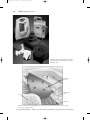
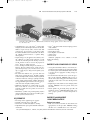
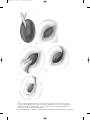
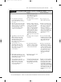
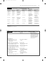
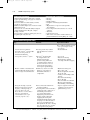
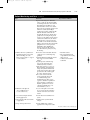
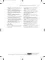
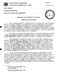
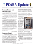
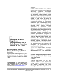
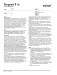
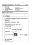
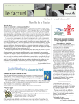
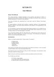
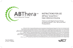
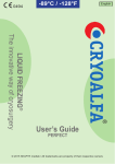
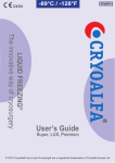
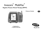
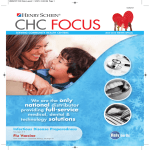
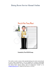
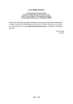
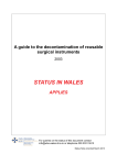
W0452-08_141
2/24/05
4:44 PM
Page 1129
http://evolve.elsevier.com
PROCEDURE
141
Vacuum-Assisted Closure™ (V.A.C.)®
System for Wounds
P U R P O S E :
To apply subatmospheric (negative) pressure
to the wound bed to stimulate granulation and reduce edema, thus enhancing
wound healing.
Eleanor R. Fitzpatrick
Mary Beth Flynn Makic
PREREQUISITE NURSING
KNOWLEDGE
• Negative-pressure wound therapy, or Vacuum-Assisted
Closure™ (V.A.C.®) therapy (V.A.C.® Kinetic Concepts
Inc., San Antonio, TX) (Fig. 141-1), is an exclusive
system for wound closure that applies subatmospheric
(negative) pressure evenly over a wound bed (Fig. 141-2).
This mechanical stress creates a noncompressive force
on the wound bed that dilates the arterioles, increasing
the effectiveness of local circulation and enhancing the
proliferation of granulation tissue.1 The system also
enhances lymphatic flow and removal of excessive fluid,
decreasing wound edema and bacterial load at the wound
site, further aiding wound healing1 (Fig. 141-3A and B).
• Wound healing is best achieved through adequate cleansing, debridement, and dressing of the wound bed based
on patient and wound characteristics.
• Wounds heal by either primary or secondary intention
(see Fig. 136-1). Most clean or clean/contaminated surgical wounds heal by primary intention. Suturing each layer
of tissue approximates the wound edges. These wounds
typically heal quickly and require minimal wound care.
Contaminated surgical or traumatic wounds (open
wounds) heal by secondary intention.
• Wounds healing by secondary intention granulate from
the base of the wound to the skin surfaces; care must be
taken to allow for uniform granulation and prevention of
open pockets/tunneling.
• Open wounds must be clean and moist to promote effective and efficient wound healing. To that end, open
wound care strives to maintain a clean, moist wound bed
that allows for effective wound healing under the support
of a dressing.
• Openly granulating wounds heal more slowly, must remain
moist to enhance tissue granulation, and may be more
painful for the patient.
• Open wounds may have excessive wound drainage,
requiring application of absorptive dressings, protection
of periwound skin, and more frequent dressing changes
to facilitate healing.
• The V.A.C.® system is indicated for wounds in which
subatmospheric pressure may promote wound healing—
for example, chronic, acute, traumatic, subacute, and
dehisced wounds, diabetic ulcers, pressure ulcers, flaps,
and grafts). The V.A.C.® will draw wound edges together
and create a less edematous, clean, vascularized wound
bed. The wound may fully heal, or improved adherence
of the flap or graft closure can be achieved.
• The V.A.C.® has been approved by the Food and Drug
Administration (FDA) for clinical use in the treatment of
these wounds. A newer system called the Mini VAC® is
portable and battery-powered and can be carried by the
patient, allowing for increased mobility. The V.A.C.®
device requires an electrical outlet for therapy; however,
some units have limited battery reserve. Optimal therapy
is achieved by delivering uninterrupted therapy at least
22 out of 24 hours (battery power is maintained).
From Lynn-McHale Wiegand, D.J., & Carlson, K. K. (2005). AACN Procedure Manual for Critical Care, 5th ed. St. Louis: Elsevier.
1129
W0452-08_141
1130
2/24/05
4:44 PM
Page 1130
Unit VII Integumentary System
Figure 141-1 Components of the VacuumAssisted Closure™ System. (Kinetic Concepts, Inc.,
San Antonio, TX.)
V.A.C. foam
dressing
Bone
Subcutaneous
tissue
Capillaries
Figure 141-2 V.A.C.® Therapy. Fluid, exudate, and debris removed from wound bed. (Courtesy: Kinetic
Concepts, Inc., San Antonio, TX.)
From Lynn-McHale Wiegand, D.J., & Carlson, K. K. (2005). AACN Procedure Manual for Critical Care, 5th ed. St. Louis: Elsevier.
W0452-08_141
2/24/05
4:44 PM
Page 1131
141
Vacuum-Assisted Closure™ (V.A.C.)® System for Wounds
1131
A
B
Figure 141-3 A, Wound defect. B, Wound defect with V.A.C.® therapy applied. (Courtesy: Kinetic Concepts,
Inc., San Antonio, TX.)
• Contraindications to use of the V.A.C.® system include
malignancy in the wound margins, untreated osteomyelitis,
fistulas to organs or body cavities, necrotic tissue with
eschar present, exposed arteries or veins in the wound.
Precautions should be used for wounds with active bleeding, difficult wound hemostasis, or patients taking
anticoagulants.25
• Negative therapy may be applied to the wound by selecting continuous or cycling pressure (intervals of 5 min
on/2 min off). After this period the dressing is removed
and the wound is cleansed, assessed, and prepared for
V.A.C.® therapy to continue if needed.
• V.A.C.® dressings are usually changed every 48 hours.3,5,7,13
However, infected wound beds may require more frequent dressing changes (every 12 hours); V.A.C.® dressings over grafts may be changed less frequently (every
3-5 days).3,5,7,13
• The wound bed should be free of necrotic tissue and
debris prior to applying the V.A.C.® In highly exudating
wounds, draining from the wound bed may be significant
in the first 24-48 hours of therapy, requiring monitoring
of urine output and hemodynamic stability. Studies have
not suggested fluid replacements have been necessary to
ensure hemostasis in highly exudating wounds.1,13
• Nutritional requirements for wound healing are great.
These needs must be assessed, met, and monitored
frequently as fluids and some proteins are removed via
V.A.C.® therapy.
• Inflammatory cytokines and protein-degrading enzymes
impair wound healing and can be removed with negativepressure systems such as the V.A.C.®
EQUIPMENT
•
•
•
•
Personal protective equipment (gown, goggles)
Nonsterile and sterile gloves; sterile field
Sterile water or normal saline (NS) for cleansing
Liquid skin barrier to protect periwound skin and/or hydrocolloid wafer
• V.A.C.® collection chamber and suction pump (commercially available)
• Sterile foam dressing
• Sterile adhesive drape
• Noncollapsible evacuation tube
• Adhesive drape
• Sterile scissors
Additional equipment to have available, as needed,
includes the following:
• Razor
PATIENT AND FAMILY EDUCATION
• Assess patient and family readiness to learn and any factors that may affect learning. It is also important to identify
how best the patient learns. ➸Rationale: Allows the nurse
to develop the most appropriate teaching strategy for
each patient.
• Provide information about the V.A.C.® system, the procedure, and the equipment. ➸Rationale: May decrease or
alleviate anxiety by assisting patient and family to understand the procedure, why it is needed, and the preferred
outcomes.
• Explain the procedure and the reason for changing wound
dressing. ➸Rationale: Decreases patient anxiety and
discomfort.
• Discuss patient’s role in dressing change procedure and
maintenance of V.A.C.® ➸Rationale: Elicits patient cooperation; prepares patient for wound management on
discharge.
PATIENT ASSESSMENT
AND PREPARATION
Patient Assessment
• Fully assess wound to determine its characteristics and
appropriateness for the procedure. ➸Rationale: Insures
that there is no contraindication to use of the V.A.C.®
From Lynn-McHale Wiegand, D.J., & Carlson, K. K. (2005). AACN Procedure Manual for Critical Care, 5th ed. St. Louis: Elsevier.
W0452-08_141
2/24/05
1132
•
•
•
•
•
4:44 PM
Page 1132
Unit VII Integumentary System
system. Provides data that can be used for comparison at
successive dressing changes.
Monitor for signs and symptoms of wound infection,
including the following:
❖ Erythema at drainage site
❖ Heat
❖ Edema
❖ Pain
❖ Elevated temperature and white blood cell count
❖ Wound drainage becoming cloudy and foul-smelling
❖ Increasing in amount of wound exudate
➸Rationale: Although negative-pressure wound therapy
assists with removal of excessive fluid, thus reducing the
presence of bacteria in the wound bed, assessment for
signs and symptoms of wound infection is necessary,
especially in compromised patients.
Determine baseline pain assessment of the patient.
➸Rationale: Provides data that can be used for comparison with past procedure assessment data. Allows the
nurse to plan for pre- and intraprocedure analgesia.
Determine baseline nutritional status and fluid volume
status. ➸Rationale: Fluids and protein may be lost during
V.A.C.® therapy.
Assess past medical history, especially related to problems with bleeding, fistula formation, or malignancy.
➸Rationale: The use of the V.A.C.® may be contraindicated
in these conditions.
Assess current medications specifically related to anticoagulant use. ➸Rationale: Identify possible areas of caution
which should be monitored with V.A.C.® use.
• Assess current laboratory values especially coagulation
studies and protein levels. ➸Rationale: Identifies abnormalities possibly associated with risks or areas to monitor
related to V.A.C.® use.
Patient Preparation
• Ensure patient and family understanding of preprocedural teaching. Reinforce teaching points as needed.
➸Rationale: Evaluates understanding of previously
taught information and provides a conduit for questions.
• Validate presence of patent intravenous access.
➸Rationale: Access may be needed for administration of
analgesic medications.
• If debridement or other invasive intervention is to be performed in conjunction with the V.A.C.® procedure, ensure
that informed consent has been obtained. ➸Rationale:
Allows patient to make decision with appropriate
information and health care providers can document this.
• Position the patient in a manner which will facilitate
dressing application and patient comfort. ➸Rationale:
Prepares patient to undergo procedure.
• Sedate the patient or administer prescribed analgesics if
needed. ➸Rationale: Improve comfort level and tolerance
of the procedure. Decreases patient anxiety and discomfort. Typically pain medication is not required for V.A.C.®
therapy; however, if the patient required analgesia for
previous dressing therapy, pain medications may be
required for V.A.C.® therapy.1,5
Procedure for Vacuum-Assisted Closure™ (V.A.C.)® System for Wounds
Steps
Rationale
1. Wash hands and don gloves.
Reduces possibility of transmission
of microorganisms.
2. Establish a sterile field with all cleansing
supplies and materials for appropriately
sized V.A.C.® dressing.
The V.A.C.® dressing size should be
chosen so that it will fill the entire
wound cavity.
3. Position the patient to facilitate cleansing
and dressing application.
Provides for patient comfort and
allows for visualization and access
to the wound.
4. Cleanse the wound according to orders
(see Procedure 136) and/or institution
protocol. (Level VI: Clinical studies in
a variety of patient populations and
situations to support recommendations.)
Exudate and debris are removed prior
to dressing application. This process
facilitates healing and decreases
bacterial burden.4,21-22
5. Physician or advanced practice nurse
may debride (see Procedure 137) necrotic
tissue or eschar if applicable. (Level VI:
Clinical studies in a variety of patient
populations and situations to support
recommendations.)
Healthy, vascularized tissue is
reached, and with a clean wound
bed there is enhanced granulation
tissue development.4,6,15,21-23
Special Considerations
If extensive debridement is
necessary, it may require an
operative suite.
From Lynn-McHale Wiegand, D.J., & Carlson, K. K. (2005). AACN Procedure Manual for Critical Care, 5th ed. St. Louis: Elsevier.
W0452-08_141
3/11/05
8:33 AM
Page 1133
141
Vacuum-Assisted Closure™ (V.A.C.)® System for Wounds
1133
Procedure for Vacuum-Assisted Closure™ (V.A.C.)® System for Wounds—Continued
Steps
Rationale
Special Considerations
6. Shave hair on the border around the
wound (if needed) and thoroughly cleanse
skin surrounding wound. (Level I:
Manufacturer’s recommendations only.)
Improves dressing adherence.
7. Dry and prepare the periwound tissue as
appropriate. Skin degreasing/medical
cleansing agents may be necessary to
apply to periwound tissue. (Level I:
Manufacturer’s recommendations only.)
Moisture from perspiration, oil, or
body fluids may cause difficulty in
achieving an air-tight seal with
V.A.C.® dressing.
8. Choose V.A.C.® soft foam (white), a
hydrophilic (water-attracting) material
or the black foam (polyurethane), a
hydrophobic (water-repelling) dressing
(Table 141-1). (Level I: Manufacturer’s
recommendations only.)
White, polyvinyl alcohol foam is
denser with smaller pores, which
restricts granulation tissue growth
into the foam and may be used in
cases in which black foam cannot
be tolerated due to pain.
Larger pores in black foam are
considered to be most effective in
stimulating granulation tissue and
wound contraction.
The white V.A.C.® dressing
holds moisture but also
allows exudate to be removed
through it. It is nonadherent
and can be used in tunnels
and shallow undermining due
to its higher tensile strength.
The black V.A.C.® dressing does
not hold moisture and allows
exudates to be removed. Its
design results in rapid growth
of new granulation.
9. Remove gloves. Wash hands. Apply
sterile gloves.
10. Open the V.A.C.® dressing onto sterile,
dry surface; inspect it for any defects.
Faulty dressings should be
replaced.
11. Cut the V.A.C.® foam with sterile
scissors in a location away from the
wound (Fig. 141-4A). (Level IV:
Limited clinical studies to support
recommendations.)
This prevents small particles from
the dressing falling into the
wound. The dressing should be
cut to fit the size and shape of
the wound, including tunnels
and undermined areas
(Fig. 141-4B). Tunneling can
result in a cyst or abscess when
the main body of a wound heals
Any exposed tendons, nerves,
or blood vessels should be
protected by moving muscle or
fascia over exposed structures
or by placing a layer of
nonadherent dressing over them.
Procedure continues on the following page
Table 141-1 Recommended Guidelines for Foam Use
V.A.C.® Polyurethane (Black Foam)
Deep, acute wounds with moderate
granulation tissue growth
Deep wounds with extremely rapid growth
in granulation tissue
Deep pressure ulcers
Superficial wounds
Shallow chronic ulcers
Postgraft therapy
Compromised flaps
Fresh flaps
Tunneling/sinus tracks/undermining
Diabetic ulcers
Dry wounds
Deep trauma wounds
Superficial trauma wounds
V.A.C.® Soft Foam
Both
Either
X
X
X
X
X
X
X
X
X
X
X
X
X
Responsible physician or advanced practice nurse should be consulted for individual patient conditions. Consult device user manual and manufacturer’s recommended
guidelines before use.
Courtesy Kinetic Concepts, Inc., San Antonio, TX.
From Lynn-McHale Wiegand, D.J., & Carlson, K. K. (2005). AACN Procedure Manual for Critical Care, 5th ed. St. Louis: Elsevier.
W0452-08_141
2/24/05
A
4:44 PM
Page 1134
B
D
C
E
Figure 141-4 A and B, Cut the V.A.C.® foam to appropriate size to fill the wound defect. C, Apply V.A.C.®
foam into wound bed. D, Apply V.A.C.® tubing to foam in the wound. E, Cover foam and 3 to 5 cm of surrounding
healthy tissue with transparent dressing drape to ensure an occlusive seal. (Courtesy Medical Media Department,
Thomas Jefferson University Hospital, Philadelphia, PA.)
From Lynn-McHale Wiegand, D.J., & Carlson, K. K. (2005). AACN Procedure Manual for Critical Care, 5th ed. St. Louis: Elsevier.
W0452-08_141
3/11/05
8:35 AM
Page 1135
141
Vacuum-Assisted Closure™ (V.A.C.)® System for Wounds
1135
Procedure for Vacuum-Assisted Closure™ (V.A.C.)® System for Wounds—Continued
Steps
Rationale
Special Considerations
and closes the entrance to the
tunnel. Bacterial invasion and
impaired healing results from
unfilled dead space.4,24
12. Size and trim the dressing drape
to cover the foam plus a 3-5 cm
border of intact skin. Do not discard
excess drape.
Excess drape may be needed later
as a patch.
Drape can be placed over
prepared skin or the barrier.
13. Gently place the foam into the wound
cavity covering the entire wound base
and sides as well as areas of tunneling
and undermining (Fig. 141-4C).
(Level IV: Limited clinical studies
to support recommendations.)
Capillaries could be compressed
if packed too tightly, and pressure
on newly formed granulation
tissue may prevent or delay
healing.4,24 If periwound skin is
fragile, use a skin preparation
(Matisol®, No-Sting®) prior to
drape application or frame the
wound with a skin barrier or
hydrocolloid dressing. This
affords protection for periwound
skin.
More than one dressing may be
needed for larger wounds. More
than one piece of foam may be
used to fill the wound bed.
Foam pieces should be in
contact but not overlapping each
other to allow equalization of
negative pressure applied to the
wound bed by the suction
device.1,3,5,7,13
14. Apply tubing to foam in the wound.
Tubing can be laid on top of the
foam or inside the foam dressing.
The tubing should be positioned
away from bony prominences
(Fig. 141-4D). (Level I: Manufacturer’s
recommendations only.)
This will prevent the development
of pressure.
For deeper wounds the tubing
should be repositioned regularly
to minimize pressure on wound
edges. Cushion skin under
tubing with excess foam.
15. Cover foam and 3 to 5 cm of
surrounding healthy tissue with
drape to ensure an occlusive seal
(Fig. 141-4E). (Level IV: Limited
clinical studies to support
recommendations.)
The vacuum will not function
without an occlusive seal. The
drape may also help maintain
a moist wound environment.2
The drape is vapor-permeable
and allows for gas exchange.
It also protects the wound from
external contamination. Foam
will contract into wound bed if
seal is obtained. If foam does
not contract, reassess outer
dressing for possible leaks
in the system or dressing
seal.1,3,5,7,13
16. Lift the tubing and pinch 1 to 3 cm of
drape together under the tubing to help
hold the tubing away from the skin and
pad underneath the tubing.
This will reduce pressure on the
skin from the tubing.
17. Avoid stretching the drape and
compressing the foam into the wound
with transparent occlusive drape.
Simply cover and seal around the
foam. (Level I: Manufacturer’s
recommendations only.)
Avoids tension and shearing forces
on wound and surrounding tissue.
Allows distribution of pressure
throughout all wounds with the
use of one pump.
More than one wound of similar
pathology in close proximity can
be managed with one negativepressure pump. Such wounds
can be bridged by placing the
V.A.C.® drape on intact skin and
a strip of foam from one wound
bed to the other. All edges of the
foam should be in contact and
the tubing placed in a central
location.
Procedure continues on the following page
From Lynn-McHale Wiegand, D.J., & Carlson, K. K. (2005). AACN Procedure Manual for Critical Care, 5th ed. St. Louis: Elsevier.
W0452-08_141
2/24/05
1136
4:44 PM
Page 1136
Unit VII Integumentary System
Procedure for Vacuum-Assisted Closure™ (V.A.C.)® System for Wounds—Continued
Steps
Rationale
18. Secure tubing with an additional
piece of drape or tape (pad
underneath tubing) several
centimeters away from the
dressing. (Level I: Manufacturer’s
recommendations only.)
Prevents pull on the primary
dressing area, which can cause
leaks.
19. Use excess drape to patch leaks and
secure borders as needed.
Leaks prevent activation of the
V.A.C.® system.
Applying the V.A.C.® Device
1. Remove canister from the sterile
packaging and push it into the V.A.C.®
unit until it clicks.
If the canister is not engaged
properly, it will not function and
an alarm will sound.
Special Considerations
2. Connect the dressing tubing to the
canister tubing, making sure both
clamps are open.
Closed clamps prevent activation
of the negative pressure.
3. Place the V.A.C.® unit on a level
surface or hang from the footboard.
(Level I: Manufacturer’s
recommendations only.)
The V.A.C.® unit will alarm and
deactivate therapy if the unit is
tilted beyond 45 degrees.
4. Press the green-lit power button.
Activates subatmospheric pressure
therapy.
In less than 1 min of operation,
the V.A.C.® dressing will
collapse unless leaks are
present.
5. Adjust the V.A.C.® unit settings based
on individual patient’s needs. Variable
negative-pressure settings can be chosen,
as well as a continuous or intermittent
mode (Table 141-2). (Level IV: Limited
clinical studies to support
recommendations.)
Continuous vacuum mode removes
cellular edema and debris,
enhancing perfusion through
vessels previously
compressed.1,10,12,14,17,18
Healing inhibitory factors present in
this fluid are also removed.20,26
After edema has been sufficiently
withdrawn, the intermittent mode
promotes granulation tissue
formation and prevents wound
dehydration.1,8,16,17
Negative pressures ranging from
50 to 175 mm Hg can be
chosen, depending on the
amount of exudate and
granulation tissue within
the wound and the type of
foam used (see Table 141-2).
Premedicate patient if necessary.
Application of negative-pressure
systems may cause mild
discomfort for the patient during
initial activation of the V.A.C.®
system.1,11
Blood flow may be inhibited by
application of higher negative
pressures.
If you suspect a leak (small
leaks may create a whistling
noise), gently press around the
tubing to better seal the drape.
Excess drape can also be used
to patch over leaks. Air leaks
most often occur around tubing.
6. Slowly increase negative pressure on
suction device to desired setting of
125 mm Hg. (Level I: Manufacturer’s
recommendations only.)
Negative pressure may be initiated
at 75 mm Hg and gradually
increased to 125 mm Hg. Maintain
therapy continuously.
Subatmospheric pressure therapy
should not be off for more than
2 hr per day.
Treatment is discontinued when
goals for V.A.C.® therapy are
achieved or after 1 to 2 weeks
without improvement in the
condition of the wound.
Physician or advanced practice
nurse will order desired
negative-pressure settings.
Lower pressure settings may be
ordered for special wound beds
(meshed grafts); higher pressure
settings (125-175 mm Hg) may
be ordered for high-effluent
wounds.1,3,5,7,13
V.A.C.® therapy is delivered for
48 hr. After this period the
dressing is removed and the
wound cleansed, assessed, and
prepared for V.A.C.® therapy to
From Lynn-McHale Wiegand, D.J., & Carlson, K. K. (2005). AACN Procedure Manual for Critical Care, 5th ed. St. Louis: Elsevier.
W0452-08_141
2/24/05
4:44 PM
Page 1137
141
Vacuum-Assisted Closure™ (V.A.C.)® System for Wounds
1137
Table 141-2 Recommended Guidelines for Treating Wound Types with V.A.C.® System
Target Pressure for
Black Polyurethane
Dressing
Target Pressure
Polyvinylalcohol
Soft Foam
Intermittent (5 min
on/2 min off) for
rest of therapy
Intermittent (5 min
on/2 min off) for
rest of therapy
Continuous
125 mm Hg
125-175 mm Hg
Every 48 hr (every
12 hr with infection)
125 mm Hg
125-175 mm Hg
(titrated up for
increased drainage)
125 mm Hg; (titrated
up for increased
drainage)
Every 48 hr (every
12 hr with infection)
125 mm Hg
Subsequent
Cycles
Wound Type
Initial Cycle
Acute/traumatic
wound
Continuous for
first 48 hr
Surgical wound
dehiscence
Continuous for
first 48 hr
Meshed graft
Continuous
Pressure ulcer
Continuous for
first 48 hr
Chronic ulcer
Continuous
Intermittent (5 min
on/2 min off) for
rest of therapy
Continuous
Fresh flap
Continuous
Continuous
125-150 mm Hg
Compromised
flap
Continuous
Continuous
125 mm Hg
75-125 mm Hg
50-125 mm Hg
125-175 mm Hg
(titrated up for
increased drainage)
125-175 mm Hg
(titrated up for
increased drainage)
125-175 mm Hg
(titrated up for
increased drainage)
125-175 mm Hg
(titrated up for
increased drainage)
Dressing Change
Interval
None; remove
dressing after
4-5 days when
using either foam
Every 48 hr (every
12 hr with infection)
Every 48 hr (every
12 hr with infection)
Every 72 hr (every
12 hr with infection)
Every 48 hr (every
12 hr with infection)
Responsible physician or advanced practice nurse should be consulted for individual patient conditions. Consult device user manual and manufacturer’s recommended
guidelines before use.
Courtesy Kinetic Concepts, Inc., San Antonio, TX.
Procedure for Vacuum-Assisted Closure™ (V.A.C.)® System for Wounds—Continued
Steps
Rationale
Special Considerations
continue if needed. If infection
is present, the dressing change
interval should be every 12 to
24 hr. Negative pressure may
enhance bacterial clearance
from the wound.9,11,19
Dressing Removal
1. To remove the dressing, raise the
tubing connector above the level
of the pump unit.
Drains fluid from tubing into
canister.
2. Tighten clamps on the dressing tube.
Prevents leakage.
3. Separate canister tube and dressing tube
by disconnecting the connector.
Allows canister to be emptied
and/or discarded.
4. Allow the pump unit to pull the exudate
in the canister tubing into the canister;
then tighten clamps on the canister tube.
Removes any remaining fluid
from the dressing.
5. Press Therapy button Off.
Deactivates pump.
6. Gently stretch drape horizontally and
slowly pull up from skin. Do not peel.
Gently remove.
Decreases patient discomfort
and potential for skin and
wound trauma.
7. Discard disposables according to
institution policy.
Reduces transmission of
microorganisms; universal
precautions.
8. Document procedure in patient record.
From Lynn-McHale Wiegand, D.J., & Carlson, K. K. (2005). AACN Procedure Manual for Critical Care, 5th ed. St. Louis: Elsevier.
W0452-08_141
2/24/05
1138
4:44 PM
Page 1138
Unit VII Integumentary System
Expected Outcomes
Unexpected Outcomes
• Wound healing/granulation enhanced by consistent
negative-pressure therapy; early signs of contraction
of wound margins
• Decreased volume of wound exudate (over time)
and absence of foul odor or color
• Enhanced wound healing because of effective wound
fluid/edema removal
• Decrease in size of wound with ability for surgical
closure with flap/graft or skin graft; complete
healing of wound
• Decreased time to satisfactory healing (may decrease
hospital length of stay and cost)
•
•
•
•
•
•
•
•
•
•
Infection
Bleeding
Fistula formation
Disruption of underlying tissue/structures
Pain
Misplacement over exposed vessel, ligaments,
other structures
Lack of improvement in wound after 1 to 2 weeks
of therapy
Tissue loss
Ischemia and necrosis
Skin erosion or maceration around wound sites, or
pressure breakdown at dressing tubing site, or both
Patient Monitoring and Care
Steps
Rationale
Reportable Conditions
These conditions should be reported
if they persist despite nursing
interventions.
• Tissue breakdown
1. Assess location of wound and
placement of V.A.C.® evacuation tube
to avoid excessive pressure on
surround tissue/structures.
Excessive pressure may result in
tissue breakdown at evacuation
tube site.
2. Assess patency of V.A.C.® system.
The V.A.C.® dressing should
be collapsed when seal is
maintained and negative
pressure is being delivered in
a consistent manner. Alarms on
the device indicate loss of seal;
raised foam dressing indicates
loss of negative-pressure therapy.
• Loss of seal
• Raised foam dressing
• Wound drainage suddenly
decreasing in amount or stopping
3. Monitor condition of wound bed and
periwound skin with dressing changes;
observe for signs of wound infection.
Identifies any evidence of wound
healing or of any changes or
abnormalities indicative of
complications.
• Erythema at drainage site
• Heat, edema, pain
• Elevated temperature and white
blood cell count
• Cloudy or foul-smelling wound
drainage
• Increased wound drainage
• Excess bleeding
• Discolored tissue in wound bed
• Macerated periwound skin
• New tunneling or undermining
4. Change the dressing every 48 hr.
If infection is present, increase the
frequency of dressing change to
every 12 to 24 hr. (Level II:
Theory-based, no research data to
support recommendations:
recommendations from expert
consensus group may exist.)
Removes infectious material from
• Signs or symptoms of infection
a healing wound bed.
(erythema, heat, discolored or
If dressing adheres to the wound base,
purulent drainage, fever)
consider imposing a single layer of
nonadherent porous material
(e.g. wide-meshed Vaselineimpregnated gauze) between the
dressing and the wound when
reapplying the dressing. The
nonadherent material must have wide
enough pores to allow unrestricted
passage of air and fluid. Because
tissue growth into the V.A.C.® dressing
may cause adherence, also consider
more frequent dressing changes.
From Lynn-McHale Wiegand, D.J., & Carlson, K. K. (2005). AACN Procedure Manual for Critical Care, 5th ed. St. Louis: Elsevier.
W0452-08_141
2/24/05
4:44 PM
Page 1139
141
Vacuum-Assisted Closure™ (V.A.C.)® System for Wounds
1139
Patient Monitoring and Care—Continued
Steps
Rationale
Reportable Conditions
If previous dressings were difficult to
remove, make sure the dressing tubing
is unclamped; introduce 10 to 30 ml of
NS solution into the tubing to soak
underneath the foam for 15 to 30 min.
NS can be injected directly into the foam
while low vacuum (50 mm Hg) is
applied to the dressing. Clamp the tube
when the NS starts to flow into the
dressing tube. Wait 15 to 30 min; then
gently remove dressing. If the patient
experiences pain during dressing change,
1% lidocaine solution may be ordered
by the physician or advanced practice
nurse. This can be introduced down the
tubing or injected into the foam with the
pump turned on at a lower pressure
(50 mm Hg). After instilling the
lidocaine, clamp the tube and wait
15 to 20 min before gently removing
the dressing.
5. Monitor the mode (continuous or
intermittent) and level of suction
(50-175 mm Hg). (Level IV:
Limited clinical studies to support
recommendations.)
Removal of edema and debris alleviates
compressive forces, thus improving
perfusion.
Suctioning fluid from within the wound
may remove factors that inhibit
healing.25
Negative-pressure wound therapy
decreases the bacterial load of
deliberately infected wounds.1
Application and release of force on
tissue stimulates cell proliferation
and protein synthesis. Mechanical
stretch on the tissue by the negative
pressure draws the wound toward
the center closing the defect.16
Once edema of the wound has
resolved (typically after 48 hr
of continuous therapy) the
intermittent mode allows a more
aggressive stimulus for granulation
tissue formation, possibly due to
rhythmic perfusion or increased
cell mitosis stimulated by a rest
and stimulation cycle.1,8,17
6. Maintain an air tight seal.
(Level I: Manufacturer’s
recommendation only.)
Loss of an air-tight seal can result
in a decreased amount of drainage
removal and in desiccation of the
wound.
7. Use sterile technique with new
dressing application.
Prevents bacterial contamination of
system.
8. Label dressing with date and time
of application.
Identifies when system should be
changed.
9. Keep canister position level.
Prevents malfunction of suction
apparatus and an inoperative status.
• Patient discomfort
• Excess granulation tissue
overgrowth into the dressing
when removed
• Continued edema within
wound bed
Procedure continues on the following page
From Lynn-McHale Wiegand, D.J., & Carlson, K. K. (2005). AACN Procedure Manual for Critical Care, 5th ed. St. Louis: Elsevier.
W0452-08_141
2/24/05
1140
4:44 PM
Page 1140
Unit VII Integumentary System
Patient Monitoring and Care—Continued
Steps
Rationale
Reportable Conditions
10. The V.A.C.® canister should be changed
when full (unit will alarm) or at least
weekly. Label with date and time
of change.
Controls odor.
• Wound drainage becoming
foul-smelling and cloudy
11. Monitor amount of wound drainage.
(Level II: Theory-based; no research
data to support recommendations:
recommendations from expert
consensus group may exist.)
If a wound produces excessive fluid,
the patient may experience a fluid
imbalance, requiring intravenous
or oral fluid replacements. Excess
drainage may also result in some
protein loss. Nutritional consult
to replace protein loss from wound
exudates may be indicated.
• Increasing amounts of
drainage
• Tachycardia
• Hypotension
• Oliguria
• Decreasing serum protein
levels
12. Encourage patient hygiene, shower, or
bath during V.A.C.® dressing changes.
Documentation
Documentation should include the following:
•
•
•
•
•
•
•
Patient and family education
Patient tolerance of the procedure
Condition of the wound bed and periwound skin description
Characteristics of wound drainage
Degree of suction (mm Hg) and continuous or intermittent mode
Nursing interventions
Premedication given and patient’s response to the
pain medication
References
1. Argenta, L.C., and Morykwas, M.J. (1997). Vacuum-assisted
closure: A new method for wound control and treatment:
clinical experience. Ann Plastic Surg, 38, 563-76.
2. Banwell, P.E. (1999). Topical negative pressure therapy in
wound care. J Wound Care, 8, 79-84.
3. Bates-Jensen, B.M., et al. (2001). Management of the wound
environment with advanced therapies. In: Sussman, C., and
Bates-Jensen, B.M., eds. Wound Care. 2nd ed. Gaithersburg,
MD: Aspen Publication, 272-92.
4. Bergstrom, N., et al. (1994). Treatment of pressure ulcers.
Clinical Practice Guideline, No. 15. Rockville, MD: U.S.
Department of Health and Human Services, Public Health
Service, Agency for Healthcare Policy and Research, AHCPR
Publication No. 95-0652.
5. Chua-Patel, C.T., et al. (2000). Vacuum-assisted wound
closure. Am J Nurs, 100, 45-8.
6. d’Udekem, Y., et al. (1998). Radical debridement and omental
transposition for post-sternotomy mediastinitis. Cardiovasc
Surg, 6, 415-8.
7. Evans, D., and Land, L. (2001). Topical negative pressure for
treating chronic wounds. The Cochrane Database of
Systematic Reviews, 4:CD001898.
8. Fabian, T.S., et al. (2000). The evaluation of subatmospheric
pressure and hyperbaric oxygen in ischemic, full-thickness
wound healing. Am Surg, 66, 1136-43.
9. Ford, C.N., et al. (2001). Interim analysis of a prospective,
randomized trial of a vacuum-assisted closure versus the
• Wound debridement procedure (if applicable), wound
cleansing procedure completed, dated, and timed
• Size of the wound measured by length, width, and depth
(consider obtaining a photograph of the wound, depending
on institution policy)
• Size and type of V.A.C.® foam dressing applied and total
number placed in the wound
• Unexpected outcomes, reportable conditions
healthpoint system in the management of pressure ulcers.
Ann Plast Surg, 49, 55-61.
10. Genecov, D.G., et al. (1998). A controlled subatmospheric
pressure dressing increases the rate of skin graft donor site
re-epithelialization. Ann Plast Surg, 40, 219-25.
11. Hersh, R.E., et al. (2001). The vacuum-assisted closure
device as a bridge to sternal closure. Ann Plast Surg,
46, 250-4.
12. Joseph, E., et al. (2000). A prospective randomized trial of
vacuum-assisted closure versus standard therapy of chronic,
nonhealing wounds. Wounds, 12, 60-7.
13. Kirby, J.P., et al. (2002). Novel uses of a negative pressure
wound care system. J Trauma, 53, 117-21.
14. McCallon, S.K., et al. (2000). The effectiveness of vacuumassisted closure vs. saline moistened gauze in the healing of
post-operative diabetic foot wounds. Ostomy/Wound Manage,
46, 28-35.
15. Meara, J.G., et al. (1999). Vacuum-assisted closure in the
treatment of degloving injuries. Ann Plast Surg,
42, 589-94.
16. Morykwas, M.J., and Argenta, L.C. (1997). Nonsurgical
modalities to enhance healing and care of soft tissue wounds.
J South Ortho Assoc, 6, 279-88.
17. Morykwas, M.J., et al. (1997). Vacuum-assisted closure: a
new method for wound control and treatment: Animal studies
and basic foundation. Ann Plast Surg, 38, 553-62.
18. Mullner, T., et al. (1997). The use of negative pressure to
promote the healing of tissue defects: A clinical trial using the
vacuum seal technique. Br J Plast Surg, 50, 194-9.
From Lynn-McHale Wiegand, D.J., & Carlson, K. K. (2005). AACN Procedure Manual for Critical Care, 5th ed. St. Louis: Elsevier.
W0452-08_141
2/24/05
4:44 PM
Page 1141
141
Vacuum-Assisted Closure™ (V.A.C.)® System for Wounds
19. Obdeijn, M.C., et al. (1999). Vacuum-assisted closure in the
treatment of poststernotomy mediastinitis, Ann Thorac Surg,
68, 2358-60.
20. Philbeck, T.E., et al. (1999). The clinical and cost
effectiveness of externally applied negative pressure wound
therapy in the treatment of wounds in home healthcare
Medicare patients. Ostomy/Wound Manage, 45, 41-50.
21. Rodeheaver, G., et al. (1994). Wound healing and wound
management: Focus on debridement. Adv Wound Care, 7,
22-4, 26-9, 32-6.
22. Sibbald, R.G., et al. (2000). Preparing the wound
bed-debridement, bacterial balance and moisture balance.
Ostomy/Wound Manage, 46, 14-35.
23. Steed, D.L., et al. (1996). Effect of extensive debridement and
treatment on the healing of diabetic foot ulcers. Diabetic
Ulcer Study Group. J Am Coll Surg, 183, 61-4.
24. Stotts, N. (1997). Co-factors in impaired wound healing.
In: Krasner, D., and Kane, D., eds. Chronic Wound Care:
A Clinical Source Book for Healthcare Professionals. 2nd ed.
Wayne, PA: Health Management Publications, Inc.
25. Wysocki, A.B. (1996). Wound fluids and the pathogenesis of
chronic wounds. J WOCN, 23, 283-90.
26. www.kci1.com/clinicalevidence/indexVAC. Accessed
May 17, 2004.
Additional Readings
Barker, D.E., et al. (2000). Vacuum pack technique of temporary
abdominal closure: A 7-year experience with 112 patients.
J Trauma, 48, 201-7.
1141
Casey, G. (2003). Nutritional support in wound healing. Nurs
Stand, 17, 55-6, 58.
Dolynchuk, K. (2000). Best practices for the prevention and
treatment of pressure ulcers. Ostomy/Wound Manage,
46, 38-52.
Erdman, D., et al. (2001). Abdominal wall defect and enterocutaneous fistula treatment with the vacuum-assisted (V.A.C.)
system. Plast Reconstr Surg, 108, 2066-8.
Falanga, V., ed. (2000). Text Atlas of Wound Management.
London: Martin Dunitz Ltd.
Falanga, V., ed. (2001). Cutaneous Wound Healing. London:
Martin Dunitz Ltd.
Irion, G. (2002). Comprehensive Wound Management. Thorofare,
NJ: Slack Incorporated.
Kinetic Concepts, Inc. (2001). V.A.C. Physician and Caregiver
Reference Manual. San Antonio: Kinetic Concepts, Inc.
Kloth, L.C., and McCulloch, J.M., eds. (2002). Wound Healing:
Alternatives in Management. 3rd ed. Philadelphia: F.A. Davis
Company.
Krasner, D.L., et al. (1999). Nursing management of chronic
wounds. Nurs Clin N Am, 34, 933-949.
Krasner, D.L. (2002). Managing wound pain in patients with
vacuum-assisted closure devices. Ostomy/Wound Manage,
48, 38-43.
Mendez-Eastman, S. (2001). Guidelines for using negative
pressure wound therapy. Adv Skin & Wound Care,
14, 314-22.
Mendez-Eastman, S. (1999). Use of hyperbaric oxygen and negative
pressure therapy in the multidisciplinary care of a patient with
nonhealing wounds. J Wound Ostomy Continence Nurs, 26, 67-76.
http://evolve.elsevier.com
From Lynn-McHale Wiegand, D.J., & Carlson, K. K. (2005). AACN Procedure Manual for Critical Care, 5th ed. St. Louis: Elsevier.