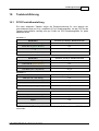
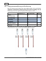
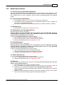
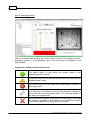
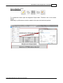
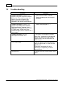
1
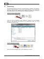
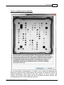
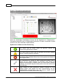
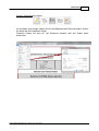
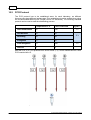
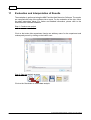
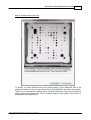
®
MutaCHIP FOOD
DNA-Macroarray Kit zur Untersuchung von genetisch bedingten
Nahrungsmittelintoleranzen
Nur für in-vitro Diagnostik
KF391007
10
KF390007
20
Immundiagnostik AG, Stubenwald-Allee 8a, 64625 Bensheim, Germany
www.immundiagnostik.com
Tel.: +49 (0)6251/ 701900
[email protected]
Fax: +49 (0)6251/ 849430
2
Inhaltsverzeichnis
1 Verwendungszweck
3
2 Einleitung
3
3 Testprinzip
4
4 Kitbestandteile
5
5 Erforderliche Materialien
5
6 Lagerung und Haltbarkeit
5
7 Arbeitsbedingungen
6
8 Hinweise und Vorsichtsmaßnahmen
6
9 Probengewinnung
6
10 Testdurchführung
7
1 PCR
...................................................................................................................................
Produktherstellung
7
2 Probenzusammenführung
...................................................................................................................................
und Verdünnung
8
3 MutaChip
...................................................................................................................................
Protokoll
9
11 Auswertung
10
12 Troubleshooting
14
© 2012 Immundiagnostik AG / Version 2.0
Verwendungszweck
1
3
Verwendungszweck
Das MutaCHIP® FOOD Kit ist ein molekularbiologischer Test zur Untersuchung von
Mutationen, die zu Nahrungsmittelintoleranzen führen. Hierfür wird die genomische DNA
mittels MutaCHIP untersucht. Folgende Marker werden durch den Kit detektiert:
Nahrungsmittelintoleranz
Marker
Alkoholintoleranz
ADH2*2
ALDH2*2
Fruktoseintoleranz
ALDB 149
ALDB 174
ALDB 334
Saccharoseintoleranz
SI 117
SI 340
SI 620
SI 1098
Glucose-6-Phosphat-Dehydrogenase
Defizienz
2
G6PD Mediterranean (S188P)
Hämochromatose
H63D
C282Y
Laktoseintoleranz
-13910 T/C
Zöliakie
HLA-DQ2 (DQA1 0501, DQB1 0201)
Einleitung
Nutrigenetik- und Nutrigenomik-Studien liefern Erkenntnisse über die Mechanismen der
Nährstoff-Gen-Wechselwirkungen und können zur Entwicklung von neuen Strategien zur
individualisierten Behandlung ernährungsbedingter Krankheiten, wie z.B. enzymatisch
bedingter Nahrungsmittelintoleranzen, beitragen. Eine enzymatisch bedingte
Nahrungsmittelintoleranz ist die Unfähigkeit eines Organismus bestimmte
Nahrungsbestandteile zu metabolisieren. Ursache hierfür sind Mutationen in jenen Genen,
die für die entsprechenden Enzyme codieren. Zu diesen Nahrungsmittelintoleranzen
gehören
Alkohol-,
Fruktose-,
Laktose-,
Saccharoseintoleranz,
hereditäre
Hämochromatose,
Zöliakie
und
Glucose-6-Phosphat-Dehydrogenase-Defizienz.
Alkoholintoleranz resultiert aus einem gestörten Abbau von Ethanol, wenn die Enzyme
Alkohol-Dehydrogenase und Aldehyd-Dehydrogenase durch Mutationen verändert sind.
Durch Gefäßerweiterungen bedingt, treten Symptome wie Gesichtsröte (Flushing),
Herzrasen, Muskelschwäche, gesteigerte Herzfrequenz oder Hitzegefühl im Magen auf.
Fruktoseintoleranz resultiert aus der reduzierten Aktivität des Enzyms Aldolase B, so
dass Fruktose nicht richtig verdaut werden kann. Wird diese unverdaute Fruktose
aufgenommen kommt es zu Symptomen wie Unterleibsschmerzen, Appetitlosigkeit und
Übelkeit. Bei einer Laktoseintoleranz wird der mit der Nahrung aufgenommene
Milchzucker Laktose nicht mehr verdaut. Ursache hierfür ist das Fehlen des Enzyms
Laktase, das den Milchzucker normalerweise in Galaktose und Glukose spaltet.
Symptome sind Blähungen, Bauchkrämpfe, Übelkeit, Erbrechen und/oder Durchfall.
Saccharoseintoleranz resultiert aus der reduzierten Aktivität des in der
Bürstensaummembran des Dünndarms lokalisierten Enzyms Sucrase-Isomaltase. Der
© 2012 Immundiagnostik AG / Version 2.0 / 06.12.2012
4
Verzehr von Saccharose führt bei Betroffenen zu Symptomen wie Durchfall,
Bauchschmerzen und/oder Erbrechen. Glucose-6-Phosphat-Dehydrogenase-Defizienz
führt zu einem Mangel des Enzyms Glucose-6-Phosphat-Dehydrogenase, welches die
Erythrozyten vor oxidativen Schäden schützt. Lebensmittel und Medikamente, die oxidativ
wirken, können zu frühkindlicher Hyperbilirubinämie und einer akuten bzw. chronischen
Hämolyse führen.Hämochromatose ist eine autosomal rezessive Krankheit, bei der
überschüssiges Eisen im Körper angereichert wird und letztendlich bei fehlender
Diagnostik zu Multiorganversagen führen kann. Zöliakie ist eine solche
Nahrungsmittelunverträglichkeit, bei der das im Weizen enthaltene Gluten eine
Autoimmunreaktion im Körper hervorruft. Diese Reaktion äußert sich durch Symptome,
wie Durchfall, Verstopfung und Müdigkeit.
Referenzen:
SIMOPOULOS, Artemis P.: Nutrigenetics/Nutrigenomics. In: Annu Rev Public
Health 31 (2010), Apr, S. 53–68
MONTALTO, Massimo; SANTORO, Luca; D’ONOFRI, Ferruccio; et al. : Adverse
reactions to food: allergies and intolerances. In: Dig Dis 26 (2008), Nr. 2, S. 96–
103
EDENBERG Howard J.; “The Genetics of Alcohol Metabolism, Role of Alcohol
Dehydrogenase and Aldehyde Dehydrogenase Variants. In: Alcohol Research &
Health, 30 (2007), Nr. 1, S. 5-13
WONG D.: Hereditary fructose intolerance. In: Molecular Genetics and Metabolism
85 (2005), Nr. 3, S. 165-167
SWAGERTY, Daniel L..; WALLING, Anne D.; KLEIN, Robert M..: Lactose
Intolerance. In: American Familiy Physician 65 (2002), Nr. 9, S. 1845-1850
RITZ, Valentina; ALFALAH, Marwan; ZIMMER, Klaus-Peter; et al.: Congenital
sucrase-isomaltase deficiency because of an accumulation of the mutant enzyme
in the endoplasmic reticulum. In: Gastroenterology 125 (2003), Dec, Nr. 6, S.
1678–1685
BEUTLER, Ernest: G6PD deficiency. In: Blood 84 (1994), Dec, Nr. 11, S. 3613–
3636
3
Testprinzip
Zur Analyse der Mutationen werden die variablen Bereiche der jeweiligen Gene mittels
PCR amplifiziert, wobei frisch extrahierte genomische DNA des Patienten als Matrize
dient. Das Amplifikationsprodukt wird anschließend auf den MutaCHIP gegeben und nach
mitgeliefertem Protokoll bearbeitet. Die Analyse mittels Präzipitationsreaktion lässt eine
eindeutige Genotypisierung aller auf den MutaCHIP gespotteten Allele zu.
Das amplifizierte Produkt wird auf den MutaCHIP gegeben und bindet an den dort
immobilisierten Sonden. Durch einen Waschschritt werden unspezifisch gebundene
Fragmente wieder entfernt. Im Anschluss wird das Enzym hinzugegeben, welches an den
Sonden-Fragment-Komplex bindet. Nach Zugabe des Substrats tritt eine Fällungsreaktion
an den Stellen, an denen noch DNA gebunden ist, auf. Das farbige Präzipitat wird mit dem
Imagereader detektiert und von der dazugehörigen Software ausgelesen und bewertet.
© 2012 Immundiagnostik AG / Version 2.0 / 06.12.2012
Testprinzip
4
5
Kitbestandteile
Jedes Testkit enthält die folgenden Reagenzien zur Durchführung von 10 bzw. 20
MutaCHIP FOOD Assays, sowie eine Gebrauchsanweisung.
Bezeichnung
Gefäßgröße
Menge
Primer Mix A (grüner Deckel)
2 mL Schraubgefäß
1
Primer Mix B (gelber Deckel)
2 mL Schraubgefäß
1
Primer Mix C (roter Deckel)
2 mL Schraubgefäß
1
Primer Mix D (blauer Deckel)
2 mL Schraubgefäß
1
PCR Buffer (inkl. Polymerase)
1,5 mL Gefäß
1
Hybridisation Buffer
30 mL Plastikgefäß
1
Washing Buffer
60 mL Plastikgefäß
1
Enzyme Mix
4 mL Plastikgefäß
1
30 mL Plastikgefäß (braun)
1
2 mL Schraubgefäß
1
1,5 mL Gefäß
10 / 20
/
1
Substrate
DNA Ref -13910 T/C
MutaCHIP®s
Gebrauchsanweisung
5
Erforderliche Materialien
Benötigte Geräte - von PharmGenomics erhältlich:
Macroarray Analysing Tool (MAAT):
o Notebook + NutriGenomics Software
o MutaCHIP Imagereader
o Thermocycler für PCR (Peqlab Primus 25 advanced)
o Thermomixer mit Kühlfunktion (BIOR Mixing Block MB-102)
Benötigte Geräte und Verbrauchsmaterialien - nicht mitgeliefert:
Pipetten:
o 0.1 - 2.5 µL
o 0.5 - 10 µL
o 10 - 200 µL
o 100 - 500 µL
0,2 ml PCR Gefäße (steril)
6
Lagerung und Haltbarkeit
Alle Komponenten sind bei 2-8 °C zu lagern.
Das Substrat ist unbedingt vor Lichteinwirkung zu schützen.
Im Inneren des Beutels befinden sich 5x MutaCHIPs mit jeweils geöffnetem
Deckel. Wenn ein Lichtschutzfolien-Beutel geöffnet wurde, müssen die Deckel der
darin verbleibenden MutaCHIPs unbedingt geöffnet bleiben, da das Schutzgas
entweicht.
© 2012 Immundiagnostik AG / Version 2.0 / 06.12.2012
6
Die MutaCHIPs können im wieder lose (nicht luftdicht) verschlossenen (keine
Klebestreifen verwenden!) Lichtschutzfolien-Beutel mehrere Wochen bei
Raumtemperatur (RT) gelagert werden. Dabei einen dunklen und trockenen (vor
Feuchtigkeit schützen) Ort zur Aufbewahrung auswählen.
Um selbst minimale Performanceverluste zu vermeiden, empfehlen wir jedoch
den Verbrauch eines geöffneten MutaCHIP Beutels möglichst innerhalb von zwei
Wochen!
Die MutaCHIPs sind gegen direkte Sonneneinstrahlung und Staub zu schützen.
7
Arbeitsbedingungen
Die MutaCHIPs dürfen niemals zentrifugiert werden.
Die Oberfläche des MutaCHIPs darf nicht mit der Pipettenspitze berührt werden
Der MutaCHIP darf nur mit den im Protokoll erwähnten Substanzen verwendet
werden.
8
Hinweise und Vorsichtsmaßnahmen
Die Vorschriften und Grundsätze für molekularbiologisches Arbeiten müssen eingehalten
werden.
Der Test ist zur Verwendung mit frisch extrahierter genomischer DNA aus EDTAVollblut als Ausgangsmaterial geeignet. Nur dann können optimale Ergebnisse
sichergestellt werden.
Mischen Sie keine Reagenzien aus unterschiedlichen Lots.
Die MutaCHIPs sind:
o nur für den Einmalgebrauch bestimmt
o nur zur in-vitro Diagnostik zu verwenden
Der MutaCHIP darf während den Arbeitsschritten nicht austrocken!
Der MutaCHIP ist für den Gebrauch mit dem MAAT und der dazugehörigen
Software von PharmGenomics ausgelegt.
Die Arbeitsschritte zügig durchführen.
Alle Ausgangslösungen während des Arbeitens kühlen.
Das MutaCHIP-Gefäß mit zwei Händen öffnen. Dabei ist darauf zu achten, dass
kein Druck auf den MutaCHIP ausgeübt wird.
9
Probengewinnung
Als Matrize für die PCR Amplifikation dient genomische DNA aus EDTA-Vollblut. Die DNAKonzentration sollte zwischen 15-30 ng/µl liegen. Die Reinheit (OD260/280) der DNA
sollte höher als 1.8 sein.
Für den Assay darf nur hochmolekulare (frisch extrahierte) DNA verwendet
werden.
© 2012 Immundiagnostik AG / Version 2.0 / 06.12.2012
Probengewinnung
10
Testdurchführung
10.1
PCR Produktherstellung
7
Die unten stehenden Tabellen zeigen die Zusammensetzung für eine Analyse der
verschiedenen Gene pro 25 µL, respektive 25,5 µL Reaktionsansatz. Um die PCR für alle
Targets durchzuführen, benötigt man pro Probe vier PCR Reaktionsgefäße, für jeden
MasterMix eines.
MasterMix A:
Bestandteil
Volumen pro 25,5 µL- Reaktionsansatz
DNA (min. 30 - max. 60 ng)
2 µL
Primer Mix A (grüner Deckel)
8 µL
MgCl2
3 µL
PCR Buffer (inkl. Polymerase)
12,5 µL
MasterMix B:
Bestandteil
Volumen pro 25 µL- Reaktionsansatz
DNA (min. 30 - max. 60 ng)
2 µL
Primer Mix B (gelber Deckel)
8 µL
Wasser nucleasefrei
2,5 µL
PCR Buffer (inkl. Polymerase)
12,5 µL
MasterMix C:
Bestandteil
Volumen pro 25 µL- Reaktionsansatz
DNA (min. 30 - max. 60 ng)
2 µL
Primer Mix C (roter Deckel)
8 µL
Wasser nucleasefrei
2,5 µL
PCR Buffer (inkl. Polymerase)
12,5 µL
MasterMix D:
Bestandteil
Volumen pro 25,5 µL- Reaktionsansatz
DNA (min. 30 - max. 60 ng)
2 µL
Primer Mix D (blauer Deckel)
8 µL
MgCl2
3 µL
PCR Buffer (inkl. Polymerase)
12,5 µL
Die Proben in den Thermocycler stellen und das in 10.2 beschriebene PCR Protokoll
verwenden.
© 2012 Immundiagnostik AG / Version 2.0 / 06.12.2012
8
10.2
Probenzusammenführung und Verdünnung
Das PCR-Protokoll muss für jedes Labor erneut etabliert werden, da die verschiedenen
Thermocycler unterschiedliche Heizraten haben. Diese Etablierung entfällt, wenn der
empfohlene Thermocycler verwendet wird (Peqlab Primus 25 advanced). Für die
Etablierung kann mit dem folgenden Standard-Protokoll begonnen werden:
Temperatur in °C
Zeit in Sekunden
Start
Denaturierung
95
120
Denaturierung
94
30
Annealing
58
30
Elongation
72
30
Finale Elongation
72
420
Store at
8
8
Zyklen
1x
36 x
1x
Nach Ablauf der PCR wird aus allen vier Reaktionsgefäßen (1-4) jeweils 4 µL PCR
Produkt entnommen und in ein neues PCR Reaktionsgefäß (5) überführt.
© 2012 Immundiagnostik AG / Version 2.0 / 06.12.2012
Testdurchführung
10.3
9
MutaChip Protokoll
A) Vorbereitung des Hybridisierungspuffers
Falls der Hybridisation Buffer trüb oder flockig geworden ist, kann dieser für wenige Sekunden in der
Mikrowelle (240 W) oder in einem Wasserbad erwärmt und durch Schwenken homogenisiert werden,
bis er wieder klar ist. Vor dem Verwenden muss der Puffer auf Raumtemperatur (RT) abgekühlt
werden.
B) Vorbereitung der DNA Proben
Die vorbereitete Probe für 2 min. bei 95°C im Thermocycler denaturieren.
Anschließend wird die denaturierte Probe mit Hybridisation Buffer auf 100 µL aufgefüllt (16
µL Probe + 84 µL Hybridisation Buffer)
Das Gemisch vollständig in den MutaCHIP pipettieren ohne dabei den Boden zu berühren
C) Hybridisierung
Hybridisieren der Probe bei 55°C, 550rpm für 30 min.
D) Waschschritte nach der Hybridisierung
Den Thermoshaker auf 50°C temperieren.
Achtung: Während des Herunterkühlens des Thermoshakers muss der MutaCHIP unbedingt
aus dem Shaker entnommen werden! Der Hybridisation Buffer muss auf dem MutaCHIP
verbleiben bis die Zieltemperatur erreicht ist!
Achtung: Auf korrekte Einstellung des Volumens achten!
Den Hybridisation Buffer aus Schritt C) vollständig entfernen
500 µL Washing Buffer vorsichtig auf den MutaCHIP pipettieren
Waschen des MutaCHIPs bei 50°C, 550 rpm für 5 min.
E) Enzymkonjugation
Den Thermoshaker auf 21°C temperieren.
Achtung: Während des Runterkühlens des Thermoshakers muss der MutaCHIP unbedingt
aus dem Shaker entnommen werden! Der Washing Buffer muss auf dem MutaCHIP
verbleiben bis die Zieltemperatur erreicht ist!
Achtung: Auf korrekte Einstellung des Volumens achten!
Den Washing Buffer aus Schritt D) vollständig entfernen
100 µL Enzyme Mix vorsichtig auf den MutaCHIP pipettieren
Konjugation des MutaCHIPs bei 21°C, 550 rpm für 15 min.
F) Zweiter Waschschritt
Achtung: Auf korrekte Einstellung des Volumens achten!
500 µL Washing Buffer vorsichtig auf den MutaCHIP pipettieren
Waschen des MutaCHIPs bei 21°C, 550 rpm für 5 min.
G) Färbung
Achtung: Auf korrekte Einstellung des Volumens achten! Der Chip darf während und nach dem
Färben nicht geschüttelt werden!
Den Washing Buffer aus Schritt F) vollständig entfernen.
100 µL Substrate in den MutaCHIP geben und für 5 min bei 21°C inkubieren. Dazu den
MutaCHIP in den Thermoshaker überführen (keine Schüttelfunktion aktivieren)
Danach das Substrate wieder vollständig entfernen (Pipette) und umgehend 500 µl Washing
Buffer hinzugeben
MutaCHIP in den Imagereader überführen (auf korrekte Platzierung achten), Bild erstellen
und mit der Analyse/ Auswertung beginnen (siehe folgendes Kapitel)
© 2012 Immundiagnostik AG / Version 2.0 / 06.12.2012
10
11
Auswertung
Die Auswertung erfolgt über das MAAT und die NutriGenomics Software. Die Ergebnisse
werden mit Hilfe der Software in einem Report zusammengestellt. Für die Auswertung
des Chips folgen Sie der folgenden kurzen Anleitung. Für weitere und detailliertere
Informationen sehen Sie bitte im NutriGenomics Software Handbuch nach.
Schritt 1: Ein neues Projekt erstellen
Klicken Sie auf die Schaltfläche Neues Experiment. Vergeben Sie einen beliebigen
Namen für das Experiment und speichern Sie es anschließend, indem Sie auf die
Schaltfläche Speichern klicken.
Schritt 2: Analyseprozess starten
Klicken Sie auf die Schaltfläche Start, um die Datenanalyse zu starten.
© 2012 Immundiagnostik AG / Version 2.0 / 06.12.2012
Auswertung
11
Schritt 3: Qualitätsprüfung des MutaCHIPs
Um ein einwandfreies Analyseergebnis zu erhalten, muss zunächst die Bildqualität des
MutaCHIPs überprüft werden. Staubpartikel auf der Unterseite des MutaCHIPs können die
Analyse beinträchtigen. Diese können durch das säubern mit einem weichen und
feuchten Tuch entfernt werden. Klicken Sie auf die Schaltfläche Analyse fortsetzen, falls
die Bildqualität der in der oberen Abbildung entspricht.
© 2012 Immundiagnostik AG / Version 2.0 / 06.12.2012
12
Schritt 4: Genotypisierungsergebnisse
Nach der vollständigen Datenanalyse können Sie die Ergebnisse im Analysemodul /
Genotypisierungsmodul oder im Diagnosebericht abrufen. Die dabei verwendete Symbole
werden in der unten stehenden Tabelle erläutert.
Symbole der Software und ihre Bedeutung
Der Patient trägt auf beiden Allelen die gesunde Variante der
untersuchten genetischen Variation.
Der Patient trägt auf einem Allel die gesunde und auf dem anderen
Allel die mutierte genetische Variation.
Der Patient trägt auf beiden Allelen die mutierte genetische Variation in
homozygoter Form.
Die Signalwerte der Sonden für diese genetische Variation sind zu
gering, um ein valides Ergebnis zu erzeugen. Dies könnte auf
Sequenzveränderungen in direkter Nähe zur untersuchten Variation
hindeuten. Die anderen Signale werden jedoch durch den Ausfall nicht
beeinträchtigt.
Beide Sonden für diese genetische Variation erzeugen ein nicht
gültiges Signal. Dies könnte auf eine Verschmutzung auf der
Chipoberfläche zurückzuführen sein. Die anderen Signale werden
jedoch durch den Ausfall nicht beeinträchtigt.
© 2012 Immundiagnostik AG / Version 2.0 / 06.12.2012
Auswertung
13
Schritt 5: Diagnostischer Report
Um die Daten auszuwerten, lassen Sie sich den diagnostischen Report anzeigen. Klicken
Sie hierfür auf die Schaltfläche Report.
Zusätzlich können Sie auch ein .pdf Dokument erstellen oder den Report direkt
ausdrucken.
© 2012 Immundiagnostik AG / Version 2.0 / 06.12.2012
14
12
Troubleshooting
Problem
Lösung
Software Meldung: Warnung! Die
Prüfen Sie das Enzym und/oder Substrat.
Signale des Biotinreferenzmarkers sind zu Wiederholen Sie den Assay mit neuem
niedrig. Dies kann ein Zeichen für einen
Enzym/Substrat.
fehlgeschlagenen oder nicht ausgeführten
Konjugationsschritt sein oder das Enzym
ist nicht mehr funktional. Das System
muss gestoppt werden.
Software Meldung: keine Fehlerangabe
ErrorCode - 3011
Der Reader ist nicht richtig angeschlossen.
Halten Sie „Esc“ für 3 Sekunden gedrückt
und schließen Sie den Reader in der
richtigen Art und Weise an - bitte wenden
Sie sich an das Benutzerhandbuch des
PGDx-Systems.
Schlechte Bildqualität
Reinigen Sie die Bodenunterseite des
Arraygefäßes. Benutzen Sie ein
weichesTuch, das mit Desinfektionsmittel
angefeuchtet ist.
Wiederholen Sie das Foto (es kann
notwendig sein diesen Schritt mehrfach zu
wiederholen).
Unscharfes Bild
Reinigen Sie vorsichtig die Kamera mit
einem Tuch oder einem Wattstäbchen.
© 2012 Immundiagnostik AG / Version 2.0 / 06.12.2012
®
MutaCHIP FOOD
DNA Macroarray Kit for the Detection of genetically conditioned
Food Intolerances
For in vitro diagnostics only
KF391007
10
KF390007
20
Immundiagnostik AG, Stubenwald-Allee 8a, 64625 Bensheim, Germany
www.immundiagnostik.com
Tel.: +49 (0)6251/ 701900
[email protected]
Fax: +49 (0)6251/ 849430
2
Content
1 Application
3
2 Introduction
3
3 Concept of the Assay
4
4 Components
5
5 Materials
5
6 Storage and Shelf life
5
7 Working Conditions
6
8 Considerations and Precautions
6
9 Sample Collection
6
7
10 Test Procedure
1 PCR
...................................................................................................................................
Product Generation
7
2 PCR
...................................................................................................................................
Protocol
8
3 MutaCHIP
...................................................................................................................................
Protocol
9
11 Evaluation and Interpretation of Results
10
12 Trouble shooting
14
© 2012 Immundiagnostik AG / Version 2.0
Application
1
3
Application
The MutaCHIP® FOOD Kit is a biomolecular test for the detection of mutations leading to
food intolerances. For this purpose the genomic DNA is analysed using a MutaCHIP and
the MAAT-System (Macro Array Analyzing Tool). The following Markers are covered by the
test:
Alcohol intolerance
ADH2*2
ALDH2*2
Fructose intolerance
ALDB 149
ALDB 174
ALDB 334
Sucrase isomaltase deficiency
SI 117
SI 340
SI 620
SI 1098
Glucose-6-Phosphate-Dehydrogenase
deficiency
2
G6PD Mediterranean (S188P)
Hemochromatosis
H63D
C282Y
Lactose intolerance
-13910 T/C
Celiac disease
HLA-DQ2 (DQA1 0501, DQB1 0201)
Introduction
Nutrigenetic- and nutrigenomic-studies provide knowledge about the mechanisms of
nutrient-gene-interactions and can contribute to the development of new strategies for the
individualised treatment of diet-related diseases, like for example enzymatic-related food
intolerances. Enzymatic-related food intolerance is the inability of an organism to
metabolise specific nutritional components. Reasons for this are mutations in those
genes coding for the corresponding enzymes. Part of these food intolerances are alcohol,
fructose and lactose intolerance, sucrase isomaltase and glucose-6-phophatdehydrogenase deficiency, hereditary hemochromatosis and celiac disease. Alcohol
intolerance results from a disturbed degradation of ethanol if the enzymes alcoholdehydrogenase and aldehyde-dehydrogenase are altered by mutation. Due to the vascular
dilation, symptoms like flushing, tachycardia, muscle weakness, increased heart rate or
sensation of heat in the stomach occur. Fructose intolerance results from a reduced
activity of the enzyme aldolase B, causing an inability to correctly digest fructose. The
undigested fructose gives rise to symptoms like abdominal pain, loss of appetite and
nausea. In case of lactose intolerance the milk sugar, ingested with the food, is not
digested. Basis for this is the lack of the enzyme lactase, which usually splits the lactose
in galactose and glucose. Symptoms are flatulence, stomach cramps, nausea, vomiting
and/or diarrhea. Saccharose intolerance results from the reduced activity of the enzyme
sucrase-isomaltase, localised in the brush border membrane of the small intestine. The
consumption of saccharose by intolerant subjectsgives rise to diarrhea, abdominal pain
and/or vomiting. Glucose-6-phophate-dehydrogenase-deficiency is the deficit of the
enzyme glucose-6-phosphate-dehydrogenase, which protects the erythrocytes from
oxidative damages. Groceries and medicaments, which act oxidatively, can lead to
© 2012 Immundiagnostik AG / Version 2.0 / 06.12.2012
4
infantile hyperbilirubinemia and acute or chronic hemolysis. Hemochromatosis is a
autosomal recessive disease leading to increased deposition of excessive iron. This
leads to multiple organ failure, if a early diagnosis is absent.Celiac disease is a food
intolerance in which the gluten, as part of the wheat, leads to a autoimmune
References:
SIMOPOULOS, Artemis P.: Nutrigenetics/Nutrigenomics. In: Annu Rev Public
Health 31 (2010), Apr, S. 53–68
MONTALTO, Massimo; SANTORO, Luca; D’ONOFRI, Ferruccio; et al. : Adverse
reactions to food: allergies and intolerances. In: Dig Dis 26 (2008), Nr. 2, S. 96–
103
EDENBERG Howard J.; “The Genetics of Alcohol Metabolism, Role of Alcohol
Dehydrogenase and Aldehyde Dehydrogenase Variants. In: Alcohol Research &
Health, 30 (2007), Nr. 1, S. 5-13
WONG D.: Hereditary fructose intolerance. In: Molecular Genetics and Metabolism
85 (2005), Nr. 3, S. 165-167
SWAGERTY, Daniel L..; WALLING, Anne D.; KLEIN, Robert M..: Lactose
Intolerance. In: American Familiy Physician 65 (2002), Nr. 9, S. 1845-1850
RITZ, Valentina; ALFALAH, Marwan; ZIMMER, Klaus-Peter; et al.: Congenital
sucrase-isomaltase deficiency because of an accumulation of the mutant enzyme
in the endoplasmic reticulum. In: Gastroenterology 125 (2003), Dec, Nr. 6, S.
1678–1685
BEUTLER, Ernest: G6PD deficiency. In: Blood 84 (1994), Dec, Nr. 11, S. 3613–
3636
3
Concept of the Assay
To analyse the mutations a PCR is performed, which amplifies the variable parts of the
different gene using freshly extracted genomic DNA as template. The amplification
product is transferred to the MutaCHIP and is treated following the provided protocol. The
analysis allows distinct genotyping of all variants of the genes that are spotted onto the
MutaCHIP.
The amplified product binds onto the MutaCHIP to the immobilised probes. In a washing
step the unspecific fragments are removed from the surface. Subsequently, the Enzyme
Mix is added to the MutaCHIP, coupling with the probe-target complex. After addition of the
substrate, a precipitate will form. This precipitate is then detected by the Image reader and
the signals are evaluated by the software.
© 2012 Immundiagnostik AG / Version 2.0 / 06.12.2012
Concept of the Assay
4
5
Components
Each test kit contains the following components for 10 or 20 MutaCHIP FOOD assays
and the instruction manual:
Description
5
Size of Reaction Tube or Flask Number
Primer Mix A (green lid)
2 mL Reaction Tube
1
Primer Mix B (yellow lid)
2 mL Reaction Tube
1
Primer Mix C (red lid)
2 mL Reaction Tube
1
Primer Mix D (blue lid)
2 mL Reaction Tube
1
PCR Buffer (incl. Polymerase)
2 mL Reaction Tube
1
Hybridisation Buffer
30 mL Reaction Tube
1
Washing Buffer
60 mL Plastic Flask
1
Enzyme Mix
4 mL Plastic Flask
1
Substrate
30 mL Plastic Flask
1
DNA Ref -13910 T/C
2 mL Reaction Tube
1
MutaCHIPs
1.5 mL Reaction Tube
20
Instruction manual
/
1
Materials
Required materials that can be ordered separately from PharmGenomics:
Macroarray Analysing Tools (MAAT):
o Notebook + NutriGenomics Software
o MutaCHIP image reader
o Thermocycler (Peqlab Primus 25 advanced)
o Thermoshaker with cooling function(BIOR Mixing Block MB-102)
Required Materials – not provided
pipettes:
o 0.1 - 2.5 µL
o 05 - 10 µL
o 10 - 200 µL
o 100 - 500 µL
0.2 mL PCR tubes (sterile)
6
Storage and Shelf life
All components are stored at 2-8 °C.
The substrate has to be strictly protected against light exposure.
The bags contain each 5x MutaCHIPs with open lids. After unsealing a bag, the
lids of the remaining MutaCHIPs have to remain open as the protection gas
escapes.
© 2012 Immundiagnostik AG / Version 2.0 / 06.12.2012
6
The MutaCHIPs can be stored in the loosely (not airtight) closed (do not use tape)
light-protection bag up to several weeks and room temperature (RT). For storage
choose a dry and dark place.
To avoid even minimal loss of performance, we recommend using up one bag of
MutaCHIPs within two weeks.
The MutaCHIPs have to be protected against direct exposure to sunlight and dust.
7
Working Conditions
Never centrifuge the MutaCHIPs.
Do not touch the surface of the MutaCHIPs with a pipette.
Use only substances that are mentioned in the protocol.
8
Considerations and Precautions
The guidelines and principles for working in a biomolecular laboratory have to be followed.
The test is suitable for genomic DNA freshly extracted from whole EDTA blood as
starting material. Only then optimal results are guaranteed.
Do not mix reagents from different lots.
The MutaCHIPs are:
o for single-use only
o only for in vitro diagnostics
Do not let the MutaCHIP dry out while performing the analysis!
The MutaCHIP is designed for the use with the MAAT and the software provided by
PharmGenomics.
Perform all steps in a timely manner.
Keep all stock solutions cooled while working.
Always open the MutaCHIP with both hands. Do not apply pressure to the tube.
9
Sample Collection
The template for PCR amplification is genomic DNA from EDTA whole blood. The DNA
concentration should be between 15 and 30 ng/µL. The minimum DNA purity (A260/A280
ratio) should be higher than OD 260/280 1.8.
For the assay high molecular (freshly extracted) DNA has to be used.
© 2012 Immundiagnostik AG / Version 2.0 / 06.12.2012
Sample Collection
10
Test Procedure
10.1
PCR Product Generation
7
The table shows the workflow for a amplification of the different genes in a 25 µL reaction.
For the PCR, four different tubes are required, one tube per PCR Mix.
MasterMix A:
Components
Volume per 25.5 µL reaction
DNA (min. 30 - max. 60 ng )
2 µL
Primer Mix A (green lid)
8 µL
MgCl2
3 µL
PCR Buffer (incl. Polymerase)
12.5 µL
MasterMix B:
Components
Volume per 25 µL reaction
DNA (min. 30 - max. 60 ng )
2 µL
Primer Mix B (yellow lid)
8 µL
PCR H20
2.5 µL
PCR Buffer (incl. Polymerase)
12.5 µL
MasterMix C:
Components
Volume per 25 µL reaction
DNA (min. 30 - max. 60 ng )
2 µL
Primer Mix C (red lid)
8 µL
PCR H20
2.5 µL
PCR Buffer (incl. Polymerase)
12.5 µL
MasterMix D:
Components
Volume per 25.5 µL reaction
DNA (min. 30 - max. 60 ng )
2 µL
Primer Mix D (blue lid)
8 µL
MgCl2
3 µL
PCR Buffer (incl. Polymerase)
12.5 µL
The samples are placed in the Thermocycler and the program described in 10.2 is to be
applied.
© 2012 Immundiagnostik AG / Version 2.0 / 06.12.2012
8
10.2
PCR Protocol
The PCR protocol has to be established anew for each laboratory, as different
thermocyclers have different heating rates. This establishment can be omitted when using
the recommended thermocycler (Peqlab Primus 25 advanced). The following standard
protocol can be used to start the establishing process:
Temperature in °C
Time in Seconds
Start Denaturation
95
120
Denaturation
94
30
Annealing
58
30
Elongation
72
30
Final Elongation
72
420
Store at
8
8
Cycles
1x
36 x
1x
After the PCR, 4 µL PCR products from all four tubes (1-4) will be transferred into a new
PCR reaction tube (5).
© 2012 Immundiagnostik AG / Version 2.0 / 06.12.2012
Test Procedure
10.3
9
MutaCHIP Protocol
A) Preparation of the Hybridisation Buffer
If the Hybridisation Buffer is turbid or a precipitate can be seen, it has to be heated in a microwave
for several seconds (240 W) or in a water bath. Gently shake the Hybridisation Buffer until it gets
clear, to homogenise it again. Before usage, it has to be cooled to room temperature (RT).
B) Preparation of DNA samples
Heat up the thermoshaker to 55°C
In a 200µl reaction tube add 4 µl of each PCR product (A, B,C and D)
Denature the DNA at 95°C for 2 min
Add 84 µl of Hybridisation Buffer to the sample
Load the sample on the MutaCHIP, without touching the bottom of the chip
C) Hybridisation
Perform the hybridisation at 55°C, 550 rpm for 30 min
D) Washing steps after Hybridisation
Set the thermoshaker to 50°C.
Note: During the cooling period the MutaCHIP has to be removed from the thermoshaker!
The Hybridisation Buffer has to remain on the MutaCHIP until the target temperature is
reached!
Caution: Pay attention to the correct adjustment of volume!
Completely remove the Hybridisation Buffer from step C)
Add carefully 500 µL of Washing Buffer onto the MutaCHIP
Wash the MutaCHIP at 550 rpm and 50°C for 5 min
E) Enzyme conjugation
Set the thermoshaker to 21°C.
Note: During the cooling period the MutaCHIP has to be removed from the termoshaker!
The Washing Buffer has to remain on the MutaCHIP until the target temperature is reached!
Caution: Pay attention to the correct adjustment of volume!
Completely remove the Washing Buffer from step D)
Add 100 µL of Enzyme Mix to the MutaCHIP
Incubate the MutaCHIP at 550 rpm and 21°C for 15 min
F) Washing step after Enyzme conjugation
Caution: Pay attention to the correct adjustment of volume!
Completely remove the Enzyme Mix from step E)
Add of 500 µL Washing Buffer to the MutaCHIP
Wash the MutaCHIP at 550 rpm and 21°C for 5 min
G) Precipitation
Caution: Pay attention to the correct adjustment of volume! Do not shake the MutaCHIP during
the precipitation!
Completely remove the Washing Buffer from step F)
Add 100 µL of Substrate to the MutaCHIP and incubate for 5 min at 21°C. To do so, put the
MutaCHIP into the thermoshaker (Do not activate shaking function)
Thereafter, remove the Substrate completely (pipette) and immediately add 500 µL of
Washing Buffer
Place the MutaCHIP into the image reader (pay attention to the correct orientation), take a
picture and start with the analysis/evaluation of the results (see following chapter)
© 2012 Immundiagnostik AG / Version 2.0 / 06.12.2012
10
11
Evaluation and Interpretation of Results
The evaluation is performed using the MAAT and the NutriGenomics Software. The results
are compiled with help of the software into a report. For the evaluation of the chip, follow
the short instructions below. For further and detailed information, please refer to the
NutriGenomics Software handbook.
Step 1: Create a new project
Click on the button New experiment. Assign an arbitrary name for the experiment and
subsequently save it by clicking on the button save.
Step 2: Start the analysis process
Click on the Start button to initiate data analysis.
© 2012 Immundiagnostik AG / Version 2.0 / 06.12.2012
Evaluation and Interpretation of Results
11
Step 3: Quality check of the chip
To ensure a correct analysis result, the picture quality of the MutaCHIP has to be
checked. Particles of dust on the bottom side of the MutaCHIP can affect the analysis.
These can be removed by cleaning with a soft and wet tissue. Follow the instructions
given in the screen shown here. Press Continue analysis if the quality of the picture is
comparable to the figure above.
© 2012 Immundiagnostik AG / Version 2.0 / 06.12.2012
12
Step 4: Genotyping results
After the complete data analysis, the results can be access in the analysis module /
genotyping module or in the diagnostic report. The used icons are explained in the
following table.
Symbole der Software und ihre Bedeutung
The patient carries on both alleles the healthy variant of the
investigated genetic variants.
The patient carries on one allele the healthy and on the other allele the
mutated genetic variant.
The patient carries on both alleles the mutated genetic variation in
homozygous form.
The signal values of the probes for this genetic variation are too weak
for a valid result. This could be caused by other sequence variations in
close proximity to the investigated variant. The remaining signals of
the assay are not influenced.
Both probes for this genetic variant show an invalid signal. This might
be caused by impurities or dust particles on the MutaCHIP surface.
The remaining signals of the assay are not influenced
© 2012 Immundiagnostik AG / Version 2.0 / 06.12.2012
Evaluation and Interpretation of Results
13
Step 5: Diagnostic report
To evaluate the results open the diagnostic Report Main. Therefore click on the button
Report.
Additionally a .pdf document can be created or the report can be directly printed.
© 2012 Immundiagnostik AG / Version 2.0 / 06.12.2012
14
12
Trouble shooting
Problem
Solution
Software Message: Warning! The
signals of the biotin reference markers
are too low. This could be a sign of a
failed or missed conjugation step.
Alternatively the enzyme could be
degraded. The system will be stopped.
Check enzyme and/or substrate.
Software Message: Warning! The
signals of the DNA probes indicate a
failed amplification. However, there
might be a homozygous deletion of the
CYP2D6 gene. Please refer to the
handbook under section "Detection of *5
homozygous"
Please read paragraph 11.1
Software Message: No error
description ErrorCode -3011
Poor image quality
Repeat the assay with new enzyme/
substrate
The reader is not plugged in properly:
Keep “Esc” pressed for 3 seconds and
plug in the reader in the right manner –
please refer to the user manual of the
PGDx System
Clean the bottom side of the MutaCHIP
using a cotton swab or a cloth wet with
disinfectant
Repeat the photograph (it can be
necessary to repeat this procedure
several times)
Blurred picture
Carefully clean the camera with a cotton
swab
© 2012 Immundiagnostik AG / Version 2.0 / 06.12.2012