1
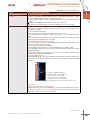
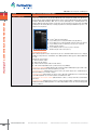
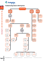
1
Troubleshooting Guide for DNA Digestion
1. FastDigest™ & CONVENTIONAL RESTRICTION ENZYMES
Digestion problem
1
Incomplete
digestion, no
digestion
2
Unexpected
cleavage
pattern
Assess enzyme activity
with control
DNA
1.1
Inactive
enzyme
Active
enzyme
1.2
Suboptimal
reaction
conditions
1.3
Unsuitable or
contaminated
DNA
1.2.2
Improper
enzyme
dilution
Inhibition
1.3.2
Absence of
recognition
sites
1.2.4
Excess
glycerol
1.3.3
Cleavage
blocked by
methylation
1.2.5
Suboptimal
DNA
concentration
1.3.4
Structure of
DNA
substrate
1.3.4.1
Supercoiled
plasmid
DNA
www.fermentas.com
204
No inhibition
1.3.1
Contaminants
in DNA
solution
1.3.4.2
Proximity of
site to DNA
ends
www.fermentas.com/doubledigest
2.1
Star
activity
3.1
Gel shift
2.2
Contamination
with another
RE
3.2
Contaminated
reagents
2.3
Contamination
with another
DNA
2.4
Incomplete
DNA digestion
Evaluate
inhibition by
template DNA
solution
1.2.1
Suboptimal
digestion
protocol
1.2.3
Improper
reaction
assembly
1.4
Water
contains
impurities
3
Diffuse DNA
bands on gel
1.3.4.3
At least two
sites required
www.fermentas.com/research
2.5
Gel shift
2.6
Unexpected
sites in
template DNA
1.3.4.4
Site
preference
www.fermentas.com/reviewer
1.
& CONVENTIONAL RESTRICTION ENZYMES
Troubleshooting Guide for DNA Digestion
Table 1.26. Troubleshooting Guide for DNA Digestion.
Problem
Possible cause and recommended solution
1. Incomplete digestion or no
digestion
Assess enzyme activity
The restriction enzyme may lose activity due to improper storage or handling. Perform a digestion reaction with
1 µg of a standard control DNA, e.g. Lambda DNA (dam–, dcm–) (#SD0021).
1.1. Inactive enzyme.
If the enzyme does not cut the control DNA:
• Check the expiration date.
• Verify that the enzyme has been stored at -20°C.
• Check the temperature of your freezer. Do not allow the temperature go below -20°C as the enzyme may
freeze and multiple freeze thaw cycles (more than 3 cycles) may result in reduced enzyme activity.
1.2. Suboptimal reaction conditions.
1.2.1. Suboptimal digestion protocol.
Follow digestion protocol specified for the restriction enzyme and type of substrate DNA.
• Use the recommended reaction buffer supplied with the restriction enzyme. For double digestions with conventional restriction enzymes, follow the recommendations of the DoubleDigest™ engine at
www.fermentas.com/doubledigest.
• Use additives where required.
• Perform the reaction at the optimal temperature specified for the restriction enzyme; refer to Table 1.8 on
p.162 for data on the activity of thermophilic enzymes at 37°C. For double digestions with mesophilic and
thermophilic conventional restriction enzymes, first digest with the mesophilc enzyme (1 h), then increase the
temperature and incubate for an additional hour.
• Ensure the volume of the reaction mixture was not reduced due to evaporation during incubation; the increase
in salt concentration may reduce enzyme activity. For thermophilic enzymes use a heat block with a hot bonnet,
e.g. a PCR cycler.
1.2.2. Improper enzyme dilution.
• Dilute restriction enzymes with Dilution Buffer for Restriction Enzymes (#B19). Restriction enzymes diluted with
this buffer are stable for at least 3-4 weeks at -20°C (for more information see p.170).
• Never dilute enzymes in water or 10X reaction buffer.
• Never dilute enzymes in 1X reaction buffer in the absence of DNA.
1.2.3. Improper reaction assembly.
• The restriction enzyme should always be the last component added to the reaction mixture.
• The restriction enzyme may be inactivated if added directly to a 10X reaction buffer.
1.2.4. Excess glycerol in the reaction mixture.
• The glycerol concentration in the reaction mixture should not exceed 5%. Thus, the volume of the restriction
enzyme added to the mixture should not exceed 1/10 of the total reaction volume.
• Enzymes sensitive to high glycerol concentration include: Alw21I, BpiI, Bsp68I, BspTI, Eco32I, Eco91I, EcoRI,
Hin6I, HinfI, Mph1103I, Mva1269I and NcoI.
1.2.5. Suboptimal DNA concentration.
The optimal range of DNA concentration in the reaction mixture is 0.02-0.1 µg/µl.
1.3. Unsuitable DNA template or contaminated DNA solution.
If the enzyme is active in the control digest, assay the substrate DNA solution for inhibitory contaminants in a
mixing experiment with control template, e.g. Lambda DNA (dam–, dcm–) (#SD0021). Perform a control digest
with two templates: control template and sample template in one reaction mixture. Do not exceed the optimal
DNA concentration in the reaction mixture (0.02-0.1 µg/µl).
• The sample template is contaminated if neither the control DNA template nor sample template is digested.
(see 1.3.1).
• The sample template is not contaminated if the control DNA template is digested but the sample template is
not. Poor digestion of the experimental template is caused by errors in the DNA sequence (see 1.3.2), methylation effects (see 1.3.3) or structure of the DNA substrate (see 1.3.4)
Note
Always ensure that the control DNA contains a recognition site for the enzyme present in the reaction. For
example, there is no NotI recognition site in lambda DNA.
1
(continued on next page)
Bulk quantities and custom formulations available upon request
205
Table 1.26. Troubleshooting Guide for DNA Digestion.
Possible cause and recommended solution
1. Incomplete digestion or no
digestion
1.3.1. Contaminants in the DNA solution.
• Template DNA may contain residual SDS, EDTA, proteins, salts or nucleases. Repurify the template using a spin
column purification kit or by phenol/chloroform extraction and ethanol precipitation (see.p 356). DNA A260/280
ratio should be 1.8-2.0. To remove EDTA and salts, wash the pellet with 70% cold ethanol.
• For reliable and reproducible plasmid miniprep purity, use the GeneJET™ Plasmid Miniprep Kit (#K0503).
• For digestion of unpurified PCR products, dilute DNA at least 3-fold in the recommended 1X restriction enzyme
buffer see protocols in p.165 or p.43 (for FastDigest™ enzymes).
• If the template DNA has been purified using silica or resin suspensions, remove all remaining particles by
centrifugation for 10 min at 10,000 rpm and ensure that no resin is carried over while transferring the DNA
solution into a new tube.
1.3.2. The substrate DNA does not contain a recognition sequence for the restriction enzyme.
• Re-check the DNA sequence and cloning strategy.
• Determine if the restriction enzyme selected requires more than one site per target DNA for 100% activity (see
also 1.3.4.3).
• Check literature for known site preferences for the restriction enzyme (see also 1.3.4.4).
• If the recognition sequence had been introduced by PCR primers, verify that the primer sequence contains the
recognition site.
1.3.3. Methylation effects.
Restriction enzyme is inhibited by methylation of the recognition site.
1.Identify which type of DNA methylation can occur on the recognition site and determine if the methylation
impairs or blocks DNA digestion with the enzyme. See Digestion of Methylated DNA on p.171 and use the
Tables 1.14-1.21 on pp.172-177.
2.If methylation impairs or blocks DNA cleavage:
• propagate your plasmid in an E.coli dam–, dcm– strain (the E.coli GM2163 dam–, dcm– strain; #M0099, is
available upon request with the purchase of any Fermentas product),
• use the REsearch™ engine at www.fermentas.com/research or check the Fermentas catalog for the availability
of a restriction enzyme isoschizomer not sensitive to DNA methylation.
A restriction enzyme which requires a methylated recognition sequence (DpnI) was used to digest unmethylated DNA.
In the case of DpnI, the neoschizomers Bsp143I or MboI can be used to digest non-methylated DpnI recognition sites. Alternatively, propagate your plasmid in E.coli dam+ strains (most conventional laboratory strains are
dam+. Please refer to p.450 for an overview about the genotypes of some common E.coli strains).
Note
When PCR is carried out with standard dNTPs and non-methylated primers the resulting DNA product is NOT
methylated.
1.3.4. Structure of substrate DNA.
1.3.4.1. Supercoiled plasmid DNA.
Use FastDigest™ enzymes which are qualified for supercoiled DNA and provide specific recommendations for
each enzyme (see Table 1.3 on p.44).
For some conventional restriction enzymes, additional units are required to digest supercoiled plasmids completely (e.g. 5-10 u (1 µl) of restriction enzyme per 1 µg of DNA), check the notes in the catalog description of
the enzyme or refer to the Certificate of Analysis.
1.3.4.2. Proximity of the recognition sequence to the DNA ends.
Some restriction enzymes cleave DNA poorly, if the recognition site is too close to the end of the DNA molecule.
• For FastDigest™ enzymes refer to Table 1.3 on p.44 or the product description to determine the effectiveness
of restriction enzymes at the ends of DNA.
• For conventional restriction enzymes refer to Tables 1.9 (p.162) and Table 1.10 (p.164).
• Consider direct cloning of your PCR product into a cloning vector, e.g. CloneJET™ PCR Cloning Kit (#K1221) or
InsTAclone™ PCR Cloning Kit* (#K1213).
* Available in certain countries only.
1.3.4.3. Restriction Enzyme requires at least two sites per DNA molecule to obtain optimal activity.
Some restriction enzymes such as AarI, BveI, Cfr42I, Eam1104, Eco57I, EcoRII, LweI, SfiI require at least two
target sites per DNA molecule for efficient cleavage (for more details see Site Preferences by Restriction Enzymes on p.202). If there is only one recognition site per DNA molecule, add a DNA oligonucleotide containing
the recognition site.
1.3.4.4. Site Preferences by Restriction Enzymes.
The DNA sequence surrounding the recognition site may influence the efficiency of digestion. Some DNA sites
are cleaved slowly or not cleaved at all (for more details see p.202) due to the surrounding sequence. Use additional units (5-10 u) of the restriction enzyme per 1 µg of DNA or determine if an isoschizomer has superior
cleavage efficiency (see Table 1.27 on p.209 or REsearch™ engine at www.fermentas.com/research).
1. FastDigest™ & CONVENTIONAL RESTRICTION ENZYMES
1
Problem
(continued on next page)
www.fermentas.com
206
www.fermentas.com/doubledigest
www.fermentas.com/research
www.fermentas.com/reviewer
1.
& CONVENTIONAL RESTRICTION ENZYMES
Troubleshooting Guide for DNA Digestion
Table 1.26. Troubleshooting Guide for DNA Digestion.
Problem
Possible cause and recommended solution
1. Incomplete digestion or no
digestion
1.4. Water contains impurities.
Compare you results using commercially available nuclease free, molecular biology grade water, e.g. Water,
nuclease-free (#R0581). Check the quality of the water used in you lab.
• Check the pH and conductivity of water. The pH of high quality water should be 5.5-6.0 with a resistance of
>18 MΩ.
• Centrifuge (10 min, 10,000 rpm) 1 ml of water and check if there is a visible pellet.
• Determine if the water contains nucleases or bacterial contamination (see 3.2 for control reactions).
2. Unexpected cleavage pattern
2.1. Star activity (relaxed specificity) of restriction enzyme (see p.206 for more details).
• Reduce the units of restriction enzyme (not more than 10 u of restriction enzyme or 1 µl of FastDigest™ restriction enzyme per 1 µg DNA).
• Use the recommended reaction buffer.
• Ensure that the glycerol concentration in the reaction mixture does not exceed 5%.
• Reduce the incubation time. For FastDigest™ enzymes – refer to Table 1.3 on p.44 for maximum incubation
times.
• Ensure the volume of the reaction mixture was not reduced due to evaporation during incubation; the resulting
increase in glycerol concentration may cause star activity.
2.2. Contamination with another restriction enzyme.
The restriction enzyme or buffer may be contaminated with another restriction enzyme due to improper handling. Use a new tube of enzyme and/or buffer.
2.3. Contamination with another substrate DNA.
The sample DNA contains a mixture of two or more different DNAs. Prepare new sample of DNA.
• For plasmid DNA preparation pick one isolated colony of recombinant E.coli and purify with GeneJET™ Plasmid
Miniprep Kit (#K0503).
• For PCR products: check the product purity on an agarose gel. If necessary, purify the PCR product prior to
digestion with the DNA Gel Extraction Kit (#K0513).
2.4. Incomplete DNA digestion (see 1).
Different DNA structures like nicked, supercoiled, dimeric molecules will always show different mobility on gels
compared to same size DNA size standards, as an example, see the picture below for migration of plasmid
DNA forms:
1
bp
10000
8000
6000
5000
4000
3500
3000
2500
2000
1500
1000
750
500
250
1
2
3
1
2
–
–
–
3
GeneRuler™ 1 kb DNA Ladder (#SM0311)
Undigested plasmid pUC19 2,7 kb DNA, forms:
upper band (~5 kb) – dimeric plasmid
below, less visible (~4 kb) – nicked plasmid
lowest band (~1.9 kb) – supercoiled plasmid
Linearized plasmid pUC19 (2,7 kb) – migrates according to its size
2.5. Gel shift (see 3.1).
2.6. Unexpected recognition sites in template DNA.
Newly generated target sites in constructed DNA may be overlooked. Recheck your DNA sequence and cloning strategy. Refer to Tables 1.23, 1.24 or 1.25 for Newly Generated Recognition Sequences (pp.186-201) to
identify all the cleavage sites present in the substrate DNA.
(continued on next page)
Bulk quantities and custom formulations available upon request
207
Table 1.26. Troubleshooting Guide for DNA Digestion.
Possible cause and recommended solution
3. Diffused DNA bands
3.1. Gel shift.
Enzyme that remains bound to the substrate DNA will affect the electrophoretic mobility of the digestion products. Restriction enzymes AarI, AloI, BdaI, BseXI, BveI, CseI, Eco57I, Eco57MI, EcoRII, FaqI, GsuI, LweI, MboII,
FastDigest™ MboII, MnlI, FastDigest™ MnlI, SchI, TsoI, TstI are particularly prone to remaining bound to the
substrate DNA. This will result in a band or smear above the expected band (see picture below). Use 6X DNA
Loading Dye & SDS Solution (#R1151) for sample preparation or heat the digested DNA in the presence of 1X
SDS prior to electrophoresis.
1. FastDigest™ & CONVENTIONAL RESTRICTION ENZYMES
1
Problem
M
2
3
4
3.2. Contaminated reagents.
Any restriction digestion reaction components may become contaminated with nucleases due to improper
handling or storage. Nuclease contamination causes DNA degradation, which appears as diffused DNA bands
on a gel.
Perform four control reactions:
I – without restriction enzyme,
II – with a new vial of buffer,
III – without restriction enzyme, with a new vial of buffer,
IV – with commercially available water e.g. Water, nuclease-free (#R0581).
• Contaminated sample DNA (diffused bands in all controls). Re-purify the DNA sample by spin column or phenol/chloroform extraction and ethanol precipitation (see p.356).
• Contaminated enzyme (diffused bands in controls 2 and 4). The enzyme may become contaminated due to
improper handling. Use a new vial of enzyme.
• Contaminated buffer (diffused bands in controls 1 and 4). Bacterial contamination of the reaction buffer will
cause DNA degradation. Use a new vial of buffer. Store all buffers at -20°C.
• Contamination of both enzyme & buffer (diffused bands in controls 1, 2 and 4). Follow the recommendations
given above.
• Contaminated water (diffused bands in controls 1,2 and 3). Bacterial or DNase contamination in improperly
handled water will cause DNA degradation. Use commercially available nuclease free molecular biology grade
water (e.g. #R0581).
www.fermentas.com
208
1
M – GeneRuler™ DNA Ladder Mix (#SM0331)
1 – 0.5 µg λ DNA prepared for loading with 6X DNA Loading Dye (#R0611)
2 – 0.5 µg λ DNA prepared for loading with 6X DNA Loading Dye & SDS Solution (#R1151)
3 – 0.5 µg λ DNA digested with TsoI (#ER1991), probe prepared for loading with
6X DNA Loading Dye (#R0611)
4 – 0.5 µg λ DNA digested with TsoI, probe prepared for loading with
6X DNA Loading Dye & SDS Solution (#R1151)
www.fermentas.com/doubledigest
www.fermentas.com/research
www.fermentas.com/reviewer