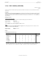
1
User's Guide ™ DIATABS DIAGNOSTIC TABLETS FOR BACTERIAL IDENTIFICATION 7th Ed. 2007 TAASTRUPGAARDSVEJ 30 DK-2630 TAASTRUP DENMARK Phone: +45 43 99 33 77 Telefax: +45 43 52 73 74 e-mail: [email protected] DIAGNOSTICA DIATABS ™ 05-2007 Page 2 of 130 USER’s GUIDE DIATABS™ DIAGNOSTIC TABLETS FOR BACTERIAL IDENTIFICATION 7th Ed. 2007 Contents 1 Introduction ................................................................................................................................................................4 2 Bacterial identification using Diatabs.........................................................................................................................5 3 Diatabs in alphabetical order ......................................................................................................................................9 3.1 ACETAMIDE HYDROLYSIS (ACM)...........................................................................................................9 3.2 ALKALINE PHOSPHATASE (Alk P).........................................................................................................10 3.3 AMINOPEPTIDASES ..................................................................................................................................12 3.3.1 3.3.2 3.3.3 3.3.4 3.3.5 GAMMA-GLUTAMYL AMINOPEPTIDASE (γ-GLU).................................................................13 LEUCINE AMINOPEPTIDASE (LAP) ..........................................................................................16 PROLINE AMINOPEPTIDASE (PRO) ..........................................................................................18 PYRROLIDONYL AMINOPEPTIDASE (PYR) ............................................................................20 TRYPSIN (BAA) (TRYP)................................................................................................................23 3.4 Anaerobes, Presumptive Identification with Oxgall (bile), Brilliant Green and Antibiotic Tablets..............27 3.5 ARGININE DIHYDROLASE (ADH) ..........................................................................................................29 3.6 BACITRACIN LOW (BaL)..........................................................................................................................31 3.7 BACITRACIN 40 UNITS (BACIT) Neo-Sensitabs .....................................................................................32 3.8 BETA LACTAMASE (Acido)......................................................................................................................33 3.9 Beta-lactamases (ESBL, AmpC, MBL) detection using Neo-Sensitabs and Diatabs....................................34 3.10 ESCULIN HYDROLYSIS (ESC) BILE ESCULIN (BE).............................................................................38 3.11 C-390.............................................................................................................................................................41 3.12 CITRATE (CIT)............................................................................................................................................42 3.13 CYCLOHEXIMIDE (CYC)..........................................................................................................................43 3.14 DEFEROXAMINE (DEFRX).......................................................................................................................44 3.15 DOUBLE TEST Diatabs ...............................................................................................................................46 3.15.1 3.15.2 3.15.3 3.15.4 3.15.5 LDC / INDOLE (LDC/IND) ...........................................................................................................47 ODC / INDOLE (ODC/IND) ..........................................................................................................48 PGUA / INDOLE (PGUA/IND) .....................................................................................................50 UREASE / INDOLE (URE/IND)....................................................................................................51 UREASE / TDA (URE/TDA) .........................................................................................................53 3.16 GENTAMICIN 250 µg (GN250), KANAMYCIN 500 µg (KA500), STREPTOMYCIN 500 µg (ST500) Neo-Sensitabs................................................................................................................................................54 3.17 FACTOR X, V, and X+V..............................................................................................................................56 3.18 FOSFOMYCIN 70 µg (FOSFO) Neo-Sensitabs ...........................................................................................58 3.19 FURAZOLIDONE 50 µg (FURAZ) MUPIROCIN 10 µg (MUPIR) Neo-Sensitabs....................................60 3.20 GLYCOSIDASES.........................................................................................................................................62 3.20.1 3.20.2 3.20.3 3.20.4 3.20.5 BETA-N-ACETYLGLUCOSAMINIDASE (ß-NAG) ...................................................................64 ALPHA-FUCOSIDASE (α-FUC) ..................................................................................................66 BETA-FUCOSIDASE (ß-FUC)......................................................................................................69 ALPHA-GALACTOSIDASE (α-GAL)..........................................................................................70 ONPG - Beta-Galactosidase (ONPG) ...........................................................................................71 © Copyright Rosco Diagnostica A/S DIATABS ™ 05-2007 Page 3 of 130 3.20.6 3.20.7 3.20.8 3.20.9 ALPHA GLUCOSIDASE (α-GLU) BETA-GLUCOSIDASE (ß-GLU)........................................73 BETA-GLUCURONIDASE (PGUA).............................................................................................75 ALPHA-MANNOSIDASE (α-MAN) ............................................................................................76 BETA-XYLOSIDASE (ß-XYL).....................................................................................................77 3.21 HIPPURATE HYDROLYSIS (HIP).............................................................................................................79 3.22 INDOXYL ACETATE (IAC) .......................................................................................................................82 3.23 LYSINE DECARBOXYLASE (LDC) ORNITHINE DECARBOXYLASE (ODC) ...................................84 3.24 METRONIDAZOLE 5 µg (MTR.5) .............................................................................................................86 3.25 METRONIDAZOLE 50 µg (MTR50) ..........................................................................................................87 3.26 NITRATE REDUCTION (NO3) ...................................................................................................................88 3.27 NOVOBIOCIN 5 µg (NOVO5) Neo-Sensitabs ............................................................................................90 3.28 O/129 (Vibriostaticum) (O/129)....................................................................................................................92 3.29 OPTOCHIN (OPT) OXGALL (OXG) ..........................................................................................................94 3.30 OXIDASE (OXI)...........................................................................................................................................96 3.31 POLYMYXINS 150 µg (CO150) Neo-Sensitabs .........................................................................................97 3.32 PORPHYRIN (d-Ala) (ALA)........................................................................................................................99 3.33 PS. AERUGINOSA SCREEN (PSAER) ....................................................................................................102 3.34 PYRAZINAMIDASE (PZA) ......................................................................................................................103 3.35 S.P.S. (SPS).................................................................................................................................................105 3.36 SUGAR FERMENTATION Tests (SFT)....................................................................................................107 3.37 TDA or INDOLE (TDA or IND) ................................................................................................................111 3.38 TELLUR (TEL)...........................................................................................................................................113 3.39 TETRATHIONATE REDUCTASE (TTR) ................................................................................................114 3.40 TRIBUTYRIN (TRIB) ................................................................................................................................115 3.41 UREASE (URE)..........................................................................................................................................117 3.42 VOGES-PROSKAUER (VP)......................................................................................................................120 4 Reagents .................................................................................................................................................................123 5 Useful TABLES for bacterial identification / differentiation .................................................................................124 6 Alphabetic INDEX of Abbreviations and Codes....................................................................................................128 © Copyright Rosco Diagnostica A/S DIATABS ™ 05-2007 Ch. 1 Page 4 of 130 1 Introduction DIATABS™ DIAGNOSTIC TABLETS for BACTERIAL IDENTIFICATION The DIATABS™, Diagnostic Tablets (D.T.), developed by Rosco are identification tests made available as individual tablets, which allow the microbiologist a free choice of the most appropriate tests for identification. Most of the Diatabs™ are rapid tests (chromogenic enzymatic reactions, and modified conventional tests). The tablets may be used as single tests to show isolated microbial properties or as part of cost-effective systems of their own. This User's Guide describes more than 80 different types of tests for identification of the clinically important groups of bacteria, and has been written by J.B. Casals on behalf of Rosco Diagnostica. The 7th Ed. 2007 of the DIATABS™ User's Guide contains updated text, tables and references, all necessary information when using Diatabs tablets for identification of bacteria and yeasts. A new chapter have been added with information how beta-lactamases (ESBL, AmpC, MBL) may be detected by phenotypical profiles using Neo-Sensitabs and Diatabs. There are new tables including new described species, such as Staphylococcus pseudintermedius, Streptococcus pseudopneumoniae and new genus such as Aggregatibacter and Parabacteroides. Finally, we would like to quote The Manual of Clinical Microbiology 8th Ed. 2003, page 893: “Individually available tablets … (Rosco Diagnostic Tablets …) are much cheaper than commercial kits, they can be applied in a number of situations and allow flexibility in tailoring the set to best suit special needs”. The User's Guide is available at our website www.rosco.dk and updated information is continuously included. ROSCO Diagnostica A/S is welcoming any feedback and questions on bacterial identification from users directly ([email protected]) or through our representatives. ROSCO DIAGNOSTICA A/S © Copyright Rosco Diagnostica A/S DIATABS ™ 05-2007 Ch. 2 Page 5 of 130 2 Bacterial identification using Diatabs Rosco Item No. Diatabs Use Chapter 55721 52011 55921 50111 50211 50411 50711 52121 56211 Acetamide Hydrolysis (25) Adonitol (50) Alkaline Phosphatase (25) Alpha-Fucosidase (50) Alpha-Galactosidase (50) Alpha-Glucosidase (50) Alpha Mannosidase (50) l-Arabinose (25) Arginine Dihydrolase (ADH) (50) Non-Fermenters Enterobacteriaceae/Staphylococci etc. Staphylococci/Anaerobes/Gemella Anaerobes/Streptococci Non-fermenters/Streptococci/Anaerobes Non-fermenters/Anaerobes/Gardnerella Listeria spp. /Arcanobacterium/Actinomyces Enterobacteriaceae/Staphylococci etc. Staphylococci/Streptococci/Non-fermenters/ Vibrionaceae/Lactic bacteria (Vanco R) 3.1 3.36 3.2 3.20.2 3.20.4 3.20.6 3.20.8 3.36 3.5 40211 70812 50021 59921 50311 Bacitracin Low (50) Bacitracin 40 U Neo-Sensitabs (50) Beta-N-Acetylglucosaminidase (25) Beta-Fucosidase (25) Beta-Galactosidase (ONPG) (50) 3.6 3.7 3.20.1 3.20.3 3.20.5 50511 50611 Beta-Glucosidase (50) Beta-Glucuronidase (PGUA) (50) 45521 50811 Beta-Lactamase (25) Beta-Xylosidase (50) 40421 10041 40511 Bile Esculin (25) Boronic Acid (25) Brilliant Green (50) Group A-streptococci/Gardnerella Screening Haemophilus spp. Anaerobes/Streptococci/Actinomyces Streptococcus anginosus group Neisseria/Enterobacteriaceae/Non-Fermenters/ Anaerobes/Actinobacillus/Pasteurella Staphylococci/Streptococci E.coli/Enterobacteriaceae/Anaerobes/ Streptococci/Arcanobacterium Haemophilus/Neisseria/Staphylococci Enterobacteriaceae/Capnocytophaga/ Acinetobacter Enterococci, Lactic bacteria (Vanco R) Detection of AmpC Anaerobes 41611 C-390 40 µg (50) Cellobiose (25) Citrate (50) Cloxacillin 500 µg (25) Colistin 10 µg (50) Cycloheximide (50) Pseudomonas aeruginosa Enterobacteriaceae/Staphylococci etc. Enterobacteriaceae/Non-Fermenters Detection of AmpC Anaerobes/Neisseria/Non-Fermenters Candida spp. 3.11 3.36 3.12 3.9 3.4 3.13 59611 Deferoxamine 250 µg (50) Dipicolinic Acid Dulcitol (25) Staph. epidermidis/Staph. hominis, Non-Fermenters Detection of MBL Enterobacteriaceae/Staphylococci etc. 3.14 3.9 3.36 56611 42611 42511 42711 74212 Esculin Hydrolysis (50) Factor V (50) Factor X (50) Factor X+V (50) Fosfomycin Neo-Sensitabs (50) Fructose (25) Furazolidone Neo-Sensitabs (50) Streptococci/Enterococci/Yersinia Haemophilus Haemophilus Haemophilus Staphylococci, Corynebacteria Enterobacteriaceae/Staphylococci etc. Staphylococci/Micrococci/Enterococci/ Corynebact. 3.10 3.17 3.17 3.17 3.18 3.36 3.19 56511 10031 41811 58921 74412 The numbers in brackets indicate the number of tablets per vial/cartridge. © Copyright Rosco Diagnostica A/S 3.20.6 3.20.7 3.8 3.20.9 3.10 3.9 3.4 DIATABS ™ 05-2007 Ch. 2 Page 6 of 130 Rosco Item No. Diatabs Use Chapter 46711 43012 52611 Galactose (25) Gamma-Glutamyl Aminopeptidase (50) Gentamicin 250 µg Neo-Sensitabs (50) Glucose (50) Enterobacteriaceae/Staphylococci etc. Meningococci/Helicobacter HLR enterococci Enterobacteriaceae/Staphylococci etc. 3.36 3.3.1 3.16 3.36 56711 Hippurate Hydrolysis (50) Campylobacter/Gardnerella/Streptococci/ Facklamia/Abiotrophia 3.21 59551 Campylobacter/Helicobacter 52711 Indoxyl Acetate (15) Inositol (25) Inulin (50) Enterobacteriaceae/Staphylococci etc. 3.22 3.36 3.36 43112 Kanamycin 500 µg Neo-Sensitabs (50) Anaerobes/HLR enterococci 3.16 52811 58411 46811 Lactose (50) LDC/Indole (50) Leucine Aminopeptidase (50) 3.36 3.15.1 3.3.2 56811 Lysine Decarboxylase (LDC) (50) Enterobacteriaceae/Staphylococci etc. Enterobacteriaceae/Salmonella ID Campylobacter/Corynebacteria/ CAT neg. Gram+cocci Enterobacteriaceae/Vibrionaceae/ Corynebacteria 52911 53011 53111 53211 59711 43611 75712 Maltose (50) Mannitol (50) Mannose (50) Melibiose (50) Metronidazole 5 µg (50) Metronidazole 50 µg (50) Mupirocin 10 µg Neo-Sensitabs (50) Enterobacteriaceae/Staphylococci etc. Enterobacteriaceae/Staphylococci etc. Enterobacteriaceae/Staphylococci etc. Enterobacteriaceae/Staphylococci etc. Anaerobes Gardnerella Staphylococci/Micrococci/Enterococci/ Corynebact. 3.36 3.36 3.36 3.36 3.24 3.25 3.19 43711 46312 Nitrate Reduction (50) Novobiocin 5 µg Neo-Sensitabs (50) Staphylococci/Non-Fermenters, Anaerobes Staphylococci/Peptostrep./Pediococci 3.26 3.27 45411 59121 50311 O/129 (Vibriostaticum) 150 µg (50) ODC/Indole (25) ONPG (Beta Galactosidase) (50) 3.28 3.15.2 3.20.5 44211 57011 Optochin (50) Ornithine Decarboxylase (ODC) (50) 44311 45711 Oxgall (Bile) (50) Oxidase (50) Vibrionaceae, Corynebacteria Enterobacteriaceae/Citrobacter spp. Neisseria/Enterobacteriaceae/Non-Fermenters/ Anaerobes/Actinobacillus/Pasteurella Pneumococci Staph. lugdunensis/Enterobacteriaceae/ Haemophilus/Corynebacteria Anaerobes Enterobacteriaceae/Non-Fermenters/Neisseria 59011 77512 57321 46911 59311 59811 47011 PGUA/Indole (50) Polymyxins 150 µg Neo-Sensitabs (50) Porphyrin (d-ALA) (25) Proline Aminopeptidase (50) Ps. aeruginosa Screen (50) Pyrazinamidase (50) Pyrrolidonyl Aminopeptidase (50) E. coli Staph. aureus/Shewanella/Kingella Haemophilus/Gram positive cocci Neisseria/Anaerobes/Clostridium difficile Ps. aeruginosa Corynebacteria/Yersinia enterocolitica Group A-streptococci/Enterobacteriaceae/ Peptostreptococci/Staphylococci/ Lactic bacteria (Vanco R) 3.15.3 3.31 3.32 3.3.3 3.33 3.34 3.3.4 The numbers in brackets indicate the number of tablets per vial/cartridge. © Copyright Rosco Diagnostica A/S 3.23 3.29 3.23 3.29 3.30 DIATABS ™ 05-2007 Ch. 2 Page 7 of 130 Rosco Item No. Diatabs Use Chapter Raffinose (50) l-Rhamnose (50) Ribose (25) Enterobacteriaceae/Staphylococci etc. Enterobacteriaceae/Staphylococci etc. Enterobacteriaceae/Staphylococci etc. 3.36 3.36 3.36 53711 44611 44712 53811 Salicin (25) Sorbitol (25) S.P.S. (50) Streptomycin 500 µg Neo-Sensitabs (50) Sucrose (25) Enterobacteriaceae/Staphylococci etc. Enterobacteriaceae/Staphylococci etc. Gardnerella/Peptostreptococci HLR enterococci Enterobacteriaceae/Staphylococci etc. 3.36 3.36 3.35 3.16 3.36 57811 TDA or Indole (50) 3.37 45011 57421 53911 48821 Tellur (50) Tetrathionate Reductase (25) Trehalose (50) Tributyrin (25) 47211 Trypsin (50) Enterobacteriaceae/Anaerobes/ Actinobacillus/Pasteurella Enterococcus faecalis Enterobacteriaceae/Non-Fermenters Enterobacteriaceae/Staphylococci etc. Moraxella catarrhalis/Non-Fermenters/ Corynebacteria Non-Fermenters/Anaerobes/ Capnocytophaga 57511 Urease (50) 57611 Urease/Indole (50) 57911 Urease/TDA (50) 79312 57711 Vancomycin 5 µg Neo Sensitabs (50) Voges-Proskauer (50) Anaerobes/Enterococci Enterobacteriaceae/Streptococci/ Staphylococci 3.4 3.42 54021 d-Xylose (25) Enterobacteriaceae/Staphylococci etc. 3.36 Meningococci/Helicobacter Campylobacter/Corynebacteria/ CAT neg Gram+cocci Clostridium difficile/Neisseria/ Peptostreptococci Group A-streptococci/Enterobacteriaceae/ Peptostreptocci/Staphylococci/Enterococci/ Lactic bacteria (Vanco R) Non-Fermenters/Porphyromonas/ Capnocytophaga 3.3.1 3.3.2 53311 53411 Enterobacteriaceae/Staphylococci/ Anaerobes/Non-Fermenters Enterobacteriaceae/Anaerobes/ Actinobacillus/Pasteurella Enterobacteriaceae 3.38 3.39 3.36 3.40 3.3.5 3.41 3.15.4 3.15.5 AMINOPEPTIDASES: 46711 46811 Gamma-Glutamyl Aminopeptidase (50) Leucine Aminopeptidase (50) 46911 Proline Aminopeptidase (50) 47011 Pyrrolidonyl Aminopeptidase (50) 47211 Trypsin (50) 3.3.3 3.3.4 3.3.5 DOUBLE TEST TABLETS: 58411 59121 59011 57611 LDC/Indole (50) ODC/Indole (25) PGUA/Indole (50) Urease/Indole (50) 57911 Urease/TDA (50) Enterobacteriaceae Enterobacteriaceae/Citrobacter spp. E. coli Enterobacteriaceae/Anaerobes/ Actinobacillus/Pasteurella Enterobacteriaceae The numbers in brackets indicate the number of tablets per vial/cartridge. © Copyright Rosco Diagnostica A/S 3.15.1 3.15.2 3.15.3 3.15.4 3.15.5 DIATABS ™ 05-2007 Ch. 2 Page 8 of 130 Rosco Item No. Diatabs Use Chapter Campylobacter/Helicobacter Moraxella catarrhalis/Non-Fermenters/ Corynebacteria 3.22 3.40 Anaerobes/Streptococci/Actinomyces Streptococci/Prevotella/Porphyromonas Streptococcus anginosus group Streptococci/Prevotella/Clostridia Neisseria/Enterobacteriaceae/ Non-Fermenters/Anaerobes/ Actinobacillus/Pasteurella Non-Fermenters/Gardnerella/Anaerobes Staphylococci/Streptococci E. coli/Anaerobes/Streptococci/ Arcanobacterium Listeria spp./Arcanobacterium/ Actinomyces Enterobacteriaceae/Capnocytophaga/ Acinetobacter/Propionibacteria. 3.20.1 3.20.2 3.20.3 3.20.4 3.20.5 Enterobacteriaceae/Staphylococci etc. Enterobacteriaceae/Staphylococci etc. Enterobacteriaceae/Staphylococci etc. Enterobacteriaceae/Staphylococci etc. Enterobacteriaceae/Staphylococci etc. Enterobacteriaceae/Staphylococci etc. Enterobacteriaceae/Staphylococci etc. Enterobacteriaceae/Staphylococci etc. Enterobacteriaceae/Staphylococci etc. Enterobacteriaceae/Staphylococci etc. Enterobacteriaceae/Staphylococci etc. Enterobacteriaceae/Staphylococci etc. Enterobacteriaceae/Staphylococci etc. Enterobacteriaceae/Staphylococci etc. Enterobacteriaceae/Staphylococci etc. Enterobacteriaceae/Staphylococci etc. Enterobacteriaceae/Staphylococci etc. Enterobacteriaceae/Staphylococci etc. Enterobacteriaceae/Staphylococci etc. Enterobacteriaceae/Staphylococci etc. Enterobacteriaceae/Staphylococci etc. Enterobacteriaceae/Staphylococci etc. 3.36 3.36 3.36 3.36 3.36 3.36 3.36 3.36 3.36 3.36 3.36 3.36 3.36 3.36 3.36 3.36 3.36 3.36 3.36 3.36 3.36 3.36 ESTERASES/LIPASES: 59551 48821 Indoxyl Acetate (15) Tributyrin (25) GLYCOSIDASES: 50021 50111 59921 50211 50311 Beta-N-Acetylglucosaminidase (25) Alpha-Fucosidase (50) Beta-Fucosidase (25) Alpha-Galactosidase (50) ONPG (Beta Galactosidase) (50) 50411 50511 50611 Alpha-Glucosidase (50) Beta-Glucosidase (50) Beta-Glucuronidase (PGUA) (50) 50711 Alpha-Mannosidase (50) 50811 Beta-Xylosidase (50) 3.20.6 3.20.6 3.20.7 3.20.8 3.20.9 SUGAR FERMENTATION TESTS: 52011 52121 52611 52711 52811 52911 53011 53111 53211 53311 53411 53621 53711 53811 53911 54021 Adonitol (50) l-Arabinose (25) Cellobiose (25) Dulcitol (25) Fructose (25) Galactose (25) Glucose (50) Inositol (25) Inulin (50) Lactose (50) Maltose (50) Mannitol (50) Mannose (50) Melibiose (50) Raffinose (50) l-Rhamnose (50) Ribose (25) Salicin (25) Sorbitol (50) Sucrose (50) Trehalose (50) d-Xylose (25) The numbers in brackets indicate the number of tablets per vial/cartridge. © Copyright Rosco Diagnostica A/S DIATABS ™ 05-2007 Ch. 3 Page 9 of 130 3 Diatabs in alphabetical order 3.1 ACETAMIDE HYDROLYSIS (ACM) REF No. 55721 Test for demonstration of the ability of bacterial strains to hydrolyse acetamide. Mainly used in differentiation of nonfermenting gram-negative rods. Procedure Prepare a dense bacterial suspension (at least McFarland No. 4) of the strain to be tested in 0.25 ml saline in a tube. Add one Acetmide Hydrolysis Diagnostic Tablet and close the tube. Incubate at 35-37 °C for 18-24 hours - some positive reactions may be recorded already after 4-6 hours. Reading of the tests Positive reaction: Negative reaction: Red Yellow, orange Results Acetamide hydrolysis is useful in the differentiation within the fluorescent group of Pseudomonas: Pseudomonas aeruginosa Pseudomonas fluorescens Pseudomonas putida ACM + 0+ 0 For the differentiation of Comamonas acidovorans (+) from Comamonas testosteroni (0). Most strains of Burkholderia cepacia are positive and most strains of St. maltophilia are negative. Most strains of Alcaligenes (faecalis, denitrificans and Achr. xylosoxidans) are positive, while other non-fermenters are negative. Non-fermenters ACM positive Ps. aeruginosa Com. acidovorans Burkh. cepacia Alc. faecalis Alc. denitrificans Achr. xylosoxidans ACM negative Ps. fluorescens Ps. putida Com. testosteroni Sten. maltophilia Quality Control DIATABS (Active ingredients) Acetamide hydrolysis (Acetamide) Positive Ps. aeruginosa ATCC 27853 Negative E. coli ATCC 25922 References 1) Palleroni, N.J.: Pseudomonas in "Bergey's Manual of Systematic Bacteriology", Vol. 1, 141-199, 1984. © Copyright Rosco Diagnostica A/S DIATABS ™ 05-2007 Ch. 3 Page 10 of 130 3.2 ALKALINE PHOSPHATASE (Alk P) REF No. 55921 Contain the chromogenic substrate: 4-nitrophenyl phosphatedi (2-amino-2-ethyl-1,3-propanediol) salt that in the presence of alkaline phosphatase releases free 4-nitrophenol (yellow colour). Procedure Prepare a dense bacterial suspension (at least McFarland No. 4) of the strain to be tested in 0.25 ml saline in a tube. Add one Alkaline Phosphatase Diagnostic Tablet and close the tube. Incubate at 35-37°C for a maximum of 4 hours. Reading of the tests Positive reaction: Negative reaction: Strong yellow Colourless or slight yellow Incubation longer than 4 hours may give a false positive reaction. Results 1) Staphylococci Most strains of S. aureus and S. epidermidis and S. schleiferi show a positive reaction, while most strains of S. hominis, S. haemolyticus, and S. warneri show a negative reaction. S. aureus S. epidermidis S. haemolyticus S. hominis S. lugdunensis S. schleiferi S. warneri HCF 100 0 0 0 87 100 0 Alk P (4h) PYR (1h) + 0 + 0 0 100 0 0+ + 0 100 + 89 0 V ODC 0 0+ 0 0 + 0 0 URE 95 86 0 + 81 0 + DEFRX R (≤14 mm) S (≥16 mm) R S R R R Poly R (≤12 mm) S S (≥14 mm) S S S S 2) Differentiation of Gemella spp. and Rothia mucilaginosa (PYR +, LAP +) Gemella bergeriae Gemella haemolysans Gemella morbillorum Gemella sanguinis Rothia mucilaginosa Alk P 0 + 0 + 0 SUC 0 V + + + SOR 0 0 0+ + 0 NO3 0 0 · 0 + VP 0 V 0 V + BaciLow R R R R S Alk P = Alkaline Phosphatase D.T., PYR (1h) = Pyrrolidonyl Aminopeptidase D.T. (1h incubation), ODC = Ornithine Decarboxylase D.T., DEFRX = Deferoxamine D.T., URE = Urease D.T., Poly = Polymyxins 150 µg Neo-S, MAL = Maltose D.T., SUC = Sucrose D.T., SOR = Sorbitol D.T., HCF = Human clumping factor, NO3 = Nitrate Reduction D.T., VP = Voges Proskauer D.T., BaciLow = Bacitracin Low D.T. (S ≥ 10 mm, R < 10 mm) 3) Useful also in the differentiation of non-fermenters, viridans streptococci, and anaerobes. Quality Control DIATABS (Active ingredients) Alkaline Phosphatase Positive E. coli ATCC 25922 (p-Nitrophenyl-Phosphate) © Copyright Rosco Diagnostica A/S Negative S. haemolyticus ATCC 29970 DIATABS ™ 05-2007 Ch. 3 Page 11 of 130 References 1) Devriese L.A. et al: Streptococcus hyointestinalis sp. nov. from the gut of swine. Intl. J. Syst. Bacteriol. 38, 440-1, 1988. 2) Lindsay J.A., Riley T.V.: Susceptibility to desferrioxamine: a new test for the identification of Staphylococcus epidermidis. J. Med. Microbiol. 35, 45-48, 1991. 3) Collins M.D. et al: Description of Gemella sanguinis sp. nov. isolated from human clinical specimens. J. Clin. Microbiol. 36, 3090-3, 1998. 4) Leung M.J.: Case of Staph. schleiferi endocarditis and a simple scheme to identify clumping factor positive staphylococci. J. Clin. Microbiol. 37, 3353-6, 1999. © Copyright Rosco Diagnostica A/S DIATABS ™ 05-2007 Ch. 3 Page 12 of 130 3.3 AMINOPEPTIDASES General description Bacteria may be differentiated on their ability to enzymatically hydrolyze a series of aminopeptidase substrates. The procedure is based upon the enzymatic liberation of beta naphthylamine (beta-NA) from an l-aminoacid- beta-NA substrate. The liberated beta-NA is identified by its reaction with Aminopeptidase reagent producing a red colour in case of positive reactions. Range The actual range of aminopeptidases (substrates) comprises: Gamma-Glutamyl Aminopeptidase (γ-GLU) (46711) Leucine Aminopeptidase (LAP) (46811) Proline Aminopeptidase (PRO) (46911) Pyrrolidonyl Aminopeptidase (PYR) (47011) Trypsin (BAA) (TRYP) (47211) Procedure Prepare a dense “milky” bacterial suspension (at least McFarland No. 4) from the strain to be tested in 0.25 ml saline in a tube. Add one tablet of aminopeptidase substrate and close the tube. Incubate at 35-37 °C for 4 hours. In som cases, overnight incubation is required. After incubation add 3 drops of Aminopeptidase reagent (92231) and read the colour reaction within 5 minutes. Reading of the tests Positive reaction: Negative reaction: 4h Red/orange Yellow Overnight Red Yellow/orange The test may also be read by exposing the tube (no reagent added) to a Wood’s lamp (at 360 nm). A blue fluorescence in the supernatant indicates a positive reaction. References General 1) Peterson E.H., Hsu E.J.: Rapid detection of selected gram-negative bacteria by aminopeptidase profiles. J. Food Sci. 43, 1853-1856, 1978. 2) Watson R.R.: Substrate specificities of aminopeptidases: a speci fic method for microbial differentiation. Methods Microbiol. 9, 1-4, 1976. 3) Euzeby J.P.: Activité peptidasique vis a vis des aminoacyl-beta-naphtilamides de quelques especies du genre Bartonella. Dictionaire de Bacteriologie Veterinaire, Sept. 1999. © Copyright Rosco Diagnostica A/S DIATABS ™ 05-2007 Ch. 3 Page 13 of 130 3.3.1 GAMMA-GLUTAMYL AMINOPEPTIDASE (γ-GLU) REF No. 46711 The test is based on enzymatic release of beta-naphthylamine from the gamma-glutamyl-beta-naphthylamide substrate. Beta-naphthylamine is detected by its reaction with Aminopeptidase reagent giving a red colour. Procedure Prepare a dense bacterial suspension with a turbidity of at least McFarland No. 4 of the strain to be tested in 0.25 ml saline in a tube. Add one Gamma-glutamyl aminopeptidase tablet and close the tube. Incubate at 35-37 °C for 4 hours. After incubation add 3 drops Aminopeptidase reagent (92231) and read the colour reaction within 5 minutes. See also Aminopeptidase, general description, page 12. Reading of the tests (4h) Positive reaction: Negative reaction: Red/orange Yellow Results 1) Neisseria/Moraxella (HTM + (V), OXI +, CAT +0) N. menigitidis N. gonorrhoeae N. lactamica N. cinerea N. polysaccharea M. catarrhalis Kingella denitrificans γ-GLU + 0 0 0 0 0 0 NA35 0+ 0 +0 + + + · SUP V + V 0 0 + 0 TRIB 0 0 0 0 0 + 0 Remarks Co 10 R ONPG + Co 10 S Co 10 R NO3 + Co 10 R, NO3 +, CAT 0 Most N. gonorrhoeae are PRO (proline aminopeptidase) positive, but approx. 5 % are negative (5). 2) Acinetobacter A. baumanii/calcoaceticus A. lwoffi γ-GLU +0 0 β-XYL + 0 γ-GLU + 0 0 IAC 0 0wk + 3) Helicobacter Helicobacter pylori Helicobacter cinaedi Helicobacter fennelliae γ−GLU = Gamma-Glutamyl Aminopeptidase D.T., NA35 = Growth in nutrient agar at 35 °C, SUP = Superoxol (30 % H2 O2) hydrogen peroxide, TRIB = Tributyrin D.T., HTM = Growth on Modified Thayer Martin medium, OXI = Oxidase D.T., CAT = Catalase, β-XYL = Beta-Xylosidase D.T., IAC = Indoxyl Acetate D.T., Co 10 = Colistin 10 µg (S≥10 mm). © Copyright Rosco Diagnostica A/S DIATABS ™ 05-2007 Ch. 3 Page 14 of 130 4a) Identification of clinically most common Nocardia spp. (3). 4 hours' tests except URE PYR 0 0 0 + γ-GLU + + + + N. nova + 0 + N. paucivorans 0 + 0 N. abscessus * N. brasiliensis N. cyriacigeorgica * N. farcinica α-GLU α-MAN + 0 + + + 0 + 0 URE + + 0 +0 45 °C 0 0 + + 0 V 0+ 0 0 + Remarks Imip S/R Imip R AMC I/R AMC S, Tobra R, Ery R Tobra I/R, AMC I/R, Ery S *) Members of the N. asteroides complex. PYR = Pyrrolidonyl Aminopeptidase D.T., γ-GLU = Gamma-Glutamyl-Aminopeptidase D.T., α-GLU = AlphaGlucosidase D.T., α-MAN = Alpha-Mannosidase D.T., URE = Urease D.T., 45 °C = growth at 45 °C. 4b) Identification of Nocardia spp. by antibiogram (6) N. abscessus N. brasiliensis N. cyriacigeorgica N. farcinica N. nova AMOX S RS V R S AMC S S I/R S I/R CEFTR S V S R V IMIP V R S S S CLARI TOBRA V S R S R S R R S V AMOX = Amoxycillin Neo-S, AMC = Amoxycillin+Clavulanate Neo-S, CEFTR = Ceftriaxone Neo-S, IMIP = Imipenem Neo-S, CLARI = Clarithromycin Neo-S, Tobra = Tobramycin Neo-S, Ery = Erythromycin Neo-S. 5) Salmonella and Shigella (4) serotypes S. typhimurium S. enteritidis S. blegdam S. berta S. gallinarum/pullorum S. typhi S. arizonae S. diarizonae S. dublin S. naestved S. kiel Shigella dysenteriae - serotypes 3 to 9 - serotypes 1, 2 and 10 Shigella sonnei Salmonella serotype III a Salmonella serotype III b γ-GLU Remarks 80 % + + + + + 0 PGUA 0 + PGUA + 0 0 0 + 0 0 0 + γ-GLU = Gamma-Glutamyl-Aminopeptidase D.T. Quality Control DIATABS (Active ingredients) Gamma-Glutamyl Aminopeptidase Positive P. aeruginosa ATCC 27853 (Gamma-Glutamyl-β-Naphthylamide) © Copyright Rosco Diagnostica A/S Negative E. coli ATCC 25922 DIATABS ™ 05-2007 Ch. 3 Page 15 of 130 References (γ-GLU) 1) Mc Nulty C.A.M et al: Rapid identification of Campylobacter Pylori by preformed enzymes. J. Clin. Microbiol. 25, 1683-6, 1987. 2) Nebreda T. et al: Urethritis caused by Neiss. meningitidis serogroup C. Clin. Microbiol. Infect. 5, 57-8, 1999. 3) Wauters G et al: Distribution of Nocardia species in clinical samples and their routine rapid identification in the laboratory. J. Clin. Microbiol. 43, 2624-8, 2005. 4) Giammanco G. et al: Interet taxonomique de la recherche de la γ-glutamyltransferase chez les Enterobacteriaceae. Ann Microbiol. (Inst. Pasteur) 131A, 181-7, 1980. 5) Blackmore T. et al: Characterization of prolyl-iminopeptidase-deficient N. gonorrhoeae. J. Clin Microbiol., 43, 4189-90, 2005. 6) Glupczynski Y. et al: Determination of antimicrobial susceptibility patterns of Nocardia spp. from clinical specimens by E-test. Clin Microbiol. Infect. 12, 905-912, 2006. © Copyright Rosco Diagnostica A/S DIATABS ™ 05-2007 Ch. 3 Page 16 of 130 3.3.2 LEUCINE AMINOPEPTIDASE (LAP) REF No. 46811 The test is based on enzymatic release of beta-naphthylamine from the 1-leucine-beta-naphtahylamid substrate. Betanaphthylamine is detected by its reaction with Aminopeptidase reagent giving a red colour. Procedure Prepare a dense bacterial suspension with a turbidity of at least McFarland No. 4 of the strain to be tested in 0.25 ml saline in a tube. Add one Leucine aminopeptidase tablet and close the tube. Incubate at 35-37 °C for 4 hours. After incubation add 3 drops of Aminopeptidase Reagent (92231) and read the colour reaction within 5 minutes. See also Aminopeptidase, general description, page 12. Reading of the tests (4h) Positive reaction: Negative reaction: Red/orange Yellow Results 1) Catalase negative gram-positive cocci LAP + + 0 + 0wk + + Streptococcus spp. Enterococcus spp. Aerococcus viridans Aerococcus urinae Leuconostoc spp. Pediococcus spp. Dolosigranulum pigrum PYR 0 + + 0 0 0 + Van5 S SR S S R R S ADH V +0 0 0 0 +0 V BE 10 99 60 0 90 + 0 Remarks PGUA + LAP = Leucine Aminopeptidase D.T., PYR = Pyrrolidonyl Aminopeptidase D.T., Van5 = Vancomycin 5 µg Neo-S (S ≥15 mm, R ≤13 mm), ADH = Arginine Dihydrolase D.T., BE = Bile Esculin D.T., PGUA = Beta-Glucuronidase D.T. 2) Corynebacteria nonlipophilic fermentative (most common) Most strains are: CAT +, MOT 0, Fosfo R, Mupi R, PRO +0, COL R, NALI R. C. amycolatum * C. argentoratense C. coylae C. diphtheriae C. glucuronolyticum * C. kutscheri C. minutissinum C. pseudotuberculosis C. renale group C. striatum * C. ulcerans C. xerosis PZA + + + 0 + +0 + 0 +0 + 0 + LAP 0 82 · V + + + 0 0 82 62 88 URE V 0 0 0 67 + 0 + + 0 + 0 NO3 +0 0 0 +0 V +0 0 V 0 + 0 60 * Resistant or multidrugresistant © Copyright Rosco Diagnostica A/S MAL 80 0 0 + 26 + + + 0 0 + + O/129 R S R S S · S R · S V S Remarks dry CAMP 0 CAMP + PGUA+, CAMP + PYR + NAG +0 CAMP rev, α-GLU 0+ PGUA + creamy CAMP rev, α-GLU + dry yellowish, α-GLU +0 DIATABS ™ 05-2007 Ch. 3 Page 17 of 130 3) Corynebacteria nonlipophilic nonfermentative Most strains are: CAT +, MOT 0, Fosfo R, Mupi R, PRO +0. C. afermentans (ANF-1) C. auris Turicella otididis (ANF-1 like) C. propinquum C. pseudodiphtericum LAP 0 + + NO3 0 0 0 60 +0 + + DNase 0 0 + Colonies smooth dry creamy Remarks URE + PZA = Pyrazinamidase D.T., LAP = Leucine Aminopeptidase D.T., NO3 = Nitrate Reduction D.T., MAL = Maltose D.T., O/129=O/129 150 µg D.T. (S ≥ 16 mm, R < 16 mm), NAG = Beta-N-acetylglucosominidase D.T., DNase, URE = Urease D.T., CAT = Catalase, MOT = motility, Fosfo = Fosfomycin Neo-S (R = no zone), Mupi = Mupirocin Neo-S (R = no zone). 4) Globicatella and Aerococcus (3) G. sanguinis A. viridans Enteroc. avium Strept. uberis Gram stain pairs/chains clusters/tetrads short chains short chains PYR 75 100 95 100 LAP 0 0 89 100 Inulin 93 7 7 100 PYR = Pyrrolidonyl Aminopeptidase D.T., LAP = Leucine Aminopeptidase D.T., Inulin D.T. 5) Aerococci (4) (Vancomycin S, CAT neg. cocci, dividing in 2 planes) Aer. viridans Aer. urinae Aer. sanguinicola Aer. christensenii Aer. urinaehominis PYR + 0 + 0 0 LAP 0 + + + 0 VP 0+ 0 0 + 0 MAL V 0 + 0 + SUC + + + 0 + PYR = Pyrrolidonyl Aminopeptidase D.T., LAP = Leucine Aminopeptidase D.T., VP = Voges-Proskauer D.T., MAL = Maltose D.T., SUC = Sucrose D.T. Quality Control DIATABS (Active ingredients) Leucine Aminopeptidase Positive S. bovis ATCC 15351 Negative Aerococcus viridans ATCC 700406 (L-Leucine-β-naphthylamide-HCL) References (LAP) 1) Devriese L.A. et al: Identification of Enterococcus species isolated from foods of animal origin. Intl. J. Food Microbiol. 26, 187-97, 1995. 2) Renaud F.N.R. et al: Identification of Turicella otitidis isolated from a patient with otorrhea associated with surgery: differentation from Coryneb. afermentans and Coryneb. auris. J. Clin. Microbiol. 34, 2625-7, 1996. 3) Lynn Shewmaker P. et al: DNA relatedness, phenotypic characteristics and antimicrobial susceptibilities of Globicatella sanguinis strains. J. Clin. Microbiol. 39, 4052-7, 2001. 4) Facklam R. et al: Phenotypic description and antimicrobial susceptibilities of Aerococcus sanguinicola isolates from human clinical samples. J. Clin. Microbiol. 41, 2587-92, 2003. 5) Christensen J.J. et al: Aerococcus urinae: polyphasic characterization of the species. APMIS, 113, 517-525, 2005. © Copyright Rosco Diagnostica A/S DIATABS ™ 05-2007 Ch. 3 Page 18 of 130 3.3.3 PROLINE AMINOPEPTIDASE (PRO) REF No. 46911 The test is based on enzymatic release of beta-naphthylamine from the l-proline-beta-naphthylamide substrate. Betanaphthylamine is detected by its reaction with Aminopeptidase reagent giving a red colour. Procedure Prepare a dense bacterial suspension with a turbidity of at least McFarland No. 4 of the strain to be tested in 0.25 ml saline in a tube. Add one Proline aminopeptidase tablet and close the tube. Incubate at 35-37 °C for 4 hours. After incubation add 3 drops of Aminopeptidase Reagent (92231) and read the colour reaction within 5 minutes. See also Aminopeptidase, general description, page 12. Reading of the tests (4h) Positive reaction: Negative reaction: Red/orange Yellow Results 1a) Identification of Clostridium difficile Most clostridia are: Kana 500 SR, Vanco 5 S, Col R, CAT 0. PRO C. difficile + C. inocuum * 0 C. perfringens C. ramosum C. sordelli C. bifermentans C. septicum 0 0 + + 0 CCFA Remarks growth + ONPG 0, PYR 0, Alk P 0, Amox S, Merop S, Imipenem I/R + Vanco I/R, Teico S (van B) ONPG +, PYR +, Alk P + ⋅ ⋅ 0 0 ⋅ PRO = Proline Aminopeptidase D.T., CCFA growth = Growth on CCFA medium, ONPG = ONPG BetaGalactosidase D.T., Alk P = Alkaline Phosphatase D.T., PYR = Pyrrolidonyl Aminopeptidase D.T., Amox = Amoxycillin Neo-S, Merop = Meropenem Neo-S, Imipenem = Imipenem Neo-S, Vanco = Vancomycin Neo-S, Teico = Teicoplanin Neo-S. * C. inocuum shows intrinsic low level resistance to vancomycin (MIC 8-16 µg/ml) with Van 5 zones < 18 mm. 1b) Rapid ID of common lecithinase positive Clostridium spp. C. novyi type A C. perfringens C. bifermentans C. sordelli IND 0+ 0 + + URE 0 0 0 + PRO 0 0 + + NAG 0 + V 0 Remarks swarm PYR + ESC 0 + + 0 PRO 0 0 + 0 LIP + 0 + 0 Remarks Strong beta haem. 1c) Rapid ID of swarming clostridia C. novyi type A C. septicum C. sporogenes C. tetani IND 0+ 0 0 +0 URE = Urease D.T., ESC = Esculin Hydrolysis D.T., LIP = Lipase. © Copyright Rosco Diagnostica A/S PYR +0, NAG 0 DIATABS ™ 05-2007 Ch. 3 Page 19 of 130 1d) Differentiation among Clostridia producing large cytotoxins C. difficile C. sordelli C. novyi A PRO + + 0 IND 0 + 0+ URE 0 + 0 LEC 0 + + Swarm 0 0 + IND = Indole D.T., URE = Urease D.T., LEC = Lecithinase. 2) Differentiation of Peptostreptococci and similar (most current clinical isolates) P. anaerobius P. asaccharolyticus M. micros F. magna P. stomatis A. vaginalis P. harei PRO + 0 +0 0 0 0 0 PYR 0 0 + + 0 0 0 GLU + 0 0 0 + + 0 α-GLU + 0 0 0 + V 0 IND 0 +0 0 0 0 0 0 SPS Alk P 0 S (≥12 mm) R + R + R V 0 S (≥15 mm) R V R 0 Remarks LAP + PRO = Proline Aminopeptidase D.T., PYR = Pyrrolidonyl Aminopeptidase D.T., α-GLU = Alpha-Glucosidase D.T., IND = Indole D.T., SPS = SPS D.T., GLU = Glucose D.T., Alk P = Alkaline Phosphatase D.T. 3) Identification of Candida albicans (4 hours incubation) (5) Candida albicans C. dublinensis Candida spp. (A) Candida spp. (B) PRO NAG + + + 0 +0 +0 0 0 α-GLU (2h) + 0 - 42 °C + 0 - where (A) comprises: C. guilliermondii, C. lipolytica, C. lusitaniae, C. norvegensis, C. parapsilosis, Tor. candida. where (B) comprises: C. glabrata, C. kruseii, C. pseudotropicalis, C. rugosa (NAG 0+), C. tropicalis (NAG 0+). PRO = Proline Aminopeptidase D.T., NAG = Beta-N-Acetylglucosaminidase D.T., α-GLU(2h) = AlphaGlucosidase D.T. (2 hours' incubation), 42 °C = Growth at 42 ° in Sabouraud Glucose Agar. Quality Control DIATABS (Active ingredients) Proline Aminopeptidase Positive P. aeruginosa ATCC 27853 Negative Cl. perfringens ATCC 12917 (L-proline ß-Naphthylamide-HCl) References (PRO) 1) Garcia A, Garcia T, Pérez J.L.: Proline aminopeptidase test for rapid screening of Clost. difficile. J. Clin. Microbiol. 35, 3007, 1997. 2) Fedorko D.F. et al: Use of cycloserine-cefoxitin-fructose-agar (CCFA) and l-proline aminopeptidase in the rapid identification of Clostridium difficile. J. Clin. Microbiol. 35, 1258-9, 1997. 3) Bourgault A.M. et al: Should all stool specimens be routinely tested for Clost. difficile. Clin. Microbiol. Infect. 5, 219-22, 1999. 4) Murdoch D.A.: Gram-positive anaerobic cocci. Clin. Microbiol. Reviews. 11, 81-120, 1998. 5) Niimi K. et al: Distinguishing Candida species by ß-N-acetylhexosaminidase activity. J. Clin. Microbiol. 39, 2089-97, 2001. 6) Yuli Song et al: Development of a flow chart for identification of gram-positive anaerobic cocci in the clinical laboratory. J. Clin. Microbiol., 45, 512-516, 2007. © Copyright Rosco Diagnostica A/S DIATABS ™ 05-2007 Ch. 3 Page 20 of 130 3.3.4 PYRROLIDONYL AMINOPEPTIDASE (PYR) REF No. 47011 Some bacteria may be differentiated by their ability to enzymatically hydrolize an 1-pyrrolidonyl-beta-naphthylamide substrate. The liberated beta-naphthylamine is identified by reaction with Aminopeptidase reagent producing a red colour in case of positive reactions. Procedure Prepare a dense “milky” suspension (at least McFarland No. 4) of the strain to be tested in 0.25 ml saline in a tube. Add one PYR Diagnostic Tablet, close the tube and incubate at 35-37 °C for 4 hours or up to 18-24 hours. In special cases an incubation period of 1 or 2 hours is used. After incubation add 3 drops of Aminopeptidase Reagent (92231) and read the colour reaction within 5 minutes. Reading of the test (4h) Positive reaction: Negative reaction: Red/orange Yellow Results 1) Streptococci (2 hours incubation) S. pyogenes (haem A) Enterococci Other streptococci PYR (2h) + + 0 2a) Most common human staphylococci S. aureus S. epidermidis S. haemolyticus S. hominis S. lugdunensis S. schleiferi PYR (1h) 0 0 + 0+ + +0 ODC 0 0+ 0 0 + 0 Alk P (4h) + + 0 0 0+ + POLY R (≤12 mm) V S (≥14 mm) S S S VP (4h) + 0wk + Poly R (≤12 mm) S (≥14 mm) S (≥14 mm) Pigment + 0 0 DEFRX R (≤14 mm) S (≥16 mm) R S R R 2b) Coagulase positive staphylococci S. aureus S. intermedius S. pseudintermedius PYR (1h) 0wk + + 2c) Staphylococci (18-24h) S. aureus S. epidermidis PYR (18-24h) + 0 PYR = Pyrrolidonyl Aminopeptidase D.T., ODC = Ornithine Decarboxylase D.T., VP = Voges-Proskauer D.T., Poly = Polymyxins 150 µg Neo-Sensitabs (S ≥ 14 mm, R ≤ 12 mm), Alk P = Alkaline Phospotase D.T., DEFRX = Deferoxamine D.T. (S ≥ 16 mm, R ≤ 14 mm). 3a) Salmonella/Citrobacter (4 hours) Salmonella spp. Citrobacter spp. PYR 0 + © Copyright Rosco Diagnostica A/S DIATABS ™ 05-2007 Ch. 3 Page 21 of 130 3b) Enterobacteriaceae (4 hours or overnight) PYR + Citrobacter, Klebsiella, Enterobacter, Serratia spp., and most Yersinia spp. Edwarsiella. E. coli, Shigella, Salmonella, Hafnia spp., and all the Proteae 4) Arcanobacterium A. pyogenes A. haemolyticum 5) 0 α−MAN VP (24h) 0 + + 0 PYR 82 0 TRIB 0 70 XYL + 0 Vancomycin resistant lactic cocci/coccobacilli from humans Enterococcus Pediococcus Leuconostoc Lactobacillus confusus PYR + 0 0 0 BE + + +0 0 ADH +0 + 0 + Van5 SR R R R PYR = Pyrrolidonyl Aminopeptidase D.T., α−MAN = Alpha-Mannosidase D.T., VP(24h) = Voges-Proskauer D.T. (incubation 24 h), TRIB = Tributyrin D.T. and XYL = Xylose D.T., BE = Bile Esculin D.T., ADH = Arginine Dihydrolase D.T., Van5 = Vancomycin 5 µg Neo-S ( S ≥15 mm, R ≤13 mm). 6) Differentiation of H2S positive (TTR +) members of Enterobacteriaceae Citrobacter spp. Edwardsiella tarda Leminorella spp. Proteus spp. Salmonella subsp. I Trabulsiella guamensis PYR + 0 · 0 0 0 LDC 0 + 0 0 + + ARA + 0 + 0 + + URE V 0 0 + 0 0 ONPG + 0 0 0 0 + PYR = Pyrrolidonyl Aminopeptidase D.T., LDC = Lysine Decarboxylase D.T., ARA = Arabinose D.T., URE = Urease D.T., TTR = Tetrathionate Reductase D.T. 7) Most common resistant non-fermenters Ps. aeruginosa Ps. fluorescens Ps. putida Achr. xylosoxidans Alc. faecalis Burkh. cepacia complex Acin. baumanii (OXI 0) St. maltophilia (OXI 0) TRIB +0 0+ 0+ 0 0 + + + PYR + 62 0 + 0 0 0 0 TRYP ACM TTR ADH + + + + + 0+ 0+ + + 0 0 + 0 +0 + 0 0 + + 0 0 +0 0 0 0 0 0 0 + 0 +0 0 COL PSAER Remarks S R S S S S 69 S S S DEFRX S R S NO3 0, ß-XYL +0 SR S IMIP R, V S α-MAN +, LDC + TRIB = Tributyrin D.T., PYR = Pyrrolidonyl Aminopeptidase D.T., ACM = Acetamide Hydrolysis D.T., TTR = Tetrathionate Reductase D.T., ADH = Arginine Dihydrolase D.T., COL = Colistin 10 µg D.T. (S ≥ 12 mm), PSAER = Ps. aerigunosa Screen D.T. (R ≤ 14 mm), NO3 = Nitrate Reduction D.T., ß-XYL = Beta-Xylosidase D.T., α-Man = Alpha-Mannosidase D.T., IMIP = Imipenem Neo-S, LDC = Lysine Decarboxylase D.T., DEFRX = Deferoxamine D.T. (S ≥ 16 mm, R ≤ 14 mm). © Copyright Rosco Diagnostica A/S DIATABS ™ 05-2007 Ch. 3 Page 22 of 130 8) Differentiation between Burkholderia, Ralstonia and Pandoraea spp. (Colistin R) Burkholderia cepacia complex Ralstonia spp. Pandoraea spp. Burkholderia gladioli OXI + + 67 PYR 0 + ⋅ LDC +0 0 0 ONPG +0 0 0 URE V + + Alk P +0 0 + 0 0 0 + +0 67 Remarks Merop R, Genta R, Tobra R, LAP +, CAT +, MOT + OXI = Oxidase D.T., PYR = Pyrrolidonyl Aminopeptidase D.T., LDC = Lysine Decarboxylase D.T., URE = Urease D.T., Alk P = Alkaline Phosphatase D.T., Merop = Meropenem Neo-S, Genta = Gentamicin Neo-S, Tobra = Tobramycin Neo-S, LAP = Leucine Aminopeptidase D.T., CAT = catalase, MOT = motility. Quality Control DIATABS (Active ingredients) Pyrrolidonyl Aminopeptidase Positive Enterobacter cloacae ATCC 13047 Negative E. coli ATCC 25922 (L-Pyrrolidonyl-ß-Naphthylamide) References (PYR) 1) Wellstood S.A.: Rapid, Cost-Effective Identification of Group A Streptococci and Enterococci by Pyrrolidonyl-betaNaphthylamide Hydrolysis. J. Clin. Microbiol. 125, 1805-1806, 1987. 2) Mulczyk M., Szewczuk A.: Pyrrolidonyl peptidase in bacteria: a new colorimetric test for differentiation of Enterobacteriaceae. J. Gen. Microbiol. 61, 9-13, 1970. 3) Casals J.B., Pringler N.: The value of 3 tests in the identification of staphylococci: pyrrolidonyl aminopeptidase (PYR) and susceptibility towards polymyxins and furazolidone. Staphylococci Symposium. Society Appl. Bacter. Edinburgh, July 1989. 4) Mackey T. et al: Identification of vancomycin - resistant lactic acid bacteria isolated from humans. J. Clin. Microbiol. 31, 2499-2501, 1993. 5) Chagla A.H. et al: Evaluation of the l-Pyrrolidonal-β-NA hydrolysis Test for the differentiation of members of the families Enterobacteriaceae and Vibrionaceae. J. Clin. Microbiol. 31, 1946-8, 1993. 6) Devriese L.A. et al: Identification of Enterococcus species isolated from foods of animal origin. Intl. J. Food Microbiol. 26, 187-197, 1995. 7) Mahoudeau I. et al: Frequency of isolation of Staph. intermedius from humans. J.Clin. Microbiol. 35, 2153-4, 1997. 8) Kahlmeter G. et al: S. lugdunensis- orsakar inte bara endokardit, 1998. © Copyright Rosco Diagnostica A/S DIATABS ™ 05-2007 Ch. 3 Page 23 of 130 3.3.5 TRYPSIN (BAA) (TRYP) REF No. 47211 The test is based on enzymatic release of beta-naphthylamine from the benzoyl-arginin-beta-naphthylamide substrate. Beta-naphthylamine is detected by its reaction with Aminopeptidase reagent giving a red colour. The test is equivalent to the benzile arginine arilmidase (BAA) test. Procedure Prepare a dense bacterial suspension with a turbidity of at least McFarland No. 4 of the strain to be tested in 0.25 ml saline in a tube. Add one Trypsin tablet and close the tube. Incubate at 35-37 °C for 4 hours. After incubation add 3 drops of Aminopeptidase Reagent (92231) and read the colour reaction within 5 minutes. See also Aminopeptidase, general description, page 12. Reading of the tests (4h) Positive reaction: Negative reaction: Red/orange Yellow Results 1) Porphyromonas P. asaccharolytica P. gingivalis P. endodentalis P. catoniae TRYP 0+ + 0 + α-FUC +0 0 0 + IND +0 + + 0 Downes et al (2) use the following Rosco D.T. in the identification of anaerobic gram-negative bacilli: α-FUC, NAG, β-XYL, α-GLU, TRYP, ESC, ONPG and URE. 2) Capnocytophaga spp. (Vanco 5 R, Kana 500 S, Colistin R, Trypsin +0) C. gingivalis C. sputigena C. haemolytica C. ochracea C. granulosa C. cynodegmi (DF-2-like) C. canimorsus (DF-2) OXI CAT 0 0 0 0 0 + + TRYP β-XYL NAG N03 + + 0 + 0 + + 0 + ⋅ 0 ⋅ ⋅ ⋅ 0 +0 ⋅ + ⋅ + + 0 +0 + 0 0 + 0 © Copyright Rosco Diagnostica A/S Remarks ADH +, PYR +, SUC + ADH +, SUC 0 DIATABS ™ 05-2007 Ch. 3 Page 24 of 130 3) IDENTIFICATION OF NON-FERMENTERS, where TRYP (BAA) is a major test (5) A) OXI 0 Stenotrophomonas maltophilia PYR TRYP 0 + ACM TRIB αMAN +0 ALkP α-GLU LDC IMP + R + TTR Remarks TTR +0 ADH DEF(S) COL(S) Remarks B) OXI +, PYR +, TRYP + Ps. aeruginosa Ps. fluorescens Sh. putrefaciens Sh. algae Elisab. meningoseptica Sphing. paucimobilis + 0+ 0 0 0 0 3 0 100 + 100 100 0 0 30 · + + + 0+ + + 0 0 + + 0 0 0 0 R R R R R R 100 100 100 R R 19 Sphing. multivorum O. anthropi Inquilinus limosus 0 0 ⋅ 100 0 100 + V V 0 ⋅ ⋅ 0 36 0 R R ⋅ R 93 R C) OXI +, PYR +, TRYP 0, NO3 + Ralstonia picketii Ralstonia mannitolilytica Com. acidovorans Com. testosteroni Achr. denitrificans Achr. xylosoxidans 0 0 + 0 +0 +0 0 0 0 0 0 0 0 0 0 0 0 0 0 0 + + +0 +0 0+ 0 0 0 0 0 100 R R R R R R R R 100 100 69 Achr. piechaudii V 0 0 ⋅ 0 R 100 Burkh. gladioli ⋅ ⋅ ⋅ ⋅ ⋅ R R D) OXI +, PYR 0, TRYP + Ps. fluorescens (PYR 62) Sphing. paucimobilis Brev. diminuta Brev. vesicularis Ps. stutzeri Ps. alcaligenes Ps. pseudoalcaligenes Ps. putida 0+ 0 0 0 0 wk 0 0 0 0 100 100 100 0 0 0 0 0 + 0 + +0 0 0 0 0+ 0 0 0 V 0 0 0 + 0 0 0 0 + + + R R 92 100 R 59 · R 100 19 R R 100 100 100 100 E) OXI +, PYR 0, TRYP 0 Burkh. cepacia complex *) Alc. faecalis Bord. bronchiseptica Olig. ureolytica +0 + 0 0 87 3 0 0 30 0 0 0 0 +0 0 V 0 0 0 0+ 13 100 R · R 100 100 100 Olig. urethralis 0 0 0 0 0 100 100 Pandoraea spp. 0 + 0 0 V · R PSAER (R) PSAER (S) IND +0 IND 0, ß-XYL +, URE 0, Pigm + IND 0, URE + IND 0, PRO +, NAG + ß-GLU +, NO3 0, ONPG +, res, mucoid MAN 0, NO3 + NO3 0, MAN + PRO 0 PRO 0 PRO + PRO 0+, MOT +, XYL + PRO 0, NO3+, TRIB + ONPG +, OXI 0, Pigm +0 NO3 0 NO3 0 NO3 +, PRO 0 PRO + NO3 0 LDC +0, ONPG +0 γ−GLU +, URE+R, NO3 + γ−GLU +, URE 0, NO3 0 Merop R, LDC 0, ONPG 0, LAP +, CAT +, MOT + TRYP = Trypsin D.T., α−FUC = Alpha-Fucosidase D.T., β-XYL = Beta-Xylosidase D.T., NAG = Beta-N-Acetylglusosaminidase D.T., NO3 = Nitrate Reduction D.T., IND = Indole D.T., PYR = Pyrrolidonyl Aminopeptidase D.T., TRIB = Tributyrin D.T., αMAN = Alpha-Mannosidase D.T., LDC = Lysine Decarboxylase D.T., IMP = Imipenem Neo-S (R = no zone), ACM = Acetamide Hydrolysis D.T., Alk P = Alkaline Phosphatase D.T., α−GLU = AlphaGlucosidase D.T., TTR = Tetrationate Reductase D.T., ADH = Arginine Dihydrolase D.T., DEF = Deferoxamine D.T. (S ≥ 16 mm, R ≤ 14 mm), COL = Colistin 10 µg D.T. (S ≥ 13 mm, R ≤ 10 mm), PSAER = Ps. aeruginosa Screen D.T. © Copyright Rosco Diagnostica A/S DIATABS ™ 05-2007 Ch. 3 Page 25 of 130 (S ≥ 16 mm, R ≤ 14 mm), MAN = Mannitol D.T., NO3 = Nitrate Reduction D.T., PRO = Proline Aminopeptidase D.T., VP = Voges Proskauer D.T., TTR = Tetrathionate Reductase D.T. , OXI = Oxidase D.T., CAT = catalase, SUC = Sucrose D.T., MOT = motility, URE = Urease D.T., Merop = Meropenem Neo-S, ONPG = ONPG Beta-Galactosidase D.T., LAP = Leucine Aminopeptidase D.T. res = multiresistant. *) Burkholderia cepacia complex (PYR 0, TRYP 0) and similar organisms Most strains are GLU +, ADH 0, OXI +wk. NO3 4 94 31 4 47 ⋅ V V LDC 100 53 (V) 99 100 100 0 100 V ODC 30 0 71 100 0 0 0 0 42 °C 43 100 84 0 100 + 26 V B. pyrrocinia (IX) B. ubonensis (X) Pandoraea spp. +0 · 11 + · 0 0 · 0 · · 89 0 · 0 · · 0 +0 · 0 B. gladioli R. picketti 33 17 0 0 0 0 4 83 100 0 77 0 0 0 B. fungorum + 0 0 0 0 0 0 B. cepacia genom (I) B. multivorans (II) B. cenocepacia (III) B. stabilis (IV) B. vietnamensis (V) B. dolosa (VI) B. ambifaria (VII) B. antina (VIII) ONPG PIGM 100 82 98 2 99 17 0 0 100 0 + ⋅ 100 V V 0 SUC 91 0 90 0 94 0 95 V Remarks ß haem 84 cream colonies XYL +, MAL +, LACT + PRO +, NAG +0 Alk P +, LAP +, CAT +, MOT +, MAL 0, Merop R, OXI 0, COL R, PYR + Alk P 0, PYR +, DEFRX S, MAL + Alk P +, LAP +, CIT +, TRIB +0 Note: Most common (> 80 %) in cystic fibrosis patients are B. multivorans and B. cenocepacia (6). NO3 = Nitrate Reduction D.T., LDC = Lysine Decarboxylase D.T., ODC = Ornithine Decarboxylase D.T., 42 °C = growth at 42 °C, PIGM = Pigment production (brown or yellow), SUC = Sucrose D.T., MAL = Maltose D.T., OXI = Oxidase D.T., XYL = Xylose D.T., LACT = Lactose D.T., PRO = Proline Aminopeptidase D.T., NAG = Beta-N-Acetylglucosaminidase D.T., COL = Colistin 10 µg Neo-S (S ≥ 13 mm, R ≤ 10 mm) DEFRX = Deferoxamine D.T. (S ≥ 16 mm, R ≤ 14 mm). GLU = Glucose D.T., ADH = Arginine Dihydrolase D.T., Alk P = Alkaline Phosphatase D.T., LAP = Leucine Aminopeptidase D.T., CAT = catalase, MOT = motility, Merop = Meropenem Neo-S, CIT = Citrate D.P., TRIB = Tributyrin D.T. 4) Differentiation of Ps. fluoresens, Ps. putida, and Ps. stuzeri (Tryp +) P. fluorescens P. putida P. stutzeri PYR V 0 0 α-GLU 0 0 +0 TRIB + 0 + PYR = Pyrrolidonyl Aminopeptidase D.T., α-GLU = Alpha-Glucosidase D.T., TRIB = Tributyrin 5) Differentiation of Burkh. cepacia complex from B. gladioli, Ralstonia pickettii and R. manitolilytica Burkh. cepacia complex B. gladioli R. pickettii R. manitolilytica PYR 0 + + + OXI + wk 0 + + ONPG +0 + 0 0 DEF R R S R COL R R R R MAN + PYR = Pyrrolidonyl Aminopeptidase D.T., OXI = Oxidase D.T., ONPG = ONPG D.T., DEF = Deferoxamine D.T., COL = Colistin D.T. © Copyright Rosco Diagnostica A/S DIATABS ™ 05-2007 Ch. 3 Page 26 of 130 Quality Control DIATABS (Active ingredients) Trypsin Positive S. maltophilia ATCC 13637 Negative E. coli ATCC 25922 (Na-Benzoyl-DL-Arginine-ß-Naphthylamide) References (TRYP) 1) Summanen P. et. al: Wadsworth Anaerobic Bacteriology Manual. 5th. Ed. Advanced Identification Methods (Level III) pages 49, 50, 65, 93, 158-159 (1993). 2) Downes J. et al: Evaluation of the Rapid ID 32 A system for identification of anaerobic Gram-negative bacilli, excluding the Bacteroides fragilis group. Clin. Microbiol. and Infect. 5, 319-326, 1999. 3) Henry D.A.: Phenotypic methods for determining genomovar status of the Burkholderia cepacia complex. J. Clin. Microbiol. 39, 1073-8, 2001. 4) Coenye T. et al: Taxonomy and identification of the Burkholderia cepacia complex. J. Clin. Microbiol. 39, 3427-36, 2001. 5) Laffineur K. et al: Biochemical and susceptibility tests useful for identification of non-fermenting gram negative rods. J. Clin. Microbiol. 40, 1085-7, 2002. 6) Reik R. et al: Distribution of Burkholderia cepacia complex species among isolates recovered from persons with or without cystic fibrosis. J. Clin. Microbiol. 43, 2926-8, 2005. © Copyright Rosco Diagnostica A/S DIATABS ™ 05-2007 Ch. 3 Page 27 of 130 3.4 Anaerobes, Presumptive Identification with Oxgall (bile), Brilliant Green and Antibiotic Tablets A simple screening method is described for separating the major groups of common anaerobic bacteria. Procedure Oxgall D.T. (bile) (44311), Brilliant Green D.T. (40511) and the antibiotic tablets: Vancomycin 5µg Neo-Sensitabs (45111), Kanamycin 500 µg Neo-Sensitabs (43111), Colistin 10 µg D.T. (41811), and Rifampicin 30 µg Neo-Sensitabs (26112) are placed on a plate of FAA + 5% blood or supplemented Brucella Blood Agar, which has been inoculated with an inoculum corrsponding to 0.5 McFarland. The plates are incubated anaerobically and the inhibition zones are read after 24-48 hours. Results Bact.fragilis group Prev.melaninogen./oralis Porphyromonas spp. Bact.ureolyticus* Fusob.mortiferum/varium Other Fusobacteria Bilophila wadsworthia Gram positive cocci Gram negative cocci Clostridia spp. Prevotella massiliensis Sutterella wadsworthiensis Dialister pneumosintes Synergistes spp. CAT Oxgall Brilliant Vanco Kana Colistin Rifa Fosfo MOT Remarks (bile) Green 5 µg 500 µg 10 µg Neo-S Neo-S V R S R R R S R 0 0 S S R R SR S R 0 0 S S S R R S R 0 0 S S R S S V 0 . 0 R R R S S R S 0 V V R R S S V S 0 + R R S S + SIM . . . S V R S R . . . . SR R S S S . . . . V S V R SR R . . . 0 R S OXI + . . . . . . R S S 0 0 R . . . 0 S R S R . . . . 0 RS R S R . . . . R = resistant, S = sensitive, SR = most strains sensitive, V = variable, CAT = Catalase, OXI = Oxidase, MOT = motility. * Bact.ureolyticus is nitrate and urease positive. For Brilliant Green, Kanamycin 500 µg, and Colistin 10 µg: Sensitive ≥10 mm; Resistant <10 mm. For Vancomycin 5 µg: Sensitive ≥20 mm; Resistant <18 mm. For Rifampicin 30 µg Neo-Sensitabs: Sensitive ≥16 mm; Resistant <16 mm. For Oxgall (bile): Sensitive: any zone; Resistant: no zone. The Oxgall tablets, after incubation, are normally surrounded by a large zone of hemolysis. Organisms growing within this zone of hemolysis (resistant to oxgall) often produce a cloudy precipitate in the agar medium. Screening of gram negative anaerobes: Bact. fragilis group Prevotella spp. Porphyromonas spp. Fusobacterium spp. Prevotella massiliensis Vancomycin Kanamycin 5 µg 500 µg R R R R S R R S R S Colistin 10 µg R SR R S . © Copyright Rosco Diagnostica A/S Fosfomycin Remarks R R R S . OXI 0 OXI 0 OXI 0 OXI 0 OXI + DIATABS ™ 05-2007 Ch. 3 Page 28 of 130 Quality Control DIATABS (Active ingredients) Oxgall 1000 µg Sensitive Resistant S. aureus ATCC 25923 B. fragilis ATCC 25285 Brilliant Green 100 µg B. fragilis ATCC 25285 F. necrophorum ATCC 25556 Colistin 10 µg E. coli ATCC 25922 S. aureus ATCC 25923 (Oxgall) (Colistin sulphate) References 1) Draper D.L., Barry A.L.: Rapid identification of Bacteroides fragilis with bile and antibiotic disks. J. Clin. Microbiol. 5, 439-443, 1977. 2) Leigh D.A., Simmons K.: Identification of non-sporing anaerobic bacteria. J. Clin. Pathol. 30, 991-992, 1977. 3) Halebian S. et al: Rapid method that aids in distinguishing Gram-positive from Gram-negative anaerobic bacteria. J. Clin. Microbiol. 13, 444-448, 1981. 4) Murray P.R., Citron D.M.: General Processing of Specimens for Anaerobic Bacteria, pp. 488-504 (499-500) in “Manual of Clinical Microbiology” 5th ed., Balows et al (eds.), ASM, 1991. 5) Bernard D. et al: Bilophila wadsworthia bacteremia in a patient with gangrenous appendicitis. CID, 18, 1023-4, 1994. 6) Anaerobic Gram-negative bacteria, p. 888-896 in Manual of Clinical Microbiology 8th ed. Yolken R.H. et al (eds), ASM 2003. © Copyright Rosco Diagnostica A/S DIATABS ™ 05-2007 Ch. 3 Page 29 of 130 3.5 ARGININE DIHYDROLASE (ADH) REF No. 56211 L-arginine is broken down in a two step process: first from l-arginine to l-citrulline (ADH) followed by a citrulline splitting system. The over-all reaction results in the formation of l-ornithine, CO2 and NH3 from the substrate l-arginine, resulting in an alcalinization of the medium and a change of colour of the indicator from yellow to red. Procedure Prepare a dense “milky” suspension (at least McFarland No. 4) from the strain to be tested in 0.25 ml saline in a tube. Add one ADH Diagnostic Tablet and 3 drops of sterile paraffin oil. Close the tube and incubate at 35-37 °C for 4 hours or up to 18-24 hours. Reading of the test Positive reaction: Negative reaction: Red Yellow After overnight incubation, positive reaction: strong red; negative reaction: yellow or orange. In most cases overnight incubation is necessary. Results 1) Enterobacter Positive: Usually negative: Enterobacter cloacae Other Enterobacter spp. ADH 97 0 0 99 0 E. cloacae E. aerogenes E. intermedium E. sakazakii E. agglomerans MR 5 5 100 5 50 Remarks α-GLU + ODC 0 ADH = Arginine Dihydrolase D.T., MR = Methyl Red, α-GLU = Alpha-Glucosidase D.T., ODC = Ornithine Decarboxylase D.T. 2) Streptococci/Enterococci Positive: Negative: E. faecalis, E. faecium, E. durans, E. gallinarum, E. casseliflavus. Group D streptococci (Strept. bovis, Strept. equinus) E. avium, E. raffinosus. 3) Non-fermenters Positive: Negative: Ps. aeruginosa, Ps. fluorescens, Ps. putida, Ps. pseudoalcaligenes, Ps. alcaligenes, Ps. stutzeri, Cryseom. luteola (Ve-1). St. maltophilia, Sphing. paucimobilis, Shew. putrefaciens, Flavobacterium spp., Brev. vesicularis, Com. acidovorans, Com. testosteroni, Pasteurella multocida, Ralst. pickettii, Alcaligenes spp., Brev. diminuta, Burkh. cepacia, Oligella spp. 4) Staphylococci Usually positive: Usually negative: S. aureus, S. haemolyticus, S. schleiferi, S. simulans, S. warneri, S. capitis. S. hominis, S. lugdunensis, S. saprophyticus, S. xylosus, S. cohnii, S. sciuri, S. lentus © Copyright Rosco Diagnostica A/S DIATABS ™ 05-2007 Ch. 3 Page 30 of 130 5) Identification of lactic bacteria (Vancomycin R) Gas in MRS (glucose) 0 Homofermentative + Heterofermentative ADH ADH + + 0 coccoid rods rods PYR Lactobacillus Lactobacillus coccoid L. confusus + Enterococcus 0 coccoid rods Lactobacillus Leuconostoc 0 Pediococcus 6) Differentiation of NVS (Abiotrophia and Granulicatella spp (4)) Abiotrophia defectiva Gran. adjacens Gran. elegans Gran. balaenopterae ADH 0 0 + + PGUA 0 +0 0 0 NAG 0 0 0 + α-GAL + 0 0 . NVS = nutritionally variant streptococci, ADH = Arginine Dihydrolase D.T., PGUA = Beta-Glucuronidase D.T., PYR = Pyrrolidonyl Aminopeptidase D.T., MR = methyl red, MRS = Man, Sharp, Rogosa broth, NAG = N-Acetylglucosaminidase D.T., α-GAL = Alpha-Galactosidase D.T. Quality Control DIATABS (Active ingredients) Arginine Dihydrolase Positive P. aeruginosa ATCC 27853 Negative K. pneumoniae ATCC 13883 (L-Arginine HCl) References 1) Mackey T. et al: Identification of Vancomycin-resistant lactic bacteria isolated from humans. J. Clin. Microbiol. 31, 2499-2501, 1993. 2) Mohr O’Hara et al: Isolation of Enterobacter intermedium from the gallbladder of a patient with cholecystitis. J. Clin. Microbiol. 36, 3055-6, 1998. 3) Sato S. et al: Abiotropia elegans comprise 8% of the nutritionally variant streptococci isolated from the human mouth. J. Clin. Microbiol. 37, 2553-6, 1999. 4) Christensen J.J., Facklam R.R.: Granulicatella and Abiotrophia species from human clinical specimens. J. Clin. Microbiol. 39, 3520-3, 2001. © Copyright Rosco Diagnostica A/S DIATABS ™ 05-2007 Ch. 3 Page 31 of 130 3.6 BACITRACIN LOW (BaL) REF No. 40211 Contain a lower amount of bacitracin (0.4 units) than Bacitracin Neo-Sensitabs, and are specially intended for differentiation of the Lancefield group A beta haemolytic streptococci from other beta-haemolytic streptococci. The test is performed on TSA Blood Agar inoculated with the strain to be tested (growth just confluent). Bacitracin Low Diagnostic Tablets will with group A beta-haemolytic streptococci produce inhibition zones: >15mm, while most beta-haemolytic streptococci from other groups will show smaller or no inhibition zones. Some false sensitive results are seen mainly with streptococci group C and G. Results 1) Streptococci Group A streptococci: Other streptococci: >15 mm <14 mm Bacitracin resistant clones of S. pyogenes (group A) were isolated from Belgian and Spanish patients (3,4). Confirm S. pyogenes using the PYR test. Most bacitracin resistant S. pyogenes (A) are resistant to erythromycin and clindamycin. 2) Gardnerella vaginalis The test is performed on Mueller-Hinton II agar + 5% blood with an inoculum equivalent to McFarland 0.5 Gardnerella vaginalis: Bifidobacteria: Lactobacilli: Streptococci: ≥ 10 mm, PRO + < 10 mm < 10 mm < 10 mm 3) Throat cultures Arcanobact. haemolyticum Strept. pyogenes A Strept. group C/G Pneumococci BaL R SR RS R PYR 0 + 0 0 MUPIR R S S S OPT R R R S (≤ 16 mm) (≥ 18 mm) BaL = Bacitracin low D.T., PYR = Pyrrolidonyl Aminopeptidase D.T., MUPI = Mupirocin Neo-S (S ≥ 16 mm, R < 16 mm), OPT = Optochin D.T. Quality Control DIATABS (Active ingredients) Bacitracin low 0.4 U Sensitive S. pyogenes ATCC 12344 Resistant S. bovis ATCC 15351 References 1) Stoner R.A.: Bacitracin and coagglutination for grouping of beta-haemolytic streptococci. J. Clin. Microbiol. 7, 463466, 1978. 2) Bellon J., Weise B., Verschraegen G., de Meyere M.: Selective Streptococcal Agar Versus Blood Agar for Detection of Group A Beta- Hemolytic Streptococci in Patients with Acute Pharyngitis. J. Clin. Microbiol. 29, 20842085, 1991. 3) Malhotra-Kumar S. et al: Bacitracin-resistant clone of Streptococcus pyogenes isolated from pharyngitis patients in Belgium. J. Clin. Microbiol. 41, 5282-4, 2003. 4) Montes M. et al: Characterization and evolution of a macrolide and bacitracin-resistant S. pyogenes clone in Spain: 1999-2005. 46th ICAAC, abstract C2-0201, 2006. © Copyright Rosco Diagnostica A/S DIATABS ™ 05-2007 Ch. 3 Page 32 of 130 3.7 BACITRACIN 40 UNITS (BACIT) Neo-Sensitabs REF No. 70812 Chocolate blood-agar with a Bacitracin 40 units Neo-Sensitabs is useful for the isolation of Haemophilus spp. in sputum samples. The test is based on the resistance of Haemophilus spp. to high concentrations of bacitracin. Gram positive cocci will show large zones of inhibition around the Bacitracin 40 units tablet, while Haemophilus strains grow near the edge of the tablet (1,2). Results Haemophilus spp. Screening of Haemophilus spp. in throat/sputum cultures Growth very near the tablet edge Streptococci/Staphylococci Growth far from the tablet BACITRACIN 40 U References 1) Möller L.V.M. et al: N-acetyl-d-glucosamine medium improves recovery of H. influenzae from sputa of patients with cystic fibrosis. J. Clin. Microbiol. 31, 1952-4, 1993. 2) Nye K.S. et al: Incorporated chocolate blood agar and chocolate blood agar plus a bacitracin disk in the isolation of H. influenzae from sputum. J. Med. Microbiol. 50, 472-5, 2001. © Copyright Rosco Diagnostica A/S DIATABS ™ 05-2007 Ch. 3 Page 33 of 130 3.8 BETA LACTAMASE (Acido) REF No. 45521 The beta lactamase test (acidometric) is suitable for detecting the production of beta lactamase by the following strains: Haemophilus, Neisseria gonorrhoeae, and staphylococci. The test is based on the opening of the beta lactam ring of the substrate (penicillin G) by beta lactamase, resulting in an acidic compound which changes the colour of the indicator (bromcresol purple) from violet to yellow. Procedure Prepare a heavy (at least McFarland No. 4) bacterial suspension in 0.25 ml water or saline in a small tube by picking colonies of the test organism from an overnight plate. A Beta Lactamase Diagnostic Tablet is added. Incubate at 35-37 °C. Reading of the tests Positive reaction: Negative reaction: The supernatant turns yellow (or brownish) within 15-20 min.* Violet * The reaction time may vary depending upon species, age of culture and the individual strain. A test should not be called negative unless no colour change has taken place in 4 hours. Beta Lactamase Induction It should be noted that some staphylococci will not show beta lactamase production, unless the enzyme has been induced by exposure to a beta lactam antimicrobial. In such cases, use growth adjacent to beta lactam antimicrobial tablets (oxacillin, methicillin) or from agar containing beta lactams. The use of the Beta Lactamase test with strains of Enterobacteriaceae is debatable, because there is lack of correlation between enzyme detection and resistance to beta lactam antibiotics, such as ampicillin, carbenicillin or cephalosporins. Store at 2-8 °C. Before opening the vial, keep it at room temperature for 1 hour; after opening store at room temperature for up to 2 months. Quality Control DIATABS (Active ingredients) Beta-Lactamase (Acido) Positive S. aureus ATCC 29231 Negative S. aureus ATCC 25923 (Penicillinprocaine 4 mg, Penicillin G sodium) References 1) Shannon K., Phillips I.: Beta-lactamase by 3 simple methods: intralactam, nitrocefin and acidometric, J. Antimicrob. Chemother. 6, 617-621, 1980. 2) Wegener H.C. et al: Antimicrobial susceptibility of Staph. hyicus isolated from exudative epidermitis in pigs. J. Clin. Microbiol. 32, 793-5, 1994. © Copyright Rosco Diagnostica A/S DIATABS ™ 05-2007 Ch. 3 Page 34 of 130 3.9 Beta-lactamases (ESBL, AmpC, MBL) detection using Neo-Sensitabs and Diatabs The detection of different resistance mechanisms in bacteria has in the last years been highlighted by many publications and national recommendations are available in many countries. Antimicrobial resistance mechanisms including the beta-lactamases are continuously developing and new methods for detection are coming up. Rosco has a broad range of products that in combination may detect different beta-lactamases by phenotypic profiles. Diatabs Code REF No. Cloxacillin 500 µg Diatabs Boronic Acid 250 µg Ceftazidime+DPA 30+250 µg * Dipicolinic Acid 250 µg * CL500 BORON 10031 10041 D.P.A * Pipeline products. For investigational and research use only. Neo-Sensitabs - CLSI potencies New cartridges (cartridges with spring) Code REF No. Aztreonam 30 µg Cefepime 30 µg Cefotaxime 30 µg Cefoxitin 30 µg Cefpodoxime 10 µg Ceftazidime 30 µg Ceftriaxone 30 µg Amoxycillin+Clavulanate 20+10 µg Cefepime+Clavulanate 30+10 µg Cefotaxime+Clavulanate 30+10 µg Cefpodoxime+Clavulanate 10+1 µg Ceftazidime+Clavulanate 30+10 µg Imipenem 10 µg Imipenem+EDTA 15+750 µg Ticarcillin+Clavulanate 75+10 µg Meropenem 10 µg AZT30 FEP30 CTX30 CFO30 CPD10 CAZ30 CTR30 AMC30 FEP+C CTX+C CPD+C CAZ+C IMI10 IM+ED TIM85 MRP10 63612 63712 63912 62912 63212 64012 64212 60112 64812 64712 80912 64612 61212 80512N 64412 64312 Cartridges without spring Code REF No. Aztreonam 30 µg Cefepime 30 µg Cefoxitin 60 µg Ceftazidime 30 µg Ceftriaxone 30 µg Amoxycillin+Clavulanate 30+10 µg Cefepime+Clavulanate 30+10 µg Ceftazidime+Clavulanate 30+10 µg Imipenem 15 µg Imipenem+EDTA 15+750 µg Ticarcillin+Clavulanate 75+15 µg Meropenem 10 µg AZTRM CFEPM CFOXT CEZDI CETRX AM+CL CP+CL CZ+CL IMIPM IM+ED TI+CL MEROP 70712 71212 71712 72212 72612 70212 79512 72312 74612 80512 78812 75312 Neo-Sensitabs © Copyright Rosco Diagnostica A/S DIATABS ™ 05-2007 Ch. 3 Page 35 of 130 Detection of ESBLs using Neo-Sensitabs 1) Enterobacteriaceae (Fig 1) Strains showing cefotaxime and/or ceftazidime MICs ≥ 2 µg/ml, showing reduced susceptibility to amoxycillin + clavulanate should be tested further for the presence of ESBLs. Mueller-Hinton agar plates are inoculated with the strain to be tested, and Neo-Sensitabs applied onto the agar; Cefotaxime, Ceftazidime and Cefepime Neo-Sensitabs at a distance of approx. 20 mm (edge to edge) from Amoxycillin + Clavulanate Neo-Sensitabs or using their combinations: Cefotaxime + Clavulanate, Cefepime + Clavulanate and Ceftazidime + Clavulanate Neo-Sensitabs. A keyhole or ghost (synergy) zone between Amox + Clav and any of Cefotaxime, Ceftazidime or Cefepime NeoSensitabs indicates the presence of an ESBL. When using the combination disks, a ≥ 5 mm larger zone for any combination compared to the corresponding single antimicrobial indicates the presence of an ESBL. Always use products from the same range with and without clavulanic acid, e.g. cephalosporins in new cartridges must only be compared to same antimicrobial with clavulanic acid in new cartridges. Cefpodoxime and Cefpodoxime + Clavulanate may be used for screening purposes. Klebsiella oxytoca hyperproducing K-1 beta-lactamase may show a false positive result (potentiation of cefotaxime and/or cefepime). Only when the strain is resistant to ceftazidime and shows synergism between ceftazidime and clavulanate should it be reported as ESBL positive. CAZ FEP 20 mm CTAX FEP 20 mm CAZ 20 mm AMC CAZ+CL 15 mm TIC+CL FEP+CL CAZ+CL 15 mm AZT 15 mm FEP+CL CTAX +CL Fig 1. ESBL - Enterobacteriaceae Fig 2. ESBL - Non-fermenters CTAX Cefotaxime, CTAX+CL Cefotaxime+Clavulanic acid, CAZ Ceftazidime, CAZ+CL Ceftazidime+Clavulanic acid, FEP Cefepime, FEP+CL Cefepime+Clavulanic acid, AMC Amoxycillin+Clavulanic acid, AZT Aztreonam, TIC+CL Ticarcillin+ Clavulanic acid. 2) Non-fermenters (Fig 2) Particularly P. aeruginosa and A. baumannii may possess several types of beta-lactamases. Non-fermenters showing reduced susceptibility to ceftazidime and/or cefepime and/or aztreonam should be tested for the presence of ESBLs. Apply Ceftazidime, Cefepime and Aztreonam Neo-Sensitabs. At a distance of approx. 15 mm (edge to edge) from them apply Ticarcillin + Clavulanate Neo-Sensitabs. Separately from them apply Ceftazidime + Clavulanate and Cefepime + Clavulanate Neo-Sensitabs. A keyhole zone or ghost zone (synergism) between Ticarcillin + Clavulanate and any of Ceftazidime, Cefepime or Aztreonam Neo-Sensitabs indicates the presence of an ESBL With the combination disks a ≥ 5 mm larger zone for Ceftazidime + Clavulanate or Cefepime + Clavulanate compared to the single antimicrobials indicates the presence of an ESBL. Always use products from the same range with and without clavulanic acid, e.g. cephalosporins in new cartridges must only be compared to same antimicrobial with clavulanic acid in new cartridges. © Copyright Rosco Diagnostica A/S DIATABS ™ 05-2007 Ch. 3 Page 36 of 130 Detection of AmpC Beta-lactamases using Neo-Sensitabs and Diatabs Enterobacteriaceae (Fig 3) Strains suspicious of possessing plasmid-mediated AmpC beta-lactamases are cefoxitin resistant and have reduced susceptibility to ceftazidime, while currently they are susceptible to cefepime and the carbapenems. Apply Ceftazidime and Cefoxitin Neo-Sensitabs. At a distance of 10 mm (edge to edge) from each apply Cloxacillin 500 ug Diatabs. Apply Ceftazidime + Clavulanate and Cefotaxime + Clavulanate Neo-Sensitabs. At a distance of 10 mm (edge to edge) apply Boronic Acid Diatabs. A keyhole or ghost zone (synergism) between Cloxacillin 500 µg and any of Ceftazidime or Cefoxitin indicates the presence of an AmpC beta-lactamase. A keyhole or ghost zone (synergism)between Boronic Acid and any of Ceftaxime+Clavulanate or Cefepime + Clavulanate, indicates the presence of an AmpC beta-lactamase. Inducible AmpC beta-lactamases will show antagonism (distorted zone) between Cefoxitin and Ceftazidime NeoSensitabs. Strains producing plasmid-mediated inducible AmpC enzymes will also show antagonism between cefoxitin and ceftazidime. In order to differentiate plasmidic AmpC and chromosomal AmpC, look at the zones around Cefotaxime, Ceftazidime and Aztreonam Neo-Sensitabs. The presence of scattered colonies (resistant mutants) at the edge of the zones indicates plasmid-mediated AmpC, while the presence of well-defined zone-edges indicates chromosomal beta-lactamase. CAZ 10 mm FOX IMI 10 mm CLOXA IMI+EDTA 10 mm AMC 10 mm 15mm AZT CAZ CAZ MER 10 mm DPA CAZ+CL 10 mm CTAX +CL BOR Fig 4. Metallo-Beta-Lactamases + Carbapenemases Fig 3. AmpC FOX Cefoxitin, CLOXA Cloxacillin, CAZ Ceftazidime, AZT Aztreonam; CAZ+CL Ceftazidime+Clavulanic acid, CTAX+CL Cefotaxime+Clavulanic acid BOR Boronic acid, AMC Amoxycillin+Clavulanic acid, IMI Imipenem , IMI+EDTA Imipenem+EDTA, MER Meropenem, DPA Dipicolinic acid. Detection of Carbapenemases using Neo-Sensitabs and Diatabs Test only strains resistant to ceftazidime (inhibition zone < 16 mm), because strains susceptible to ceftazidime do not produce carbapenemases. Most of the strains show also some kind of reduced susceptibility to carbapenems (imipenem/meropenem MICs ≥ 2 µg/ml). 1) Metallo-beta-lactamases (Fig 4) Apply Imipenem and Imipenem + EDTA Neo-Sensitabs at a distance of 10 mm from each other. Apply Meropenem at a 10 mm distance from Imipenem + EDTA. Apply Ceftazidime Neo-Sensitabs and Dipicolinic acid Diatabs at a distance of 10 mm from each other. A keyhole or ghost zone (synergism) between Ceftazidime and Dipicolinic acid indicates the presence of a metallobeta-lactamase. This technique is the most recommended. A difference of ≥ 5 mm larger zone with Imipenem + EDTA compared to Imipenem alone indicates the presence of a metallo-beta-lactamase. Some false positives may occur due to the activity of EDTA alone against some strains. Therefore a keyhole or ghost zone (synergism) between Imipenem and Imipenem + EDTA and/or between Imipenem + EDTA and meropenem is a good confirmatory test. © Copyright Rosco Diagnostica A/S DIATABS ™ 05-2007 Ch. 3 Page 37 of 130 2) Other Carbapenemases (Fig 4) Strains showing a negative metallo-beta-lactamase test may produce other carbapenemases. They are Class A enzymes such as IMI, KPC-1 to KPC-4, GES-2 to GES-5, isolated from Enterobacteriaceae, particularly Klebsiella pneumoniae. Besides Class D enzymes, such as oxacillinases (OXA-23, OXA-24, OXA-51-OXA-58) isolated mainly from A. baumannii. An assay for carbapenemase production is described in User's Guide Neo-Sensitabs (under detection of class A and D carbapenemases). Briefly: A MH plate is inoculated with E. coli ATCC 25922 and Imipenem and Meropenem NeoSensitabs applied onto the plate. A suspension of the organism to be tested is applied with a loop making a streak of 15-20 mm on each side of Imipenem/Meropenem Neo-Sensitabs. After incubation, a distortion of the zone of inhibition around the streaks indicates the presence of a carbapenemase. Strains producing Class A enzymes are isolated more frequently. They show currently imipenem MICs ≥ 4 µg/ml and resistance to ceftazidime and aztreonam. In most cases, they will show synergy between imipenem and clavulanate (amox+clav). Some Class A enzymes (NmcA, Sme-1 to Sme-3, IMI-1, IMI-2) are inducible and these strains will show antagonism between Imipenem and Ceftazidime Class D enzymes (oxacillinases) are responsible for many A. baumannii outbreaks. They show currently imipenem/meropenem MICs ≥ 2 µg/ml and are ceftazidime resistant. Synergism between imipenem and clavulanate is weak or non-existent. References 1) Vercauteren E. et al: Comparison of screening methods for detection of ESBLs and their prevalence among blood isolates of E. coli and Klebsiella spp. in a Belgian Teaching Hospital. J. Clin. Microbiol. 35, 2191-2197, 1997. 2) de Gheldre Y. et al: National epidemiologic survey of Enterobacter aerogenes Belgian hospitals from 1996 to 1998. J. Clin. Microbiol. 39, 889-896, 2001. 3) Tzelepi E. et al: Detection of ESBL in clinical isolates of Enterobacter cloacae and E. aerogenes. J. Clin. Microbiol. 38, 542-546, 2000. 4) Mirelis B. et al: A simple phenotypic method for the differentiation between acquired and chromosomal AmpC beta-lactamases in E. coli. Enferm. Infecc. Microbiol. Clin. 24, 370-372, 2006. 5) Wonkeun Song et al: Use of Boronic acid methods to detect the combined expression of plasmid mediated AmpC beta-lactamases and ESBLs in clinical isolates of Klebsiella spp., Salmonella and P. mirabilis. Diagn. Microbiol. Infect. Dis..57, 315-318, 2007. 6) Prof P. Nordmann: Evaluation de tests phenotypiques de detection de cephalosporinases integrant l'utilisation des disques de cloxacilline et d'acide boronique. Oct. 2006. Internal study. 7) Ruppe E. et al: First detection of the Ambler Class C1 AmpC beta-lactamase in Citrobacter freundii by a new simple double-disk synergy test. J. Clin. Microbiol. 44, 4204-4207, 2006. 8) Cornaglia G. et al: Metallo-beta-lactamases as emerging resistance determinants in gram-negative pathogens: open issues. Int. J. Antimicr. Ag. 29, 380-388, 2007. 9) Franklin C. et al: Phenotypic detection of carbapenem-susceptible metallo-beta-lactamase-producing gramnegative-bacilli. J. Clin. Microbiol. 44, 3139-3144, 2006. 10) Moland E.S. et al: Prevalence of newer beta-lactamases in gram-negative clinical isolates collected in the U.S. from 2001 to 2002. J. Clin. Microbiol. 44, 3318-3324, 2006. 11) Deshpande L.M. et al: Emergence of serine carbapenases (KPC and SME) among clinical strains of Enterobacteriaceae in the U.S. Medical Centers: Report from the MYSTIC Program (1999-2005). Diagn. Microbiol. Infect. Dis. 56, 367-372, 2006. © Copyright Rosco Diagnostica A/S DIATABS ™ 05-2007 Ch. 3 Page 38 of 130 3.10 ESCULIN HYDROLYSIS (ESC) BILE ESCULIN (BE) REF No. 56611 REF No. 40411 Both tests are based on the demonstration of esculetin released by hydrolysis of esculin. Esculetin reacts with iron to form a brown/black phenolic iron complex. The Bile Esculin Test is mainly used in differentiating Group D streptococci and enterococci (positive) from other streptococci (negative). Esculin Hydrolysis is useful in the differentiation of Streptococci, Enterobacteriaceae, non-fermenters, etc. Procedure 1 Make a dense suspension of the strain to be tested in 0.25 ml physiological saline with a turbidity of at least McFarland No. 4 in a small tube. Add one Diagnostic Tablet and close the tube. Incubate a 35-37 °C for 4 hours (or up to 24 hours). Reading of the tests Positive reaction: Negative reaction: Black/grey Colourless/light grey Procedure 2 The Diagnostic Tablets are placed onto a blood agar plate inoculated with the strain to be tested. The plate is incubated at 35-37 °C overnight. Reading of the tests Positive reaction: Negative reaction: The tablet and the colonies around it turn black/grey and there is no zone of inhibition (Bile Esculin). The tablet remains white and the colour of the colonies has not changed. A zone of inhibition may appear around the Bile Esculin tablet. Results 1) Yersinia enterocolitica pathogenic serotype Yersinia enterocolitica (pathogenic serotype) Yersinia enterocolitica (non pathogenic) Yersinia spp. ESC 0 SAL 0 PZA 0 + + + V V + ESC = Esculin Hydrolysis D.T., SAL = Salicin D.T. and PZA = Pyrazinamidase D.T. All tests performed at 25 °C. 2) Identification of vancomycin resistant cocci/coccobacilli from humans Enterococcus Pediococcus Leuconostoc Lactobac. confusus BE + + +0 0 PYR + 0 0 0 ADH +0 +0 0 + Van5 S/R R R R BE = Bile Esculin D.T., PYR = Pyrrolidonyl Aminopeptidase D.T., ADH = Arginine Dihydrolase D.T. and Van5 = Vancomycin 5 µg Neo-S (S≥15 mm, R≤12 mm). © Copyright Rosco Diagnostica A/S DIATABS ™ 05-2007 Ch. 3 Page 39 of 130 3) Differentiation of S. bovis I/II, S. mutans and E. faecalis BE + + V + S. gallolyticus (S. bovis I) S. bovis II (S. bovis) S. mutans E. faecalis PYR 0 0 0 + SORB 0 0 + + α-GAL + ⋅ ⋅ 0 MAN + 0 + +0 BE = Bile Esculin D.T., PYR = Pyrrolidonyl Aminopeptidase D.T., SORB = Sorbitol D.T., MAN = Mannitol D.T. α-GAL = Alpha-Galactosidase D.T. 4) Identification of Actinomyces and related species from human sources Most strains are: Vanco 5 S, Kana 500 SR, Col R, Metro RS, Cipro R. PYR PIGM CAT NO3 CAMP URE A. europaeus A. dentalis A. funkei A. georgiae A. gerencseriae 0 0 0 0 0 0 0 0 0 0 + 0 + V V 0 ⋅ + 0 0 0 0 0 0 0 A. graevenitzii A. israelii A. meyeri A. naeslundii A. neuii subsp neuii A. neuii subsp anitratus A. odontolyticus A. radicidentis A. radingae A. turicensis A urogenitalis A. viscosus Arcanob. bernardiae Arcanob. haemolyticum Arcanob. pyogenes Actinobaculum schaalii Actinob. urinale + 0 0 0 0 0 0 0 0 0 + + V + V V + 0 0 0 + 0 + + V 0+ + + 0 0 + 0 0 0 0 + 0 0 0 + 0 0 + + V 0 + + 0 0 + + 0 0 0 0 0 0 0 ESC αFUC PGUA αGLU NAG ONPG ARA Remarks + 0 + 0 + 0 SUC 0, RAF 0 + 0 + + 0 + 0 0 + + + + 0 + 0 0 + + + + 0 + 0 0 0 0 0 + 0 0 0 + 0 V 0 + 0 0 0 0 0 0 V + + + + + + 0 + 0 0 0 + + + + + + 0 + +0 0 + 0 0 0 + 0 0 0 0 +rev 0 0 0 0 0 0+ 0 0 V wk + 0+ + 0 0 0 V 0 + V 0 0 + + 0 0+ V + + + + + + + 0 0 V 0 + 0 V + + + + 0 + V 0 + 0 0 + 0 wk 0 wk 0 0 0 0 wk 0 0 0 0 0 0 + 0 + + 0 0 + 0 0 + 0 ⋅ + 0 0 + 0 0 0 ⋅ SUC +, RAF 0 SUC +, RAF +0 αMAN +. αMAN 0 PRO + PIGM = Pigment, CAT = catalase, NO3 Nitrate reduction D.T., CAMP = CAMP reaction, URE = Urease D.T. ESC = Esculin Hydrolysis D.T., αFUC = Alpha-Fucosidase D.T., αGLU = Alpha-Glucosidase D.T., NAG = BetaN-Acetylglucosaminidase D.T., ARA = Arabinose D.T., Vanco 5 = Vancomycin 5 µg Neo-S (S ≥ 20 mm, R ≤ 18 mm), Kana 500 = Kanamycin 500 µg Neo-S (S ≥ 10 mm, R < 10 mm), Col = Colistin 10 µg Neo-S (S ≥ 10 mm, R < 10 mm), Metro = Metronidazole 5 µg D.T. (S ≥ 15 mm, R = no zone), PGUA = BetaGlucuronisase D.T., PRO = Proline Aminopeptidase D.T., PYR = Pyrrolidonyl Aminopeptidase D.T., Cipro = Ciprofloxacin Neo-S. Quality Control DIATABS (Active ingredients) Esculin Hydrolysis Positive K. pneumoniae ATCC 13883 (Esculin) © Copyright Rosco Diagnostica A/S Negative E. coli ATCC 25922 DIATABS ™ 05-2007 Ch. 3 Page 40 of 130 References 1) Banton C.E. et al: Abccess caused by vancomycin-resistant Lactobacillus confusus. J.Clin. Microbiol. 29, 2063-4, 1991. 2) Farmer III J.J. et al: Pyrazinamidase, CR-MOX Agar, Salicin fermentation-Esculin hydrolysis and d-xylose fermentation for identifying pathogenic serotypes of Yersinia enterocolitica. J. Clin. Microbiol. 30, 2589-94, 1992. 3) Sarkonen N. et al: Phenotypic identification of Actinomyces and related species isolated from human sources. J. Clin. Microbiol. 39, 3955-61, 2001. 4) Santala A.M. et al: Evaluation of four commercial test systems for identification of Actinomyces and some closely related species. J. Clin. Microbiol. 42, 418-420, 2004. © Copyright Rosco Diagnostica A/S DIATABS ™ 05-2007 Ch. 3 Page 41 of 130 3.11 C-390 REF No. 41611 An antimicrobial agent, 9-chloro-9-(4-diethylaminophenyl)-10- phenylacridan (C-390) has demonstrated exceptional selective properties for Pseudomonas aeruginosa (1,2,3). C-390 Diagnostic Tablets contain 40 µg diffusible amount per tablet, and are useful for the identification of Pseudomonas aeruginosa. C-390 is packed in cartridges of 50 tablets that may be used with a dispenser. Procedure Place one C-390 Diagnostic Tablet on an inoculated plate (Mueller-Hinton Agar) for sensitivity testing. Incubate at 35-37 °C for 18-24 hours. Read the diameter of the inhibition zone in mm. Iso-sensitest Agar may also be used. Results Semi-confluent growth Pseudomonas aeruginosa: zone <12 mm Confluent growth (Kirby-Bauer) no zone Other Pseudomonas spp. and non-fermenters: zone ≥15 mm ≥12 mm Some strains of Alcaligenes xylosoxidans may give small zones of inhibition with C-390 Diagnostic Tablets. Quality Control DIATABS (Active ingredients) C-390 40 µg Sensitive Resistant S. maltophilia ATCC 13637 P. aeruginosa ATCC 27853 E. coli ATCC 25922 (18-26 mm) (No zone of inhibition) References 1) Davis J.R. et al.: “4-h Identification of Pseud. aeruginosa with 9-chloro-9- (4-diethylaminophenyl) -10phenylacridan”. J. Clin. Microbiol. 17, 1054-1056, 1983. 2) Araj G.F.: “Use of 9-chloro-9-(4-diethylaminophenyl) -10-phenylacridan as a primary medium for recovery of Pseud. aeruginosa from clinical specimens”. J. Clin. Microbiol., 20, 330-333, 1984. 3) Yu P.K.W. et al.: “Comparison of C-390 and cetrimide in the identification of Pseud. aeruginosa”. Abstract 624. ICAAC 1985. 4) Casals J.B., Pringler N.: “Identification of Pseudomonas aeruginosa with a C-390 Diagnostic Tablet”, 4th European Congress of Clinical Microbiology, Nice, 1989, poster 515. 5) von Graevenitz A. et al.: “Isolation of an unclassified non-fermentative gram-negative rod from a patient on continuous peritoneal dialysis”. Eur. J. Clin. Microbiol. Infect. Dis. 12, 568-570, 1993. 6) Anthony M. et al: Genetic analysis of Ps. aeruginosa isolates from the sputa of Australian adult cystic fibrosis patients. J. Clin. Microbiol. 40, 2772-2778, 2002. © Copyright Rosco Diagnostica A/S DIATABS ™ 05-2007 Ch. 3 Page 42 of 130 3.12 CITRATE (CIT) REF No. 56511 Diagnostic Tablets for testing alcalinization of citrate. Mainly used in the identification of Enterobacteriaceae and nonfermenting gram-negative bacteria. Procedure Prepare a dense bacterial suspension (at least McFarland No. 4) of the strain to be tested in 0.25 ml saline in a tube. Add one Citrate Diagnostic Tablet and close the tube. Incubate at 35-37 °C for 18-24 hours. Positive reactions can sometimes be observed after 4-6 hours incubation. Reading of the test Positive reactions: Negative reactions: Red Yellow/orange Results Citrate may be used in the differentiation of Enterobacteriaceae. CIT positive Citrobacter spp. Enterobacter spp. Serratia spp. Providencia spp. Klebsiella pneumoniae/oxytoca + + + + + CIT negative E. coli Shigella spp. Edwardsiella spp. Morganella morganii Proteus vulgaris Yersinia spp. 0 0 0 0 0+ 0 Quality Control DIATABS (Active ingredients) Citrate Positive P. aeruginosa ATCC 27853 Negative Proteus vulgaris ATCC 13315 (Citrate) References 1) Farmer III J.J. et al: Biochemical identification of new species and biogroups of Enterobacteriaceae isolated from clinical specimens. J. Clin. Microbiol. 21, 46-76, 1985. © Copyright Rosco Diagnostica A/S DIATABS ™ 05-2007 Ch. 3 Page 43 of 130 3.13 CYCLOHEXIMIDE (CYC) REF No. 58921 Cycloheximide (actidione) is a chemical substance which shows activity against several species of fungi. Cycloheximide Diagnostic Tablets contain 15 µg of diffusible amount per tablet. The difference in sensitivity of Candida species to cycloheximide may be useful in the identification of these strains. Procedure Place one Cycloheximide Diagnostic Tablet on an inoculated plate (Modified Shadomy agar) for sensitivity testing. Incubate at 30-37 °C for 18-24 hours. Read the diameter of the inhibition zone in mm. Reading of the tests Sensitive: Resistant: zone ≥ 25 mm (MIC ≤ 16 µg/ml) zone < 25 mm Results The following Candida species are sensitive: C.(Tor.) glabrata, C. krusei, C. lusitaniae. Other sensitive fungi are: Cryptococcus spp., Saccharomyces cerevisiae. The following Candida species are found resistant: C. albicans, C. pseudotropicalis, C. tropicalis, C. parapsilosis, C. guilliermondii. Other resistant fungi are: Trichosporon spp. and Geotrichum candidum. Within the resistant strains, we may differentiate between strains showing a) no zone of inhibition and b) a small zone of inhibition (< 25 mm). a) No zone: b) Small zone: C. albicans, C. pseudotropicalis C. guilliermondii, C. parapsilosis, C. tropicalis Quality Control DIATABS (Active ingredients) Cycloheximide 15 µg Sensitive C. krusei ATCC 6258 Resistant C. albicans ATCC 64548 (Cycloheximide) References 1) Salkin I.F.: New medium for differentiation of Candida albicans from Candida stellatoidea. J. Clin. Microbiol. 9, 551-553, 1979. 2) Sobczak H.: A simple disk-diffusion test for differentiation of yeast species. J. Med. Microbiol. 20, 307-316, 1985. © Copyright Rosco Diagnostica A/S DIATABS ™ 05-2007 Ch. 3 Page 44 of 130 3.14 DEFEROXAMINE (DEFRX) REF No. 59611 Deferoxamine is a siderophore that has been used in the differentiation of coagulase negative staphylococci. Deferoxamine Diagnostic Tablets contain 250 µg diffusible amount per tablet and are useful for the identification of Staphylococcus epidermidis and Staphylococcus hominis. Principle Deferoxamine is an iron-chelating agent. Most staphylococci need iron in the media for growth. S. aureus grows well under conditions of iron restriction, while most coagulase negative staphylococci need certain amounts of iron in the medium. Deferoxamine Diatabs chelates most of the iron around the tablet and consequently particularly S. epidermidis and S. hominis cannot grow in the vicinity of Deferoxamine Diatabs, resulting in an inhibition zone, while other staphylococci are not affected. Procedure Place one Deferoxamine Diagnostic Tablet on an inoculated plate (Mueller-Hinton II or similar) for sensitivity testing. Incubate at 35-37 °C overnight. Read the diameter of the inhibition zone. Please note: 1) Use agar media without blood. Blood-agar media are useless for this test (iron-chelating). 2) Measure the zone up to colonies of normal size. Particulary with S. epidermidis semi-inhibited colonies are found inside the inhibition zone. They should be disregarded. Results 1) Staphylococci Staphylococcus epidermidis Staphylococcus. hominis Staphylococcus lutrae Other staphylococci * DEFRX Zone of inhibition in mm ≥ 16 mm (S) ≥ 16 mm (S) ≥ 16 mm (S) ≤ 14 mm (R) * Other staphylococci includes: S. aureus, S. hæmolyticus, S. warneri, S. simulans, S. capitis, S. lugdunensis, S. schleiferi, S. auricularis, S. saprophyticus, S. xylosus, S. cohnii. DEFRX = Deferoxamine D.T. 2) Coagulase negative staphylococci, human (Powerful discriminating tests) S. epidermidis S. hominis S. simulans S. haemolyticus S. schleiferi S. lugdunensis S. saprophlyticus S. cohnii S. xylosus S. warneri S. capitis DEFRX Fosfo Novo PYR (1h) 0 S (≥16 mm) S (≥30 mm) S (≥14 mm) S R (<28 mm) S 0 S + R (≤14 mm) S R R S + R S S + R S S + R R 0 R (≤13 mm) R S R 0 R S R + R R S 0 R R (no zone) S 0 ODC 0 0 0 , HCF 0 0 , HCF + 0 0 0 0 0 0 + DEFRX = Deferoxamine D.T., Fosfo = Fosfomycin Neo-S, Novo = Novobiocin 5 µg D.T., ODC = Ornithine Decarboxylase D.T, PYR(1h) = Pyrrolidonyl Aminopeptidase D.T. (Incubation 1 hour), HCF =Human Clumping Factor. © Copyright Rosco Diagnostica A/S DIATABS ™ 05-2007 Ch. 3 Page 45 of 130 3) CNS mastitis staphylococci S. lugdunensis S. simulans S. warneri S. haemolyticus S. epidermidis S. hominis CNS (Novo R) DEFRX Novo Fosfo R (≤14mm) S (≥14 mm) S (≥30 mm) R S S R S R R S R (<28mm) S S (≥16 mm) S S S R R R (<13mm) V DEFRX = Deferoxamine D.T., Novo = Novobiocin 5 µg D.T., Fosfo = Fosfomycin Neo-S. 4) Coagulase positive staphylococci S. aureus S. intermedius S. pseudintermedius S. schleiferi (coagulans) S. hyicus S. delphini S. lutrae DEFRX Poly VP (4h) + R (≤14mm) R (≤12mm) R 0 S (≥14 mm) R + S (≥14 mm) R S + R S 0 R S 0 0 S (≥16 mm) S MAL + 0w + 0 0 + + TRE + + + 0 + 0 + PYR (1h) 0 wk + + + · · · DEFRX = Deferoxamine D.T., Poly = Polymyxin Neo-S, VP(4h) = Voger Proskauer D.T. (4 hours incubation), MAL = Maltose D.T., TRE = Trehalose D.T., PYR (1h) = Pyrrolidonyl Aminopeptidase D.T. (1 h incubation). 5) Ralstonia/Cupriavidus (7,8) (Wautersia) Most strains are: CAT +, Oxi +, PYR +, TRYP 0. Ralstonia pickettii R. mannitolilytica Cupriavidus insidiosus Cupriavidus gilardii Cupriavidus pauculus (IVc-2) COL10 DEFRX R S R S R R S R S R MAN 0 + 0 0 0 Alk P 0 0 0 + + URE +0 + wk 0 +R COL10 = Colistin 10 µg (S ≥13 mm, R ≤ 10 mm), DEFRX = Deferoxamine D.T., (S ≥16 mm), MAN = Mannitol D.T., Alk P = Alkaline Phosphatase D.T., URE = Urease D.T., +R = rapid positive, CAT = catalase, OXI = Oxidase, PYR = Pyrrolidonyl Aminopeptidase D.T., TRYP = Trypsin D.T. Quality Control DIATABS (Active ingredients) Deferoxamine 250 µg Sensitive S. epidermidis ATCC 12228 Resistant S. aureus ATCC 25923 (Deferoxamine mesylate) References 1) Lindsay J.A., Riley T.V.: Susceptibility to desferrioxamine: a new test for the identification of Staphylococcus epidermidis. J. Med. Microbiol., 35, 45-48, 1991. 2) Devriese L.A. et al: A simple identification scheme for coagulase negative staphylococci from bovine mastitis”. Research in Vet. Science 57, 240-4, 1994. 3) Mulder J.G.: A simple and inexpensive method for the identification of Staph. epidermidis and Staph. hominis. Eur. J. Clin. Microbiol. Infect. Dis. 14, 1052-6, 1995. 4) Foster G. et al: Staph. lutrae sp. nov. of new coagulase-positive species isolated from otters. Intl. J. Syst. Bacteriol. 47, 724-6,1997. 5) Kahlmeter G. et al: S.lugdunensis - orsakar inte bara endokardit, 1998. 6) Nuttall N.: Identification of clinically significant coagulase negative staphylococci. Workshop 4th South Pacific Congress 9-13 October 1995. 7) De Baere T. et al: Classification of Ralstonia pickettii biovar 31 "thomasii" strains and of new isolates related to nosocomial recurrent meningitis as Ralstonia mannitolilytica sp. nov. IJSEM 51, 547-558, 2001. 8) Vay C. et al: Bacteremia due to Cupriavidus pauculus (formerly CDC group IVc-2) in a haemodyalisis patient. Clin. Microbiol. Newsletter, 29, 30-32, 2007. © Copyright Rosco Diagnostica A/S DIATABS ™ 05-2007 Ch. 3 Page 46 of 130 3.15 DOUBLE TEST Diatabs Double Test Diatabs permit performing two tests using one tablet. Double test reactions are read as follows: After incubation for 4 hours or (18-24 hours) at 35-37°C a) the first reaction is read without reagent addition providing the first test result, and b) in the same tube the second reaction is read after reagent addition, providing the second test result. The following Double Test Diatabs are currently available: LDC/Indole ODC/Indole PGUA/Indole Urease/Indole Urease/TDA Enterobacteriaceae Enterobacteriaceae E. coli Enterobacteriaceae, Non-Fermenters Enterobacteriaceae The use of simplified rapid testing results in up to 75 % reduction in cost of reagents and technologist time, with a decrease in time to reporting. © Copyright Rosco Diagnostica A/S DIATABS ™ 05-2007 Ch. 3 Page 47 of 130 3.15.1 LDC / INDOLE (LDC/IND) REF No. 58411 Double Test tablet for Lysine decarboxylase (LDC) and Indole test, mainly for use in the identification of Enterobacteriaceae. Procedure Prepare a dense suspension (at least McFarland No. 4) of the strain to be tested in 0.25 ml saline in a small tube. Add one diagnostic tablet and 3 drops of paraffin oil and close the tube. The oil overlayer provides anaerobic conditions necessary to avoid false positive reactions for the lysine decarboxylase test. Incubate at 35-37 °C for 3-4 hours (or up to 24 hours). Reading of the tests Lysine decarboxylase (LDC) NB! The Lysine decarboxylase test must be read before adding reagent for the Indole test. Positive reaction: Negative reaction: Blue/violet Yellow, green, grey After overnight incubation only strong blue or violet is positive! Indole After reading the LDC test add 3 drops of Kovacs’ reagent (92031), shake gently and wait for 3 minutes. Look only at the colour of the surface layer. Positive reaction: Negative reaction: Red (surface layer) Yellow Results 1) Screening for Salmonella/Shigella (1). LOUIS Test (3 hours) LDC + + 0 0 + ONPG + 0 0 0 0 URE 0 0 + + 0 IND + + + 0 0 0 0 0 0 0 0 0 + 0 + 0 0 Possible ID E. coli Step 1 Discard Proteus spp. Morganella Salmonella Discard Shigella spp. (LDC neg. Salmonella) Shigella spp. Shigella sonnei or Sh. dysent. I Confirm by serology Confirm by serology Confirm by serology Confirm by serology Step 2 Neg. Discard Neg. Discard Neg. Discard Neg. Discard Quality Control DIATABS (Active ingredients) LDC/Indole (L-Lysine, L-Tryptophane) Positive E. coli ATCC 25922 (LDC pos., IND pos.) Negative Proteus vulgaris ATCC 13315 (LDC neg., IND pos.) K. pneumoniae ATCC 13883 (LDC pos., IND neg.) References 1) Wilson G.: Rapid and economical method for biochemical screening of stool isolates for Salmonella and Shigella species. J.Clin. Microbiol. 42, 4821-3, 2004. © Copyright Rosco Diagnostica A/S DIATABS ™ 05-2007 Ch. 3 Page 48 of 130 3.15.2 ODC / INDOLE (ODC/IND) REF No. non-stock (59121) Double Test tablet for Ornithine decarboxylase (ODC) and Indole test, mainly for use in the identification of Enterobacteriaceae. Procedure Prepare a dense suspension (at least McFarland No. 4) of the strain to be tested in 0.25 ml saline in a small tube. Add one diagnostic tablet and 3 drops of paraffin oil and close the tube. The oil overlayer provides anaerobic conditions necessary to avoid false positive reactions for the ornithine decarboxylase test. Incubate at 35-37 °C for 3-4 hours (or up to 24 hours). Reading of the tests Ornithine decarboxylase (ODC) NB! The Ornithine decarboxylase test must be read before adding reagent for the Indole test. Positive reaction: Negative reaction: Blue/violet Yellow, green, grey After overnight incubation only strong blue or violet is positive! Indole After reading the ODC test add 3 drops of Kovacs’ reagent (92031), shake gently and wait for 3 minutes or more. Look only at the colour of the surface layer. Positive reaction: Negative reaction: Red (surface layer) Yellow Results 1) Differentiation of Citrobacter spp. C. freundii C. koseri C. amalonaticus C. braaki C. farmeri C. gillenii C. murliniae C. sedlakii C. werkmanii C. youngae ODC 0 + + + + 0 0 + 0 0+ IND 0 + + 0 + 0 + + 0 0 DUL 0+ V 0 V 0 0 + + 0 +0 ESC 0 0+ 0+ 0 0+ V V + 0 0 MALON 0 + 0 0 0 + 0 + V 0+ MEL + 0 0 + + +0 V + 0 0 RAF +0 0 0 0 + 0+ 0+ 0 0 0 Remarks ADON +0 ß-XYL V ODC/IND = ODC/Indole D.T., DUL = Dulcitol D.T., ESC = Esculin Hydrolysis D.T., MALON = Malonate, MEL = Melibiose D.T., RAF = Raffinose D.T., ADON = Adonitol D.T., β-XYL = Beta-Xylosidase D.T. © Copyright Rosco Diagnostica A/S DIATABS ™ 05-2007 Ch. 3 Page 49 of 130 2) Differentiation of biotypes of H. influenzae (4) Biotype I Biotype II Biotype III Biotype IV (H. quentini) Biotype V Biotype VI Biotype VII Biotype VIII ODC + 0 0 + + + 0 0 IND + + 0 0 + 0 + 0 URE + + + + 0 0 0 0 ODC/IND = ODC/Indole D.T., URE = Urease D.T. 3) Differentiation of most common Vibrio spp. (human interest) Most Vibrio spp. are OXI +, O/129 S, NO3 +. Inoculum on 2.5 % NaCl solution, incubation at 30 °C. IND Vibrio cholerae Vibrio mimicus Vibrio metschnikovii Vibrio hollisae Photobacterium (Vibrio) damsela Vibrio fluvialis/ V. furnisii Vibrio alginolyticus Vibrio parahaemolyticus V. vulnificus bio 1 V. vulnificus bio 2 V. vulnificus bio 3 Vibrio harveyi ADH LDC ODC ONPG ARA MAN PRO VP + + 20 + 0 0 0 60 0 +0 + + 35 0 50 + + 0 0 0 + +0 50 0 0 0 0 0 + 0 + + + 0 0 0+ 0 · 0 0 75 0 + 0 +0 0+ +0 + + V + + +0 0 0 0 0 0 0 0 + + + + + + 0 50 + +0 0 + 0 40 0 0+ V V V 0 +0 0 80 0 0 0 0 + + + +0 0 0 50 + + + + · · · 0 + 0 0 0 0 50 COL Remarks R TTR 0 SR S OXI 0, NO3 0 + S NAG 0 , PYR + R S PYR + S RS RS R R R R TRYP +, TTR + TRYP +, TTR + SORB 0 SORB + SORB 0 ADH = Arginine Dihydrolase D.T., LDC = Lysine Decarboxylase D.T., ARA = Arabinose D.T. , MAN = Mannitol D.T., PRO = Proline Aminopeptidase D.T., VP = Voges Proskauer D.T., COL = Colistin 10 µg Neo-S (S ≥ 13 mm, R = ≤ 10 mm), TTR Tetrathionate Reductase D.T., NO3 = Nitrate Reduction D.T., PYR = Pyrrolidonyl Aminopeptidase D.T., TRYP = Trypsin D.T., O/129 D.T. (S ≥ 16 mm, R < 16 mm). Quality Control DIATABS (Active ingredients) ODC/Indole (L-Ornithine, L-Tryptophane) Positive E. coli ATCC 25922 (ODC. pos, IND pos.) Negative K. pneumoniae ATCC 13883 (ODC neg., IND neg.) References 1) Brenner D.J. et al: Classification of Citrobacteria by DNA hybridization: Designation of C. farmeri sp. nov., C. youngae sp. nov., C. braakii sp. nov., C. werkmanii sp. nov., C. sedlakii sp. nov. and 3 unnamed Citrobacter genomospecies. Intl. J. Syst. Bacteriol. 43, 645-658, 1993. 2) Janda M.J. et al: Biochemical identification of Citrobacteria in the clinical laboratory. J. Clin. Microbiol. 32, 1850-4, 1994. 3) Brenner D.J. et al: Biochemical identification of Citrobacter species defined by DNA hybridization and description of Citrobacter gillenii sp. nov. and C. murliniae sp. nov.. J. Clin. Microbiol. 37, 2619-24, 1999. 4) Campos J.M.: Haemophilus. Manual of Clinical Microbiology 6th ed. chapter 45, 557-565, 1995. 5) Vibrio Key differential Tests. Manual of Clinical Microbiology 8th ed., 707-712, 2003. 6) Mak G.C. et al: Reduced levofloxacin susceptibility and tetracycline resistance in a clinical isolate of Haemophilus quentini identified by 16S or RNA sequencing. J. Clin. Microbiol. 43, 5391-5392, 2005. © Copyright Rosco Diagnostica A/S DIATABS ™ 05-2007 Ch. 3 Page 50 of 130 3.15.3 PGUA / INDOLE (PGUA/IND) REF No. 59011 Double Test tablet for Beta Glucuronidase (PGUA) and Indole test, mainly for use in the identification of Escherichia coli e.g. from urinary tract infections. Approx. 94 % of E. coli are positive for PGUA and approx. 99 % are positive for Indole. The use of simplified identification systems saves laboratory resources, results in up to 75% reduction in cost of reagents and technologist time with a reduction in time to reporting (4). Procedure Prepare a dense suspension (at least McFarland No. 4) of the strain to be tested in 0.25 ml saline in a small tube. Add one diagnostic tablet and close the tube. Incubate at 35-37 °C for 3-4 hours (or up to 24 hours). Reading of the tests Beta Glucuronidase (PGUA) NB! The Beta Glucuronidase test must be read before adding reagent for the Indole test. Positive reaction: Negative reaction: Yellow Colourless Indole After reading the PGUA test add 3 drops of Kovacs’ reagent (92031), shake gently and wait for 3 minutes. Look only at the colour of the surface layer. Positive reaction: Negative reaction: Red (surface layer) Yellow Quality Control DIATABS (Active ingredients) PGUA/Indole (p-Nitrophenyl-β-D-Glucuronic acid, L-Tryptophane) Positive E. coli ATCC 25922 (PGUA pos., IND pos.) Proteus vulgaris ATCC 13315 (PGUA neg., IND pos.) Negative Enterobacter cloacae ATCC 13047 (PGUA neg., IND neg.) References 1) Iritani B. et al: Evaluation of a rapid tube assay for presumptive identification of E. coli from veterinary specimens”. J. Clin. Microbiol. 26, 564-6, 1988. 2) Casals J.B., Pringler N.: Rapid Identification of E. coli with a Double Test Tablet: Beta Glucuronidase (PGUA)/Indole. 4th European Congress of Clinical Microbiology, Nice, 1989, poster 514. 3) Domínguez A., Alcaide F., Pulido A., Ayats J., Pérez J.L., Martín R.: Use of a Commercial Double-Test Tablet (Rosco PGUA/Indole) for Screening of Escherichia coli. Diagn. Microbiol. Infect. Dis. 15, 291-294, 1992. 4) York M.K. et al.: Multilaboratory validation of rapid spot tests for identification of E.coli. J. Clin. Microbiol. 38, 3394-8, 2000. © Copyright Rosco Diagnostica A/S DIATABS ™ 05-2007 Ch. 3 Page 51 of 130 3.15.4 UREASE / INDOLE (URE/IND) REF No. 57611 Double Test tablet for the Urease test and the Indole test; both tests are commonly used in identification of e.g. Enterobacteriaceae and non- fermenting gram-negative bacteria. Procedure Prepare a dense suspension (at least McFarland No. 4) of the strain to be tested in 0.25 ml saline in a tube. Add one diagnostic tablet and close the tube. Add. 3 drops of paraffin oil and incubate at 35-37 °C for 4 hours (or 18-24 hours). For “non-fermenters” overnight incubation is recommended. Reading of tests Urease NB! The urease test must be read before adding reagent for the Indole test. Positive reaction: Negative reaction: Red/purple Yellow After overnight incubation only strong red/purple is positive! Indole After reading the Urease test add 3 drops of Kovacs’ reagent (92031), shake gently and wait for 3 minutes. Look only at the colour of the surface layer. Positive reaction: Negative reaction: Red (surface layer) Yellow/orange Results 1) Differentiation of Actinobacillus spp. from Pasteurella spp./Mannheimia spp. (CAT +, OXI +) Actinobacillus spp. Pasteurella spp. Mannheimia spp. Haemophilus spp. URE + 0+ 0 V IND 0 + 0 V α-GLU V + 0 0 SUC + + + 0 Remarks Factor X/V + α-GLU = Alpha-Glucosidase D.T. SUC = Sucrose D.T., Factor X D.T., Factor V D.T. 2) Differentiation of Pasteurella spp. (human interest) Most strains are: OXI +, CAT +, NO3 +, ADH 0, URE 0, ESC 0, O/129 S, MOT 0. "P. caballi" P. canis bio 1 P. canis bio 2 P. dagmatis P. langaaensis P. multocida ssp. multocida P. multocida ssp. septica P. multocida ssp. gallicida Taxon 45 Bisgaard P. stomatis Gallibacterium anatis Avibacterium avium Avibacterium gallinarum CAT IND 0 + + + 0 + + + + + + +0 + 0 + 0 + 0 +0 + + + +0 0 0 0 URE ODC ONPG MAL TRE MAN SOR Remarks 0 0 0 + 0 0 0 0 0 0 0 0 0 + + + 0 0 +0 +0 + + + 0 0 0 + 0 0 0 + 0 0 0 0 0 + 0 0 +0 0 0 + 0 0+ 0+ 0+ 0+ 0 V 0 + 0 +0 + + 0 +0 + 0 0 + +0 + + + 0 0 0 + +0 + + 0 0 + 0 0 0 0 0 0 0 + 0 + + 0 V 0 0+ DUL 0, α-GLU + DUL 0, α-GLU + DUL +, α-GLU 0 α-GLU 0+, SUC 0 α-GLU + CAT = catalase, URE/IND = Urease/Indole D.T., ODC = Ornithine Decarboxylase D.T., MAL = Maltose D.T., TRE = Trehalose D.T., MAN = Mannitol D.T., SOR = Sorbitol D.T., DUL = Dulcitol D.T., α-GLU = AlphaGlucosidase. © Copyright Rosco Diagnostica A/S DIATABS ™ 05-2007 Ch. 3 Page 52 of 130 3) Differentiation of clostridia producing neurotoxins (Gel +) C. tetani C. botulinum type B Clostridium spp. RKD IND 75 0 + LEC 0 0 + ESC 0 + 0 NO3 0 0 + URE 0 + + Gel = gelatinase, LEC = lecithinase, ESC = Esculin Hydrolysis D.T., NO3 = Nitrate Reduction D.T., URE/IND = Urease/Indole D.T. Quality Control DIATABS (Active ingredients) Urease/Indole (Urea, L-Tryptophane) Positive Morganella morganii ATCC 25830 (URE pos., IND pos.) K. pneumoniae ATCC 13883 (URE pos., IND neg.) Negative E. coli ATCC 25922 (URE neg., IND pos.) References 1) Ashhurst-Smith C. et al.: Actinobacillus equuli septicemia: an unusual zoonotic infection. J. Clin. Microbiol. 36, 2789-90, 1998. 2) Euzéby J.P. Dictionnaire de bacteriologie veterinaire. March 2001. 3) Gerards S.H. et al: Pasteurella multocida ssp. multocida and P. maltocida ssp. septica. Differentiation by PCR fingerprinting and α-glucosidase activity. J. Clin. Microbiol. 39, 2558-64, 2001. 4) Aparma Dixit et al: Characterization of Clostridium spp. RKD producing botulinum–like neurotoxin. System. Appl. Microbiol. 28, 405-414, 2005. © Copyright Rosco Diagnostica A/S DIATABS ™ 05-2007 Ch. 3 Page 53 of 130 3.15.5 UREASE / TDA (URE/TDA) REF No. 57911 Double Test tablet for the Urease test and the Tryptophane deaminase test (TDA). The tablet is mainly used in identification of Enterobacteriaceae and is especially useful in differentiation of the Proteus-MorganellaProvidencia-group (TDA positive) from the rest of the family. Procedure Prepare a dense suspension (at least McFarland No. 4) of the strain to be tested in 0.25 ml saline in a small tube. Add one diagnostic tablet and close the tube. Incubate at 35-37 °C for 3-4 hours (or 18-24 hours). Reading of the tests Urease NB! The Urease test must be read before adding reagent for the Tryptophane deaminase test. Positive reaction: Negative reaction: Red/purple Yellow After overnight incubation only strong red/purple is positive! Tryptophane deaminase (TDA) After reading the Urease test add 2 drops of Ferric Chloride 10% solution and read within 5 minutes. Positive reaction: Negative reaction: Red/brown Yellow/orange Indole-positive strains may produce an orange colour due to indole production. This is a negative reaction. Results Proteus spp. Morganella spp. Providencia spp. Other Enterobacteriaceae URE +R +R V V TDA + + + 0 +R = rapid positive reaction Quality Control DIATABS (Active ingredients) Urease/TDA (Urea, L-Tryptophane) Positive Proteus vulgaris ATCC 13315 (URE pos., TDA pos.) K. pneumoniae ATCC 13883 (URE pos., TDA neg.) © Copyright Rosco Diagnostica A/S Negative E. coli ATCC 25922 (URE neg., TDA neg.) DIATABS ™ 05-2007 Ch. 3 Page 54 of 130 3.16 GENTAMICIN 250 µg (GN250), KANAMYCIN 500 µg (KA500), STREPTOMYCIN 500 µg (ST500) Neo-Sensitabs REF No. 43012 REF No. 43112 REF No. 44712 High content tablets for detection of high level resistance (HLR) towards the aminoglycosides in enterococci and streptococci. Kanamycin 500 µg is also useful in the presumptive identification of anaerobes. In several countries approx. 50 % of E. faecalis isolates are highly resistant to streptomycin (MIC >2000 µg/ml) and HLR to gentamicin is increasing rapidly. Low content discs and automatized methods have difficulties in detecting this kind of resistance. Procedure The media recommended are: Mueller-Hinton II without blood for enterococci and M-H II with 5% blood for streptococci. The inoculum is standardized as for routine sensitivity testing (0.5 McFarland). Reading of the tests Zone diametres and the corresponding MIC values are as follows: Gentamicin 250 µg Kanamycin 500 µg Streptomycin 500 µg Zone diameter high level resistant < 14 mm (HLR) < 14 mm (HLR) < 14 mm (HLR) Equivalent MIC > 500 µg/ml > 1000 µg/ml > 1000 µg/ml In general we may conclude: a) If a strain shows HLR to Streptomycin: this aminoglycoside will not show synergistic killing in combination with a penicillin (or vancomycin). b) If a strain shows HLR to Kanamycin: this aminoglycoside and amikacin cannot be used. c) If a strain shows HLR to Gentamicin: then the strain is HLR to all aminoglycosides, except streptomycin. Streptomtycin might be useful, if the strain does not show HLR to streptomycin. E. faecium shows intrinsic resistance towards kanamycin, tobramycin and netilmicin due to the production of the enzyme AAC (6'). Consequently there is no synergy with beta-lactams. Quality Control NEO-SENSITABS Potency Code Gentamicin Streptomycin 250 µg 500 µg GN 250 ST500 E. faecalis ATCC 51299 E. faecalis ATCC 29212 no zone (R) no zone (R) 17-23 - MH-agar, inoculum McF 0.5, incubation 35 °C 16-18 hours. © Copyright Rosco Diagnostica A/S DIATABS ™ 05-2007 Ch. 3 Page 55 of 130 References 1) Amsterdam D.: Simple detection of high level resistance of Enterococcus faecalis to aminoglycosides. An alternative to synergy testing. The Antimicrobic Newsletter 5, 36-38, 1988. 2) Spiegel C.A.: Laboratory Detection of High-Level Aminoglycoside Aminocyclitol Resistance in Enterococcus spp. J. Clin. Microbiol. 26, 2270-2274, 1988. 3) Huycke M.M. et al: Bacteremia Caused by Hemolytic, High-Level Gentamicin- Resistant Enterococcus faecalis. A.A.C. 35, 1626-1634, 1991. 4) Sahm D.F. et al: Detection of High-Level Aminoglycoside Resistance in Enterococci Other Than Enterococcus faecalis. J. Clin. Microbiol. 29, 2595-2598, 1991. 5) Torres C. et al: Detection of aminoglycoside-penicillin synergy against Enterococcus faecium using high control aminoglycoside disks. Eur. J. Clin. Microbiol. Infect. Dis. 14, 878-82, 1995. © Copyright Rosco Diagnostica A/S DIATABS ™ 05-2007 Ch. 3 Page 56 of 130 3.17 FACTOR X, V, and X+V REF No. 42511 REF No. 42611 REF No. 42711 Contain growth factors for the differentiation of Haemophilus spp.: Hemin (X-Factor) and NAD (V-Factor). Principle of the Test Haemophilus influenzae requires both X-Factor and V-Factor for growth, while Haemophilus parainfluenzae requires V-Factor only. Growth around the diagnostic tablets (and not on the rest of the plate) is taken as evidence of requirement for either growth factor alone or both factors together. Procedure Make a suspension in saline (approx. 0.5 McFarland) of colonies from an agar plate and swab the suspension on a medium free of the two growth factors (e.g. TSA agar). Place the diagnostic tablets containing X-, V-, and X+V-Factors onto the agar; Factor X and Factor V at a distance of approx. 2 cm from each other and Factor X+V somewhat further away from these. Incubate the plate in 5-10% CO2 at 35-37 °C for 18-24 hours. Reading of the Test a) Haemophilus influenzae Growth is seen only around the Factor X+V tablet and between the Factor X and Factor V tablets (Fig. 1). Fig. 1 V X X+V The area of growth between the Factor X and the Factor V tablets is closer to the Factor X tablet due to higher diffusability of V-Factor than X- Factor, giving a semicircle of growth around the X-Factor tablet. Haemophilus influenzae strains with very small V-Factor requirements (0.04 mg/liter or similar) may give a full circle of growth around the Factor X tablet. b) Haemophilus parainfluenzae Growth is seen only around the Factor V and the Factor X+V tablets (Fig. 2). Fig. 2 V X+V X The growth zones around Factor X+V are considerably larger than those seen for Haemophilus influenzae due to higher diffusability of Factor V compared to Factor X. © Copyright Rosco Diagnostica A/S DIATABS ™ 05-2007 Ch. 3 Page 57 of 130 Choice of medium The medium should be tested with known cultures of H. influenzae and H. parainfluenzae to make sure it is adequate for the test avoiding the following problems: a) The medium lacks adequate amounts of other nutrients essential for growth of Haemophilus spp. TSA-agar (e.g. BBL) has been recommended for the test allowing growth of more strains than the less nutrious Mueller-Hinton agar (Doern & Chapin, 1984). Other media may be used, but must be checked for content of X-and VFactors (see b) and c)). b) The medium contains hemin (X-Factor) Haemophilus influenzae will show the reaction of a strain requiring only V-Factor and can be misidentified as Haemophilus parainfluenzae. Similar reactions can be seen as a result of carry-over from chocolate agar when preparing the inoculum for the test. Check with known H. influenzae strains to assure there is no growth around the Factor V tablet. c) The medium contains NAD (V-Factor) Haemophilus influenzae requires only small amounts of V-Factor (approx. 0.04 - 0.2 mg/liter (Evans et al., 1974)), and some media contain sufficient amounts for growth (e.g. from yeast extract (CASO-Agar Merck No. 5458)). On these media H. influenzae gives the pattern of a strain requiring only X-Factor - growth around Factor X and Factor X+V tablets with growth zones of equal size. Small contents of V-Factor will not usually interfere with the reaction of H. parainfluenzae as this species requires considerably higher concentrations of V-Factor (approx. 1-5 mg/liter (Evans et al., 1974)). These media may be used for the test if growth around the Factor X tablet is disregarded. The growth pattern around the Factor V and Factor X+V tablets will be correct. Quality Control DIATABS (Active ingredients) Factor V (b-Nicotinamide adenine dinucleotide sodium) Factor X (Hemin chloride) H. influenzae ATCC 49247 H. parainfluenzae ATCC 7901 Factor X + V References 1) Doern G.V., Chapin K.C: Laboratory Identification of Haemophilus influenzae: Effects of Basal Media on the Results of the Satellitism Test and Evaluation of the Rap ID NH System. J. Clin. Microbiol. 20, 599- 601, 1984. 2) Evans N.M., Bell S.M., Smith D.D.: New Satellitism Test for Isolation and Identifica-tion of Haemophilus influenzae and Haemophilus parainfluenzae in Sputum. J. Clin. Microbiol. 1, 89-95, 1975. 3) Evans N.M., Smith D.D., Wicken A.J.: Haemin and nicotinamide adenine dinucleotide requirements of Haemophilus influenzae and Haemophilus parainfluenzae. J. Med. Microbiol. 7, 359-365, 1974. 4) Kilian M., Sørensen I., Frederiksen W.: Biochemical characteristics of 130 recent isolates from Haem. influenzae meningitis. J. Clin. Microbiol. 9, 409-412, 1979. 5) Santanam, P.: A Modified Method for Differentiation of Haemophilus influenzae from Haemophilus parainfluenzae. Eur. J. Clin. Microbiol. 3, 150-151, 1984. 6) Quentin R., Musser J.M., Mellouet M., Sizaret P.-Y., Selander R.K., Goudeau A.: Typing of Urogenital, Maternal, and Neonatal Isolates of Haemophilus influenzae and Haemophilus para-influenzae in Correlation with Clinical Source of Isolation and Evidence for a Genital Specifity of H. influenzae Biotype IV. J. Clin. Microbiol. 27, 22862294, 1989. © Copyright Rosco Diagnostica A/S DIATABS ™ 05-2007 Ch. 3 Page 58 of 130 3.18 FOSFOMYCIN 70 µg (FOSFO) Neo-Sensitabs REF No. 74212 We have been using Fosfomycin 70 µg Neo-Sensitabs for a long time in our laboratory as an aid in the identification of staphylococci. We find, in accordance with Iwantscheff (1988), that the staphylococci may be divided into three groups: a) strains resistant to fosfomycin (S. capitis), b) strains with intermediate sensitivity, and c) the most sensitive strains. The degree of sensitivity to fosfomycin differs for some species that are otherwise closely related, e.g. S. saprophyticus is considerably more resistant than the other novobiocin resistant species, S. xylosus and S. cohnii. Procedure Sensitivity testing is performed on Mueller-Hinton II Agar with an inoculum equivalent to McFarland 0.5. Incubation at 35-37 °C overnight. Results 1) Human staphylococci FOSFO no zone a) S. capitis S. capitis ssp. ureolyticus S. caprae b) S. hominis, S. haemolyticus, S. warneri, S. saprophyticus small zone < 28 mm c) S. aureus, S. epidermidis, S. lugdunensis, S. schleiferi, S. xylosus, S. cohnii, S. cohnii ssp. urealyticum, S. simulans* zone > 30 mm * S. simulans show growth of resistant colonies inside the inhibition zone (≥ 40 mm). 2) Coagulase negative mastitis staphylococci S. hyicus S. chromogenes S. simulans S. warneri S. haemolyticus S. epidermidis S. hominis CNS Novo R NOVO5 S (≥14 mm) S S S S S S R (<13 mm) DEFRX R (≤14 mm) R R R R S (≥16 mm) S R FOSFO Remarks S (≥30 mm) S S R (≤28 mm), URE + R URE 0 S R V NOVO5 = Novobiocin 5 µg Neo-S, DEFRX = Deferoxamine D.T., FOSFO = Fosfomycin 70 µg Neo-S. 3) Corynebacteria and Listeria are resistant to fosfomycin, therefore Fosfomycin 70 µg Neo-Sensitabs may be used on blood agar plates for isolation/screening of diphtheroids (growth near the edge of the tablet). © Copyright Rosco Diagnostica A/S DIATABS ™ 05-2007 Ch. 3 Page 59 of 130 References 1) Iwantscheff A.: In-vitro activity of fosfomycin against different Staphylococci species. J. Antimicrob. Chemother. 21, 379-381, 1988. 2) Devriese L.A. et al: A simple identification scheme for coagulase negative staphylococci from bovine mastitis. Research in Vet. Science 57, 240-4, 1994. 3) Foster G. et al: Staph. lutrae sp. nov., a new coagulase positive species isolated from otters. Intl. J. Syst. Bacteriol. 47, 724-6, 1997. 4) Leung M.J.: Colony variation in Staphylococcus lugdunensis. J. Clin. Microbiol. 36, 3096-8, 1998. 5) von Gravenitz A. et al: Coryneform bacteria in throat cultures of healthy individuals. J. Clin. Microbiol. 36, 2087-8, 1998. © Copyright Rosco Diagnostica A/S DIATABS ™ 05-2007 Ch. 3 Page 60 of 130 3.19 FURAZOLIDONE 50 µg (FURAZ) MUPIROCIN 10 µg (MUPIR) Neo-Sensitabs REF No. 74412 REF No. 75712 Furazolidone and Mupirocin are useful in the differentiation of staphylococci (sensitive) from micrococci (resistant). Besides, they are useful in the differentiation of enterococci and some coryneform bacteria. Procedure Sensitivity testing of staphylococci or micrococci is performed on Mueller- Hinton II Agar without blood with an inoculum equivalent to McFarland 0.5 using Furazolidone 50 µg Neo-Sensitabs and Mupirocin 10 µg Neo-Sensitabs. Strains that cannot grow on this agar may be tested on Mueller-Hinton II agar with added 5 % blood. Incubate at 35-37 °C overnight. If only one test is used, we recommend Furazolidone 50 µg Neo-Sensitabs. Results 1) Differentiation of staphylococci from micrococci/kitococcus: 1a) Sensitive: (S) Resistant: (R) FURAZ and MUPIR ≥ 16 mm: staphylococci < 16 mm: micrococci Above interpretation is also valid for semi-confluent growth on Iso-Sensitest, DST, PDM II and Danish Blood Agar. 1b) Staphylococcus spp. Micrococcus spp. Kitococcus spp. FURAZ S R R OXA V S R ADH V 0 + 2) Differentiation of enterococci 2a) Enterococcus faecalis Enterococcus faecium Other enterococci 2b) Enterococcus gallinarum Enterococcus faecalis Enterococcus faecium Enterococcus casseliflavus (flavescens) Enterococcus durans Enterococcus hirae Enterococcus caccae MUPIR FURAZ NOVO5 R (NZ) S R (<13 mm) S R (NZ) S (≥14 mm) SR S S FURAZ MUPIR MOT PIGM MGP XYLR α-GAL ARA CLINDA Remarks S S R (NZ) S S R (NZ) S S + 0 0 + 0 0 0 + + 0 0 + + 0 0 0 + 0 V + + 0 + + R R S R S · · R R · 0 0 0 0 0 0 · 0 + 0 · 0 0 +0 0 0 0 0 S · · MAN + SUC 0 MAN 0, SUC +0 MAN 0, ADH 0, TEL R, NAG +, ß-MAN +,VP + MUPIR = Mupirocin 10 µg Neo-S, FURAZ = Furazolidone 50 µg Neo-S, NOVO5 = Novobiocin 5 µg Neo-S, MOT = motility, NZ = no zone, PIGM = pigment, MGP = Methyl-α-D-glucopyranoside, RS = Most strains resistant, XYLR = Rapid Xylose D.T. (incub. 2 h at 37 °C, McF 3) (9), α-GAL = Alpha-Galactosidase D.T., OXA = Oxacillin Neo-S, ADH = Arginine Dihydrolase D.T., ARA = l-Arabinose, CLINDA = Clindamycin Neo-S, MAN = Mannitol D.T., SUC = Sucrose D.T., TEL = Tellur 500 µg D.T., ß-MAN = Beta-mannosidase, NAG = N-acetyl glucosaminidase D.T., VP = Voges-Proskauer D.T., © Copyright Rosco Diagnostica A/S DIATABS ™ 05-2007 Ch. 3 Page 61 of 130 3) Coryneform bacteria C. minutissimum C. amycolatum FURAZ S (zone) R (no zone) O/129 S R LAP + 0 O/129 = O/129 150 µg D.T., LAP = Leucine Aminopeptidase. 4) Throat cultures Arcanobact. haemolyticum Streptococcus pyogenes (A) Streptococcus group C/G BaL MUPIR R R S S R(V) S PYR 0 + 0 BaL = Bacitracin low 0.4 U N.D. (S > 15 mm), MUPIR = Mupirocin 10 µg Neo-S (R = no zone), PYR = Pyrrolidonyl Aminopeptidase D.T. 5) Agents of Zoonotic infections. Differentiation of streptococci, corynebacteria and listeria (11) Arconob. pyogenes Strept. suis Erysipel. rhusiopathiae Streptococci spp. Corynebacteria spp. Listeria spp. MUPI R S ⋅ S R R VANCO FOSFO S S S S R S S S S R S R PYR + +0 +0 0+ 0+ 0 PRO + ⋅ V ⋅ +0 0 PGUA + +´0 ⋅ 0+ 0+ 0 H2S 0 0 + ⋅ ⋅ ⋅ Haem. Beta Alpha CAT 0 α-MAN 0 α-MAN + MUPI = Mupirocin Neo-S (S ≥ 16 mm and R < 16 mm), VANCO = Vancomycin Neo-S (S ≥ 16 mm and R < 16 mm), FOSFO = Fosfomycin Neo-S (S ≥ 16 mm and R < 16 mm), PYR = Pyrrolidonyl Aminopeptidase D.T., PRO = Proline Aminopeptidase D.T., PGUA = Beta-Glucuronidase D.T., H2S = Hydrogen sulphide production. Haem = Haemolysis. References 1) Ezekiel P.A., Baker J.S.: Evaluation of a furazolidone-peptone Agar and a Furazolidone Disc Diffusion method for differentiating staphylococci from micrococci. Annual Meeting ASM, 1983, Abstract C-367. 2) De la Fuente et al: Comparison of methods for routine separation of coagulase negative staphylococci from micrococci isolated from sheep. Comp. Immunol. Microbiol. Infect. Dis. 9, 347-353, 1986. 3) Casals J.B., Pringler N.: The value of 3 tests in the identification of staphylococci: Pyrrolidonyl aminopeptidase (PYR) and Susceptibility towards Polymyxins and Furazolidone. Staphylococci Symposium, Society for Applied Bacteriology, Edinburgh, July 1989. 4) Casals J.B., Pringler N.: Identification of staphylococci using a combination of chromogenic substrates and sensitivities towards Furazolidone, Novobiocin and Colistin. Workshop on Pathogenesis of Wound and BiomaterialAssociated Infections, Lund University, 1989. 5) Wegener H.C.: Diagnostic value of phage typing, antibiogramme typing, and plasmid profiling of S. hyicus from piglets with exudative dermatitis. J. Vet. Med. 40, 13-20, 1993. 6) Chesneau O. et al: Staphylococcus pasteuri sp. nov. Isolated from human, animal and food specimens. Intl. J. Syst. Bacteriol. 43, 237-44, 1993. 7) Früh M. et al: Use of second-line biochemical and susceptibility tests for the differential identification of coryneform bacteria. Clin. Microbiol. Infect. 4, 332-8, 1998. 8) Iwen P.C. et al: Evaluation of the revised Microscan dried overnight gram-positive identification panel to identify Enterococcus species. J. Clin.. Microbiol. 37, 3756-8, 1999. 9) Chen D. K. et al: Evaluation of d-Xylose and 1% Methyl-α-D-glucopyranoside fermentation tests for distinguishing Ent. gallinarum from Ent. faecium. J. Clin. Microbiol. 38, 3652-5, 2000. 10) Qamer S. et al: Use of colony morphology to distinguish different enterocccal strains and species in mixed culture from clinical specimens. J. Clin Microbiol 41, 2644-6, 2003. 11) Ide L. et al: Arcanobact. pyogenes: spondylodiscitis in a Veterinary Surgeon: a plea for cooperation between medical and veterinary microbiologists in identification of causal agents of zoonotic infections. Clin. Microbiol. Newsletter 28, 163-7, 2006. © Copyright Rosco Diagnostica A/S DIATABS ™ 05-2007 Ch. 3 Page 62 of 130 3.20 GLYCOSIDASES General description The chromogenic glycosidases tests are based upon enzymatic release of yellow-coloured nitrophenol from the substrates. Because the tests detect preformed enzymes non-growing suspensions can be used, and the tests are thus applicable also to microorganisms that do not grow in conventional test media. The tests are rapid and relatively inexpensive. Range The range of Glycosidase Diatabs comprises: Beta-N-Acetylglucosaminidase (NAG) Alpha-Fucosidase (α-FUC) Beta-Fucosidae (ß-FUC) Alpha-Galactosidase (α-GAL) Beta-Galactosidase (ONPG) Alpha-Glucosidase (α-GLU) Beta-Glucosidase (ß-GLU) Beta-Glucuronidase (PGUA) Alpha-Mannosidase (α-MAN) Beta-Xylosidase (ß-XYL) (50021) (50111) (59921) (50211) (50311) (50411) (50511) (50611) (50711) (50811) Procedure Prepare a dense “milky” bacterial suspension (at least McFarland No. 4 ) from the strain to be tested in 0.25 ml saline in a tube. Add one diagnostic tablet and close the tube. Incubate at 35-37 °C for 4 hours or overnight. Reading of the tests Positive reaction: Negative reaction: Yellow Colourless When testing Beta-N-Acetylglucosaminidase only a strong yellow colour should be recorded as positive. With strains that produce a yellow pigment (e.g. Enterob. agglomerans, Flavobacterium, Xanthomonas) or a red pigment (Serratia) use the bacterial suspension without the tablet (negative control) as control of colour, in order to facilitate the readings. The tests are useful in identification of a wide variety of bacterial strains, including Enterobacteriaceae, non-fermenters, staphylococci, streptococci, anaerobes, neisseria, and haemophilus. References General 1) Kilian M., Bülow P.: Rapid diagnosis of Enterobacteriaceae. Detection of bacterial Glycosidases. Acta Path. Microbiol. Scand. Sect. B., 84, 245- 251, 1976. 2) Corbel M.J. et al: Identification of “Haemophilus somnus” by rapid tests for preformed enzymes. Letters in Appl. Microbiol. 3, 13-15, 1986. 3) Haapasalo M., Ranta H., Shah H. et al: Biochemical and Structural Characterization of an Unusual Group of Gramnegative, Anaerobic Rods from Human Periapical Osteitis. J. Gen. Microbiol. 132, 417-426, 1986. 4) Haapasalo M.: Bacteroides buccae and Related Taxa in Necrotic Root Canal Infections. J. Clin. Microbiol. 24, 940944, 1986. 5) Bruun B., Ursing J.: Phenotypic Characterization of Flavobacterium meningosepticum Strains Identified by DNADNA Hybridization. Acta Path. Microbiol. Scand. Sect. B, 95, 41-47, 1987. 6) Murray P.R., Citron D.M.: General Processing of Specimens for Anaerobic Bacteria pp. 488-504 (500) in “Manual of Clinical Microbiology”, 5th ed., ASM, 1991. 7) Kerr K.G., Rotowa N.A., Hawkey P.M., Lacey R.W.: Evaluation of the Rosco system for the identification of Listeria species. J. Med. Microbiol. 35, 193-196, 1991. 8) Jousimies-Somer H.R. et al: Bacteroides, Porphyromonas, Prevotella, Fusobacterium and other anaerobic gramnegative bacteria. Manual Clin. Microbiology 6th Ed., ASM, 603-618, 1995. 9) Sumanen P., Barow E.J., Citron D.M., Strong C, Wexler H.M., Finegold S.M. Wodsworth Anaerobic Bacteriology Manual 5th Ed. Advanced Identification Methods (Level III) pages 65, 93, 152, 1993. © Copyright Rosco Diagnostica A/S DIATABS ™ 05-2007 Ch. 3 Page 63 of 130 10) Dumaz B. et al: Enzymatic profiles of Prevotella, Porphyromonas and Bacteroides species obtained with the APlZYM system and Rosco Diagnostic Tablets. Clin. Infect. Dis. 20 (suppl. 2) S192-S194, 1995. 11) Rautio M. et al: Characteristics of an unusual anaerobic pigmented gram negative rod isolated from normal and inflamed appendices. Clin. Infect. Dis. 25, Suppl. 2, S107-S110, 1997. 12) Summanen P. et al: Wadsworth Anaerobic Bacteriology Manual. 5th ed. pages 49-50, 65, 93, 152, 157-9, 1993. © Copyright Rosco Diagnostica A/S DIATABS ™ 05-2007 Ch. 3 Page 64 of 130 3.20.1 BETA-N-ACETYLGLUCOSAMINIDASE (ß-NAG) REF No. 50021 Results 1) Streptococci (milleri) S. intermedius S. anginosus/constellatus NAG + 0 2) Actinomyces Most strains are: Vanco S, Col R, Metro RS, Cipro R. A. europaeus A. radingae A. turicensis NAG 0 + 0 ONPG + + 0 PZA 0 + 0 PRO + + + 0 42 °C + 0 ⋅ ⋅ 3) Identification of C. albicans (4 h) Candida albicans C. dublinensis A) Candida spp. B) Candida spp. NAG +0 + 0 0 XYL + 0 ⋅ ⋅ 2h αGLU + 0 ⋅ ⋅ where A) comprises: C. guilliermondii, C. lipolytica, C. lusitaniae, C. norvegensis, C. parapsilosis, Tor. Candida. where B) comprises: C. glabrata, C. krusei, C. pseudotropicalis, C. rugosa (NAG 0+), C. tropicalis (NAG 0+). NAG = Beta-N-Acetylglucomaninidase D.T., ONPG = ONPG D.T., PZA = Pyrazinamidase D.T., PRO = Proline Aminopeptidase D.T., 42 °C = Growth at 42 °C in Sabouraud Glucose Agar, XYL = d-Xylose D.T. 4) Differentiation of most current Candida species (4 hours) C. albicans C. glabrata C. krusei C. parapsilosis C. tropicalis C. dublinensis Alk P 0 0 + 0 + ⋅ NAG + 0 0 0 0+ +´0 PRO + 0 (V) 0 + 0 + TRE V + 0 0 V ⋅ SUC +0 0 0 +0 +0 ⋅ CYC R (no zone) S S RS RS ⋅ 42 °C + ⋅ ⋅ ⋅ ⋅ 0 2h αGLU + ⋅ ⋅ ⋅ ⋅ 0 TRE = Trehalose D.T., SUC = Sucrose D.T., CYC = Cycloheximide D.T. (S ≥ 25 mm, R < 25 mm). AlkP = Alkaline Phosphatase D.T., 42 °C = Growth at 42 °C in Sabouraud Glucose Agar, 2h αGLU = Alpha-Glucosidase D.T. (incubation 2 hours). 5) Differentiation inside the Clostridium clostridioforme group (3,4) C. clostridioforme C. bolteae C. hathewayi C. citroniae C. aldenense NAG 0 0 + 0 0 ONPG + 0 V 0 +0 RAF + 70 + 0 + IND 0 0 0 + + RAF = Raffinose.D.T, IND = Indole D.T. © Copyright Rosco Diagnostica A/S DIATABS ™ 05-2007 Ch. 3 Page 65 of 130 Quality Control DIATABS (Active ingredients) Beta-N-Acetylglucosaminidase Positive P. aeruginosa ATCC 27853 Negative S. aureus ATCC 25923 (p-Nitrophenyl-N-acetyl-β-D-glucosaminide) References (ß-NAG) 1) Jousimies-Somer H.R. et al: Anaerobic gram-negative bacilli and cocci. Manual of Clin. Microbiology 5th Ed. ASM, 538-552, 1991. 2) Niimi K. et al: Destinguishing Candida species by ß-N-acetylhexosaminidase activity. J. Clin. Microbiol. 39, 2089-97, 2001. 3) Finegold S.M. et al: Clostridium clostridioforme: a mixture of 3 clinically imortant species. Eur. J. Clin. Microbiol. Infect. Dis. 24, 319-24, 2005. 4) Warren Y.A. et al: Clostridium aldenense and Cl. citroniae sp nov isolated from human clinical infections. J. Clin. Microbiol. 44, 2416-22, 2006. © Copyright Rosco Diagnostica A/S DIATABS ™ 05-2007 Ch. 3 Page 66 of 130 3.20.2 ALPHA-FUCOSIDASE (α-FUC) REF No. 50111 Results 1) Streptococci S. gordonii S. sanguinis α-FUC + 0 2) Anaerobe gram negative rods (Oxgall S, Brilliant Green S, Vanco 5 R, Kan R) non pigmented saccharolytic Prevotella bergensis Prevotella AIP-261-03 Prev. multisaccharivorax Prevotella disiens Prevotella oralis Prevotella bivia Prevotella buccae Prevotella buccalis Prevotella multiformis Prevotella baroniae Prevotella marshii Prevotella massiliensis ARA + ⋅ ⋅ 0 0 0 + 0 0 0 0 α-FUC 0 + 0 0 + + 0 + +0 + 0 ß-GLU 0+ + + 0 + 0 + 0 0 + 0 ESC + 0 + 0 + 0 + + 0 + 0 ß-XYL + ⋅ ⋅ 0 0 0 + 0 ⋅ ⋅ 0 0 0 0 0 0 NAG 0 + 0 + + + 0 Remarks Remarks IND 0, SUC 0, RAF 0, CEL + α-GAL +, SUC + α-GLU + Col R, CEL 0, RAF 0 CEL +, RAF + NAG +, α-GAL, ONPG + SUC 0, MAL +, GEL +, α-GLU IND +, OXI + KAN S non-saccharolyt 3) Porphyromonas human origin (Oxgall S, BrG S, Vanco 5 S) P. asaccarolytica P. gingivalis P. endodentalis P. catoniae P.(levii like) = somerae Tannerella forsythensis P. uenonis α-FUC +0 0 0 + 0 + 0 TRYP 0+ + 0 + 0 + ⋅ IND +0 + + 0 0 V + Pigm 0 PGUA +, Vanco R 4) Anaerobe gram negative rods, pigmented (OXG S, BrG S, α−GLU+, TRYP 0) Prevotella melaninogenica P. intermedia/nigrescens Prevotella denticola Prevotella loescheii Prevotella corporis Prevotella pallens α-FUC + +0 + + 0 + α-GAL + 0 + + 0 0 IND 0 + 0 0+ 0 + CEL 0 0 0 + 0 0 © Copyright Rosco Diagnostica A/S LIP + + + + 0 0 NAG + 0 + + 0 0 Remarks TRYP +0 DIATABS ™ 05-2007 Ch. 3 Page 67 of 130 5) Bacteroides fragilis group (OXG R, BrG S, Vanco R, Kana 500 R, Col R, ESC +0) α-FUC PGUA IND ß-GLU α-GAL TRE RHAM ARA CAT ⋅ ⋅ 0 ⋅ + 0 + + 0 0 0 0 0 (0) 0 0 + 0 + + 0 0 + + + + + + + + + + 0 0 + + + 0 + 0 0 + + +0 + ⋅ ⋅ + 0 + 0 + 0+ + 0 + + 0+ +0 0+ 0+ ⋅ V 0 + + + 0 0 + 0 0 0 + + + + + + 0 0 0 + 0 + 0 0 + 0 0 0 + + + +0 0 0 0 + ⋅ ⋅ ⋅ ⋅ ⋅ ⋅ ⋅ 0 + + + + + + + + + + + + + + + + + + + 0 0 + + + +wk + 0+ 0 0 0 0 0 + + 0 + +0 + + + + + + 0 + 0 + + + +0 0+ 0 0 0 0 ⋅ Bacteroides fragilis Bacteroides vulgatus Parabacteroides distasonis Bacteroides cacae Parabacteroides merdae Parabacteroides finegoldii Parabacteroides goldsteinii Bacteroides plebeius + + 0 95 0 0 0 + Bacteroides coprocola Bacteroides massiliensis Bacteroides dorei Bact. thetaiotaomicron Bacteroides ovatus Bacteroides uniformis Bacteroides stercoris Bacteroides eggerthii Bacteroides nordii Bacteroides salyersae Bacteroides intestinalis Remarks ESC 0, LAP 0 PYR + ADH 0, LAP 0 ß-XYL + NAG +, ESC +, CEL + LAP 0 LAP + NAG + ESC 0, CEL 0 SAL 0+, LAP 0 SAL + SAL + ADH +, CEL 0 SUC 0 ADH 0, CEL + SUC + SAL 0 α-FUC = Alpha-Fucosidase D.T., TRYP = Trypsin D.T., IND = Indole D.T., β-GLU = Beta-Glucosidase D.T., ESC = Esculin Hydrolysis D.T., α-GAL = Alpha-Galactosidase D.T., CEL = Cellobiose D.T., OXG = Oxgall D.T., BrG = Brilliant Green D.T., LIP = Lipase, TRE = Trehalose D.T., ARA = Arabinose D.T., CAT = Catalase, ß-XYL = Beta-Xylosidase D.T., NAG = Beta-N-Acetylglucosaminidase D.T., PYR = Pyrrolidonyl Aminopeptidase D.T., PGUA = Beta-Glucuronidase D.T., SAL = Salicin D.T., COL = Colistin 10 µg D.T. (S ≥ 13 mm, R ≤ 10 mm), α-GLU = Alpha-Glucosidase D.T., Oxgall D.T. (S ≥ 10 mm, R < 10 mm), BrG = Brilliant green D.T. (S ≥ 10 mm, R < 10 mm), Kana 500 = Kanamycin 500 Neo-S (S ≥ 10 mm, R < 10 mm) Col = Colistin 10 µg Neo-S (S ≥ 10 mm, R < 10 mm) Vanco = Vancomycin 5 µg Neo-S (S ≥ 20 mm, R ≤ 18 mm), ADH = Arginine Dihydrolase D.T., SUC = Sucrose D.T. , RAF = Raffinose D.T., OXI = Oxidase D.T., α-GAL = Alpha-Galactosidase D.T., MAN = Mannitol D.T., ONPG = ONPG.D.T., MAL = Maltose D.T., GEL = gelatinase, ADH = Arginine Dihydrolase. D.T., LAP = Leucine Aminopeptidase D.T., RHAM = l-Rhamnose D.T. 6) Pigmented gram negative rods (anaerobes) Prev. denticola/ melaninogenica/loescheii Prev. intermedia/nigrescens Prev. pallens Prev. corporis Prev. bivia Prev. disiens Prev. multiformis Porph. asaccharolytica Porph. gingivalis Porph. endodentalis Prev. massiliensis (non-pigm.) IND 0 α-GLU + ONPG + α-FUC + NAG + LIP + + + 0 0 0 0 + + + + + + + + + + 0 0 0 0 0 0 0 + 0 + 0 + 0 0 + + 0 + 0 +0 + 0 0 0 0 0 0 + 0 ⋅ 0 + 0 ⋅ + 0 0 0 0 0 0 0 0 ⋅ Remarks Vanco R Vanco R Vanco R Vanco R Vanco R, RAF 0 Vanco R Vano R, RAF + TRYP 0, Vanco S TRYP +, Vanco S TRYP 0, Vanco S Vanco R, Kana S, OXI + IND = Indole D.T., α-GLU = Alpha-Glucosidase D.T. α-FUC = Alpha-Fucosidase D.T., NAG = Beta-NAcetylglucosaminidase D.T., LIP = Lipase, TRYP = Trypsin D.T., Vanco = Vancomycin 5 µg Neo-S (S ≥ 20 mm, R ≤ 18 mm), RAF = Raffinose D.T. © Copyright Rosco Diagnostica A/S DIATABS ™ 05-2007 Ch. 3 Page 68 of 130 7) Differentiation of Bacteroides/Parabacteroides (5) Bacteroides (IND 0) Bacteroides (IND +) Parabacteroides α-FUC + V 0 IND 0 + 0 α-FUC = Alpha-Fucosidase D.T., IND = Indole D.T. 8) Differentiation of Parabacteroides (5,6) (OXG R, Vanco R, Kan 500 R, Col R) P. distasonis P. merdae P. goldsteinii P. johnsonii α-FUC 0 0 0 0 IND 0 0 0 0 ß-GLU + 0 + 0 PGUA 0 + + + PYR 0 + 0 0 CAT + 0 V + Remarks ß-XYL + α-FUC = Alpha-Fucosidase D.T., IND = Indole D.T., ß-GLU = Beta-Glucosidase D.T, PGUA = BetaGlucuronidase D.T., PYR = Pyrrolidonyl Aminopeptidase D.T., CAT = catalase. Quality Control DIATABS (Active ingredients) Alpha-Fucosidase Positive B. fragilis ATCC 25285 Negative E. coli ATCC 25922 (p-Nitrophenyl-α-L-Fucosidase) References (α-FUC) 1) Heltberg et al: The cultivation and rapid enzyme identification of DF-2. Eur. J. Clin. Microbiol. 3, 241-3, 1984. 2) Jousimies-Somer H.R. et al: Anaerobe gram-negative bacilli and cocci, Manual of Clin.Microbiology 5th Ed. ASM, 538-552, 1991, 6th Ed. ASM, 603-620, 1995, and 8th Ed. ASM 888-896, 2003. 3) Könönen E. et al: Biohemical and genetic characterization of a Prevotella intermedia/nigrescens-like organism. Intl. J. Syst. Bacteriol. 48, 39-46, 1998. 4) Könönen E. et al: Phylogenetic characterization and proposal of a new pigmented species to the genus Prevotella: Prevotella pallens sp. nov. Intl. J. Syst. Bacteriol. 48, 47-51, 1998. 5) Sakamoto M. et al: Reclassification of B. distasonis, B. goldsteinii and B. merdae as Parabacteroides distasonis gen. nov. comb. nov., Parabacteroides goldsteinii comb. nov. and Parabact. merdae comb. nov. Intl. J. System Evol. Microbiol. 56, 1599-1605, 2006. 6) Sakamoto M. et al: Parabacteroides johnsonii sp. nov., isolated from human faeces. Intl. J. System Evol. Microbiol. 57, 293-296, 2007. © Copyright Rosco Diagnostica A/S DIATABS ™ 05-2007 Ch. 3 Page 69 of 130 3.20.3 BETA-FUCOSIDASE (ß-FUC) REF No. 59921 Results 1) Streptococcus "milleri" anginosus group (ADH +, CAT 0, ESC +, VP +, MAN 0, PYR 0, SOR 0) S. anginosus S. constellatus S. constellatus subsp. pharyngis S. intermedius ß-FUC 0 0 + NAG 0 0 + ß-GLU + 0 +0 RAF V 0 0 α-GLU 0+ +0 ⋅ + + V 0+ + NAG = Beta-N-Acetylglucosaminidase D.T., ß-GLU = Beta-Glucosidase, RAF = Raffinose D.T., α-GLU = AlphaGlucosidase D.T. 2) Differentiation of Group C and G beta-haemolytic streptococci S. anginosus S. constellatus S. dysgalactiae * subsp. equisimilis S. constellatus subsp. pharyngis S. equi subsp. zooepidermicus VP + + 0 PGUA 0 0 + ß-GLU + 0 V ß-FUC 0 0 0 SORB 0 0 0 TRE +0 ⋅ + + 0 +0 + 0 ⋅ 0 + V 0 + 0 * may be alpha-haemylytic sometimes. VP = Voges Proskauer D.T., PGUA = Beta-Glucuronidase D.T., ß-GLU = Beta-Glucosidase D.T., ß-FUC BetaFucosidase D.T., SORB = Sorbitol D.T., TRE = Trehalose D.T. Quality Control DIATABS (Active ingredients) Beta-Fucosidase (p-Nitrophenyl-ß-D-fucopyranoside) Positive S. intermedius ATCC 27335 Negative E. coli ATCC 25922 References 1) Whiley R.A. et al: Phenotypic differentiation of S. intermedius S. constellatus and S. anginosus strains within the "S. milleri group". J. Clin. Microbiol. 28, 1497-1501, 1990. 2) Whiley R.A. et al: A study of small-colony, beta-haemolytic, Lancefield group C streptococci within the anginosus group: description of S. constellatus subsp. pharyngis subsp. nov., associated with the human throat and pharyngitis. I.J.S.E.M. 49, 1443-9, 1999. 3) Gray T.: Streptococcus anginosus group: clinical significance of an important group of pathogens. Clin. Microbiol. Newslettter 27, 155-9, 2005. © Copyright Rosco Diagnostica A/S DIATABS ™ 05-2007 Ch. 3 Page 70 of 130 3.20.4 ALPHA-GALACTOSIDASE (α-GAL) REF No. 50211 Results 1) Nutritionally variant streptococci (NVS) = Abiotropia and Granulicatella spp. (PYR +, LAP +) A. defectiva G. adiacens G. elegans G. balaenopterae α-GAL + 0 0 0 PGUA 0 + 0 0 NAG 0 0 0 + ADH 0 0 + + α-GAL = Alpha-Galactosidase D.T., PGUA = Beta-Glucuronidase D.T., NAG = Beta-N-acetylglucosaminidase D.T. and ADH = Arginine Dihydrolase D.T., LAP = Leucine Aminopeptidase D.T. 2) Anaerobe gram negative rods pigmented (Oxgall S, Brilliant Green S, α-GLU +, TRYP 0) Prevotella melaninogenica P. intermedia/nigrescens Prevotella denticola Prevotella loescheii Prevotella corporis Prevotella pallens α-GAL + 0 + + 0 0 α-FUC + + + + 0 + IND 0 + 0 0 0 + CEL 0 0 0 + 0 0 LIP + + + + 0 0 α-GAL = Alpha-Galactosidase D.T., α-FUC = Alpha-Fucosidase D.T., IND = Indole D.T. and CEL = Cellobiose D.T., LIP = Lipase Oxgall D.T. (S ≥ 10 mm, R < 10 mm), Brilliant green D.T. (S ≥ 10 mm, R < 10 mm), α-GLU = Alpha-Glucosidase D.T., TRYP = Trypsin D.T. 3) Differentiation of Aerotolerant Clostridium spp. (Vanco S, Kana S, Col R) Clostridium tertium Clostridium histolyticum Clostridium intestinale α-GAL + + ⋅ ONPG + 0+ ⋅ PYR 0 + ⋅ Gel 0 + 0 SORB 0 0 + HEM α/ ß 0 beta α-GAL = Alpha-Galactosidase D.T., PYR = Pyrrolidonyl Aminopeptidase D.T., Gel = Gelatine hydrolysis, SORB = Sorbitol, HEM = hemolysis. Quality Control DIATABS (Active ingredients) Alpha-Galactosidase Positive Negative E. coli ATCC 25922 P. aeruginosa ATCC 27853 (p-Nitrophenyl- α-D-Galactopyranoside 0.3) References (α-GAL) 1) Ruoff K.L.: Nutritionally variant streptococci. Clin. Microbiol. Reviews 4, 184-90, 1991. 2) Steyaert S. et al: Septicemia in neutropenic patients infected with Clostridium tertium resistant to Cefepime and other expanded-spectrum cephalosporins, J. Clin. Microbiol. 37, 3778-9, 1999. 3) Christensen J.J., Facklam R.R.: Granulicatella and Abiotrophia species from human clinical specimens. J. Clin. Microbiol. 39, 3520-3, 2001. © Copyright Rosco Diagnostica A/S DIATABS ™ 05-2007 Ch. 3 Page 71 of 130 3.20.5 ONPG - Beta-Galactosidase (ONPG) REF No. 50311 Results 1) Actinobacillus/Pasteurella Actinobacillus spp. Pasteurella spp. ONPG + 0 URE + 0+ IND 0 + 2) Actinomyces Most strains are: Vanco S, Col R, Metro RS, Cipro R. A.. europaeus A. radingae A. turicensis ONPG + + 0 NAG 0 + 0 PZA 0 + 0 ONPG = ONPG D.T., URE = Urease D.T., IND = Indole D.T., NAG = Beta-N-Acetylglucosaminidase D.T. and PZA = Pyrazinamidase D.T. 3) HACEK group and miscellaneous gram negative rods/cocobacilli (Capnocytophaga, Pasteurella spp.) γ-GLU OXI CAT SUC α-GLU ONPG TRYP 0 IND NIT Aggregatibacter aphrophilus* A. actinomycetemcomitans Cardiob. hominis Cardiob. valvarum Eikenella corrodens + + + · 0 V 0+wk + + + 0 + 0 0 0 + 0 + V + + 0 0 + 0 + 0 0 0 + 0 0 + + 0 0 0 + wk +0 0 + + 0 0 + Kingella spp. Capnocytophaga spp. Capn. canimorsus Capn. cynodegmi Past. multocida Mannh. haemolytica 0 · · · 0 · + 0 + wk + + + 0 0 + + + +0 V + 0 + + + 0 + + + V · 0 +0 + + 0 +0 · +0 + + 0 0 V 0 0 0 +0 0 0+ +0 0 + + + Remarks ALA + LAP + MAN +, TTR + MAN 0, TTR + LDC +0, ODC +, PRO + Col R α-FUC +, ADH + ODC +0, PRO 0 ODC 0, α-FUC + γ-GLU = Gamma-Glutamyl Aminopeptidase D.T., OXI = oxidase, CAT = catalase, SUC = Sucrose D.T., α-GLU = Alpha-Glucosidase D.T., TRYP = Trypsin D.T., IND = Indole D.T., NIT = Nitrate reduction, ALA = Porphyrin D.T., Col = Colistin 10 µg D.T.( S ≥ 13 mm, R ≤ 10 mm), α-FUC = Alpha-Fucosidase D.T. * Aggregatibacter aphrophilus cover the previous H. aphrophilus and H. paraphrophilus. 4) Differentiation of Actinobacillus spp. Most strains are: URE +, ONPG +, NO3, +, ADH 0, ODC 0, IND 0, O/129 S. OXI Actinobacillus hominis A. equuli ssp. equuli A. equuli ssp. haemolyticus (B-11) A. lignieresii A. pleuropneumoniae A. suis A. capsulatus A. Bisgaard taxon 8 CAT αGAL αGLU ßXYL ßGLU SOR + V + 0 +wk +wk + + + +0 0+ +0 + + +0 V + + + 0 0 + + + + V + +0 0 0 © Copyright Rosco Diagnostica A/S TRE MAN Remarks V 0 +0 0 0 V + + + + + 0+ ß-haem +0 0 V + +0 0 0 0 0 + 0 0 0 + + 0 + + 0 + + LACT + LACT 0 DIATABS ™ 05-2007 Ch. 3 Page 72 of 130 OXI CAT + + 0+ + + + V + A. arthritidis (B-9) A. genomospecies 2 A. ureae "Pasteurella pneumotropica" αGAL αGLU ßXYL ßGLU SOR + + 0 V 0 0 0 V 0 0 0 0 + 0 0+ 0 TRE MAN Remarks 0 0 0 V + + + 0 ONPG 0, URE +R IND +, ODC + OXI = Oxidase D.T., CAT = Catalase, αGAL = Alpha-Galctosidase D.T., αGLU = Alpha-glucosidase D.T., ßXYL = Beta-Xylosidase D.T., ßGLU = Beta-Glucosidase D.T., SOR = Sorbitol D.T., TRE = Trehalose D.T., MAN = Mannitol D.T., ß-haem = beta haemolysis, LACT = Lactose D.T., URE = Urease D.T., +R = rapidly positive, IND = Indole D.T., ODC = Ornithine Decarboxylase D.T. Quality Control DIATABS (Active ingredients) ONPG (Beta-Galactosidase) Positive E. coli ATCC 25922 Negative S. aureus ATCC 25923 (o-Nitrophenyl-β-D-Galactopyranoside) References (ONPG) 1) Bruun B., Ursing J. Phenotypic charaterization of Flavobacterium meningosepticum strains identified by DNADNA hybridization. Acta path. microbiol. immunol. scand. Section B, 95, 41-47, 1987. 2) Ashhurst-Smith C. et al: Actinobacillus equuli septicemia: an unsual zoonotic infection. J. Clin. Microbiol. 36, 2789-90, 1998. 3) Friis Møller A. et al: Clinical significance and taxonomy of Actinobacillus hominis. J.Clin. Microbiol. 39, 930-5, 2001. 4) Christensen H. et al: Final classification of Bisgaard taxon 9 as A. Actinobacillus arthritidis sp. nov. and recognition of a novel genomospecies for equine strains of A. lignieresii. IJSEM 52, 1239-46, 2002. 5) Christensen H. et al: Raclassification of equine isolates previously reported as A. equuli, variants of A. equuli, A. suis or Bisgaards taxon 11 and proposal of A. equuli ssp. equuli ssp. nov. and A. equuli ssp. haemolyticus ssp. nov. IJSEM 52, 1569-76, 2002. © Copyright Rosco Diagnostica A/S DIATABS ™ 05-2007 Ch. 3 Page 73 of 130 3.20.6 ALPHA GLUCOSIDASE (α-GLU) BETA-GLUCOSIDASE (ß-GLU) REF No. 50411 REF No. 50511 Results 1) Gardnerella vaginalis CAT 0, OXI 0 Gardnerella vaginalis Lactobacillus vaginalis Corynebacteria vaginalis Bifidobacterium Atopobium vaginae α-GLU + + V + 0 ß-GLU 0 ⋅ ⋅ ⋅ 0 SPS HIP S (≥10 mm) + R V R V R 0 R ⋅ Remarks PRO + PRO +, ADH +, LAP +, Metro R, Vanco S. 2) Enterobacter sakazakii (2,3) Enterobacter sakazakii Enterobacte cloacae Enterobacter aerogenes Enterobacter agglomerans α-GLU + 0 0 0 α-GLU = Alpha-Glucosidase D.T., β-GLU = Beta-Glucosidase D.T., SPS = S.P.S. D.T., HIP = Hippurate Hydrolysis D.T. 3) Differentiation of C. albicans from C. dublinensis Candida albicans Candida dublinensis α-GLU (2h) + 0 α-GLU (2h) = Alpha-Glucosidase D.T. (incubation 2 hours). 4) Phenotypic patterns of Aerococcus urinae (4) A. urinae phenotype I A. urinae phenotype II ß-GLU 0 + PGUA + 0 ESC 0 + ß-GLU = Beta-Glucosidase D.T., PGUA = Beta-Glucosidase D.T., ESC = Esculin Hydrolysis D.T. Quality Control DIATABS (Active ingredients) Alpha-Glucosidase Positive Negative S. maltophilia ATCC 13637 P. aeruginosa ATCC 27853 K. pneumoniae ATCC 13883 Morganella morganii ATCC 25830 (p-Nitrophenyl-α-D-Glucopyranoside) Beta-Glucosidase (p-Nitrophenyl-ß-D-Glucopyranoside) © Copyright Rosco Diagnostica A/S DIATABS ™ 05-2007 Ch. 3 Page 74 of 130 References (α-GLU, ß-GLU) 1) Bastida Vilá M.T. et al: Gardnerella vaginalis bacteremia in an adult male. J. Clin. Microbiol. Infect. Dis. 16, 400-1, 1997. 2) Muytiens H.L. et al: Enzymatic profiles of Enterobacter sakazakii and related species with special reference to α-glucosidase reaction and reproducibility of the test system. J. Clin. Microbiol. 20, 684-6, 1984. 3) Poterac. E. sakazakii unexpectedly widespread in some food-processing plants. ASM news, 70, 109, 2004. 4) Christensen J.J. et al: Aerococcus urinae: polyphasic characterisation of the species. APMIS 113, 517-525, 2005. © Copyright Rosco Diagnostica A/S DIATABS ™ 05-2007 Ch. 3 Page 75 of 130 3.20.7 BETA-GLUCURONIDASE (PGUA) REF. No. 50611 Beta-Glucuronidase (PGUA) Diatabs are useful in the presumptive identification of Escherichia coli. As E. coli is the ethiological agent of approx. 80 % of urinary tract infections, a simple, specific, rapid and accurate method for its identification is very useful. Procedure Prepare a dense bacterial suspension (at least McFarland No. 4) of the strain to be tested in 0.25 ml saline in a tube. Add one PGUA tablet and close the tube. Incubate at 35-37 °C for 4 hours (or overnight). Reading of the test Positive reaction: Negative reaction: Yellow Colourless Approx. 94 % of E. coli are positive for the PGUA test. Among the other Enterobacteriaceae only some Shigella and Salmonella (approx. 30 %) are found positive. Strains of Citrob. freundii and Enterobacter cloacae have been found positive in uncommon cases. Results 1) Enterobacteriaceae E. coli Salmonella spp. Shigella spp. Other PGUA 94 V V 0 2) Arcanobacterium haemolyticum biotypes A. haemolyticum smooth A. haemolyticum rough PGUA 0 97 SUC 41 0 Infection wounds respiratory PGUA = Beta-Glucuronidase D.T., SUC = Sucrose D.T. Quality Control DIATABS (Active ingredients) Beta-Glucuronidase (PGUA) Positive E. coli ATCC 25922 Negative K. pneumoniae ATCC 13883 (p-Nitrophenyl- ß -D glucuronic acid) References (PGUA) 1) Dibb W.L., Bottolfsen K.L.: Evaluation of Rosco diagnostic beta glucuronidase tablets in the identification of urinary isolates of Escherichia coli. Acta Path. Microbiol. Scand. Sect.B. 92, 261 264, 1984. 2) Hansen W., Yourassowsky E.: Detection of beta-glucuronidase in lactose-fermenting members of the family Enterobacteriaceae and its presence in bacterial urine cultures. J. Clin. Microbiol. 20, 1177, 1179, 1984. 3) Pérez J.L., Berrocal C.I., Berrocal L.: Evaluation of a commercial beta-glucuronidase test for the rapid identification of Escherichia coli. J. Applied Bacteriol. 61, 541-545, 1986. 4) Casals J.B., Pringler N.: Rapid Identification of E. coli with a Double Test Tablet: Beta Glucuronidase (PGUA)/Indole”. 4th European Congress of Clinical Microbiology, Nice, 1989, poster 514. 5) Domínguez A., Alcaide F., Pulido A., Ayats J., Pérez J.L., Martín R.: Use of a Commercial Double-Test Tablet (Rosco PGUA/Indole) for Screening of Escherichia coli. Diagn. Microbiol. Infect. Dis. 15, 291-294, 1992. 6) Vandepilte J. et al: Basic laboratory producedures in Clinical Bacteriology WHO Geneva, page 35-36 and 109, 1991. 7) Carlson P. et al: Biotypes of Arcanobacterium haemolyticum. J. Clin. Microbiol. 32, 1654-7, 1994. © Copyright Rosco Diagnostica A/S DIATABS ™ 05-2007 Ch. 3 Page 76 of 130 3.20.8 ALPHA-MANNOSIDASE (α-MAN) REF No. 50711 Results 1) Actinomyces Actinomyces gerencseriae Actinomyces israelii α-MAN + 0 2) Arcanobacterium / Listeria / Corynebacterium A. pyogenes A. haemolyticum Dermabacter hominis Listeria spp. Corynebacterium spp. Erysipelothrix spp. ß-FUC α-MAN PRO + 0 + + + + + ⋅ ⋅ +0 0 ⋅ 0 0 + + 0 0 PYR VP(24h) TRIB 82 0 + 0 V ⋅ + 0 ⋅ + ⋅ ⋅ 0 70 + ⋅ ⋅ ⋅ XYL + 0 V V ⋅ ⋅ CAT FOSFO Remarks 0 0 + + ⋅ ⋅ S S S R R ⋅ LDC + Vanco R ß-FUC = Beta-Fucosidase D.T., α-MAN = Alpha-Mannosidase D.T., PRO = Proline Aminopeptidase. D.T., PYR = Pyrrolidonyl Aminopeptidase D.T., VP (24h) = Voges Proskauer D.T. (24 hours' incubation), TRIB = Tributyrin D.T., XYL = Xylose D.T., CAT = Catalase, FOSFO = Fosfomycin Neo-S (S ≥ 30 mm, R ≤ 28 mm). Quality Control DIATABS (Active ingredients) Alpha-Mannosidase Positive L. monocytogenes ATCC 19115 Negative E. coli ATCC 25922 (p-Nitrophenyl-α-Mannopyranoside) References (α-MAN) 1) Carlson P. et. al: Alpha mannosidase: a rapid test for identification of Arcanobacterium haemolyticum. J. Clin. Microbiol. 32, 854-5, 1994. 2) von Gravenitz A: Alpha mannosidase in Arcanobacterium haemolyticum. J. Clin. Microbiol. 32, 2883, 1994. © Copyright Rosco Diagnostica A/S DIATABS ™ 05-2007 Ch. 3 Page 77 of 130 3.20.9 BETA-XYLOSIDASE (ß-XYL) REF No. 50811 Results 1) Acinetobacter Acinetobacter baumaniii /calcoaceticus A. lwoffi 2) ß-XYL +0 γGLU +0 0 0 ß-XYL 0 + 0 0 ONPG + + 0 +0 ß-XYL + + V 0 0 V V 0 PYR + + +0 + + + + 0 Propionibacteria, Metro I/R Propionibacterum acnes Propionibacterium. avidum P. granulosum P. propionicum Remarks (CAT +, IND +) (CAT 0, IND 0) 3a) Enterobacteriaceae Klebsiella spp. Enterobacter spp. Yersinia spp. Citrobacter spp. Serratia spp. Citrobacter amalonaticus Serratia rubidaea Other enterobacteriaceae 3b) Klebsiella/Enterobacter/Serratia Klebsiella spp. Enterobacter spp. Serratia spp. 4) PYR + + + ß-XYL + + 0 ODC 0 + + Capnocytophaga (PYR +, TRYP +, OXI 0, CAT 0) Capnocytophaga gingivalis Capnocytophaga sputigena Capnocytophaga ochracea ß-XYL 0 + 0 NAG 0 +0 + β-XYL = Beta-Xylosidase D.T., ONPG = ONPG D.T., CAT = catalase, IND = Indole D.T., PYR = Pyrrolidonyl Aminopeptidase D.T., NAG = Beta-N-Acetylglucosaminidase D.T., γ-GLU = Gamma-Glutamyl Aminopeptidase D.T. OCD = Ornithine Decarboxylase D.T. Quality Control DIATABS (Active ingredients) Beta-Xylosidase Positive K. pneumoniae ATCC 13883 (p-Nitrophenyl ß-D-xylopyranoside) © Copyright Rosco Diagnostica A/S Negative E. coli ATCC 25922 DIATABS ™ 05-2007 Ch. 3 Page 78 of 130 References (ß-XYL) 1) Jousimies-Somer H. et.al: Bacteroides, Porphyromonas, Prevotella, Fusobacterium and other anaerobic gramnegative bacteria. Manual Clinical Microbiology 6th Ed., 603-618, 1995. 2) Murray P.R., Citron D.M.: General processing of specimens for anaerobic bacteria. Manual Clinical Microbiology 5th Ed., 488-500, 1991. © Copyright Rosco Diagnostica A/S DIATABS ™ 05-2007 Ch. 3 Page 79 of 130 3.21 HIPPURATE HYDROLYSIS (HIP) REF No. 56711 Diagnostic Tablets for determinating the ability of bacterial strains to hydrolyze hippurate by the action of the enzyme hippurate hydrolase. The tablets contain sodium hippurate which is split into benzoic acid and the amino acid glycine. The latter is detected in the test by addition of Ninhydrin solution. Hippurate hydrolase Hippuric acid + H2O → Benzoic acid + Glycine Procedure Prepare a dense suspension of the strain (at least McFarland No. 4) to be tested in 0.25 ml saline in a tube. Add one Hippurate Hydrolysis Diagnostic Tablet, close the tube and incubate for 4 hours or overnight at 35-37 °C. For Campylobacter it is important to harvest bacteria from blood-agar (TSA or Columbia agar + blood) and to use a high inoculum. After incubation add 5 drops of Ninhydrin 3.5% sol. (91731), close the tube and reincubate for 10 minutes at 35-37 °C. Read within 5 minutes. Reading of the test Positive reaction: Negative reaction: Deep purple - blue Colourless, light yellow or occasionally a faint tinge of purple Do not reincubate longer than 10 minutes as false positives may result. Do not use reagents other than ninhydrin to make the colour reaction. The test is useful in the presumptive identification of Group B streptococci, Gardnerella vaginalis, and Campylobacter jejuni. Results 1a) Streptococcus spp. Group B streptococci Other beta-haemolytic Streptococci (except group D) HIP + 0 1b) Streptococci from subclinical mastitis S. agalactiae (B) S. dysgalactiae subsp. dysgalactiae (C) S. uberis (E, P, U, G) S. parauberis S. canis (G) HIP + 0 ESC 0 0+ PGUA V + PYR 0 0 CAMP + 0 +0 +0 0 + + +0 + 0 0+ + + V 0+ 0 + ESC = Esculin Hydrolysis D.T., PGUA = Beta-Glucuronidase D.T., PYR = Pyrrolidonyl Aminopeptidase D.T., CAMP = CAMP reaction. 2a) Campylobacter spp. Campylobacter jejuni Helicobacter westmeadii Other campylobacter / Helicobacter HIP + + 0 © Copyright Rosco Diagnostica A/S DIATABS ™ 05-2007 Ch. 3 Page 80 of 130 2b) Differentiation of enteropathogenic Campylobacters / Arcobacter butzleri C. coli C. jejuni C. lari C. upsaliensis C. fetus Arcobacter butzleri HIP 0 + 0 0 0 0 IAC + + 0 + 0 +0 CAT CLOTN + R +0 RS + R 0 wk S + S V R 25 °C 0 0 0 0 + + COL R R R R R S IAC = Indoxyl Acetate D.T., CAT = catalase, CLOTN = Cephalothin Neo-S (S ≥ 16 mm, R < 16 mm), 25 °C = Growth at 25 °C, COL = Colistin. 3) Gardnerella vaginalis CAT 0, OXI 0 Gardnerella vaginalis Bifidobacteria Lactobacilli/dipftheroids Atopobium vaginae 4) SPS S R R R (≥10 mm), PRO + PRO +, ADH +, LAP +, Metro R, Vanco S Nutritionally variant streptococci (NVS) = Abiotropia spp. and Granulicatella spp. (PYR +, LAP +) A. defectiva G. adiacens G. elegans G. baldenopterae 5) HIP + 0 V ⋅ HIP 0 0 + 0 PGUA 0 +0 0 0 α-GAL +0 0 0 0 ADH 0 0 + + ADH + V 0 0 0 SUC V + 0 + + TRE 0 0+ + + + Differentiation of Facklamia spp. Facklamia hominis Facklamia ignava Facklamia languida Facklamia sourekii Facklamia tabacinasalis HIP + + 0 + 0 HIP = Hippurate Hydrolysis D.T., PGUA = Beta-Glucuronidase D.T., α-GAL = Alpha-Galactosidase D.T., ADH = Arginine Dihydrolase D.T., SUC = Sucrose D.T., TRE = Trehalose D.T. 6) Catalase negative cocci from milk (8) S. uberis S. bovis S. dysgalactiae Aerococcus spp. Enterococcus/lactococcus LAP + + + 0 + PYR + 0 0 + + HIP + 0 0 V INU +0 V 0 0 RAF 0 + 0 V LAP = Leucine Aminopeptidase D.T., PYR = Pyrrolidonyl Aminopeptidase D.T., HIP = Hippurate Hydrolysis D.T., INU = Inulin D.T., RAF = Raffinose D.T. 7) Differentiation of Mobiluncus spp. Mobiluncus curtisii Mobiluncus mulieris HIP + 0 ADH + 0 ONPG +0 0 α-FUC 0 V α-GLU + + ADH = Arginine Dihydrolase D.T., α-FUC = Alpha-Fucosidase D.T., α-GLU = Alpha-Glucosidase D.T. © Copyright Rosco Diagnostica A/S DIATABS ™ 05-2007 Ch. 3 Page 81 of 130 Quality Control DIATABS (Active ingredients) Hippurate Hydrolysis Positive S. agalactiae ATCC 12386 Negative S. pyogenes ATCC 12344 (Hippuric acid Sodium-salt) References 1) Bastida Vilá M.T. et al: Gardnerella vaginalis bacteremia in an adult male. Eur. J. Microbiol. Infect. Dis. 16, 400-1, 1997. 2) Roggenkamp A. et al: Abiotropia elegans sp. nov. a possible pathogen in patients with culture negative endocarditis. J. Clin. Microbiol. 36, 100-4, 1998. 3) Sorlin P. et al: Recurrent ”Flexispira rappini” bacteremia in an adult patient undergoing hemodyalisis: case report. J. Clin. Microbiol. 37, 1319-23, 1999. 4) Sato S. et al: Abiotropia elegans strains comprise 8% of the nutririonally variant streptococci isolated from the human mouth. J. Clin. Microbiol. 37, 2553-6, 1999. 5) Lawson P.A. et al: Facklamia languida sp. nov. isolated from human clinical specimens. J. Clin. Microbiol. 37, 1161-4, 1999. 6) Christensen J.J. Facklam R.R.: Granulicatella and Abiotrophia species from human clinical specimens. J. Clin. Microbiol. 39, 3520-3, 2001. 7) On S.L.W.: Identification methods for Campylobacters, Helicobacters and related organisms. Clin. Microbiol. Reviews 9, 405-22, 1996. 8) Fortin M et al: Identification of catalase negative, non-beta-haemolytic gram positive cocci isolates from milk samples. J. Clin. Microbiol. 41, 106-109, 2003. 9) Hoyles L et al: Transfer of members of the genus Falcivibrio to the genus Mobiluncus and amended description of the genus Mobiluncus. System. Appl. Microbiol. 27, 72-83, 2004. © Copyright Rosco Diagnostica A/S DIATABS ™ 05-2007 Ch. 3 Page 82 of 130 3.22 INDOXYL ACETATE (IAC) REF No. 59551 Diagnostic tablets that are useful in the identification of Campylobacter spp. C. jejuni, C. coli and C. upsaliensis are positive while other Campylobacter spp. are negative. The related species Arcobacter cryaerophilus, Arcobacter butzleri, and Helicobacter fennelliae and occasionally Helicobacter cinaedi (weak pos.) also give a positive reaction while Helicobacter pylori is negative. Indoxyl Acetate is packed in vials of 15 tablets. Store at 2-8°C. Principle The diagnostic tablets are used to detect the presence of acetate esterase in microorganisms. Organisms possessing acetate esterase activity hydrolyze indoxyl acetate into acetic acid and free indoxyl. Free indoxyl reacts with oxygen, which results in a blue/green colour (positive). Procedure Prepare a dense “milky” suspension equivalent to at least McFarland No.4 from freshly-grown colonies into 0.25 ml saline in a small tube. Add one Indoxyl Acetate Diagnostic Tablet and close the tube. Incubate at 37 °C in ambient air for 4 hours or 18-24 hours. Reading of the test Positive reaction: Negative reaction: Blue, green sediment Colourless, slightly coloured supernatant (sediment not blue) Results 1) Campylobacter/Helicobacter Most strains are: OXI +, CAT +. Campylobacter jejuni Campylobacter coli Campylobacter upsaliensis Helicobacter fennelliae H. salomonis/bizzozeroni Campylobacter lari Helicobacter pylori H. felis/cynogastricus IAC + + + + + 0 0 0 γGLU V 0 · 0 + · + + 2) Helicobacter spp. isolated from human blood (CAT +, OXI +, PZA 0, Growth 25 °C 0, COL R) Helicobacter spp. VA, BC Helicobacter westmeadi Helicobacter cinaedi Helicobacter mainz Helicobacter fennelliae Flexispira rappini (CAT 0) Flexispira like (CAT +) Helicobacter canis Helicobacter pullorum IAC + 0 0wk 0 + 0 0 + 0 NO3 0 + + 0 0 0 0 0 + Susceptibility Growth URE γGLU NAL CLTN 25°C 42°C 0 S (>16mm) R 0 0 ⋅ 0 S R 0 0 ⋅ 0 0 S V 0 0 0 R S (>16mm) 0 0 ⋅ 0 0 S S 0 0 +R + R R 0 + + R R 0 0 ⋅ 0 + S R 0 + 0 + R S 0 + Remarks AlkP + HIP + AlkP 0 AlkP 0 AlkP 0 AlkP + AlkP + AlkP 0, COL S, Oxgall R, IAC = Indoxyl Acetate D.T., HIP = Hippurate Hydrolysis D.T., NO3 = Nitrate Reduction D.T., URE = Urease D.T., γGLU = Gamma-Glutamyl Aminopeptidase D.T., NAL = Nalidixan Neo-S, (S>16 mm R<16mm), CLTN = Cephalothin Neo-S (S>16mm R<16mm), AlkP = Alkaline Phosphatase D.T., CAT = catalase, +R = rapid positive, PZA = Pyrazinamidase D.T., COL = Colistin. © Copyright Rosco Diagnostica A/S DIATABS ™ 05-2007 Ch. 3 Page 83 of 130 3) Differentiation of C. curvus, C. jejuni, Wolinella succinogenes, and Helicobacter pylori (5) All strains OXI + and MOT +. Campylobacter curvus Campylobacter jenuni Wolinella succinogens Helicobacter pylori IAC + wk + 0 0 CAT 0 + 0 + URE 0 0 0 +R NO3 + + + V CAT = catalase, URE = Urease D.T., NO3 = Nitrate Reduction D.T., OXI = Oxidase D.T., MOT = motility, +R = rapidly positive. 4) Differentiation of emerging Campylobacter spp., Arcobacter and Helicobacter from stools Membrane filtration onto antibiotic-free media and incubation in an H2-enriched microaerobic atmosphere at 37 °C is a simple and costeffective protocol for the isolation of all known Campylobacter, Arcobacter and Helicobacter spp. (6 Lastovica A.J. et al.). C. jejuni subsp. jejuni C. jejuni subsp. doylei C. coli C. upsaliensis Arcobacter butzleri Arcobacter skirrowi C. fetus *) C. lari C. fennelliae H. cinaedi C. hyointestinalis C. concisus C. sputorum *) C. fetus subsp. fetus C. fetus subsp. venerealis IAC HIP + + + + + + 0 0 + 0 0 0 0 + + 0 0 0 0 0 0 0 0 0 0 0 CLTN S R 42 °C + 0 Growth McC + 0 + 0 + 0 + 0 0 0 + 0 + Rapid H2S +/0 0 0 0 0 0 0 + 0 0 0 0 + NAL CLIN S/R S S S V V R R S S R V R R S R S R V SR R S V V V S Remarks NO3 0 Aerotolerant COL S Aerotolerant COL S NO3 0, COL R NO3 +, COL R HIP = Hippurate Hydrolysis D.T., Growth McC = Growth in McConkey, NAL = Nalidixan Neo-S, CLTN = Cephalothin Neo-S, Col = colistin. NO3 = Nitrate Reduction D.T. Quality Control DIATABS (Active ingredients) Indoxyl Acetate (Indoxyl acetate) Positive Campylobacter jejuni ATCC 33291 Negative E. faecalis ATCC 51299 References 1) Mills C.K., Gherna R.L.: Hydrolysis of Indoxyl Acetate by Campylobacter Species. J. Clin. Microbiol. 25, 15601561, 1987. 2) Sorlin P. et al: Recurrent Flexispira rappini bacteriemia in an adult patient undergoing hemodialysis: case report. J. Clin. Microbiol. 37, 1319-23, 1999. 3) Weir S. et al: Recurrent bacteremia caused by a “Flexispira like” organism in a patient with X-linked agammaglobulinemia. J. Clin. Microbiol. 37, 2439-45, 1999. 4) Weir S. et al: Un uncommon Helicobacter isolate from blood: evidence of a group of Helicobacter spp. pathogenic in AIDS patients. J. Clin. Microbiol. 37, 2729-33, 1999. 5) Wetsch N.M. et al: Campylobacter curvus-associated hepatic abscesses: a case report. J. Clin. Microbiol. 44, 190911, 2006. 6) Lastovica A.J. et al: Emerging campylobacter spp.: the tip of the iceberg. Clin. Microbiology Newsletter 28, 49-55, 2006. © Copyright Rosco Diagnostica A/S DIATABS ™ 05-2007 Ch. 3 Page 84 of 130 3.23 LYSINE DECARBOXYLASE (LDC) ORNITHINE DECARBOXYLASE (ODC) REF No. 56811 REF No. 57011 The diagnostic tablets are based on a modified conventional enzyme test between the active ingredient and a colour indicator. The active ingredient in Lysine Decaboxylase Diatabs is lysine and in Ornithine Decarboxylase Diatabs ornithine. Decarboxylation of lysine by lysine decarboxylase yields cadaverine, while decarboxylation of ornithine yields putrescine. The production of these amines elevates the pH of the medium, changing the colour of the indicator from yellow to blue/violet (positive). If the organism does not produce the appropriate enzyme, the suspension remains acidic (yellow). Procedure Prepare a dense suspension (at least McFarland No. 4) of the strain to be tested in 0.25 ml saline in a tube. Add one diagnostic tablet and 3 drops of paraffin oil and close the tube. The oil overlayer provides anaerobic conditions necessary to avoid false positive reactions. Incubate at 35-37 °C for 4 hours or up to 24 hours. Reading of the test After 4 hours incubation: Positive reaction: Blue/violet Negative reaction: Yellow, green After 18-24 hours incubation: Positive reaction: Strong violet Negative reaction: Yellow, green, grey or light blue Results 1) Both tests are well-known tests in the identification of Enterobacteriaceae and Vibrionaceae. 2) Ornithine Decarboxylase is used together with Indole and Urease in biotyping of Haemophilus spp. (see page 49). 3) Ornithine Decarboxylase is used for identification of Staphylococcus lugdunensis Staphylococcus lugdunensis S. epidermidis/hominis Other CNS ODC + 0+ 0 PYR + 0 V LDC 70 + 0 ODC + + 0 DEFRX Remarks R (≤ 14 mm) S (≥ 16 mm) R 4) Coryneform bacteria Actinomyces neuii Dermobacter hominis Other fermentative coryneforms ODC = Ornithine Decarboxylase D.T., PYR = Pyrrolidonyl Aminopeptidase D.T., DEFRX = Deferoxamine D.T., LDC = Lysine Decarboxylase D.T. 5) Burkholderia cepacia complex (PYR 0, TRYP 0) (5) See under TRYPSIN, page 25. © Copyright Rosco Diagnostica A/S DIATABS ™ 05-2007 Ch. 3 Page 85 of 130 Quality Control DIATABS (Active ingredients) Lysine Decarboxylase (LDC) Positive K. pneumoniae ATCC 13883 (L-Lysine) Ornithine Decarboxylase (ODC) E. coli ATCC 25922 Negative Enterobacter cloacae ATCC 13047 K. pneumoniae ATCC 13883 (L-Ornithine HCl) References 1) Schnitzler N. et al: Staph. lugdunensis: report of a case of peritonitis and easy to perform screening strategy. J. Clin. Microbiol. 36, 812-3, 1998. 2) Kahlmeter G. et al: S. lugdunensis orsakar inte bara endokardit, 1998. 3) Leung M.J. et al: Colony variation in Staph. lugdunensis. J. Clin. Microbiol. 36, 3096-8, 1998. 4) Früh M. et al: Use of second-line biochemical and susceptibility tests for the differential identification of coryneform bacteria. Clin. Microbiol. Infect. 4, 332-8, 1998. 5) Henry D.A.: Phenotypic methods for determining genomovar status of the Burkholderia cepacia complex. J. Clin. Microbiol. 39, 1073-8, 2001. © Copyright Rosco Diagnostica A/S DIATABS ™ 05-2007 Ch. 3 Page 86 of 130 3.24 METRONIDAZOLE 5 µg (MTR.5) REF No. 59711 Susceptibility to Metronidazole 5 µg can be used as a simple method to screen for anaerobic bacteria. Procedure The Metronidazole 5 µg diagnostic tablet (9 mm) is placed on an inoculated primary agar plate. The plate is incubated at 35-37 °C in anaerobic atmosphere for 24-48 hours. Apply one Metronidazole 5 µg tablet on the primary inoculum, using enriched blood agar. The tablet must be placed on the edge of the plate, otherwise growth of extremely susceptible organisms (fusobacteria) may be suppressed completely. Primary plates should be examined after incubation for 48 h and 5 days. Cell as well as colony morphology and smell are useful in the identification process of gram positive anaerobic cocci. Reading of the test Anaerobic bacteria: Microaerophilic bacteria Aerobes: MTR.5 S (zone of inhibition ≥ 15 mm) R (no zone of inhibition) R (no zone of inhibition) Results Gram positive anaerobic cocci (peptostreptococci) must be distinguished from microaerophilic organisms (streptococci, gemella, Staph. saccharolyticus). 1) Peptostreptococci (MTR.5 susceptible) and similar (most current clinical isolates) P. anaerobius P. asaccharolyticus M. micros F. magna P. stomatis A. vaginalis P. harei GLU + 0 0 0 + + 0 α-GLU + 0 0 0 + V 0 IND 0 +0 0 0 0 0 0 PRO + 0 +0 0 0 0 0 PYR 0 0 + + 0 0 0 Alk P 0 + + V 0 V 0 SPS S R R/V R S R R Remarks (≥ 12 mm) (≥ 15 mm) LAP + GLU = Glucose D.T., α-GLU = Alpha-Glucosidase D.T., IND = Indole D.T., PRO Proline Aminopeptidase D.T. , PYR = Pyrrolidonyl Aminopeptidase D.T., Alkaline Phosphatase D.T., SPS = SPS D.T. Quality Control DIATABS (Active ingredients) Metronidazole 5 µg Sensitive B. fragilis ATCC 25285 Resistant E. coli ATCC 25922 References 1) Murdoch D.A.: Gram-positive anaerobic cocci. Clin. Microbiol. Reviews 11, 81-120, 1998. 2) Yuli Song et al: Development of a flow chart for identification of gram-positive anaerobic cocci in the clinical laboratory. J. Clin. Microbiol. 45, 512-516, 2007. © Copyright Rosco Diagnostica A/S DIATABS ™ 05-2007 Ch. 3 Page 87 of 130 3.25 METRONIDAZOLE 50 µg (MTR50) REF No. 43611 Susceptibility to Metronidazole 50 µg and S.P.S. can be used as simple means to separate four major groups of vaginal bacteria that may be confused morphologically with Gardnerella vaginalis. Procedure Use the agar diffusion method with an inoculum equivalent to McFarland 0.5 on Mueller-Hinton II agar + 5% blood. Incubate in an atmosphere with 10% CO2. Results 1) Gardnerella vaginalis CAT 0, OXI 0 Gardnerella vaginalis G. vaginalis like organisms Lactobacilli Coryneforms Bifidobacteria Atopobium vaginae MTR50 SPS α-GLU S/R (≥12 mm S) S (≥10 mm) + R R ⋅ R R + R R + S R +0 R R 0 ß-GLU 0 ⋅ ⋅ ⋅ ⋅ 0 HIP + +0 90 V 0 ⋅ Remarks PRO + PRO +, ADH +, LAP +, Vanco S MTR50 = Metronidazole 50 µg D.T., SPS = SPS D.T., α-GLU = Alpha-Glucosidase D.T., ß-GLU = BetaGlucosidase D.T., HIP = Hipurate Hydrolysis D.T. Resistance of G. vaginalis to metronidazole may have arisen from widespread use of this drug to treat bacterial vaginosis (3). Resistance to metronidazole is now common among G. vaginalis isolates. Quality Control DIATABS (Active ingredients) Metronidazole 50 µg Sensitive G. vaginalis ATCC 14018 Resistant E. coli ATCC 25922 References 1) Piot P.: Gardnerella, Streptobacillus, Spirillum, and Calymmatobacterium. pp. 483-487 in Manual of Clinical Microbiology, 5th ed., Balows A. et al (eds), ASM, 1991. 2) Bastida Vilá M.T.: Gardnerella vaginalis bacteremia in adult male. J. Clin. Microbio. Infect. Dis. 16, 400-1, 1997. 3) McLean N.W. et al: Growth inhibition of metronidazole-susceptible and metronidazole-resistant strains of Gardnerella vaginalis by lactobacilli in vitro. Appl. Environm. Microbiol. 62, 1089-2, 1996. © Copyright Rosco Diagnostica A/S DIATABS ™ 05-2007 Ch. 3 Page 88 of 130 3.26 NITRATE REDUCTION (NO3) REF No. 43711 Contain sodium molybdate and potassium nitrate. Procedure 1 Prepare a dense “milky” suspension (at least McFarland No. 4) from the strain to be tested in 0.25 ml saline in a tube. Add one Nitrate Reduction tablet and close the tube. Incubate at 35-37 °C for 4 hours or 18-24 hours. After incubation add 1 drop of N,N-Dimethyl-α-Naphthylamine sol. and 1 drop Sulfanilic acid sol. Read within 2 minutes. Reading of the test Positive reaction: Negative reaction: Red/pink Colourless, light pink Results 1) Most aerobes give a positive reaction. The following give a negative reaction: Acinetobacter Moraxella Flavobacterium some Pseudomonas spp. NO3 0 0 0 0 2) Cocco-bacillary Neisseria spp. / Moraxella / Psychrobacter / Pasteurella (Oxidase +) N. elongata subsp. glycolytica N. elongata subsp. elongata N. elongata subsp. nitroreducens N. weaveri N. bacilliformis (4) Kingella denitrificans Kingella kingae Kingella potus Moraxella catarrhalis Psychrobacter spp. Pasteurella spp. CAT 0 + 0 GLU +0 0 0 NO3 0 0 + TRIB 0 0 0 COL S S S + 0+ 0 0 0 + + + 0 0 + + + 0 + + 0 +0 + 0 0 +0 V + 0 +0 0 0 + + V ⋅ S S R R R V ⋅ S Pigment Remarks + + + 0 + wk 0 0 + 0 0 0 URE + IND +0, ODC+0, URE 0+ CAT = catalase, GLU = Glucose D.T., NO3 = Nitrate Reduction D.T., TRIB = Tributyrin D.T., COL = Colistin 10 µg D.T. (S ≥ 12 mm, R < 10 mm). Procedure 2 When testing anaerobes, the tablet can also be placed on an inoculated plate, which is incubated for 24-48 hours. After incubation 1 drop each of N,N-Dimethyl-α-Naphthylamine sol. and Sulfanilic acid sol. is added to the tablet. Reading of the test A pink or red colour is interpreted as positive indicating reduction of nitrate to nitrite. © Copyright Rosco Diagnostica A/S DIATABS ™ 05-2007 Ch. 3 Page 89 of 130 Results 1) Among anaerobes the following give a positive reaction: Bacteroides ureolyticus group Veillonella Propionibacterium acnes Some Clostridia spp. Eubacterium lentum Bilophila wadsworthia Wolinella/Campylobacter NO3 + + + + + + + 2) Differentiation of Propionibacteria (Metro I/R) Propionibacteria acnes Propionibacteria avidum Propionibacteria granulosum P. propionicum (Arachnia) NO3 +0 0 0 + IND + 0 0 0 ß-XYL 0 + 0 0 CAT + + + 0 NO3 = Nitrate Reduction D.T., IND = Indole D.T., ß-XYL=Beta-Xylosidase D.T. and CAT = catalase. Quality Control DIATABS (Active ingredients) Nitrate Reduction Positive E. coli ATCC 25922 Negative S. saprophyticus ATCC 15305 (Sodium Molybdate 40 µg, Potassium nitrate) References 1) Wideman P.A., Citronbaum D.M., Sutter V.L.: Simple disk technique for detection of nitrate reduction by anaerobic bacteria. J. Clin. Microbiol. 5, 315-319, 1977. 2) Foster G. et al: Staph. lutrae sp. nov., a new coagulase-positive species isolated from otters. Intl. J. Syst. Bacteriol. 47, 724-6, 1997. 3) Funke G. et al: Clinical Microbiology of Coryneform bacteria. Clin. Microbiol. Reviews 10, 125-159, 1997. 4) Lundgren B. et al: Two cases of endocarditis caused by Neisseria elongata subsp. nitroreducens and phenotypic differentiation from Kingella denitrificans. J. Clin. Microbiol. and Infect. 4, 514-8, 1998. © Copyright Rosco Diagnostica A/S DIATABS ™ 05-2007 Ch. 3 Page 90 of 130 3.27 NOVOBIOCIN 5 µg (NOVO5) Neo-Sensitabs REF No. 76312 May be used in the diagnostic work to differentiate the Staphylococcus saprophyticus group (causing urinary tract infections in young women) from other coagulase negative staphylococci. The S. saprophyticus group is resistant to Novobiocin 5 µg Neo-Sensitabs, while other staphylococci are sensitive. Use Mueller-Hinton II agar. In anaerobe bacteriology Novobiocin 5 µg Neo-Sensitabs may be used as a presumptive test to differentiate Peptostreptococcus anaerobius/indolicus that are sensitive: (MIC <1.6 µg/ml), from other peptostreptocci that are resistant to novobiocin (MIC >25 µg/ml), i.e. zone size below 13 mm. Procedure For anaerobes use FAA + 5% blood or supplemented Brucella Blood agar with an inoculum equivalent to McFarland 0.5. Use current susceptibility testing media for staphylococci/ pediococci. Results 1) Staphylococcus saprohyticus group McFarland 0.5 (Kirby- Bauer) Semi-confluent growth Resistant (zone) S. saprophyticus, Staphylococcus xylosus, Staphylococcus cohnii, S. cohnii subsp. urealyticum, Staphylococcus sciuri, Staphylococcus lentus < 13 mm Other staphylococci ≥ 14 mm < 15 mm Sensitive (zone) ≥ 16 mm 2) Staphylococcus hominis/epidermidis S. hominis subs. hominis S. hominis subs. novobiosepticus S. epidermidis Other CNS NOVO5 DEFRX S S R S SR V S R FOSFO R (<28 mm) R (<28 mm) MSE 9 9 S (>30 mm) V 90 V NOVO5 = Novobiocin 5 µg Neo-S, DEFRX = Deferoxamine D.T. (S ≥16mm, R ≤14mm), FOSFO = Fosfomycin Neo-S, MSE = Mannose D.T. 3) Anaerobes Sensitive (zone) Peptostreptococcus anaerobius, P. indolicus, P. heliotrinreducens, ≥ 14 mm Resistant (zone) P. asaccharolytica, F .magna, M. micros, A. prevotii, A. tetradius < 13 mm © Copyright Rosco Diagnostica A/S DIATABS ™ 05-2007 Ch. 3 Page 91 of 130 4) Pediococci Pediococcus acidilactici Pediococcus pentosaceus NOVO5 S R MAL 0 + NOVO5 = Novobiocin 5 µg Neo-S and MAL = Maltose D.T. References 1) Wren M.W.D., Eldon C.P., Dakin G.H.: Novobiocin and the differentiation of peptococci and peptostreptococci. J. Clin. Path. 30, 620-622, 1977. 2) Casals J.B., Pringler N.: Identification of staphylococci using a combination of chromogenic substrates and sensitivities towards Furazolidone, Novobiocin and Colistin. Workshop on Pathogenesis of Wound and BiomaterialAssociated Infections, Lund University, 1989. 3) Wegener H.C.: Diagnostic value of phage typing, antibiogram typing, and plasmid profiling of Staph. hyicus from piglets with exudative epidermitis J.Vet.Med. 13-20, 1993. 4) Devriese L.A.: A simple identification scheme for coagulase negative staphylococci from bovine mastitis. Research in Vet. Science 57, 240-4, 1994. 5) Weinstein M.P. et al: Clinical importance of identifying CNS isolated from blood cultures: evaluation of Microscan panels versus a conventional Reference Method. J. Clin. Microbiol. 36, 2089-92, 1998. © Copyright Rosco Diagnostica A/S DIATABS ™ 05-2007 Ch. 3 Page 92 of 130 3.28 O/129 (Vibriostaticum) (O/129) REF No. 45411 Vibrios are sensitive to the vibriostatic agent O/129 (2,4-diamino 6,7 di-isopropyl pteridine). The diffusible amount is 150 µg per tablet. The O/129 is useful for differentiation of Vibrios from Enterobacteriaceae and Aeromonas. O/129 is also useful in the differentiation of corynebacteria. Procedure A plate of Oxoid Blood Agar Base (CM271) containing 0.5% NaCl is seeded with the culture under test and the O/129 150 µg diagnostic tablet is applied. The plates are incubated for 24 hours before reading sensitivity. If commercial sensitivity agar is used instead of CM271, many of the marine vibrio strains will not grow, but in addition many enterobacteria will show a degree of sensitivity to O/129. Strains with acquired resistance against trimethoprim will also be resistant to O/129. Reading of the test Sensitive: ≥16 mm Resistant: <16 mm Results 1a) Differentiation of Aeromonas spp. Aeromonas hydrophilia Aeromonas caviae Aeromonas veronii (sobria) Aeromonas (veronii) ODC 0 0 0 + LDC + 0 + + ADH + + + 0 ARA + + 0 0 ODC +0 + 0+ 0 MAN + 0 + 0 TRYP 27 + +0 0 Remarks URE + 1b) Vibrio/Aeromonas/Plesiomonas/Photobacterium Most strains are: OXI +, GLU + Vibrio spp. Plesiomonas shigelloides Aeromonas Photobacterium spp. O/129 S S R S ADH 0+ + +0 + Note that strains showing resistance to trimethoprim or trhimethoprim + sulfa connot reliably be tested with O/129. ODC = Ornithine Decarboxylase D.T., LDC = Lysine Decarboxylase D.T., ADH = Arginine Dihydrolase D.T., ARA = Arabinose D.T., TRYP = Trypsin D.T., MAN = Mannitol D.T. 1c) Vibrio/Enterobacteriaceae/Pasteurellaceae Vibrio spp. Enterobacteriaceae Pasteurellaceae MOT + + 0 OXI + 0 + Alk P V 0 + O/129 S R S High salt Growth V No growth MOT = motility, OXI = Oxidase D.T., Alk P = Alkaline Phosphatase D.T., High salt = medium with high salt content. © Copyright Rosco Diagnostica A/S DIATABS ™ 05-2007 Ch. 3 Page 93 of 130 2) Corynebacteria nonlipophilic, fermentative Corynebacterium striatum C. minutissimum C. amycolatum (F-2) Corynebacterium xerosis 3) O/129 S S R S NAG 0 89 0 ⋅ O/129 S R S PZA 0 wk + LAP 82 + 0 +0 MAL 0 + 80 + AMP S (≥ 23 mm) S RS (< 20 mm) RS α-GLU 0 0 0+ +0 Corynebacteria Corynebacterium diphtheriae Corynebacterium imitans Corynebacterium striatum O/129 = O/129 150 µg D.T., NAG = Beta-N-Acetylglucosaminidase D.T., LAP = Leucine Aminopeptidase D.T., MAL = Maltose D.T., PZA = Pyrazinamidase D.T., AMP = Ampicillin 33 µg Neo-S, ADH = Arginine Dihydrolase D.T., OCT = Ornithine Decarboxylase D.T., α-GLU = Alpha-Glucosidase D.T., Quality Control DIATABS (Active ingredients) O/129 150 µg Sensitive Kocuria rhizophila ATCC 9341 Resistant E. coli ATCC 25922 (2,4-Diamino-6,7-diisopropylpteridine phosphate salt) References 1) Lee J.V.: Identification of Aeromonas, Vibrio and related organisms, pp. 152-165 in Identification methods for microbiologists. Skinner F.A., Lovelock D.W. (Ed.s.), Acad. Press London, N.Y. 1979. 2) Baumann P., Schubert H.W.: Vibrionaceae, page 535 in Bergey’s Ma nual of Systematic Bacteriology, vol. 1, 1984. 3) Dalgaard P.: Qualitative and quantitative characterization of spoilage bacteria from packed fish. Intl. J. Food Microbiol. 26, 319-333, 1995. 4) Abbott S.L. et al: Misidentification of unusual Aeromonas spp. as members of the genus Vibrio: a continuing problem. J. Clin. Microbiol. 36, 1103-4, 1998. 5) Früh M. et al: Use of second-line biochemical and susceptibility tests for the differential identification of coryneform bacteria. Clin. Microbio. Infect. 4, 332-8, 1998. 6) Renaud F.N.R.: Differentiation of Corynebacterium amycolatum, C. minutissimum and C. striatum by carbon substrate assimilation tests. J. Clin. Microbiol. 36, 3698-3702, 1998. 7) Abbot S.L. et al: The genus Aeromonas: biochemical characteristics, atypical reactions and phenotypic identification schemes. J. Clin. Microbiol. 41, 2348-57, 2003. © Copyright Rosco Diagnostica A/S DIATABS ™ 05-2007 Ch. 3 Page 94 of 130 3.29 OPTOCHIN (OPT) OXGALL (OXG) REF No. 44211 REF No. 44311 Optochin is an agent capable of inhibiting growth of pneumococci, but not alpha-streptococci or other streptococci. Optochin Diatabs contain 10 µg of diffusible optochin and are useful for the presumptive identification of pneumococci. Oxgall is useful being a substitute of the bile solubility test; each tablet contains 1000 µg diffusible oxgall. Procedure Pneumococci (incubated in an atmosphere containing CO2 on an agar with blood) show an inhibition zone ≥ 18 mm around Optochin diagnostic tablets, while streptococci show inhibition zones of < 16 mm. In the event of inhibition zones of 16-17 mm, the test is repeated with optimum inoculum (McFarland 0.5). Pneumococci incubated aerobically show a zone of inhibition ≥ 20 mm, but the preferred method is CO2 atmosphere (6). The optimal blood agar is TSA with 5 % sheep blood. With Oxgall, pneumococci show a zone of ≥ 19 mm. Reading of the test CO2 atmosphere: Pneumococci: Streptococci : Aerobe atmosphere: Pneumococci: Streptococci: Optochin Oxgall ≥ 18 mm < 16 mm ≥ 19 mm ≤ 17 mm ≥ 20 mm < 18 mm Results 1) Differentiation of non-beta-haemolytic streptococci S. pneumoniae Group B strep (non ß-haem) S. bovis Viridans streptococci NVS (Abiotrophia spp.) S. mitis S. pseudopneumoniae OPT S R R R R R/S R/S BE 0 0 + 0+ 0 0 0 PYR 0 0 0 0+ + 0 0 HIP 0 + · 0+ V 0 0 OXG α-GAL ADH Remarks TRE + S + + R · · R · · R · · · · · TRE 0 R · 0 TET RS, ERY RS, R 0 + TRE V OPT = Optochin D.T., BE = Bile Esculin D.T., PYR = Pyrrolidonyl Aminopeptidase D.T., HIP = Hippurate Hydrolysis D.T., NVS = Nutritionally variant streptococci, OXG = Oxgall D.T. (S ≥ 19 mm, R ≤ 17 mm), α-GAL = AlphaGalactosidase D.T., ADH = Arginine Dihydrolase D.T.,. 2) Differentiation of S. pneumoniae, S. pseudopneumoniae and S. mitis/oralis group (7,8) S. pneumoniae S. pseudopneumoniae S. mitis/oralis group OPT ADH S RS RS + + 0+ 1% Deox 0.1% Deox S R R S S S OPT = Optochin D.T., ADH = Arginine Dihydrolase D.T., 1 % Deox = 1 % sodium deoxycholate lysis, 0.1 % Deox = 0.1 % sodium deoxycholate lysis. © Copyright Rosco Diagnostica A/S DIATABS ™ 05-2007 Ch. 3 Page 95 of 130 Quality Control DIATABS (Active ingredients) Optochin 10 µg Sensitive Resistant S. pneumoniae ATCC 49619 S. bovis ATCC 15351 S. aureus ATCC 25923 B. fragilis ATCC 25285 (Ethylhydrocuprein HCl) Oxgall 1000 µg (Oxgall) References 1) Ragsdaler R.A., Sanford J.P.: Interfering effect of incubation in carbon dioxide on the identification of pneumococci by optochin discs. Appl. Microbiol. 22, 854-855, 1971. 2) Gardam M.A.: Optochin revisited: defining the optimal type of blood agar for presumptive identification of Strept. pneumoniae. J. Clin. Microbiol. 36, 833-4, 1998. 3) Ruoff K.L.: Streptococci. Manual Clinical Microbiology 6th Ed., 299-307, 1995. 4) Kellog J.A. et al: Identification of Strept. pneumoniae revisited. J. Clin. Microbiol. 39, 3373-5, 2001. 5) Christensen A. et al: Pneumococci and bile solubility. Clin. Microbiol. Infect. 6, Suppl. 1, 163, 2000. 6) Arbique J.C. et al: Accuracy of phenotypic and genotypic testing for identification of S. pneumoniae and description of S. pseudopneumoniae sp. nov. JCM 42, 4686-96, 2004. 7) Keith E.R. et al: Characteristics of S. pseudopneumoniae isolated from purulent sputum samples. J. Clin. Microbiol. 44, 923-7, 2006. 8) Balsalobre at al: Molecular characterization of disease-associated streptococci of the mitis group that are optochin susceptible. J. Clin. Microbiol. 44, 4163-71, 2006. © Copyright Rosco Diagnostica A/S DIATABS ™ 05-2007 Ch. 3 Page 96 of 130 3.30 OXIDASE (OXI) REF No. 45711 The oxidase test is useful in the presumptive identification of Neisseria as well as for miscellaneous gram-negative bacteria (Non-Fermenters, Vibrionaceae, Campylobacter, etc.). Oxidase Diatabs contain the substrate NNN’N’-tetramethyl-p-phenylenediamine 2 HCl, which is very sensitive. Procedure Lay a thick filter paper in an empty petri dish and place an Oxidase diagnostic tablet on it. Add one drop of saline on top of the tablet, wait 60 seconds and add another drop of saline on top of the tablet. When the filter paper around the tablet is wet, the colony is immediately smeared onto the wet filter paper approx. 3-8 mm apart from the edge of the tablet using a plastic or platinum loop (Nichrome and iron containing wires give false positive reactions). Reading of the test Make the reading within 2 min. of smearing the filter paper. The colony turns blue/purple when the strain is oxidase positive. Use a positive control in cases of weak positive reactions. Results Among the oxidase positive microorganisms are: Neisseria Aeromonas Vibrio most Pseudomonas spp. Alcaligenes Moraxella Plesiomonas. Pasteurella Flavobacterium Campylobacter Among the oxidase negative are: Staphylococci streptococci Enterobacteriaceae Acinetobacter Haemophilus. anaerobes Stenot. maltophilia Differentiation of Anaerobispirillum from Campylobacter: Campylobacter spp. Anaerobispirillum spp. (succiniproducens) OXI +0 0 Ery S (≥ 30 mm) R (≤ 23 mm) CAT +0 0 OXI = Oxidase D.T., Ery = Erythromycin Neo-S, CAT = catalase. Quality Control DIATABS (Active ingredients) Oxidase Positive P. aeruginosa ATCC 27853 Negative E. coli ATCC 25922 (Tetramethyl-p-phenylenediamine) References 1) Gadberry J.L., Clemmons K., Drumm K.: Evaluation of methods to detect oxidase activity in the genus Pasteurella. J. Clin. Microbiol. 12, 220- 225, 1980. 2) Tee W. et al: Three cases of Anaerobispirillum succiniproducens bacteremia confirmed by 16 S + RNA Gene sequencing. J. Clin. Microbiol. 36, 1209-13, 1998. © Copyright Rosco Diagnostica A/S DIATABS ™ 05-2007 Ch. 3 Page 97 of 130 3.31 POLYMYXINS 150 µg (CO150) Neo-Sensitabs REF No. 77512 Polymyxins 150 µg (Colistin) Neo-Sensitabs are useful in identification of staphylococci. S. aureus (zones ≤ 12 mm) is the most resistant species, but some strains of S. epidermidis, S. hyicus and a few strains of S. lugdunensis may also show small zones of inhibition (≥ 13 mm). Procedure Sensitivity testing of staphylococci is performed on Mueller-Hinton II agar without blood using McFarland 0.5 inoculum. Danish Blood Agar may be used with semiconfluent growth.. Polymyxins 150 µg Neo-Sensitabs may be added to the routine antibiogram. Incubate at 35 °C overnight. Results 1) Staphylococci McFarland 0.5 (Kirby- Bauer) Semi-confluent growth Resistant S. aureus ≤ 12 mm ≤ 15 mm Sensitive Other staphylococci ≥ 14 mm ≥ 16 mm 2) Non fermenters Shewanella algae Shewanella putrefaciens Poly R (< 18 mm) S ( ≥ 20 mm) 3) Cocco-bacillary Neisseria spp. / Moraxella / Psychrobacter / Pasteurella (Oxidase +) N. elongata subsp. clycolytica N. elongata subsp. elongata N. elongata subsp. nitroreducens N. weaveri N. bacilliformis (7) Kingella denitrificans Kingella kingae Kingella potus Moraxella catarrhalis Psychrobacter spp. Pasteurella spp. CAT 0 + 0 GLU +0 0 0 NO3 0 0 + TRIB 0 0 0 POLY S S S + 0+ 0 0 0 + + + 0 0 + + + 0 + + 0 +0 + 0 0 +0 V + 0 +0 0 0 + + V ⋅ S S R R R V ⋅ S Pigment Remarks + + + 0 + wk 0 0 + 0 0 0 URE + IND +0, ODC+0, URE 0+ CAT = catalase, GLU = Glucose D.T., NO3 = Nitrate Reduction D.T., TRIB = Tributyrin D.T., POLY = Polymyxins 150 µg Neo-S (S ≥ 20 mm, R < 18 m). Quality Control (McF 0.5) NEO-SENSITABS Code Polymyxins 150 µg CO150 E. coli ATCC 25922 Ps. aeruginosa ATCC 27853 19-24 mm 20-25 mm © Copyright Rosco Diagnostica A/S DIATABS ™ 05-2007 Ch. 3 Page 98 of 130 References 1) Heltberg O., Bruun B.: Polymyxin susceptibility in staphylococci differentiating coagulase positive and coagulase negative strains. Acta path. microbiol. immunol. Scand., Sect. B, 91, 157-161, 1983. 2) Heltberg O., Bruun B.: Recognition of coagulase negative Staph. aureus strains by primary polymyxin usceptibility testing. Acta path. microbiol. immunol. Scand., Sect. B, 92, 115-118, 1984. 3) Casals J.B., Pringler N.: The value og 3 tests in the identification of staphylococci: Pyrrolidonyl aminopeptidase (PYR) and Susceptibility towards Polymyxins and Furazolidone. Staphylococci Symposium, Society for Applied Bacteriology, Edinburgh, July 1989. 4) Casals J.B., Pringler N.: Identification of staphylococci using a combination of chromogenic substrates and sensitivities towards Furazolidone, Novobiocin and Colistin. Workshop on Pathogenesis of Wound and Biomaterial-Associated Infections, Lund University, 1989. 5) Holt H.M. et al: For infections with Shewanella alga: a bacteriologic, clinical and epidemiological study of 67 cases. Clin. Microbiol. Infect. 3, 329-333, 1997. 6) Lundgren B. et al: Two cases of endocarditis caused by Neisseria elongata subsp. nitroreducens and phenotypic differentiation from Kingella denitrificans. Clin. Microbiol. Infect. 4, 514-8, 1998. 7) Xiang Y. Han et al: Neisseria bacilliformis sp. nov. isolated from human infections. J. Clin. Microbiol. 44, 474-79, 2006. © Copyright Rosco Diagnostica A/S DIATABS ™ 05-2007 Ch. 3 Page 99 of 130 3.32 PORPHYRIN (d-Ala) (ALA) REF No. 57321 Contain delta-aminolevulinic acid for the detection of hemin (X-factor) requirement in the differentiation of Haemophilus influenzae from Haemophilus parainfluenzae. This enzymatic test is rapid and independent of several factors affecting the usual tests for growth factor requirements (e.g. presence of X-or V-factor in the test medium, “carry over” of X-factor with the inoculum from chocolate agar, lack of other essential nutrients in the test medium). Principle of the Test Haemophilus parainfluenzae does not require hemin (X-factor) for growth because it possesses enzymes for the biosynthesis of heme (Fig. 1). When supplied with delta-aminolevulinic acid, Haemophilus parainfluenzae strains synthetize porphobilinogen and porphyrins, which are detected in the test. Porphyrins show characteristic red fluorescence when exposed to long wave UV-light (360 nm). Porphobilinogen that contains a pyrrole ring produces a red colour with Kovacs' reagent (92031) (in the lower water phase). Kilian (1974) tested 134 Haemophilus strains and found a perfect agreement between the two methods of reading. delta-Aminolevulinic acid ⇓ Porphobilinogen ⇓ Porphyrins ⇓ Protoporphyrin IX ⇓ Heme Fig. 1. Main steps of the heme biosynthesis. Haemophilus influenzae that requires hemin for growth lacks the enzymes for heme synthesis and consequently does neither produce porphyrins nor porphobilinogen from delta-aminolevulinic acid (negative reaction). Procedure Prepare a dense bacterial suspension (at least McFarland No. 4) of fresh colonies (18-24 hours) of the haemophilus strain to be tested in 0.25 ml saline in a tube. Add one Porphyrin (ALA) diagnostic tablet and close the tube. Incubate at 35-37 °C for 4-6 hours or in case of negative or doubtful reactions for up to 24 hours. Reading of the test The test can be read in two ways: a) Add 4 drops of Kovacs' reagent (92031), shake and wait for up to 10 minutes. Positive reaction: The strain does not require X-factor: Red/pink colour in the lower water phase Haemophilus parainfluenzae Negative reaction: The strain requires X-factor: Colourless water phase Haemophilus influenzae After the addition of Kovacs’ reagent the tube cannot be reincubated for re-reading. For rapid results it is advisable to incubate two tubes for each strain: one for addition of reagent after 4 hours, and another for later confirmation (reincubation), if a negative test result is obtained. © Copyright Rosco Diagnostica A/S DIATABS ™ 05-2007 Ch. 3 Page 100 of 130 b) Expose to long wave UV light, 360 nm (Wood’s lamp). Positive reaction: Red fluorescence The test strain does not require X-factor: Haemophilus parainfluenzae Negative reaction: The test strain requires X-factor: No red fluorescence Haemophilus influenzae, Haemophilus haemolyticus In case of doubtful or negative reactions, the tube should be re-incubated for 18-24 hours. Other haemophilus H. ducreyi (usualliy genital sources) H. aegyptius (conjunctivitis) H. haemolyticus (oral flora) Aggregatibacter aphrophilus Aggregatibacter segnis ALA 0 0 0 + or 0 0 Other bacteria Staphylococci (CAT +) Staphylococci (CAT 0) Streptococci Aerococci Rothia mucilaginosa ALA (Kovacs') + + 0 0 + ALA = Porphyrin D.T. 1) Differentiation of Aggregatibacter spp. (ALA +, NO3 +, Alk P +, non-haem) A. actinomycetemcomitans A. aphrophilus A. segnis Haemophilus parainfluenzae ALA + + + + CAT + 0 V ⋅ ONPG 0 + V ⋅ SUC 0 + + + TRE 0 + 0 0 γ-GLU + + 0 0 CAT = Catalase, ONPG = ONPG D.T. SUC = Sucrose D.T., TRE = Trehalose D.T. , γ-GLU = Gamma-Glutamyl Aminopeptidase D.T. Quality Control DIATABS (Active ingredients) Porphyrin (d-Ala) Positive H. parainfluenzae ATCC 7901 Negative H. influenzae ATCC 49247 (d-Aminolevulinic acid) References 1) Kilian M.: A Rapid Method for the Differentiation of Haemophilus strains. Acta Path. Microbiol. Scand. Sect. B 82, 835-842, 1974. 2) Kilian M.: Haemophilus. Ch. 25 in Manual of Clinical Microbiology, 3rd ed. Lennette E.H. et al. (Eds.). American Society for Microbiology, Washington D.C., 1980. 3) Kilian M., Sørensen I., Frederiksen W.: Biochemical Characteristics of 130 Recent Isolates from Haemophilus influenzae Meningitis, J. Clin. Microbiol. 9, 409-412, 1979. 4) Tebbutt, G.M.: Evaluation of some methods for the Laboratory identification of Haemophilus influenzae. J. Clin. Pathol. 36, 991-995, 1983. 5) Wong J.D.: Porphyrin test as an alternative to benzidine test for detecting cytochromes in catalase negative grampositive cocci. J. Clin. Microbiol. 25, 2006-7, 1987. 6) Munson E. et al: Comparison of porphirin-based, growth factor based and biochemical based testing methods for identification of Haemophilus influenzae. Eur. J. Clin. Microbiol. Infect. Dis. 21, 196-203, 2002. © Copyright Rosco Diagnostica A/S DIATABS ™ 05-2007 Ch. 3 Page 101 of 130 7) Nørskov-Lauridsen N., Kilian M.: Reclassification of Actinobact. actinomycetemcomitans, H. aphrophilus, H. paraphrophilus, H. segnis as Aggregatibacter actinomycetemcomitans gen. nov. comb. nov., A. aphrophilus comb. nov. and A. segnis com. nov. and emended description of A. aphrophilus to include V-factor dependent and V-factor independent isolates. IJSEM 56, 2135-2146, 2006. © Copyright Rosco Diagnostica A/S DIATABS ™ 05-2007 Ch. 3 Page 102 of 130 3.33 PS. AERUGINOSA SCREEN (PSAER) REF No. 59311 Keeven and DeCicco (1989) found that 1,10-phenanthroline has a high selective specificity for Pseudomonas aeruginosa. Ps. aeruginosa Screen Diatabs contain 80 µg diffusible amount per tablet and are useful for the presumptive identification of Pseudomonas aeruginosa. Procedure Place one Ps. aeruginosa Screen Diagnostic Tablet on an inoculated plate (Mueller-Hinton agar or similar) for sensitivity testing. Incubate at 35-37 °C for 18-24 hours. Read the diameter of the inhibition zone in mm. Measure only the clear zone with no growth. Reading of the test Pseudomonas aeruginosa Other Pseudomonas species, non-fermenters, and Enterobacteriaceae 108 CFU/ml Confluent growth (Kirby-Bauer) ≤ 14 mm PSAER 105-106 CFU/ml Semi-confluent growth ≤ 16 mm ≥ 18 mm ≥ 20 mm Results 1) Strains from cystic fibrosis patients Ps. aeruginosa Burk. cepacia complex Achr. xylosoxidans St. maltophilia Inquilinus limosus Pandoraea spp. PSAER R S S S S S COL SR RS V V R R PYR +0 0 + 0 + ⋅ TRYP + 0 0 + + 0 TRIB +0 + 0 + ⋅ +0 Remarks ADH 0, GLU + Genta R Aminoglyc R, Quinolones R res Meropenem R, URE +, Alk P +, Genta/Tobra R, LAP +, MOT +, CAT + COL = Colistin 10 µg D.T. (S ≥ 13 mm, R ≤ 10 mm), PYR = Pyrrolidonyl Aminopeptidase D.T., TRYP = Trypsin D.T., SR = most strains are S, RS = most strains are R, res = multiresistant, TRIB = Tributyring D.T. Quality Control DIATABS (Active ingredients) Ps. aeruginosa Screen 80 µg Sensitive E. coli ATCC 25922 Resistant P. aeruginosa ATCC 27853 (1,10 Phenanthroline) References 1) Keeven J.K., De Cicco B.T.: Selective medium for Pseudomonas aeruginosa that uses 1,10-Phenanthroline as the selective agent. Appl. Environm. Microbiol. 55, 3231-3233, 1989. 2) Pringler N., Casals J.B.: Identification of Pseudomonas aeruginosa with Ps. aeruginosa Screen Diagnostic Tablets, 5th European Congress of Clinical Microbiology and Infectious Diseases, Oslo, Sep., 1991. 3) Fijita S. et al: Identification of Ps. aeruginosa by using a disk of phenanthroline and C-390 and by cell agglutination testing with monoclonal antibodies. J.Clin. Microbiol. 30, 2728-9, 1992. © Copyright Rosco Diagnostica A/S DIATABS ™ 05-2007 Ch. 3 Page 103 of 130 3.34 PYRAZINAMIDASE (PZA) REF No. 59811 Test to differentiate pathogenic corynebacteria (negative reaction) from other corynebacteria (positive reaction). The enzyme pyrazinamidase catalyzes the hydrolysis of pyrazinamide into pyrazinoic acid and ammonia. Also useful in the differentiation of Yersinia enterocolitica serotypes. Procedure Make a turbid, “milky” suspension equivalent to at least McFarland No. 8 (Corynebacteria) or No. 4 (Yersinia) of the test strain from an agar plate culture in 0.25 ml distilled water in a tube. Add one PZA Diatabs and close the tube. Incubate for 4 hours or over-night at 35-37 °C. Reading of the test After incubation add one drop of ferrous ammonium sulphate solution 5% w/v in purified water (freshly prepared or stored at -20°C). Positive reaction: Negative reaction: Orange, red Colourless, light yellow Results 1) Corynebacteria The pathogenic species C. diphtheriae, C. pseudotuberculosis and C. ulcerans give a negative reaction, while other corynebacteria give a positive or variable reaction. 2) Yersinia enterocolitica (5) Yersinia enterocolitica (pathogenic serotype) Yersinia enterocolitica non-pathogenic Yersinia spp. PZA 0 SAL 0 ESC 0 + + + + V V PZA = Pyrazinamidase D.T., SAL = Salicin D.T., ESC = Esculin Hydrolysis D.T. All tests performed at 25 °C. 3) Yersinia enterocolitica and Y. enterocolotica-like nontipeable species (6) Yersinia enterocolitica Yersinia enterocolitica-like a) Y. mollaretti b) Y. bercovieri COL on CIN agar No ground glass appearance Erose edges and ground glass appearnce ESC V SAL V PZA 0+ 0 0 0 0 + + Col on CIN agar = colony morphology on CIN–agar. All test at 25 °C. Quality Control DIATABS (Active ingredients) Pyrazinamidase Positive E. coli ATCC 25922 (Pyrazinamide) © Copyright Rosco Diagnostica A/S Negative S. aureus ATCC 25923 DIATABS ™ 05-2007 Ch. 3 Page 104 of 130 References 1) Colman G., Weaver E., Efstratiou A.: Screening tests for pathogenic corynebacteria. J. Clin. Pathol. 45, 46-48, 1992. 2) Farmer III J.J. et al: Pyrazinamidase, CR-MOX Agar, Salicin fermentation Esculin hydrolysis and D-xylose fermentation for identifying pathogenic serotypes of Yers. enterocolitica. J. Clin. Microbiol. 30, 2589-94, 1992. 3) Efstratiou A., Sesardic D., Hoy C.S., Sangrador A., Cookson B.D.: Microbiological Diagnosis of Diphtheria. Poster 448, 6th European Congress of Clinical Microbiology and Infectious Diseases, Seville, Spain, March, 1993. 4) Efstratiou A.: Laboratory Diagnosis of Diphtheria (European Region WHO). Pyrazinamidase Test and Rosco Identification System, pages 9-10, 37-42, 1994. 5) Chiesa C. et al: Identification of pathogenic serotypes of Yersinia enterocolitica. J.Clin. Microbiol. 31, 2248-50, 1993. 6) Hallanvûo S. et al: Simplified phenotypic scheme evaluated by 165 rRNA sequencing for differentiation between Y. enterocolytica and Y. enterocolytica-like spp. JCM 44, 1077-80, 2006. © Copyright Rosco Diagnostica A/S DIATABS ™ 05-2007 Ch. 3 Page 105 of 130 3.35 S.P.S. (SPS) REF No. 44611 SPS Diatabs contain 1 mg diffusible amount of sodium polyanethol sulfonate per tablet and are useful for identification of Peptostreptococcus anaerobius and Gardnerella vaginalis. Procedure SPS diagnostic tablets are placed on inoculated blood agar plates (inoculum equivalent to McFarland 0.5 - confluent growth) before incubation. 1) Peptostreptococcus anaerobius is sensitive to SPS and a zone of inhibition around the diagnostic tablet is produced (≥12 mm). Incubate anaerobically for 48 hours at 35-37 °C. Other Peptostreptococcus species are resistant to SPS (no zone of inhibition). P. anaerobius P. stomatis P. asaccharolyticus M. micros F. magna A. vaginalis P. harei SPS S (≥ 12 mm) S (≥ 15 mm) R V R R R GLU + + 0 0 0 + 0 α-GLU + + 0 0 0 V 0 IND 0 0 +0 0 0 0 0 PRO + 0 0 +0 0 0 0 PYR 0 0 0 + + 0 0 Alk P Remarks 0 0 + + V V LAP + 0 SPS = SPS D.T., GLU = Glucose D.T., α-GLU = Alpha-Glucosidase D.T., IND = Indole D.T., PRO = Proline D.T., PYR = Pyrrolidonyl Aminopeptidase D.T., Alk P = Alkaline Phosphatase D.T. Use FAA + 5% blood or Brucella Blood Agar and incubate in anaerobic atmosphere. 2) Gardnerella vaginalis is sensitive to SPS and shows an inhibition zone of ≥10 mm. Lactobacillus spp., Corynebacterium spp., Bifidobacterium spp., and vaginal streptococci are resistant. Gardnerella vaginalis Vag. lactobacilli Vag. corynebacteria Bifidobacterium Atopobium vaginae SPS S (≥ 10 mm) R R R R HIP + V V 0 ⋅ α-GLU + + V + 0 ß-GLU Remarks CAT 0, OXI 0, PRO + 0 ⋅ ⋅ ⋅ PRO +, ADH +, LAP +, 0 Metro R, Vanco S HIP = Hippurate hydrolysis D.T., ß-GLU = Beta-Glucosidase D.T. Use Mueller-Hinton II agar + 5% blood and incubate in an atmosphere with 5-10% CO2. Quality Control DIATABS (Active ingredients) S.P.S. 1000 µg Sensitive G. vaginalis ATCC 14018 (Sodium polyanethol sulfonate) © Copyright Rosco Diagnostica A/S Resistant Kocuria rhizophila ATCC 9341 DIATABS ™ 05-2007 Ch. 3 Page 106 of 130 References 1) Graves M.H., Morello J.A., Kocka F.E.: Sodium polyanetholsulfonate sensitivity of anaerobic cocci. Appl. Microbiol. 27, 1131-1133, 1974. 2) Reimer L.G., Reller L.B.: Use of a Sodium Polyanetholesulfonate Disk for the Identification of Gardnerella vaginalis. J. Clin. Microbiol. 21, 146-149, 1985. 3) Bastida Vilá M.T et al: Gardnerella vaginalis bacteremia in an adult male. Eur. J. Clin. Microbiol. Infect. Dis. 16, 400-1, 1997. 4) Murdoch D.A.: Gram-positive anaerobic cocci. Clin. Microbiology Reviews 11, 81-120,1998. 5) Yuli Song et al: Development of a flow chart for identification of gram-positive anaerobic cocci in the clinical laboratory. J. Clin. Microbiol. 45, 512-516, 2007. © Copyright Rosco Diagnostica A/S DIATABS ™ 05-2007 Ch. 3 Page 107 of 130 3.36 SUGAR FERMENTATION Tests (SFT) Diatabs for sugar fermentation contain the specific sugar substrate together with a weak buffer and an indicator (phenol red), which changes colour from red to yellow in case of a positive reaction. Range The range of sugar fermentation tests comprises: Diatabs Adonitol l-Arabinose Cellobiose Dulcitol Fructose Galactose Glucose Inositol Inulin Lactose Maltose Mannitol Mannose Melibiose Raffinose l-Rhamnose Ribose Salicin Sorbitol Sucrose Trehalose d-Xylose Code (ADO) (ARA) (CEL) (DUL) (FRU) (GAL) (GLU) (INO) (INU) (LAC) (MAL) (MAN) (MSE) (MEL) (RAF) (RHAM) (RIB) (SAL) (SOR) (SUC) (TRE) (XYL) REF No. (52011) (52121) (non-stock) (non-stock) (non-stock) (non-stock) (52611) (non-stock) (52711) (52811) (52911) (53011) (53111) (53211) (53311) (53411) (non-stock) (non-stock) (53711) (53811) (53911) (54021) Procedure Please note: Colonies should be obtained from bacteria grown on sugarfree media, such as TSA agar. Prepare a dense bacterial suspension (at least McFarland No. 4) of the strain to be tested in 0.25 ml saline in a tube. Add one sugar diagnostic tablet and close the tube. Incubate at 35-37 °C for 4 hours or overnight. Positive reaction: Negative reaction: Yellow, yellow-orange (4 hours), yellow (overnight) Red, orange-red For identification of pathogenic Neisseria we recommend the use of enzymatic tests (Gamma Glutamyl aminopeptidase Tributyrin (48821), ONPG (50311) and the Superoxol test). (46711), Results 1) Differentiation of Yersinia Yersinia enterocolitica Yersinia frederiksenii Yersinia intermedia Yersinia kristensenii Yersinia aleksiciae Yersinia aldovae Other Yersinia spp. SUC +0 + + 0 0 0 V RHAM 0 + + 0 0 + 0 RAF 0 0 + 0 0 0 V VP (25°C) + + + 0 ⋅ + 0 IND V + + V ⋅ 0 0 LDC ⋅ ⋅ ⋅ 0 + ⋅ ⋅ TRE ⋅ ⋅ ⋅ + + ⋅ ⋅ ODC ⋅ ⋅ ⋅ + + ⋅ ⋅ SUC = Sucrose D.T., RHAM = Rhamnose D.T., RAF = Raffinose D.T., VP(25°C) = Voges Proskauer D.T (at 25° C), IND = Indole D.T., TRE = Trehalose D.T., LDC = Lysine Decarboxylase D.T., ODC = Ornithine Decarboxylase D.T. © Copyright Rosco Diagnostica A/S DIATABS ™ 05-2007 Ch. 3 Page 108 of 130 2) Yersinia enterocolitica Yersinia enterocolitica (path.) Yersinia enterocolitica (path. atypical) Yersinia kristensenii SUC + PZA 0 VP + 0 0 + 0 + 0 SUC = Sucrose D.T., PZA = Pyrazinamidase D.T. and VP = Voges Proskauer D.T. All tests performed at 25 °C. 3) Enterococci resistant to vancomycin (7,8) RM 0 0 96 100 Enterococcus faecalis Enterococcus faecium Enterococcus casseliflavus Enterococcus gallinarum PIGM 0 0 95 0 ARA 0 100 92 90 XYL 0 0 ⋅ + FURAZ S R S S RM = Rapid motility (incub. 4 h at 35 °C), PIGM = Pigment, ARA = Arabinose D.T., XYL = Rapid Xylose (incub. 2 h at 37 °C, McF 3) (8), FURAZ = Furazolidone Neo-S. 4) Differentiation of Streptococcus bovis biotype I and II MAN 0 +0 Streptococcus bovis (bio II) S. gallolyticus (bovis bio I) NAG 0 +0 MAN = Mannitol D.T. and NAG = Beta-N-Acetylglucosaminidase D.T. 5) Differentiation of Gemella spp. Gemella bergeriae Gemella haemolysans Gemella morbillorum Gemella sanguinis MAL 0 + + +0 SUC 0 V + + SOR 0 0 0+ + AlkP 0 + 0 + MAL = Maltose D.T., SUC = Sucrose D.T., SOR = Sorbitol D.T. and AlkP = Alkaline Phosphatase D.T. 6) Identification of Candida glabrata (9) Candida glabrata Candida tropicalis Other Candida spp. TRE (4h) + V 0 SUC (4h) 0 + V TRE = Trehalose D.T., SUC = Sucrose D.T. Incubation 4 h at 37 °C, McF 2. 7) Very rapid (30-60 seconds) identification of Candida glabrata (10,11) Candida glabrata Candida tropicalis Other Candida spp. TRE + SUC + Clinistix Clinistix + 0 V + 0 V TRE = Trehalose D.T., SUC = Sucrose D.T. The Diagnostic Tablets are crushed in saline (1 D.T. in 2 ml saline) and the supernatant used for testing. © Copyright Rosco Diagnostica A/S DIATABS ™ 05-2007 Ch. 3 Page 109 of 130 8) Differentiation of Bacillus spp. B. subtilis B. cereus B. megaterium ANA gr. 0 + 0 NO3 + +0 0 MAN + 0 + LEC 0 + 0 ANA gr. = Anaerobic growth, LEC = Lecithinase. 9) Enterococcus/lactococcus E. faecalis E. faecium Lact. garviae SOR + V 0 ARA 0 + 0 42 °C + + 0 42°C = growth at 42 °C. Quality Control DIATABS (Active ingredients) Adonitol Positive Negative K. pneumoniae ATCC 13883 E. coli ATCC 25922 E. coli ATCC 25922 S. aureus ATCC 25923 P. aeruginosa ATCC 27853 A. lwoffii ATCC 9957 S. bovis ATCC 15351 E. coli ATCC 25922 E. coli ATCC 25922 Proteus vulgaris ATCC 13315 E. coli ATCC 25922 E. coli ATCC 25922 Morganella morganii ATCC 25830 Proteus vulgaris ATCC 13315 E. coli ATCC 25922 Proteus vulgaris ATCC 13315 Enterobacter cloacae ATCC 13047 K. pneumoniae ATCC 13883 E. coli ATCC 25922 E. coli ATCC 25922 Proteus vulgaris ATCC 13315 Enterobacter cloacae ATCC 13047 E. coli ATCC 25922 Morganella morganii ATCC 25830 Morganella morganii ATCC 25830 Morganella morganii ATCC 25830 (Adonitol) l-Arabinose (L-Arabinose) Glucose (D-Glucose monohydrate) Inulin (Inulin) Lactose (Lactose monohydrate) Maltose (Maltose monohydrate) Mannitol (D-Mannitol) Melibiose (D-Melibiose) Raffinose (D-Raffinose pentahydrate) l-Rhamnose Proteus vulgaris ATCC 13315 (L-Rhamnose) Sorbitol (D-Sorbitol) Sucrose (Saccharose) Trehalose (D-Trehalose dihydrate) d-Xylose E. coli ATCC 25922 (d-Xylose) References 1) Hollis D.G. et al: Use of the rapid fermentation tests in determining carbohydrate reactions of fastidious bacteria in clinical laboratories. J. Clin. Microbiol. 12, 620-623, 1980. 2) Farmer III J.J. et al: Pyrazinamidase, CR-MOX agar, Salicin fermentation - Esculin hydrolysis and d-xylose fermentation for identifying pathogenic serotypes of Yersinia enterocolitica. J. Clin. Microbiol. 30, 2589-94, 1992. 3) Bottone E. Yersinia enterocolitica: the charisma continues. Clin. Microbiol. Reviews, 10, 257-276, 1997. 4) Guigoule A. et al: Phenotypic and genotypic characterization of virulent Yersinia enterocolitica strains unable to ferment sucrose. J. Clin. Microbiol. 36, 2732-4,1998. 5) Devriese L.A. et al: Differentiation between Strept. gallolyticus strains of human clinical and veterinary origins and Strept. bovis strains from the intestinal tracts of ruminants. J. Clin. Microbiol. 36, 3520-3,1998. 6) Collins M. D. et al: Description of Gemella sanguinis sp. nov. isolated from human clinical specimens. J. Clin. Microbiol. 36, 3090-3, 1998. © Copyright Rosco Diagnostica A/S DIATABS ™ 05-2007 Ch. 3 Page 110 of 130 7) Hanson K.L. et al: Comparison of simple and rapid methods for identifying enterococci intrinsically resistant to vancomycin. J. Clin. Microbiol. 37, 815-7, 1999. 8) Chen D.K. et al: Evaluation of d-xylose and 1% Methyl-α-D-Glucopyramide fermentation tests for distinguishing Ent. gallinarum from Ent. faecium. J. Clin. Microbiol. 38, 3652-5, 2000. 9) Lopez J. et al. Rapid identification of Cand. glabrata based on trehalose and sucrose assimilation using Rosco Diagnostic Tablets. J. Clin. Microbiol. 39, 1172-4, 2001. 10) Parant F. et al: A one minute trehalase detection test for Candida glabrata identification. J. Mycol. Med. 11, 26-31, 2001. 11)Freydiere A.M. et al: Identification of Candida glabrata by a 30 second trehalase test. J. Clin. Microbiol. 40, 3602-5, 2002. © Copyright Rosco Diagnostica A/S DIATABS ™ 05-2007 Ch. 3 Page 111 of 130 3.37 TDA or INDOLE (TDA or IND) REF No. 57811 Double test tablet that can be used for either the Indole test or the Tryptophan deaminase test (TDA). The Tryptophan deaminase test differentiates Proteus, Morganella, and Providencia (positive reaction) from other Enterobacteriaceae (negative reaction) and thus replaces the Phenylalanine deaminase test. Procedure Prepare a dense suspension (at least McFarland No. 4) of the strain to be tested in 0.25 ml saline in a small tube. Add one diagnostic tablet and close the tube. Incubate at 35-37 °C for 4 hours or 18-24 hours. The tablet can be used for either the Indole test or the Tryptophane deaminase test. Reading of the tests A) Indole After incubation add 3 drops of Kovacs’ reagent (92031), shake gently and wait for at least 3 minutes. Look only at the colour of the surface layer. Positive reaction: Negative reaction: Red (purple, pink) (surface layer) Yellow Results 1) The indole test is a well-known test used in the identification of Enterobacteriaceae and in the differentiation of anaerobes. 2) Actinobacillus/Pasteurella Actinobacillus spp. Pasteurella spp. IND 0 + URE + 0+ ONPG + 0 3a) Differentiation of Propionibacteria (Metro I/R) Propion acnes Propion avidum Propion granulosum Propion propionicum (Arachnia) IND + 0 0 0 NO3 +0 0 0 + ß-XYL 0 + 0 0 NO3 + 0 CAT + 0 wk CAT + + + 0 3b) Propionibacterium acnes/Actinomyces Prop. acnes Actinomyces spp. IND + 0 IND = Indole D.T., URE = Urease D.T., NO3 = Nitrate Reduction D.T., B-XYL = Beta-Xylosidase D.T., and CAT = Catalase. © Copyright Rosco Diagnostica A/S DIATABS ™ 05-2007 Ch. 3 Page 112 of 130 4) Differentiation of Fusobacteria (BrG R, Kana 500 S, Col S, Fosfo S, Vanco 5 R) Microscopy IND PYR ESC Alk P Rifa Oxgall Remarks pleomorphic 0 + +0 + R (<16 mm) R pleomorphic + 0 0 + SR V subsp. necrophorum LIP + subsp. funduliformis LIP 0 fusiform Fusobacterium nucleatum + 0+ 0 0 S (≥16 mm) S (zone) NO3 0, PGUA 0 (slender) Fusobacterium varium +0 + 0 V R R (no zone) Fusobacterium gonidiaformans non fusiform + 0 0 S S HIP + ⋅ boat shape Fusobacterium naviforme + 0 0 S S PGUA + ⋅ Fusobacterium mortiferum Fusobacterium necrophorum IND = Indole D.T., PYR = Pyrrolidonyl Aminopeptidase D.T., ESC = Esculin Hydrolysis D.T., Alk P = Alkaline Phosphatase D.T., Rifa = Rifampicin Neo-S, Oxgall (S = zone, R = no zone). BrG = Brilliant Green D.T. (S ≥ 10 mm, R < 10 mm), Kana 500 = Kanamycin 500 µg (S ≥ 10 mm, R < 10 mm), Col = Colistin 10 Neo-S (S ≥ 10 mm, R < 10 mm), Fosfo = Fosfomycin Neo-S (S ≥ 20 mm), Vanco 5 = Vancomycin 5 µg Neo-S ( S ≥ 20 mm, R ≤ 18 mm). B) Tryptophane deaminase (TDA) After incubation add 2 drops of Ferric Chloride 10% solution. Read within 5 minutes. Positive reaction: Negative reaction: Brown/red Yellow/orange Indole positive strains may produce an orange colour due to indole production. This is a negative reaction. Results Proteus spp. Morganella morganii Providencia spp. Other Enterobacteriaceae TDA + + + 0 Quality Control DIATABS (Active ingredients) TDA or Indole: Positive Negative Indole Proteus vulgaris ATCC 13315 K. pneumoniae ATCC 13883 TDA Proteus vulgaris ATCC 13315 K. pneumoniae ATCC 13883 (L-Tryptophane) TDA or Indole: (L-Tryptophane) References 1) Funke G. et al: Clinical microbiology of Coryneform bacteria. Clin. Microbiol. Reviews 10, 125-159, 1997. 2) Ashhurst-Smith C. et al: Actinobacillus equuli septicemia: an unusual zoonotic infection. J. Clin. Microbiol., 36, 2789-90, 1998. © Copyright Rosco Diagnostica A/S DIATABS ™ 05-2007 Ch. 3 Page 113 of 130 3.38 TELLUR (TEL) REF No. 45011 Contain potassium tellurite and are specially intended for the differentiation of Enterococcus faecalis from other enterococci and streptococci. The diffusible amount is 500 µg per tablet. Procedure Place one Tellur diagnostic tablet on an agar plate seeded with the culture to be tested and incubate overnight at 35-37 °C. Enterococcus faecalis will normally grow on a tellurite containing substrate with black colonies, i.e. it will grow close to the edge of the Tellur diagnostic tablet showing a broad ring of black colonies, whereas other enterococci and streptococci will grow relatively far from the tablet. Reading of the test TEL E. faecalis: Streptococci and most other enterococci: Grey/black colonies, close to the edge of the tablet. < 12 mm (R) > 15 mm (S) A few Tellur resistant strains are found among E. casseliflavus, E. mundtii, E. faecium, and E. gallinarum. Quality Control DIATABS (Active ingredients) Tellur 500 µg Sensitive S. bovis ATCC 15351 Resistant E. faecalis ATCC 29212 (Potassium tellurite) Reference 1) Facklam R.R.: Recognition of group D streptococcal species of human origin by biochemical and physiological tests. Appl. Microbiol. 23, 1131-1139, 1972. © Copyright Rosco Diagnostica A/S DIATABS ™ 05-2007 Ch. 3 Page 114 of 130 3.39 TETRATHIONATE REDUCTASE (TTR) REF No. 57421 The enzyme tetrathionate reductase catalyzes the reduction of tetrathionate into thiosulphate. It has a diagnostic interest in the case of gram negative facultative anaerobes (Enterobacteriaceae, Vibrionaceae, etc.) and also in the case of gram negative strictly aerobes (Non-fermenting gram negative bacilli). Procedure Prepare a dense "milky" suspension (at least McFarland No. 4) from the strain to be tested in 0.25 ml saline in a tube. Add one TTR Diagnostic Tablet and 3 drops of sterile paraffin oil. Close the tube and incubate at 35-37 °C for 4 hours or for 18-24 hours. Reading of the test Positive reaction: Negative reaction: Yellow Red/orange The oil overlay provides anaerobic conditions necessary to avoid false positive reactions. Results 1) Enterobacteriaceae TTR positive Edwardsiella Salmonella +0 Citrobacter freundii Serratia liquefaciens Proteus spp. Morganella Providencia spp. TTR negative E. coli Shigella spp. Klebsiella spp. Enterobacter spp. Serratia marcescens (V) Yersinia enterocolitica (V) 2) Non-fermenters TTR positive Comamonas acidovorans Com testosteroni Shewanella putrefaciens Shewanella algae Sten. maltophilia Alcalig denitrificans +0 Achr. xylosoxidans Past. multocida TTR negative Ps. fluorescens Ps. putida Sphing. paucimobilis Burkh. cepacia Brev. diminuta Ralst. pickettii Acinetobacter spp. Quality Control DIATABS (Active ingredients) Tetrathionate reductase (Tetrathionate) Positive Proteus vulgaris ATCC 13315 Negative E. coli ATCC 25922 Reference 1) Richard C.: La tetrathionate-reductase (TTR) chez les bacilles a gram negatif: Interet diagnostique et epidemiologique. Bull. Inst. Pasteur, 75, 369-382, 1977. 2) Freland C. et al: Campylobacter pyloridis: etude bacteriologique et sensibilite aux antibiotiques. Path. Biologie 35, 1037-1042, 1987. 3) Le Minor L: Tetrathionate reductase, beta glucuronidase and ONPG-test in the genus Salmonella. Zentralbl. Bakteriol. (Orig A), 243, 321-5, 1979. © Copyright Rosco Diagnostica A/S DIATABS ™ 05-2007 Ch. 3 Page 115 of 130 3.40 TRIBUTYRIN (TRIB) REF No. 48821 Test for enzymatic hydrolysis of tributyrin into butyric acid and glycerol. The release of butyric acid lowers pH and results in a colour change from red to yellow. Mainly used in differentiation of Moraxella catarrhalis (positive within 4 hours) from Neisseria spp. (negative). Procedure Growth from an agar plate (oxidase positive, gram-negative diplococci) is suspended in 0.25 ml saline to achieve a turbidity corresponding to McFarland No. 4-5. Add one Tributyrin diagnostic tablet and close the tube. Incubate at 35-37 °C for 4 hours. It is also possible to read after overnight incubation. Reading of the test Positive reaction: Negative reaction: Yellow, yellow orange Red Results 1) Moraxella/Neisseria Moraxella catarrhalis Neisseria spp. TRIB + 0 2) Coryneform bacteria Coryn. glucuronolyticum Coryn. renale TRIB + 0 PGUA + 0 PGUA = Beta-Glucuronidase. 3) Differentiation of Moraxella/Psychrobacter M. catarrhalis M. nonliquefaciens M. lacunata M. osloensis M. atlantae TRIB + 0 0 0 0 GLU ⋅ ⋅ ⋅ ⋅ ⋅ 42 °C 0 + 0 + 0 URE 0 0 0 0 0 NO3 +0 + + V 0 Alk P + 0 + + + Ps. phenylpyruvicus Ps. immobilis 0 +0 + + 0 0 + + V + + V Ps. phenylpyruvicus-like + 0 0 + + ⋅ Remarks PRO + Yellow pigm., PYR 0 PYR +, McConk + Growth stim. bile Growth stim. bile DEFRX S, growth not stim. bile Growth stim. bile GLU = Glucose D.T., 42 °C = growth at 42 °C, URE = Urease D.T., NO3 = Nitrate Reduction D.T., Alk P = Alkaline Fosfatase D.T., PRO = Proline Aminopeptidase D.T., McConk. = Growth in McConkey, Growth stim. bile = Growth stimulated by bile, DEFRX = Deferoxamine D.T. (S ≥ 16 mm, R ≤ 14 mm). Quality Control DIATABS (Active ingredients) Tributyrin Positive P. aeruginosa ATCC 27853 (Tributyrin) © Copyright Rosco Diagnostica A/S Negative E. coli ATCC 25922 DIATABS ™ 05-2007 Ch. 3 Page 116 of 130 References 1) Riou J.Y. et al: Hydrolyse de la tributyrine par les Neisseria et les Branhamella. (French). Ann.Microbiol. (Inst. Pasteur), 132A, 159-169, 1981. 2) Riou J.Y., Guibourdenche: Branhamella catarrhalis. New methods of bacterial diagnosis. Drugs (suppl. 3), 1-6, 1986. 3) Christensen J.J. et al.: Branhamella catarrhalis: significance in pulmonary infections and bacteriological features. Acta path. microbiol. immunol scand, Section B, 94, 89-95, 1986. 4) Richards J.: Evaluation of a rapid method for identifying Branhamella catarrhalis. J. Clin. Pathol. 41 ,462-464, 1988. 5) Cooke R.P.D.: Laboratory diagnosis of Branhamella catarrhalis. J. Clin. Pathol. 41, 923, 1988. 6) Mannion P.T.: Tributyrin hydrolysis for identifying Branhamella catarrhalis. J. Clin. Pathol. 42, 115, 1989. 7) Perez J.L. et al: Butyrate esterase (Tributyrin) spot test, a simple method for immediate identification of Moraxella catarrhalis. J. Clin. Microbiol. 28, 2347-8, 1990. 8) Früh M. et al: Use of second line biochemical and susceptibility tests for the differential identification of coryneform bacteria. Clin. Microbiol. Infect. 4, 332-8, 1998. 9) Verduin C.M. et al: Moraxella catarrhalis: from emerging to established pathogen. Clin. Microbiol. Reviews 15, 125-144, 2002. © Copyright Rosco Diagnostica A/S DIATABS ™ 05-2007 Ch. 3 Page 117 of 130 3.41 UREASE (URE) REF No. 57511 The hydrolysis of urea is catalyzed by a specific enzyme, urease, to yield two molecules of ammonia. In the presence of the indicator phenol red there is a change of colour from yellow/orange to red/ purple in case of a positive reaction. Procedure Prepare a dense “milky” suspension (at least McFarland No. 4) from the strain to be tested in 0.25 ml saline in a tube. Add one Urease diagnostic tablet, close the tube and incubate at 35-37 °C for 4 hours or 18-24 hours. Reading of the test Positive reaction: Negative reaction: Red/purple Yellow/orange After overnight incubation only strong red or purple should be considered as a positive reaction! Results 1) Enterobacteriaceae The rapidity by which there is a change of colour (urease pos.) may have a diagnostic interest. Morg. morganii show in most cases a positive reaction within 30 min. The following species of Enterobacteriaceae usually show a positive reaction within 4 hours: Proteus spp., Morg. morganii, Enterobact. gergoviae. Klebsiella pneumoniae/Klebs. oxytoca and Yersinia spp. show also a positive reaction, but in most cases after overnight incubation. The remaining Enterobacteriaceae show a negative reaction. 2) Staphylococci The following staphylococci usually show a positive reaction: Staph. epidermidis, Staph. hominis, Staph. warneri, Staph. simulans, Staph. saprophyticus, Staph. xylosus, Staph. aureus (V), S. lugdunensis (V), Staph. capitis subsp. ureolyticus, Staph. cohnii subsp. urealyticum. The following staphylococci show a negative reaction: Staph. capitis, Staph. haemolyticus, Staph. auricularis, Staph. schleiferi, Staph. cohnii, Staph. sciuri, Staph. lentus. 3) Non-fermenters The following non-fermenters usually show a positive reaction: Flav. odoratum, Ochrobactrum anthropi (Group Vd), Sph. multivorum, CDC group IV c-2, Oligella ureolytica (IVe), Bordetella bronchiseptica, Agrob. tumefaciens (radiobacter). 4) Differentiation of Actinobacillus from Pasteurella Actinobacillus spp. Pasteurella spp. URE + 0+ IND 0 + ONPG + 0 URE = Urease D.T., IND = Indole D.T. 5) Useful in identification of gram-negative anaerobes: Bact. ureolyticus (+), Bilophila spp. (+0), Desulfomonas pigra (V). Others are negative. © Copyright Rosco Diagnostica A/S DIATABS ™ 05-2007 Ch. 3 Page 118 of 130 6) Differentiation of lipophilic corynebacteria Most strains are: CAT +, PRO +0, Fosfo R, Mupi R, MOT 0, Col R, Nali R. C. accolens C. afermentans ssp. lipophilicum C. bovis CDC group F-1 C. pseudogenitalium CDC group G C. jeikeium (JK) C. macgingleyi C. urealyticum (D-2) C. resistens URE 0 0 NO3 + 0 PZA V + HIP · 0 GLU + 0 SUC V 0 AlkP Remarks αGLU + 0 +0 LAP + 0+ + 0 V V + +0 0 + + 0 + + 0 ONPG + 0 0 0 + 0 V 0 + 0 0 + + 0 + 0 V + V +0 ⋅ + + + 0 + V 0 + 0 0 + + + V + FRU + αGLU 0, Res., FRU 0 LAP 0 O/129 RS PYR +, α-GLU 0, Res. URE = Urease D.T., NO3 = Nitrate Reduction D.T., HIP = Hippurate Hydrolysis D.T., GLU = Glucose D.T., SUC = Sucrose D.T., AlkP = Alkaline Phosphatase D.T., αGLU = Alpha-Glucosidase S.D., LAP = Leucine Aminopeptidase D.T., PZA = Pyrazinamidase D.T., O/129 = O/129 D.T. (S ≥ 16 mm, R < 16 mm), Res = multiresistant, CAT = catalase, PRO = Proline Aminopeptidase, Fosfo = Fosfomycin Neo-S (R = no zone), Mupi = Mupirocin Neo-S (R = no zone), MOT = motility, PZA = Pyrazinamindase D.T., FRU = Fructose D.T.. 7) Differentiation of Brucella spp. from similar organisms All inoculation procedures and manipulation of possible cultures of Brucella spp. should be performed wearing gloves, in a biological safety cabinet. Brucella spp. Bordetella bronchispetica Acinetobacter spp. Psychrobacter spp. Oligella ureolytica Pasteurella spp. Francisella tularensis Haemophilus influenzae CAT + ⋅ ⋅ ⋅ ⋅ ⋅ ⋅ ⋅ OXI + URE +R NO3 + + 0 + + +wk 0 V + V + +R 0+ 0 V + 0 V + + 0 + MOT Cell morphol. Remarks Tiny cbb from blood, bone marrow, 0 + 0 0 V 0 0 0 stains faint small ccb, rods broad ccb broad ccb tiny ccb medium rods tiny ccb small ccb X or V factor not req. from urine IND + X or V factor req. OXI = Oxidase D.T., URE= Urease D.T., NO3 = Nitrate Reduction D.T., MOT = motility, +R = rapid positive reaction, ccb = coccobacilli, IND = Indole D.T., wk = weak. 1) Brucella spp. appear as punctate smooth colonies, non pigmented, non haemolytic. 2) Brucella spp. do not grow on McConckey Agar. 3) Brucella spp. have a characteristic Gram stain morphology: tiny, faintly stained coccobacilli (differentiation from Psychrobacter spp.). 4) Brucella spp. are oxidase and urease positive (in most cases rapidly positive), and X or V Factors are not required for growth (differentiation from haemophilus spp.). 5) The most common misidentification of Brucella spp. is Haemophilus influenzae (requires X and V Factors) and Psychrobacter spp. (broad coccobacilli). Quality Control DIATABS (Active ingredients) Urease Positive Proteus vulgaris ATCC 13315 (Urea) © Copyright Rosco Diagnostica A/S Negative E. coli ATCC 25922 DIATABS ™ 05-2007 Ch. 3 Page 119 of 130 References 1) Jousimies-Somer H.R. et al: Anaerobic gram-negative bacilli and cocci. Manual of Clinical Microbiology 5th Ed., 538-552, 1991. 2) Summanen P. et al: Wadsworth anaerobic Bacteriology Manual 5th Ed. Advanced Identification Methods (Level III) pages (49, 50, 65, 93, 159) 1993. 3) Ashhurst-Smith C. et al: Actinobacillus equuli septicemia: an unusual zoonotic infection. J. Clin. Microbiol. 36, 2789-90, 1998. © Copyright Rosco Diagnostica A/S DIATABS ™ 05-2007 Ch. 3 Page 120 of 130 3.42 VOGES-PROSKAUER (VP) REF No. 57711 The Voges-Proskauer test is used in the differentiation among Enterobacteriaceae and in the streptococci group. Procedure Prepare a dense bacterial suspension (at least McFarland No. 4) of the strain to be tested in 0.25 ml saline in a tube. Add one Voges-Proskauer Diagnostic Tablet and close the tube. Incubate at 35-37 °C for not more than 4 hours. Before reading, add 2 drops of alpha-naphthol solution (5% in ethanol) and afterwards 1 drop of 40% KOH and shake. Reading of the test Positive reaction: Negative reaction: Red/pink Colourless (no change in colour) . Very light pink. Wait 5 min before a test is considered negative. Results 1) Enterobacteriaceae VP positive Enterobacter spp., Klebsiella pneumoniae/oxytoca, Serratia spp., Hafnia Citrobacter spp., E. coli, Klebsiella ozaenae/rhinoscleromatis, Morganella morganii, Proteus spp. (except Pr. mirabilis: V), Providencia spp., Salmonella spp., Shigella spp. 2) negative Yersinia Yersinia enterocolitica Yersinia kristensenii VP(25°C) + 0 SUC +0 0 PZA 0 + VP(25°C) = Voges Proskauer D.T. (at 25 °C), SUC = Sucrose D.T. and PZA = Pyrazinamidase D.T. 3a) Streptococci beta-haemolytic (human) (8) (Mupi S) S. pyogenes (Group A) S. anginosus (Groups A, C, G, F) Group B (S. agalactiae) S .dysgalactiae subsp. equisimilis (ACG) * S. iniae (no group) S. porcinus (E, P, U, V) S. pseudoporcinus VP(4h) PYR PGUA 0 + V + 0 0 HIP CAMP Colony Size Remarks SORB 0, MAN 0 0 0 Large 0 0 Small + 0 0 0 +0 + + 0w + 0 ⋅ Large 0 + V + +0 0+ + + ⋅ 0 0+ 0 + + ⋅ Small S. canis (G) 0 V 0 0 + Large S. equi subsp. zooepidemicus (C) Arcanobact. haemolyticum 0 0 + 0 0 Large 0 0 V 0 ⋅ * may be alpha haemolytic sometimes. © Copyright Rosco Diagnostica A/S SORB 0, MAN 0 αGAL 0, TRE +, SORB 0 SORB 0, MAN + SORB +, MAN + SORB +, MAN +, ADH +, ESC +, LAP + SORB 0, αGAL +, MAN 0 SORB +, TRE 0 Mupi R DIATABS ™ 05-2007 Ch. 3 Page 121 of 130 VP(4h) = Voges Proskauer D.T. (4 h incubation), PYR = Pyrrolidonyl Aminopeptidase D.T., PGUA = BetaGlucuronidase D.T., HIP = Hippurate Hydrolysis D.T., Mupi = Mupirocin Neo-S (S ≥ 16 mm, R < 16 mm), CAMP = CAMP test. 3b) Viridans streptococci (most current clinical isolates) Most strains are: CAT 0, LAP +, PYR 0, BE 0. A1) S. mitis group (VP 0) S. gordonii S. parasanguinis S. sanguinis S. mitis S. oralis S. suis S. gallinaceus S. massiliensis S. pneumoniae S. pseudopneumoniae ONPG α-GAL α-FUC OPT ADH TRE ESC R + + V + + + R R R/S R R + + 0+ 0 + + 0+ +0 +0 + +0 V + 0 + V 0 0 0 + 0+ + 0 V + 0 + 0 0 + R R + + + 0 + 0 + 0 + 0 + 0 S R/S +0 + + + + 0 0 ⋅ + V V V ESC + + 0 α-GAL V + 0 Remarks PGUA 0, ß-MAN +, SUC + AlkP 0, SUC + (Oxgall R), AMYG + Oxgall R, AMYG 0 PGUA +0, CAMP +, PYR +0, MAN 0 PGUA 0, MAN + HIP +, LAP +, AlkP +, MAL +, SUC 0 Oxgall S Oxgall R, TET Rs, ERY RS A2) S. sanguinis biotypes S. sanguinis bio 1 S. sanguinis bio 2 S. sanguinis bio 3 ONPG 0 V + B) S. “milleri”/anginosus group (ADH +, ESC +, MAN 0, PYR 0, VP +, SOR 0) S. anginosus S. constellatus S. constellatus subsp. pharyngis S. intermedius NAG 0 0 + ONPG 0 0 + RAF V 0 0 ß-FUC 0 0 + ß-GLU + 0 +0 + + 0+ + V PGUA 0 0 0 0 + 0 TRE 0 0 + 0 + V C) S. mutans group (ADH 0, VP +, MAN +) S. mutans S. sobrinus S. cricetus S. downei RAF +0 0 + 0 BaL ß-GLU R + R 0 S (≥10 mm) S D) Bovis group (ADH 0, most VP + and MAN 0) S. infantarius S. lutetiensis S. gallolyticus (bovis I) S. macedonicus S. pasteurianus S. bovis (II) ONPG 0 0 0 +0 + 0 α-GAL + +0 V 0+ V +0 ESC 0+ + + 0 + + MAN 0 0 + 0 0 0 Remarks MEL + MEL 0 BE + ß-MAN + BE +,VP 0 ADH = Arginine Dihydrolase D.T., α-GLU = Alpha-Glucosidase D.T., α-GAL = Alpha-Galactosidase D.T., α-FUC = Alpha-Fucosidase D.T., ESC = Esculin Hydrolysis D.T., NAG = Beta-N-Acetylglucosaminidase D.T., ß-FUC = Beta© Copyright Rosco Diagnostica A/S DIATABS ™ 05-2007 Ch. 3 Page 122 of 130 Fucosidase D.T., ß-GLU = Beta-Glucosidase D.T., RAF = Raffinose D.T., BaL = Bacitracin Low D.T., VP = Voges Proskauer D.T. (4 hours incubation), Col. dry adh. = Colonies dry adherent. OPT = Optochin 10 µg D.T., TRE = Trehalose D.T., AMYG = Amygdalin. 4a) Coagulase positive staphylococci Staphylococcus aureus S. intermedius S. pseudintermedius S. hyicus S. schleiferi coagulans VP(4h) PYR(1h) Poly PGUA + 0 wk 0 R (≤ 12 mm) 0 wk + 0 S (≥ 14 mm) + + 0 S (≥ 14 mm) 0 0 wk V + + + 0 S (≥ 14 mm) VP(4h) = Voges Proskauer D.T. (4 h. incubation), PYR = Pyrrolidonyl Aminopeptidase D.T. (1 h. incubation), Poly = Polymyxins Neo-S, PGUA = Beta-Glucuronidase D.T. 4b) Differentiation of gram positive cocci from blood cultures (most common) S. aureus St. pneumoniae Enterococci St. group A St. group B PYR (1h) αGAL 0 · 0 +0 + V + 0+ 0 0 HIP · 0 V 0 + VP + 0 + 0 + Remarks HCF + OPT S BE+, GrD HCF 0 αGAL = Alpha-Galactosidase D.T., HIP Hippurate Hydrolysis D.T., VP = Voges Proskauer D.T., PYR = Pyrrolidonyl Aminopeptidase (1 hour incubation), HCF = Human clumping factor, OPT = Optochin D.T. (S ≥ 18 mm, R < 16 mm), BE = Bile Esculin D.T., GrD = Group D. 5) Arcanobacterium spp. Arcanob. pyogenes Arcanob. haemolyticum VP(24h) α-MAN + 0 0 + PYR 82 0 TRIB 0 70 XYL + 0 VP(24h) = Voges Proskauer D.T. (24h incubation), α-MAN = Alpha-Mannosidase D.T., PYR = Pyrrolidonyl Aminopeptidase D.T., TRIB = Tributyrin D.T., XYL = Xylose D.T. Quality Control DIATABS (Active ingredients) Voges-Proskauer (Sodium Pyruvate 2 mg, Creatine) Positive Enterobacter cloacae ATCC 13047 Negative E. coli ATCC 25922 References 1) Devriese L.A. et al: Streptococcus hyointestinalis sp. nov. from the gut of swine. Intl. J. Syst. Bacteriol 38, 440-1, 1988. 2) Devriese L.A. et al: Identification of Enterococcus spp. isolated from foods of animal origin. Intl. J. Food Microbiol. 26, 187-197, 1995. 3) Mahoudeau I. et al: Frequency of isolation of Staph. intermedius from humans. J. Clin. Microbiol. 35, 2153-4, 1997. 4) Guiyoule A. et al: Phenotypic and genotypic characterization of virulent Yersinia enterocolitica strains unable to ferment sucrose. J. Clin. Microbiol. 36, 2732-4, 1998. 5) Carlson P. et al: Additional tests to differentiate Arcanobacterium haemolyticum and Actinomyces pyogenes. Zentralbl. Bakteriol. in press. 6) Claridge III J.E. et al: Genotypic and phenotypic characterization of “Streptococcus milleri” group isolates from a Veteran Administration Hospital population. J. Clin. Microbiol. 37, 3681-87, 1999. 7) Ruoff K. et al: Streptococcus in Manual of Clinical Microbiology. 7th ed. 17, 283-296, 1999. 8) Brandt C.M. et al: Characterization of blood culture isolates of Str. dysgalactiae subsp. equisimilis possessing Lancefield's group A antigen. J. Clin. Microbiol. 37, 4194-7, 1999. © Copyright Rosco Diagnostica A/S DIATABS ™ 05-2007 Ch. 4 Page 123 of 130 4 Reagents Reagents are used together with some of the Diatabs. An overview of these Diatabs is given in the table. Ninhydrin Solvent, Aminopeptidase and Kovacs' Reagents are available from Rosco. The other reagents are easily prepared. Follow the safety guidelines for the chemicals being used. For quality control use the reagent together with the recommended Diatabs when testing positive and negative QC strains. Reagent REF No. Use with Diatabs (REF No.) Aminopeptidase Reagent 92231 46711, 46811, 46911, 47011, 47211, 58011 Kovacs' Reagent 92031 57611, 58411, 59121, 59011, 57611 (IND) Ninhydrin Solvent 91731 56711 (HIP) N,N-Dimethyl-α-Naphtylamine 43711 (NO3) Sulfanilic acid solution 43711 (NO3) Ferric Chloride 10 % solution 57911, 57811 (TDA) Alpha-naphthol solution 57711 (VP) 40 % KOH 57711 (VP) Ferrous ammonium sulphate solution 59811 (PZA) N,N-Dimethyl-α-Naphthylamine: Dissolve 600 mg N,N-Dimethyl-α-Naphthylamine (Sigma D 4011 or Fluka 40860) in 30 ml Acetic acid 100 % and dilute to 100 ml with water, purified. Store in the refrigerator in a brown glass bottle away from light. Sulfanilic acid solution: Dissolve 800 mg Sulfanilic acid i 30 ml Acetic acid 100% and dilute to 100 ml with water, purified. Store in the refrigerator in a brown glass bottle away from light. Ferric Chloride 10 % solution: Dissolve 10 g ferric chloride FeCl3 · 6 H2O in water, purified to make 100 ml. Alpha-naphthol solution: Dissolve 5 g α-naphthol in 100 ml of absolute ethanol. Store in the refrigerator in a brown glass bottle away from light. 40 % KOH: Dissolve 40 g of potassium hydroxide in 100 ml of carbon dioxide free water, purified. Ferrous ammonium sulphate solution 5%: Dissolve 5 g of ferrous ammonium sulfate in 100 ml of purified water. Use only freshly prepared or stored at –20 °C. © Copyright Rosco Diagnostica A/S DIATABS ™ 05-2007 Ch. 5 Page 124 of 130 5 Useful TABLES for bacterial identification / differentiation 1) 2) 3) 4) 5) 6) 7) 8) 9) 10) 11) 12) 13) 14) 15) 16) 17) 18) 19) 20) Enterobacteriaceae Non-Fermenters Vibrio / Aeromonas / Plesiomonas Staphylococci / Micrococci / Kitococcus Enterococci Streptococci / Pneumococci Catalase Negative, Gram Positive Cocci Pediococcus / Leuconostoc / Enterococcus Arcanobacterium Neisseria / Moraxella / Psychrobacter / Brucella Haemophilus / HACEK Group / Aggregatibacter Corynebacteria Gardnerella / Mobiluncus Actinobacillus / Pasteurella Actinomyces Campylobacter / Helicobacter Bacillus Anaerobes Nocardia Yeast 1) ENTEROBACTERIACEAE Pages Identification of E. coli (PGUA/Indole) ............................................................................................... 50 Identification of Salmonella / Shigella (LOUIS test) ............................................................................ 47 Differentiation of Enterobacteriaceae ................................................................................................... 21, 77 Differentiation of Klebsiella/ Enterobacter / Serratia ........................................................................... 77 Differentiation of H2S positive (TTR +) members of Enterobacteriaceae ............................................ 21 Differentiation of Salmonella / Citrobacter .......................................................................................... 20 Differentiation of Citrobacter spp. ....................................................................................................... 48 Differentiation of Proteus / Morganella / Providencia from others ...................................................... 53, 112 Differentiation of Enterobacter ............................................................................................................. 29 Differentiation of Enterobacter sakazakii from other Enterobacter spp. ............................................. 73 Differentiation of Yersinia spp. ............................................................................................................ 107, 120 Differentiation of Yersinia enterocolitica pathogenic serotype ............................................................ 38, 103, 108 Salmonella and Shigella serotypes ....................................................................................................... 14 2) NON-FERMENTERS Differentiation of most important non-fermenters ................................................................................ 23 Differentiation of most common resistant non-fermenters ................................................................... 21 Identification of Pseudomonas aeruginosa (C-390, Ps.aeruginosa Screen) ......................................... 41, 102 Differentiation of Ps. fluorescens, Ps. putida and Ps. stutzeri .............................................................. 25 Differentiation of Acinetobacter baumanii / lwoffi ............................................................................... 13, 77 Differentiation of Burkholderia cepacia complex ................................................................................ 25 Differentiation of Ralstonia / Cupriavidus spp. .................................................................................... 45 Differentiation of Shewanella alga / Shewanella putrefaciens ............................................................. 97 Differentiation of gram negatives from cystic fibrosis patients ............................................................ 102 Differentiation between Burkholderia, Ralstonia and Pandoraea spp. ................................................ 22 Differentiation of Burkholderia cepacia complex, B. gladioli, R. picketti and R. manitolilytica ......... 25 © Copyright Rosco Diagnostica A/S DIATABS ™ 05-2007 Ch. 5 Page 125 of 130 Pages 3) VIBRIO / AEROMONAS / PLESIOMONAS Differentiation of Vibrio, Aeromonas, Plesiomonas, Photobacterium ................................................ 92 Differentiation of Aeromonas spp. ........................................................................................................ 92 Differentiation of the most common Vibrio spp. (human) .................................................................... 49 Differentiation of Vibrio/Enterobacteriaceae/Pasteurellaceae .............................................................. 92 4) STAPHYLOCOCCI / MICROCOCCI / KITOCOCCUS Identification of most important staphylococci .................................................................................... 10 Identification of most common human staphylocci .............................................................................. 20 Differentiation of coagulase negative staphylococci (human)............................................................... 44, 58 Differentiation of coagulase negative mastitis staphylococci ............................................................... 45, 58 Differentiation of coagulase positive staphylococci ............................................................................. 20, 45, 122 Differentiation of staphylococci from micrococci and kitococcus ....................................................... 60 Differentiation of S. aureus / S. intermedius / S. pseudintermedius .................................................... 20 Differentiation of S. haemolyticus / S. hominis / S. lugdunensis .......................................................... 20, 84 Identification of S. lugdunensis (ODC +, PYR +) ................................................................................ 84 Differentiation of S. saprophyticus group ............................................................................................. 90 Staph. hominis / S. epidermidis ............................................................................................................. 90 5) ENTEROCOCCI Differentiation of enterococci ............................................................................................................... 60, 113 Differentiation of enterococci resistant to vancomycin ........................................................................ 108 Differentiation of enterococci from Lactococcus garviae .................................................................... 109 6) STREPTOCOCCI / PNEUMOCOCCI Differentiation of beta-haemolytic streptococci (human) ...................................................................... 120 Identification of S. pyogenes ................................................................................................................. 20, 31 Identification of group B streptococci .................................................................................................. 79 Differentiation of the "milleri" anginosus group .................................................................................. 64, 69, 121 Identification of gram positives from throat cultures ............................................................................ 31, 61 Differentiation of viridans streptococci ................................................................................................ 121 Differentiation of S. gordonii from S. sanguinis ................................................................................... 121 Differentiation of S. bovis from S. gallolyticus ..................................................................................... 39, 108 Differentiation of S. bovis I/II, S. mutans and E. faecalis ..................................................................... 39 Identification of pneumococci .............................................................................................................. 94 Differentiation of S. pneumoniae, S. pseudopneumoniae and S. mitis/oralis group ............................. 94 Differentiation of gram positive cocci from blood cultures .................................................................. 122 Streptococci from subclinical mastitis .................................................................................................. 79 Differentiation of Group C and G beta-haemolytic streptococci .......................................................... 69 7) CATALASE NEGATIVE, GRAM POSITIVE COCCI Differentiation of the different genus ................................................................................................... 16 Catalase negative cocci from milk ........................................................................................................ 80 Differentiation of Aerococcus spp. ....................................................................................................... 17 Differentiation of Abiotrophia and Granulicatella spp. ....................................................................... 30, 70, 80 Differentiation of Gemella spp. ............................................................................................................ 10, 108 Differentiation of Facklamia spp. ......................................................................................................... 80 Differentiation of Globicatella and Aerococcus ................................................................................... 17 Phenotypic patterns of Aerococcus urinae ........................................................................................... 73 8) PEDIOCOCCUS / LEUCONOSTOC / ENTEROCOCCUS Differentiation of vancomycin resistant lactococci / coccobacilli (human) .......................................... 21, 38 Differentiation of Pediococcus spp. ...................................................................................................... 91 © Copyright Rosco Diagnostica A/S DIATABS ™ 05-2007 Ch. 5 Page 126 of 130 Pages 9) ARCANOBACTERIUM Arcanobacterium haemolyticum biotypes ............................................................................................ 75 Differentiation of Arcanobacterium pyogenes/A. haemolyticum from Dermabacter hominis/Listeria . 21, 76, 122 Throat cultures (Arcanobacterium/streptococci) .................................................................................. 31, 61 10) NEISSERIA / MORAXELLA / PSYCHROBACTER / BRUCELLA Identification / differentiation of Moraxella catarrhalis ...................................................................... 115 Differentiation of Neisseria spp. / Moraxella ....................................................................................... 13 Differentiation of cocco-bacillary Neisseria spp. / Kingella spp. / Moraxella / Psychrobacter / Pasteurella .............................................................................................................. 88, 97 Differentiation of Moraxella spp. / Psychrobacter ................................................................................ 115 Differentiation of Brucella spp. from similar organisms ...................................................................... 118 11) HAEMOPHILUS / HACEK GROUP /AGGREGATIBACTER Identification of Haemophilus (Factor X, V, X+V, ALA) .................................................................... 56, 99 Screening of Haemophilus in throat/sputum cultures ........................................................................... 32 Differentiation of the HACEK group of microorganisms (+ Capnocytophaga) ................................... 71 Differentiation of biotypes of Haemophilus influenzae ........................................................................ 49 Differentiation of Aggregatibacter spp. ................................................................................................ 100 12) CORYNEBACTERIA Differentiation of lipophilic corynebacteria .......................................................................................... 118 Differentiation of nonlipophilic – fermentative spp. ............................................................................ 16, 93 Differentiation of of nonlipophilic – nonfermentative spp. .................................................................. 17 Differentiation of C. diphtheriae from C. imitans and C. striatum ....................................................... 93, 103 Differentiation of C. minutissimum from C. amycolatum, C. striatum, C. xerosis ............................... 61, 93 Differentiation of C. glucuronolyticum from C. renale ........................................................................ 115 LDC +, ODC + coryneforms ................................................................................................................ 84 Differentiation of Corynebacterium from Listeria spp. ........................................................................ 76 13) GARDNERELLA / MOBILUNCUS Identification of Gardnerella vaginalis ................................................................................................ 31, 73, 80, 87, 105 Differentiation of Mobiluncus spp. ....................................................................................................... 80 14) ACTINOBACILLUS / PASTEURELLA Differentiation of Pasteurella spp. (human interest) ........................................................................... 51 Differentiation of Actinobacillus spp. from Pasteurella spp. ............................................................... 51, 71, 111 Differentiation of Actinobacillus spp. ................................................................................................... 71 Differentiation of Vibrio/Enterobacteriaceae/Pasteurellaceae ............................................................. 92 15) ACTINOMYCES Identification of Actinomyces and related species ............................................................................... 39 Differentiation of Actinomyces europaeus / A. radingae / A. turicensis ............................................... 64, 71 Differentiation of Actinomyces gerencseriae / A. israelii ..................................................................... 76 Differentiation of Actinomyces spp. from Propionibacterium acnes .................................................... 111 16) CAMPYLOBACTER / HELICOBACTER Differentiation of Helicobacter spp. isolated from human blood ......................................................... 82 Differentiation of H. pylori / H. cinaedi / H. fennelliae ........................................................................ 13 Differentiation of Campylobacter jejuni ............................................................................................... 79 Differentiation of enteropathogenic Campylobacters / Arcobacter butzleri ......................................... 80 Differentiation of C. curvus, C. jejuni, W. succinogenes and H. pylori ................................................ 83 Differentiation of emerging Campylobacter spp., Arcobacter and Helicobacter from stools ............... 83 © Copyright Rosco Diagnostica A/S DIATABS ™ 05-2007 Ch. 5 Page 127 of 130 Pages 17) BACILLUS Differentiation of B. subtilis / B. cereus / B. megaterium ..................................................................... 109 18) ANAEROBES Presumptive identification of anaerobes (Oxgall, Brilliant Green, Vanco 5, Kana 500, COL 10 µg) .. 27 Screening of gram-negative anaerobes (B. fragilis, Prevotella, Porphyromonas, Fusobacteria) .......... 27 Differentiation of Bacteroides fragilis group ....................................................................................... 67 Differentiation of Bacteroides / Parabacteroides .................................................................................. 68 Differentiation within Parabacteroides spp. ........................................................................................ 68 Differentiation of Fusobacterium spp. .................................................................................................. 112 Differentiation of Porphyromonas spp. ................................................................................................ 23, 66 Differentiation of Prevotella spp. ......................................................................................................... 66 Differentiation of pigmented gram-negative rods ................................................................................. 66, 67, 70 Differentiation of Peptostrecocci / Micromonas micros / Finegoldia magna ....................................... 19, 86, 105 Differentiation of lecithinase positive Clostridium spp. ....................................................................... 18 Differentiation of swarming Clostridium spp. ...................................................................................... 18 Differentiation of aerotolerant clostridia .............................................................................................. 70 Differentiation of Clostridium difficile ................................................................................................. 18 Differentiation of Clostridia-producing neurotoxins ............................................................................ 52 Differentiation of Clostridia-producing large cytotoxins ...................................................................... 19 Differentiation inside the Clostridium clostridioforme group .............................................................. 64 Differentiation of Propionibacterium spp. ........................................................................................... 77, 89, 111 Differentiation of Capnocytophaga spp. .............................................................................................. 23, 77 19) NOCARDIA Identification of clinically most common Nocardia spp. ..................................................................... 14 Identification of Nocardia spp. by antibiogram .................................................................................... 14 20) YEAST Differentiation of the most current Candida spp. ................................................................................. 64 Identification of Candida albicans (differentiation of C. dublinensis) ................................................. 19, 64 Rapid identification of Candida glabrata ............................................................................................. 108 Differentiation of C. albicans from C. dublinensis ............................................................................... 73 © Copyright Rosco Diagnostica A/S DIATABS ™ 05-2007 Ch. 6 Page 128 of 130 6 Alphabetic INDEX of Abbreviations and Codes A) ACM = Acetamide Hydrolysis Diatabs (55721) ADH = Arginine Dihydrolase Diatabs (56211) ADO = Adonitol Diatabs (52011) AER = Aerotolerant strains ALA (dALA) = Porphyrin (d-Ala) Diatabs (57321) Alk P = Alkaline Phosphatase Diatabs (55921) ARA = l-Arabinose Diatabs (52121) ANAgr = Anaerobic growth AMP (AMP33) = Ampicillin 33 µg Neo-Sensitabs (70412) AmpC = AmpC beta-lactamases B) BACIT = Bacitracin 40 U Neo-Sensitabs (70812) BaciLow (BaL) = Bacitracin Low 0.4U Diatabs (40211) BE = Bile Esculin Diatabs (40411) BrG = Brilliant green 100 µg Diatabs (40511) BOR (BORON) = Boronic Acid 250 µg Diatabs (10041) C) C-390 = C-390 40 µg Diatabs (41611) CAT = Catalase CEL = Cellobiose Diatabs (Non-stock) CCFA = CCFA medium (Clostridium difficile) CIT = Citrate Diatabs (56511) CL500 = Cloxacillin 500 µg Diatabs (10031) CLTN (CLOTN) = Cephalothin 66 µg (Cephalosporins) Neo-Sensitabs (72912) COL (Co.10) = Colistin 10 µg Diatabs (41811) Col dry adh = Colonies dry adherent CYC = Cycloheximide Diatabs (58911) D) DEF (DEFRX) = Deferoxamine 250 µg Diatabs (59611) DUL = Dulcitol Diatabs (Non-stock) E) ESBL = Extended spectrum betal-lactamases ESC = Esculin Hydrolysis Diatabs (56611) F) Fosfo (FOSFO) = Fosfomycin 70 µg (Fosfomycin+Glucose-6-Phosphat) Neo-Sensitabs (74212) FRU = Fructose Diatabs (Non-stock) α-FUC = Alpha-Fucosidase Diatabs (50111) ß-FUC = Beta-Fucosidase Diatabs (59921) Fura (FURAZ) = Furazolidone 50 µg Neo-Sensitabs (74412) G) GAL = Galactose Diatabs (Non-stock) α-GAL = Alpha-Galactosidase Diatabs (50211) Gel (GEL) = Gelatine hydrolysis Genta 250 (GN250) = Gentamicin 250 µg Neo-Sensitabs (43012) GLU = Glucose Diatabs (52611) α-GLU = Alpha-Glucosidase Diatabs (50411) ß-GLU = Beta-Glucosidase Diatabs (50511) γ-GLU = Gamma-Glutamyl Aminopeptidase Diatabs (46711) H) HCF = Human clumping factor HIP = Hippurate Hydrolysis Diatabs (56711) HLR = High Level Resistance © Copyright Rosco Diagnostica A/S DIATABS ™ 05-2007 Ch. 6 Page 129 of 130 I) IMP (IMIPM) = Imipenem 15 µg Neo-Sensitabs (74612) IAC = Indoxyl Acetate Diatabs (59551) IND (IN) = Indole Diatabs (Non-stock) INO = Inositol Diatabs (Non-stock) INU = Inulin Diatabs (52711) K) Kana 500 (KA500) = Kanamycin 500 µg Neo-Sensitabs (43112) L) LAC = Lactose Diatabs (52811) LAP = Leucine Aminopeptidase Diatabs (46811) LDC = Lysine Decarboxylase (LDC) Diatabs (56811) LEC = Lecithinase LIP = Lipase LDC/IND = LDC/Indole (Lysine decarboxylase/Indole) Diatabs (58411) M) MAL = Maltose Diatabs (52911) MALON = Malonate MAN = Mannitol Diatabs (53011) α-MAN = Alpha Mannosidase Diatabs (50711) MBL = Metallo-beta-lactamases McConk. = Growth in McConkey Agar MGP = Methyl-α-D-glucopyranoside MEL = Melibiose Diatabs (53211) MTR50 = Metronidazole 50 µg Diatabs (43611) MTR.5 = Metronidazole 5 µg Diatabs (59711) MOT = Motility MR = Methyl Red MRS = Man, Sharp, Rogosa broth. MSE = Mannose Diatabs (53111) MTM = Growth on modified Thayer-Martin medium Mupi (MUPIR) = Mupirocin 10 µg Neo-Sensitabs (75712) N) NA35 = Growth on nutrient agar at 35 °C NAG (ß-NAG) = Beta-N-Acetylglucosaminidase Diatabs (50021) NAL (NALID) = Nalidixan 130 µg Neo-Sensitabs (75812) NO3 = Nitrate Reduction Diatabs (43711) Novo (Novo-5) (NOVO5) = Novobiocin 5 µg Neo-Sensitabs (76312) NVS =Nutritionally variant streptococci O) O/129 = O/129 (Vibriostaticum) 150 µg Diatabs (45411) ODC = Ornithine Decarboxylase (ODC) Diatabs (57011) ODC/IND = ODC/Indole Diatabs (59121) ONPG = ONPG (Beta-Galactosidase) Diatabs (50311) OPT = Optochin 10 µg Diatabs (44211) OXG = Oxgall 1000 µg Diatabs (44311) OXI = Oxidase Diatabs (45711) P) PGUA (PGA) = Beta-Glucuronidase Diatabs (50611) PGUA/IND = PGUA/Indole (Beta-Glucuronidase/Indole) Diatabs (59011) PIGM = Pigment production Poly (CO150) = Polymyxins 150 µg Neo-Sensitabs (77512) PRO = Proline Aminopeptidase Diatabs (46911) PSAER (PsS) = Ps. aeruginosa Screen 80 µg Diatabs (59311) PYR = Pyrrolidonyl Aminopeptidase (PYR) Diatabs (47011) PZA = Pyrazinamidase Diatabs (59811) R) R = Resistant RS = Most strains resistant RAF = Raffinose Diatabs (53311) RHAM = Rhamnose Diatabs ( (Non-stock) RIB = Ribose Diatabs (Non-stock) RIFA (Rifa) (RIFAM) = Rifampicin 30 µg Neo-Sensitabs (77712) RM = Rapid motility (4 hours at 35 °C) © Copyright Rosco Diagnostica A/S DIATABS ™ 05-2007 Ch. 6 Page 130 of 130 S) S = Sensitive (susceptible) SR = Most strains sensitive SAL = Salicin Diatabs (Non-stock) SFT = Sugar fermentation tests SOR (SORB) = Sorbitol Diatabs (53711) SPS = S.P.S. 1000 µg Diatabs (44611) SUC = Sucrose Diatabs (53811) SUP = Superoxol (30 % H2O2) Strep 500 (ST500) = Streptomycin 500 µg Neo-Sensitabs (44712) T) TDA or IND = TDA or Indole (Tryptophan Deaminase or Indole) Diatabs (57821) TEL = Tellur 500 µg Diatabs (45011) TRE = Trehalose Diatabs (53911) TRIB = Tributyrin Diatabs (48821) TRYP = Trypsin Diatabs (47211) TTR = Tetrathionate Reductase Diatabs (57421) U) URE (UR) = Urease Diatabs (57511) URE/IND = Urease/Indole Diatabs (57611) URE/TDA = Urease/TDA (Urease/Tryptophan Deaminase) Diatabs (57911) V) V = Variable Vanco (Van.5) = Vancomycin 5 µg Neo-Sensitabs (79312) VP = Voges-Proskauer Diatabs (57711) wk = weak X) XYL = Xylose Diatabs (54021) ß-XYL = Beta-Xylosidase Diatabs (50811) +R = rapidly positive + = More than 90 % strains positive +0 = 75 - 90 % strains positive V = 26 - 74 % strains positive 0+ = 10 - 25 % strains positive 0 = Less than 10 % strains positive If a number is written in the table, it refers to the percentage of positive strains. © Copyright Rosco Diagnostica A/S