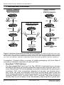
1
User Manual Retroviral Gene Transfer and Expression User Manual United States/Canada 800.662.2566 Asia Pacific +1.650.919.7300 Europe +33.(0)1.3904.6880 Japan +81.(0)77.543.6116 Clontech Laboratories, Inc. A Takara Bio Company 1290 Terra Bella Ave. Mountain View, CA 94043 Technical Support (US) E-mail: [email protected] www.clontech.com PT3132-1 (PR842519) Published 15 April 2008 Retroviral Gene Transfer and Expression User Manual Table of Contents I. Introduction 4 II. List of Components 12 III. Additional Materials Required 14 IV. Safety & Handling of Retroviruses 15 V. Plasmid Manipulations A. Propagating Plasmids 16 16 B. Generating Expression Vectors 16 VI. Culturing Packaging Cell Lines 17 A. General Considerations 17 B. Starting Cells from Frozen Stocks 17 C. Maintaining Packaging Cell Lines 18 D. Freezing Packaging Cell Lines 18 VII. Virus Production A. Transfecting Retroviral Vectors 19 20 B. Selecting Stable Virus-Producing Cell Lines 22 C. Concentrating Virus 22 D. Producing Virus from Stable Packaging Cell Clone (PT67) 22 E. Storage of Viral Stocks VIII.Determining Viral Titer A. General Considerations B.Procedure for Determining Viral Titer C. Alternative Methods 23 24 24 24 25 IX.Infecting Target Cells 26 A. General Considerations 26 B. Infecting Target Cells 26 X. Troubleshooting Guide 28 XI. References Appendix A: Culture Plate Conversions Appendix B: Titration of Antibiotic Stocks (Kill Curves) 31 34 35 Appendix C: Additional Viral Infection Methods 36 Clontech Laboratories, Inc. www.clontech.com 2 Protocol No. PT3132-1 Version No. PR842519 Retroviral Gene Transfer and Expression User Manual Table of Contents continued List of Figures Figure 1. Virus production in packaging cell lines Figure 2. Overview of transient and stable virus production Figure 3. Overview of producing infectious retrovirus 5 6 19 List of Tables Table I: Host range of packaging cell lines expressing different envelopes Table II: Packaging cell lines to use for infection Table III: Culture plate conversions 7 8 34 Note: The viral supernatants produced by these retroviral systems could, depending on your DNA insert, contain potentially hazardous recombinant virus. Due caution must be exercised in the production and handling of recombinant retrovirus. The user is strongly advised not to create retroviruses capable of expressing known oncogenes in amphotropic or polytropic host range viruses. Please refer to the appropriate regional and institutional guidelines on handling retroviruses. Please contact your on-site safety officer for specific requirements in your facility. In the United States, NIH guidelines require that retroviral production and transduction be performed in a Biosafety Level 2 facility. For more information, see appropriate HHS publications. Section IV in this User Manual contains a brief description of Biosafety Level 2 as well as other general information and precautions. http://bmbl.od.nih.gov and www.niehs.nih.gov/odhsb/biosafe/ nih/rdna-apr98.pdf Protocol No. PT3132-1 www.clontech.com Version No. PR842519 Clontech Laboratories, Inc. 3 Retroviral Gene Transfer and Expression User Manual I. Introduction Retroviral gene transfer is a technique for efficiently introducing stable, heritable genetic material into the genome of any dividing cell type (Ausubel et al., 1995; Coffin et al., 1996). This User Manual supports many Clontech packaging cell lines, retroviral vectors, and retroviral expression systems. Retroviral Gene Transfer Technology Current retroviral gene transfer technology is based on the coordinated design of packaging cell lines and retroviral expression vectors. The development of packaging lines—cell lines that package recombinant retroviral RNAs into infectious, replication-incompetent particles—created a new level of safety and control (Figure 1; Mann et al., 1983; Miller & Buttimore, 1986). To develop a packaging cell line, the viral gag, pol, and env genes—necessary for particle formation and replication—are stably integrated into the genome of the packaging cell line. The separate introduction and integration of the structural genes minimizes the chances of producing replication-competent virus due to recombination events during cell proliferation (Morgenstern & Land, 1990; Miller & Chen, 1996). Retroviral expression vectors provide the packaging signal Ψ+, transcription and processing elements, and a target gene. Inserts of up to 6.5 kb can be efficiently packaged. Transfection of the retroviral vector into a packaging cell line produces high-titer, replicationincompetent virus. The viral env gene, expressed by the packaging cell line, encodes the envelope protein, which determines the range of infectivity (tropism) of the packaged virus. Viral envelopes are classified according to the receptors used to enter host cells. For example, ecotropic virus can recognize a receptor found on only mouse and rat cells. Amphotropic virus recognizes a receptor found on a broad range of mammalian cell types. Dualtropic virus recognizes two different receptors found on a broad range of mammalian cell types. A pantropic packaging cell line provided a major advancement in retroviral gene transfer, as this cell line produces virus that can infect both mammalian and non-mammalian cells (Burns et al., 1993). Using this cell line, virions are pseudo-typed with the envelope glycoprotein from the vesicular stomatitis virus (VSV-G). Unlike other viral envelope proteins, VSV-G mediates viral entry through lipid binding and plasma membrane fusion (Emi et al., 1991). Stable expression of the VSV-G envelope protein is toxic; thus, the packaging cell line only contains the viral gag and pol genes. Virus is produced by transiently cotransfecting a retroviral expression vector and pVSV-G into a pantropic packaging cell line. Once a packaging cell line is transfected with a retroviral expression vector that contains a packaging signal, the viral genomic transcript containing the target gene and selectable marker are packaged into infectious virus within 48–72 hrs. Alternatively, you can use antibiotic selection to select cells that stably express the integrated vector. Stable virus-producing cells can be frozen and used in later experiments. Virus produced by both transient and stable Clontech Laboratories, Inc. www.clontech.com 4 Protocol No. PT3132-1 Version No. PR842519 Retroviral Gene Transfer and Expression User Manual Introduction continued I. Packaging Cell (produces viral proteins from stably integrated genes) 1) Transfection integration Retroviral vector transient expression stable expression 2) Transcription Gene X Neor DNA Gene X Neor RNA viral proteins 3) Viral proteins recognize 4) Packaging 5) Budding of infectious but replication-incompetent virus 6) Collect virus and infect target cells Figure 1. Virus production in packaging cell lines. The gag, pol and env genes required for viral production are integrated into the packaging cells genome. The vector provides the viral packaging signal, commonly denoted Ψ+, a target gene, and drug-resistance marker. transfections can infect target cells and transmit target genes; however, it cannot replicate within target cells because the viral structural genes are absent. The Retro-X™Universal Packaging System (Cat. No. 631530) is a transient packaging system that allows you to select the envelope according to the tropism needed for your experiments. It includes the GP2-293 cell line, which has the viral gag and pol genes incorporated in its genome. The remaining portion of the packaging function, the viral env gene, must be cotransfected with the retroviral expression vector bearing the gene of interest. The kit includes vectors that encode ecotropic, amphotropic, dualtropic (10A1) and pantropic (VSV-G) envelope proteins. This allows you to cater the tropism or host range of the packaged virus to your needs by determining which envelope protein is used. Protocol No. PT3132-1 www.clontech.com Version No. PR842519 Clontech Laboratories, Inc. 5 Retroviral Gene Transfer and Expression User Manual I. Introduction continued Transient Production Stable Production Plate packaging cells Plate packaging cells Transfect Transfect 8–10 hr Expression Vector 24 hr Target Cell Infection Expression Vector Plate target cells Select stable clones 48–72 hr Collect virus 2 weeks 18 hr Infect with virus Screen for hightiter clones Determine viral titer 1 week 2 weeks 2–3 days Select cells and analyze Expand & collect virus 1 week Figure 2. Overview of transient and stable virus production. To produce high-titer virus transiently, transfect a retroviral expression vector into an HEK 293-based packaging cell line. After 48–72 hours, collect virus and determine the viral titer or infect a target cell line. Alternatively, you can use antibiotic selection to develop clones that stably produce high-titer retrovirus. In addition, Clontech offers a variety of stable packaging cell lines. Table II provides a detailed overview of each cell line. • RetroPack™ PT67 Cell Line The RetroPack PT67 Cell Line (Cat. No. 631510) is derived from a mouse fibroblast (NIH 3T3) cell line designed for stably producing high-titer retrovirus. RetroPack PT67 cells package virus with a dualtropic (or polytropic) envelope, 10A1, that recognizes receptors on mouse, rat, human, hamster, mink, cat, dog, and monkey cells. Virus produced by these cells can enter target cells via two surface molecules, the amphotropic retrovirus receptor, RAM1 (Pit2), and the GALV (Pit1) receptor. Two viral receptors means that if one receptor is not abundantly expressed by a given speClontech Laboratories, Inc. www.clontech.com 6 Protocol No. PT3132-1 Version No. PR842519 Retroviral Gene Transfer and Expression User Manual I. Introduction continued TABLe I: Host range of packaging cell lines expressing different envelopes Target Cellsb Envelopesa Dualtropic Amphotropic Ecotropic Pantropicc Mouse + + + + Rat + + + + Hamster + +/– – + Mink + + – + Cat + + – + Dog + + – + Monkey + + – + Human + + – + Avian - - - + Fish - - - + Insect - - - + See Table II: Packaging Cell Lines for a description of the envelope proteins. a This listing of the most common target cells is not intended to be exclusive. Other cells which are not listed may also be infected. b c Virus packaged with the pantropic envelope also infects molusk, amphibian, ameoba and nematode cells. cies or cell type, the alternate receptor may still allow viral entry. Thus, virus packaged by RetroPack PT67 has a broad mammalian host range (Table I; Miller & Miller, 1994; Miller, 1996). These cells are best suited for stable virus production. • EcoPack2™-293 Cell Line The EcoPack2-293 Packaging Cell Line (Cat. No. 631507) is a human embryonic kidney, HEK 293-derived cell line designed for rapid, transient production of high-titer, ecotropic retrovirus (Figure 2). EcoPack2-293 cells can also be used to produce retrovirus stably. Bleomycin- and hygromycinresistance genes were used to separately introduce the viral gag-pol and env genes. Virus produced by EcoPack2-293 cells possess an ecotropic envelope (gap70), and thus can infect both mouse and rat cells (Table I). Retroviral sequence within the cell genome has been minimized, reducing the likelihood that replication-competent virus will be produced through recombination. EcoPack2-293 cells are more adherent and produce higher viral titers in comparison to our original EcoPack™-293 cells. • AmphoPack™-293 Cell Line The AmphoPack-293 Packaging Cell Line (Cat. No. 631505) is a human embryonic kidney, HEK 293-derived cell line designed for rapid, transient Protocol No. PT3132-1 www.clontech.com Version No. PR842519 Clontech Laboratories, Inc. 7 Retroviral Gene Transfer and Expression User Manual I. Introduction continued table iI: packaging cell lines Retropack™ PT67 EcoPack2™ -293 AmphoPack™ -293 GP2-293 Cell type NIH 3T3 HEK 293 HEK 293 HEK 293 Tropism Dualtropic Ecotropic Amphotropic Pantropica Env-specificb Broad mammalian Murine, rat Broad Mammalian, or non-mammalian Envelope 10A1 gap70 4070A VSV-Ga Env-specificb Receptors GALV (Pit1), RAM1 (Pit2) mCAT-1 RAM1 phosphatidylserine, phosphatidyl-inositol, & GMS gangliosidea TK DHFR Bleo Hyg Bleo Puro DHFR – Target cells Markersc gag-pol env mammalian production of high-titer, amphotropic retrovirus (Figure 2). AmphoPack-293 cells can also be used to produce high-titer retrovirus stably. Bleomycin- and puromycin-resistance genes were used to separately introduce the viral gag-pol and env genes. Therefore, the popular neomycin and hygromycin selection markers can be used to develop clones that stably produce high-titer virus. Virus produced by AmphoPack-293 cells express an amphotropic envelope (4070A), and thus can infect a broad range of mammalian cell types (Table I). • GP2-293 Packaging Cell Line The Pantropic Retroviral Expression System (Cat. No. 631512) features GP2-293, a HEK 293-based packaging cell line that stably expresses the viral gag and pol genes. To produce infectious virus, cotransfect GP2-293 with a retroviral expression vector and pVSV-G, a plasmid that expresses VSV-G from the CMV promoter (Yee et al., 1994).The VSV-G envelope must be cotransfected with the vector due to toxicity caused by the fusogenic properties of the VSV-G protein. A positive control cell line, GP2-293 Luc, allows verification that pVSV-G is functioning properly and that target cells can be infected.This system takes advantage of the envelopes ability to infect non-mammalian cells (Table I). The Retro-X™ Universal Expression System (Cat. No. 631530) also features the GP2-293 cell line. To produce infectious virus, cotransfect GP2-293 with a retroviral expression vector and the vector that encodes Clontech Laboratories, Inc. www.clontech.com 8 Protocol No. PT3132-1 Version No. PR842519 Retroviral Gene Transfer and Expression User Manual I. Introduction continued the envelope of your choice: pAmpho, pEco, p10A1, or pVSV-G. A positive control vector, pQCLIN, allows verification that the envelope vector is functioning properly and that target cells can be infected. The major advantages of the Universal Packaging System are as follows: • The system can be used with any MMLV-based vector. • The tropism can be changed to accomodate the desired cell type. • High titers are generated (106–107). • Infectious virus can be obtained in 48 hours, and the packaging system is therefore ideal for testing multiple constructs. • Excess Gag and Pol proteins are not generated, which may be detrimental (Yap et al., 2000). Retroviral Expression Vectors Clontech offers a wide range of retroviral expression vectors that can all be used with our various packaging cell lines. For more detailed descriptions of our vectors and sequence information, visit our www.clontech.com and navigate to the vector information page. All vectors contain the extended retroviral packaging signal, Ψ+, which promotes high-titer virus production. With the exception of the expression vectors in the MSCV Retroviral Expression System (Cat. No. 634401), all vectors are derived from Moloney murine leukemia virus (MMLV). Each vector contains a different antibiotic resistance marker—neomycin, hygromycin, or puromycin—allowing you to choose the cloning vector appropriate for the desired selection method. The MSCV Vectors contain a specifically designed long terminal repeat (LTR) from the murine stem cell PCMV virus. PCMV stands for PCC4-cell-passaged myeloproliferative sarcoma virus (Hilberg et al., 1987, Hawley et al., 1994).This LTR differs from the MMLV LTR by several point mutations and a deletion. These changes enhance transcriptional activation and decrease transcriptional suppression in embryonic stem and embryonal carcinoma cells. As a result, the LTR drives high-level constitutive expression of a target gene in stem cells and other mammalian cell lines (Hawley et al., 1994). The Retro-X™ Q Vectors are self-inactivating bicistronic expression vectors designed to express a target gene along with an antibiotic selection marker without the risk of promoter interference from the 5' LTR. In the case of pQCXIX two target genes may be expressed. Upon transfection into a packaging cell line, Q Vectors can transiently express, or integrate and stably express a viral genomic transcript containing the CMV immediate early promoter, gene-of-interest, IRES and antibiotic selection marker. Also included in the viral genomic transcript are the necessary viral RNA processing elements including the LTRs, packaging signal (ψ+), and tRNA primer binding site. The self-inactivating feature of the vectors is provided by a deletion in the 3' LTR enhancer region (U3). During reverse transcription Protocol No. PT3132-1 www.clontech.com Version No. PR842519 Clontech Laboratories, Inc. 9 Retroviral Gene Transfer and Expression User Manual I. Introduction continued of the retroviral transcript in the infected cell, the inactivated 3' LTR is copied and replaces the 5' LTR, resulting in inactivation of the 5' LTR promoter (CMV). The gene of interest and antibiotic resistance gene are expressed from an internal CMV promoter and co-translated, via the internal ribosome entry site (IRES), as a bicistronic message in mammalian cells (Jackson et al., 1990; Jang et al., 1988). Creator™ Compatibility for Diverse Gene Expression Studies Our retroviral systems are fully compatible with the Creator™ Gene Cloning and Expression System.This system uses Cre-loxP recombination to transfer a gene of interest directly from a single donor vector into numerous acceptor expression vectors without the need for subcloning. This strategy provides easy access to retroviral expression as well as fluorescent protein tagging, yeast two-hybrid studies, tetracycline-regulated gene expression, bacterial expression, and more. Creator™ Acceptor Vectors, such as pLP-LNCX and pLP-RevTRE, serve as entry points into our standard retroviral expression and tetracycline-regulated retroviral expression systems. See Section XII for ordering information. Further details on the Creator System, including the Creator™ DNA Cloning Kits User Manual (PT3460-1), are available at www.clontech.com. Retroviral Delivery of RNAi Constructs Our pSIREN-Retro-Q Vector is designed for gene silencing experiments based on the RNA interference phenomenon (July 2003 Clontechniques). Expression of silencing RNAs (shRNA) can decrease expression of a target gene in vivo (Xia et al., 2002). For more information about gene silencing technology, and use of the pSIREN vectors, please refer to the Knockout RNAi Systems User Manual (PT3739-1). Viral delivery of shRNAs has the following advantages: 1. Retroviral expression systems are capable of highly efficient gene delivery. • Viral vectors take advantage of viral mechanisms that allow efficient delivery of their nucleic acids to susceptible cell targets. Since recombinant viruses can infect nearly 100% of a cell population, the selection process used to enrich the population for a construct can be eliminated. If less than 100% of cells carry the construct, expression of specified gene may still be detected. • This efficiency is difficult to achieve in primary cells with transfection. • Retroviruses integrate into the host cell’s genome promoting permanent and stable gene transfer as well as persistent expression of the shRNA cassette. • Viral infection provides consistent, reproducible transfer of the sequence of interest. However, transfection efficiency can be low and inconsistent. Clontech Laboratories, Inc. www.clontech.com 10 Protocol No. PT3132-1 Version No. PR842519 Retroviral Gene Transfer and Expression User Manual I. Introduction continued 2. Copy number can be controlled with retroviral vectors.The shRNA dosage is important in maintaining gene silencing (Barton & Medzhitov, 2002). 3. Retroviral vector-based shRNAs produced within a retroviral packaging cell do not affect the titer or production of virus particles (Brummelkamp et al., 2002). 4. Because shRNA molecules anneal to specific sequences, developing vectors that target specific cell types is unnecessary because only those cells that express the targeted sequence will be affected by the vector. 5. Retroviral shRNA expression is more economical than chemical synthesis of small RNA, which is expensive for labs to do on a continuous basis. 6. Retroviral-vector based shRNA expression provides the option for stable expression. Adeno-X™ Adenoviral Gene Expression For experiments requiring transient gene expression in non-dividing or difficult-to-transfect cells, we recommend our Adeno-X™ Expression Systems.These adenovirus-based systems enable high-level protein expression in a wide variety of cell types (dividing or non-dividing) without the need for plaque purification (January 2000 & April 2003 Clontechniques). Protocol No. PT3132-1 www.clontech.com Version No. PR842519 Clontech Laboratories, Inc. 11 Retroviral Gene Transfer and Expression User Manual II. List of Components Store cell lines in liquid nitrogen (–196°C). Store all plasmids and primers at –20°C. Retro-X™ System (Cat. No. 631508) • 1 ml RetroPack PT67 Cell Line (2 x 10⁶ cells/ml) • 40 µl pLNCX2 Retroviral Vector (0.5 µg/µl) • 40 µl pLXSN Retroviral Vector (0.5 µg/µl) • 40 µl pLAPSN Retroviral Vector (0.5 µg/µl) • 100 µl pLNCX Seq/PCR Primer (20 µM) • 100 µl pLXSN Seq/PCR Primer (20 µM) Retro-X™ Q Vector Set (Cat. No. 631516) • 20 µg pQCXIN Retroviral Vector (500 ng/µl) • 20 µg pQCXIH Retroviral Vector (500 ng/µl) • 20 µg pQCXIP Retroviral Vector (500 ng/µl) • 20 µg pQCLIN Retroviral Vector (500 ng/µl) • 100 µl 5' pQC Seq/PCR Primer (20 µM) • 100 µl 3' pQC Seq/PCR Primer (20 µM) LRCX Retroviral Vector Set (Cat. No. 631511) • 20 µg pLNCX2 Retroviral Vector (0.5 µg/µl) • 20 µg pLHCX Retroviral Vector (0.5 µg/µl) • 20 µg pLPCX Retroviral Vector (0.5 µg/µl) • 100 µl 5' pLNCX Seq/PCR Primer (20 µM) • 100 µl 3' pLNCX Seq/PCR Primer (20 µM) MSCV Retroviral Expression System (Cat. No. 634401) • 1 ml RetroPack PT67 Cell Line (2 x 10⁶ cells/ml) • 20 µg pMSCVneo Retroviral Vector (0.5 µg/µl) • 20 µg pMSCVhyg Retroviral Vector (0.5 µg/µl) • 20 µg pMSCVpuro Retroviral Vector (0.5 µg/µl) • 100 µl 5' pMSCV Primer (20 µM) • 100 µl 3' pMSCV Primer (20 µM) Pantropic Retroviral Expression System (Cat. No. 631512) • 1 ml GP2-293 Packaging Cell Line (2 x 10⁶ cells/ml) • 1 ml GP-293 Luc Packaging Cell Line (2 x 10⁶ cells/ml) • 20 µg pLNHX Vector (0.5 µg/µl) • 20 µg pLXRN Vector (0.5 µg/µl) • 20 µg pLLRN Control Vector (0.5 µg/µl) • 20 µg pVSV-G Vector (0.5 µg/µl) Clontech Laboratories, Inc. www.clontech.com 12 Protocol No. PT3132-1 Version No. PR842519 Retroviral Gene Transfer and Expression User Manual II. List of Components continued Retro-X™ Universal Packaging System (Cat. No. 631530) • 1 ml GP2-293 Packaging Cell Line (2 x 10⁶ cells/ml) • 20 µg p10A1 Vector (0.5 µg/µl) • 20 µg pAmpho Vector (0.5 µg/µl) • 20 µg pEco Vector (0.5 µg/µl) • 20 µg pVSV-G Vector (0.5 µg/µl) • 20 µg pQCLIN Control Vector (0.5 µg/µl) RetroPack™ PT67 Cell Line (Cat. No. 631510) • 1 ml RetroPack PT67 Cell Line (2 x 10⁶ cells/ml) EcoPack2™-293 Cell Line (Cat. No.631507) • 1 ml EcoPack2-293 Cell Line (2 x 10⁶ cells/ml) AmphoPack™-293 Cell Line (Cat. No. 631505) • 1 ml AmphoPack-293 Cell Line (2 x 10⁶ cells/ml) Protocol No. PT3132-1 www.clontech.com Version No. PR842519 Clontech Laboratories, Inc. 13 Retroviral Gene Transfer and Expression User Manual III. Additional Materials Required • • • • • • • • • • • • • • • • • • • • • • • • Dulbecco’s Modified Eagle’s Medium (high glucose with sodium pyruvate & glutamine; Sigma Cat. No. D5796) Fetal bovine serum (FBS). Note: serum need not be heat inactivated. 200 mM L-Glutamine (Sigma Cat. No. G7513) Solution of 10,000 units/ml Penicillin G sodium and 10,000 µg/ml Streptomycin sulfate (Sigma Cat. No. P0781) Complete Medium Dulbecco’s Modified Eagle’s Medium DMEM [or Minimum Essential Medium, α Modification (α-MEM)] supplemented with 100 units/ml penicillin G sodium, 100 µg/ml streptomycin, 4 mM L-glutamine, 1 mM sodium pyruvate and 10% fetal bovine serum (FBS). G418 (Cat. No. 631307) Note: Make a 10 mg/ml active stock solution by dissolving 1 g of powder in approximately 70 ml of complete medium without supplements. Filter sterilize and store at 4°C. G418 can also be purchased as a premade solution. Hygromycin (Cat. No. 631309) Puromycin (Cat. No. 631305) Aminopterin (Calbiochem Cat. No. 454125) Hypoxanthine (Calbiochem Cat. No. 4010) Thymidine (Calbiochem Cat. No. 6060) Zeocin (Invitrogen Cat. No. R250-01) Polybrene (Hexadimethrine Bromide; Sigma Cat. No. H9268) Trypsin-EDTA (Trypsin; Sigma Cat. No. T3924) TNE (50 mM Tris-HCl [pH 7.8], 130 mM NaCl, 1 mM EDTA) Dulbecco’s phosphate buffered saline (DPBS; VWR Cat. No. 82020-066) Cell Freezing Medium (Sigma Cat. No. C6164) or DMSO (Sigma Cat. No. D2650) Tissue culture plates and flasks BD Biocoat Collagen Type I 12-well plates (BD Biosciences Cat. Nos. 354500 & 356500) Cloning cylinders (PGC Scientific Cat. No. 62-6150-40, -45) NIH-3T3 cells (ATCC Cat. No. CRL-1658) CalPhos™ Mammalian Transfection Kit (Cat. No. 631312) CLONfectin™ Transfection Reagent (Cat. No. 631301) Chloroquine (Sigma Cat. No. C6628) Clontech Laboratories, Inc. www.clontech.com 14 Protocol No. PT3132-1 Version No. PR842519 Retroviral Gene Transfer and Expression User Manual IV. Safety & Handling of Retroviruses The protocols in this User Manual require producing, handling, and storing infectious retrovirus. A thorough understanding of safe laboratory practices and potential retroviral hazards is essential. MMLV does not naturally infect human cells; however, viruses packaged from the MMLV-based vectors described here are capable of infecting human cells if packaged in a cell line with the proper tropism. This statement is also true for PCMV-based vectors. The viral supernatants produced by these retroviral systems could, depending on your retroviral insert, contain potentially hazardous recombinant virus. For these reasons, exercise due caution when producing and handling recombinant retrovirus. The user is strongly advised not to create retroviruses capable of expressing known oncogenes in amphotropic, dualtropic or pantropic packaging cell lines. Appropriate NIH, regional, and institutional guidelines apply, as well as specific guidelines for other countries. Please contact your on-site safety officer for specific requirements in your facility. In the United States, NIH guidelines require that retroviral production and transduction be performed in a Biosafety Level 2 (BL2) facility. A brief description of BL2 is given below. It is neither detailed nor complete. More information about BL2 guidelines is available at http://bmbl.od.nih.gov/contents.htm and more information about the risk group assessment for our viral systems is available at http://www4.od.nih. gov/oba/rac/guidelines/guidelines.html. If possible, observe and learn the practices described below from someone who has experience working with retroviruses. For more information, see the following reference: • Biosafety in Microbiological and Biomedical Laboratories, Fourth Edition (May 1999) HHS Pub. No. (CDC) 93-8395. U.S. Department of Health and Human Services, PHS, CDC, NIH. Practices • Perform work in a limited access area • Post biohazard warning signs • Minimize production of aerosols • Decontaminate potentially infectious wastes before disposal • Take precautions with sharps Safety equipment • Use a laminar flow hood with a HEPA filter • Wear protective laboratory coat, face protection, and double gloves Facilities • Autoclave for decontamination of solid and liquid waste • Use unrecirculated exhaust air • Stock chemical disinfectants for spills Protocol No. PT3132-1 www.clontech.com Version No. PR842519 Clontech Laboratories, Inc. 15 Retroviral Gene Transfer and Expression User Manual V. Plasmid Manipulations A. Propagating Plasmids 1.To ensure that you have a renewable source of DNA, transform each plasmid into a suitable E. coli host strain (e.g., DH5α). 2.Purify plasmids with a NucleoBond® or NucleoSpin® Plasmid Kit. Alternatively, isolate plasmids by banding on a CsCl gradient (Sambrook & Russell, 2001). B. Generating Expression Vectors Use standard molecular biology techniques to transfer your target gene into an expression vector (Sambrook & Russell, 2001). 1.Purify your gene fragment by any standard method. The cDNA or gene fragment must contain an ATG initiation codon. Adding a Kozak consensus ribosome binding site may improve expression levels in mammalian systems (Kozak, 1987). Please note that all sequences placed into a retroviral vector must be compatible with the retroviral life cycle and allow complete transcription of the fulllength viral genome. Sequences such as poly-A signals must not be included (Coffin et al., 1996). You can generate the fragment using compatible restriction sites that are on either side of the gene and in the cloning vector. If no such sites are present, use PCR to incorporate suitable restriction sites into your gene fragment. PCR fragments can be conveniently cloned into any vector using our In-Fusion™ PCR Cloning Kits. 2.Digest the vector with the appropriate restriction enzyme(s), treat with phosphatase, and purify. 3.Ligate the digested vector and the target gene fragment. 4.Transform ligation mixture into E. coli. 5.Identify the desired recombinant plasmid by restriction analysis, and confirm orientation and junctions by sequencing. Clontech Laboratories, Inc. www.clontech.com 16 Protocol No. PT3132-1 Version No. PR842519 Retroviral Gene Transfer and Expression User Manual VI. Culturing Packaging Cell Lines A. General Considerations The protocols in this section are intended for use with packaging cell lines from Clontech. • The RetroPack PT67 cell line has a very short doubling time (<16 hr). Split the culture before it becomes confluent. • The doubling time for EcoPack2-293, AmphoPack-293, and GP2-293 cell lines is 24–36 hr. • If you experience low packaging cell line viability, grow the cells for a longer period of time to allow for cell recovery and expansion. • All our packaging cell lines should be grown at 37°C in a humidified chamber with 5–10% CO2. See the Certificate of Analysis for details particular to each cell line. B. Starting Cultures from Frozen Stocks The protocols in this User Manual provide only general guidelines for mammalian cell culture techniques. Perform all steps involving cell culture using sterile technique in a suitable hood. For those requiring more information on mammalian cell culture, we recommend the following general references: • Culture of Animal Cells, Fourth Edition, ed. by R. I. Freshney (2000, Wiley-Liss) • Current Protocols in Molecular Biology, ed. by F. M. Ausubel et al. (1995, Wiley & Sons) Note: Frozen cells should be cultured immediately upon receipt or as soon thereafter as possible. Increased loss of viability may occur after shipping if culturing is delayed. 1.Transfer the vial of frozen cells from liquid N2 to a 37°C water bath until just thawed. To prevent osmotic shock and to maximize cell survival, perform the following: a. Rinse the outside of the tube with 70% ethanol. b. Add 1 ml complete medium (prewarmed to 37°C) to tube.Transfer mixture to a 15-ml tube. c. Add 5 ml complete medium and mix gently. Repeat. The final volume should be 12 ml. d. Centrifuge at 250 x g for 10 min. e. Remove supernatant. 2.Gently resuspend cells in 10 ml complete medium: DMEM [or Minimum Essential Medium, α Modification (α-MEM)] supplemented with 100 units/ml penicillin G sodium, 100 µg/ml streptomycin, 4 mM L-glutamine, 1 mM sodium pyruvate and 10% fetal bovine serum. 3.Incubate cells at 37°C with 5% CO2. Protocol No. PT3132-1 www.clontech.com Version No. PR842519 Clontech Laboratories, Inc. 17 Retroviral Gene Transfer and Expression User Manual VI. Culturing Packaging Cell Lines continued C. Maintaining Packaging Cell Lines Generally, cells should be plated at 106 per 100-mm plate and split every 2–3 days when they reach 70–80% confluency. Note: Plate HEK 293-based packaging cell lines on collagen-coated plates initially to promote adherence after thawing. These cells may be cultured on non-coated plates/ flasks after recovery; however, if adherence is poor, we recommend collagen-coated vessels for all culturing purposes including viral packaging. Split the cells as follows: 1.Remove medium, and wash cells once with room-temperature PBS. Note: If cells are over-confluent, omit the wash since the cells may detach from the plate. 2.Treat with 2 ml of trypsin-EDTA solution for 0.5–1 min. Depending on the cell line, you may need to treat the cells longer. 3.Add 3 ml of media + serum to inhibit trypsinization. 4.Resuspend cells gently by pipetting. 5.Add a predetermined portion of cells to a 100-mm plate in 10 ml of complete medium. Rock the plate to distribute the cells evenly. Note: Split RetroPack PT67 cells at a ratio of up to 1:20, and split HEK 293-based cells at a ratio of 1:10. 6.If cell viability is low, grow cells for a longer period of time, maintain higher cell densities, and verify culture conditions. D. Freezing Packaging Cell Lines Once a stable cell culture is established, we recommend that several aliquots of cells be frozen for future use. Prepare frozen aliquots of the packaging cells to ensure a renewable source as follows: 1.Expand the cell line into the desired number of flasks or plates. 2.When the desired number of flasks/plates reaches ~80% confluency, wash the cells once with PBS or HBSS, trypsinize using standard tissue-culture procedures (Freshney, 2000), add 2–4 volumes of complete medium to neutralize trypsin, and harvest cells. 3.Count the cells using a hemocytometer (Freshney, 2000), and collect by centrifugation (250 x g for 10 min at room temperature). 4.Resuspend in 4°C cell freezing medium containing 10% DMSO at 1–2 x 10⁶ cells/ml. 5.Dispense 1-ml aliquots into labeled freezing vials and place in a cell freezing container (reduces temperature ~1°C/min) at –80°C overnight. Alternatively, place the vials on ice or at –20°C for 1–2 hr, transfer to an insulated container such as a foam ice chest, and place in a –80°C freezer for several hours to overnight. 6.Transfer vials to liquid nitrogen. 7.Two or more weeks later. To confirm viability of frozen stocks, start a fresh culture of each frozen cell type, as described in Section B above. Clontech Laboratories, Inc. www.clontech.com 18 Protocol No. PT3132-1 Version No. PR842519 Retroviral Gene Transfer and Expression User Manual VII. Virus Production This section provides detailed procedures for virus production, target cell infection, and stable clone selection. For more detailed information or related protocols, see Coffin & Varmus (1996) or Ausubel et al. (1995). Figure 3 provides an overview of methods for producing high-titer virus using RetroPack PT67, EcoPack2-293, Amphopack-293, and GP2-293 cells. Transient Virus Production Stable Virus Production RetroPack™ PT67 1. Transfect with retroviral vector. 2. Select stable clones. 3. Determine viral titer. 4. Infect target cells. EcoPack2™-293 & AmphoPack™-293 1. Transfect with retroviral vector 2. Determine viral titer. or 2. Select stable clones. 3. Infect target cells. 3. Determine viral titer. 4. Infect target cells. GP2-293 1. Cotransfect with retroviral vector & envelope vector (pVSV-G, pEco, pAmpho, or p10A1). or 1. Transfect with retroviral vector (omit envelope vector). 2. Select stable clones. 2. Determine viral titer. 3. Concentrate virus (optional, for VSV-G) 3. Before each infection, transiently transfect with envelope vector. 4. Concentrate virus (optional) 4. Infect target cells 5. Determine viral titer 6. Infect target cells Figure 3. Overview of producing infectious retrovirus. Protocol No. PT3132-1 www.clontech.com Version No. PR842519 Clontech Laboratories, Inc. 19 Retroviral Gene Transfer and Expression User Manual VII. Virus Production continued A. Transfecting Retroviral Vectors Transfect by any standard method. We routinely use 60-mm plates for culturing packaging cell lines; See Additional Materials Required. Typically, transfections are done in smaller volumes than culturing. For maximal transfection efficiencies, we recommend the CalPhos™ Mammalian Transfection Kit (Cat. No. 631312). For maximal transfection efficiency in liposome-mediated transfections, we recommend Clonfectin™ Transfection Reagent (Cat. No. 631301). To optimize your transfection protocol, you can transfect the host cell line with a noninducible reporter expression vector, such as our pLAPSN included in our Retro-X™ System (Cat. No. 631508), or Living Colors™ Vectors, and assay for reporter gene activity. After choosing a method of transfection, optimize cell density (usually 60–80% confluency or 1–2 x 10⁶ cells/60 mm plate), the amount and purity of the DNA, media conditions, and transfection time. If a transfection method is already established in your laboratory, proceed with those conditions. Keep optimized parameters constant to obtain reproducible results. 1. Clone your target gene into a retroviral expression vector, or use the provided control vector for control experiments. Note: Use only high quality plasmid DNA. We recommend using a NucleoBond or NucleoSpin Plasmid Kit. 2.12–24 hr before transfection, plate packaging cells on a 60-mm plate at 60–80% confluency (1–2 x 10⁶ cells/60-mm plate). Note: Adding 25 µM chloroquine just prior to transfection may increase transfection efficiency 2–3 fold. Prepare a 25 mM stock of chloroquine in distilled water and filter sterilize. 1–2 hr before transfection, replace medium with medium containing chloroquine (Pear et al., 1993). 3. Transfect each 60-mm plate with the following amount of plasmid DNA: For RetroPack™ PT67, EcoPack™ 2-293 & AmphoPack™-293: ~5–10 μg of plasmid DNA For GP2-293: ~5 μg of expression vector and ~5 μg envelope vector Notes: • When using GP2-293 cells, envelope vector must be cotransfected. • When using a CaPO4-based transfection method, the final volume of transfection mixture should not exceed 0.5 ml for a 60-mm plate or 1 ml for a 100-mm plate. More than 1 ml of CaPO4 precipitants can be toxic to cells. Add the transfection solution to the medium and evenly distribute the solution on the cells. If toxicity is observed, perform transfection with 0.5 ml of the transfection mix. • 6–8 hr after transfection, you may perform glycerol shock treatment to increase the uptake of DNA (Freshney, 2000). Clontech Laboratories, Inc. www.clontech.com 20 Protocol No. PT3132-1 Version No. PR842519 Retroviral Gene Transfer and Expression User Manual VII. Virus Production continued 4.For RetroPack PT67 cells, aspirate culture medium 10–24 hr after transfection. Wash RetroPack PT67 cells twice with PBS, and add 3 ml of complete medium. Proceed to Section VII.B. 5. For HEK 293-based cell lines, aspirate culture medium 8–10 hr after transfection, and add 3 ml of complete medium. 6. Incubate the culture for an additional 48–72 hr to allow viral titer to increase. The viral titer reaches a maximum ~48 hr after transfection and is generally at least 30% of the maximum beyond 72 hr after transfection. Alternative Method: Infecting packaging cells (Ping-Pong) This method can be used to deliver the viral construct to the packaging cell line, an objective that can be accomplished by transfection, electroporation, or even infection (Note: Retro-X Q vectors can only be delivered by transfection). This method can also eliminate the need for selecting individual clones when making stable packaging cell lines (Parente & Wolfe, 1996). Alternatively, infect the packaging cells with virus obtained from another packaging cell line. Table II details the appropriate packaging cell lines to use for infection. A protocol for infection follows in Section VIII. This approach produces high-titer virus for several reasons: • More cells acquire the construct, and copy number is higher and more consistent (1–2 copies per cell per single round of infection depending upon titer of virus stock). • Virus-producing clones derived from transduced cells are more stable than those derived from transfected cells (Parente & Wolfe, 1996). • Allows the host-range of a vector to be changed. Important Notes: • This method requires previously transfected, virus-producing packaging cells. • You cannot infect cells that are already expressing the same or similar (eg. Ampho and 10A1) envelope protein. For example, virus produced from RetroPack™ PT67 cells cannot efficiently infect AmphoPack™-293 cells and vice versa. • Virus packaged in GP2-293 cells can be used to infect any other cell line depending on the envelope (pVSV-G, pEco, pAmpho or p10A1) that was cotransfected with the expression vector. • Virus produced by EcoPack2-293 cells can only infect mouse and rat cells, such as RetroPack PT67 cells. Protocol No. PT3132-1 www.clontech.com Version No. PR842519 Clontech Laboratories, Inc. 21 Retroviral Gene Transfer and Expression User Manual VII. Virus Production continued B. Selecting Stable Virus-Producing Cell Lines 1.Prior to using antibiotics to establish stable cell lines, you must titrate antibiotic stocks to determine the optimal concentration for selection (see Appendix B). This procedure is commonly called a kill curve. 2.Plate transfected packaging cells in selection medium 24–36 hr after transfection. 3.Culture cells for one week with the appropriate antibiotic. 4.Isolate large, healthy colonies and transfer them to individual plates or wells. Note: We generally isolate clones using cloning cylinders or cloning disks. The selected cell populations usually produce titers of ~ 105 cfu/ ml. If you require higher titer clones, pick individual clones for propagation. Determine viral titer as described in Section VIII. You must screen 20–50 clones to isolate a clone of acceptably high titer. Once clones are isolated, withdraw antibiotic from the medium. C. Concentrating Virus (VSV-G enveloped virions only, Burns et al., 1994) 1.Remove cell debris and aggregated virus by low speed centrifugation for 5 min at 4°C. 2.Pellet the virus at 50,000 x g for 90 min at 4°C. Remove the supernatant. 3.Resuspend the virus to 0.5–1% of the original volume in TNE (See Additional Materials required), and incubate overnight at 4°C. Note: If desired, perform a second round of ultracentrifugation (Steps 1–2). 4. Determine the viral titers of pre- and post-concentrated viral supernatants. 5. Infect target cells (Section IX.B.3). D. Producing Virus from Stable Packaging Cell Clone (PT67) 1.Remove clone from liquid nitrogen and follow thaw procedures outlined in Section VI.B. 2.Culture the clone, until cell culture reaches the desired culture volume. 3.Retaining one plate for the continuation of the culture, plate the remaining cells at 60–80% confluency in the desired number of culture vessels. 4.Viral supernatants can then be harvested in 24 hr intervals until cells are no longer viable. Discard all cells once the virus has been harvested. Clontech Laboratories, Inc. www.clontech.com 22 Protocol No. PT3132-1 Version No. PR842519 Retroviral Gene Transfer and Expression User Manual VII. Virus Production continued E. Storage of Viral Stocks 1.Once viral supernatant has been collected, briefly centrifuge sample to remove cellular debris at 500 x g for 10 min. Pool all similar stocks at this time. 2.Aliquot cleared supernatant into single-use tubes to avoid multiple freeze-thaw cycles. 3.Store tubes at –70°C. No cryoprotectant is required. Note: Avoid multiple freeze-thaw cycles, since titers can drop as much as 2–4 fold with each cycle (Higashikawa & Chang, 2001; Kwon et al., 2003). Protocol No. PT3132-1 www.clontech.com Version No. PR842519 Clontech Laboratories, Inc. 23 Retroviral Gene Transfer and Expression User Manual VIII.Determining Viral Titer A.General Considerations Determining the viral titer is necessary for three reasons: • Confirmation that viral stocks are viable. • Determination of the proper transduction conditions for your particular cell type by adjusting the MOI for the desired transduction efficiency. (i.e., control of copy number) MOI = No. of virus particles per target cell • Determination of the maximum number of target cells that can be infected for a given virus volume. B.Procedure for Determining Viral Titer 1.Plate NIH 3T3 cells one day prior to beginning this procedure. Plate cells in 6-well plates at a density of 0.5–1 x 10⁵ cells per well. Add 2 ml of medium per well. 2.Prepare 20 ml of complete medium and add 60 µl of 4 mg/ml polybrene. Note: Polybrene is a polycation that reduces charge repulsion between the virus and the cellular membrane. 3.Collect virus-containing medium from packaging cells. 4.Filter medium through a 0.45-µm cellulose acetate or polysulfonic (low protein binding) filter. Do not use a nitrocellulose filter because nitrocellulose binds proteins in the retroviral membrane and destroys the virus. This is the viral stock. 5.Prepare six 10-fold serial dilutions as follows: a. Add 1.35 ml of medium (Step 2) to each of six 1.5-ml microcentrifuge tubes. b. Add 150 µl of virus-containing medium (Step 4) to the first tube. Mix. c. Transfer 150 µl of viral stock dilution from tube 1 to tube 2. Continue serial dilutions by transferring 150 µl of eac hsuccessive dilution to the next prepared tube. 6.Infect NIH 3T3 cells by adding 1 ml of the diluted virus medium (Step 5) to the wells. Final polybrene concentration will be 4 µg/ ml in ~3 ml. 7.If you used pLAPSN from the Retro-X™ System for virus production, stain cells after 48 hr by assaying for alkaline phosphatase expression. Use any standard alkaline phosphatase assay (Ausubel et al., 1995). For other vectors, subject cells to antibiotic selection 24 hr after infection for one week. Clontech Laboratories, Inc. www.clontech.com 24 Protocol No. PT3132-1 Version No. PR842519 Retroviral Gene Transfer and Expression User Manual VIII.Determining Viral Titer continued See Appendix B for Kill Curve information. 8.The viral titer corresponds to the number of colonies present at the highest dilution that contains colonies, multiplied by the dilution factor. For example, the presence of four colonies in the 106 dilution would represent a viral titer of 4 x 10⁶. 4 colony forming units (cfu) x 106 = 4 x 10⁶ cfu/ml For virus produced from RetroPack PT67, EcoPack2-293, AmphoPack-293, and GP2-293 cells, a good viral titer is >106 cfu/ ml. C.Alternative Methods We recommend that you determine viral titer by infecting NIH 3T3 cells with serially diluted viral supernatants produced with a control vector such as pLAPSN, part of our Retro-X™ System (Cat. No. 631508). Infect both NIH 3T3 cells and your target cells. See Section IX for instructions on infecting NIH 3T3 cells. Infecting your target cell line will give you a rough, but rapid, estimation of infection success. You can use your cells of choice to determine viral titer (e.g., HeLa or Mink cells), but NIH 3T3 cells are widely accepted as the standard target cell for titering retrovirus because of the efficiency at which these cells become infected.The same virus preparation can give different "apparent" titers on different cells lines due to differential receptor expression and cell cycle rates. For more information on determining viral titer, please refer to Ausubel, et al. (1995). The method described in this manual is a standard gene transducing unit assay that measures the functional titer of a particular virus stock—the virus ability to infect is assayed. Another method is a drug-resistance colony assay, in which antibiotic selection of the infected cells gives rise to a countable number of colonies after roughly 10–14 days. Some variations of this method describe: a transduction, followed by a shorter selection period (3 days; Byun et al., 1996), recently-infected target cells (Tafuro et al., 1996; Miyao et al., 1995), and in situ PCR (PRINS; Claudio et al., 2001) with similar results. Other markers include LacZ, EGFP (Cashion et al., 1999, Muldoon et al., 1997), and luciferase. Although it relates directly to the infectious viral particles, functional titer does not provide a consistent measurement of virion concentration because it depends upon the transduction efficiency of the cell line being used to determine titer. Therefore, direct quantitation for determining virus particle concentration may be more desirable. Also, physical quantitation lends itself for more high-throughput applications, such as screening of stable virus-producing clones for high-titer variants. Direct quantitation of virus concentration in supernatant does not rely on antibiotic selection and therefore all viruses, regardless of sequence can be quantitated. Methods for the direct quantitation Protocol No. PT3132-1 www.clontech.com Version No. PR842519 Clontech Laboratories, Inc. 25 Retroviral Gene Transfer and Expression User Manual VIII.Determining Viral Titer continued of virus particles include slot blots (Nelson et al., 1998; Murdoch, et al., 1997; Onodera, et al., 1997) and PCR applied to viral supernatants (Quinn & Trevor, 1997; Morgan et al., 1990). Reverse transcriptase activity has also been used (Goff et al., 1981). Some have used protein production (encoded by the retroviral transgene) from packaging cells as a method for screening high titer clones, but this method is flawed because protein production does not correlate with the number of infectious virions (Tasaki et al., 1997). IX. Infecting Target Cells A. General Considerations The following protocols are general recommendations for infecting adherent cells, such as NIH 3T3 or HeLa. Use them as a starting point for determining optimal conditions for your experiments. If these protocols do not work for your cell type, please refer to Appendix C for alternative infection methods. Important: Multiple rounds of infection can improve your results by increasing the number of infected cells as well as increasing the copy number per cell. Virus produced with the VSV-G envelope can be concentrated by ultracentrifugation to titers of up to 109 cfu/ml (See Section VII.C). B. Infecting Target Cells 1. Plate the target cells 12–18 hr before infection, at a cell density of 1–2 x 10⁵ per 60-mm plate. If you will be using infected cells for a biological assay, ensure that the control cells are treated with an insert-free virus under identical conditions. Note: The viral pre-integration complex enters the nuclei of actively dividing cells only. 2. For infection, collect medium from packaging cells and filter medium through a 0.45-µm cellulose acetate or polysulfonic (low protein binding) filter. Do not use a nitrocellulose filter because it binds proteins in the retroviral membrane and destroys the virus. Optional: For VSV-G enveloped virus, you can concentrate virus as described in Section VII.C. 3.Add virus to target cells. Until you have determined the viral titer, use as much virus-containing medium as possible for the infection. Store remaining viral supernatant at –80°C. Clontech Laboratories, Inc. www.clontech.com 26 Protocol No. PT3132-1 Version No. PR842519 Retroviral Gene Transfer and Expression User Manual IX. Infecting Target Cells continued Notes: • Titer will decrease ~2–4-fold per freeze-thaw cycle. •The optimal final concentration of polybrene may need to empirically determined but generally falls within a range of 2–12 µg/ml. • Excessive exposure to polybrene (>24 hr) can be toxic to cells. Alternatively, perform infections sequentially, ~12 hr apart. Doing so increases the efficiency of infection, but also increases copy number. Cellular receptors can be occupied by soluble envelope and/or non-functional virions. Therefore, to ensure that cellular receptors will be unoccupied by viral envelope, allow cells to rest for a minimum of 12 hr between each infection. 4.Add polybrene to a final concentration of 4–8 μg/ml. 5.Replace medium with fresh medium after 24 hr of incubation. 6. To determine the efficiency of infection, subject a small subpopulation of cells to antibiotic treatment.The infected cells should be used for experiments or for selection as soon as possible, but not earlier than 24 hr after the last infection. The growth of some target cells is strongly affected by media conditioned by the packaging cells. You can take certain precautions to avoid an adverse effect induced by the packaging cell-derived supernatants: • Dilute virus-containing media at least 2-fold with fresh medium. • Expose target cells to the virus for 4–6 hr and then replace with fresh medium. • For cells that prove more difficult to infect, please see references located in Appendix C. Protocol No. PT3132-1 www.clontech.com Version No. PR842519 Clontech Laboratories, Inc. 27 Retroviral Gene Transfer and Expression User Manual X. Troubleshooting Guide A. Cloning DNA does not cut as expected Incomplete digest: repeat digest with more enzyme, a different enzyme lot, or for a longer period of time. Low yield of plasmid Retroviral constructs use a low copy pBR322 ori. Grow more liquid culture and purify using lowcopy purification procedures. Plasmid is difficult to grow or clone. Plasmid may rearrange due to presence of LTR's. Switch to alternate E. coli strain for unstable DNA sequences. B. Packaging Cells Poor viability upon thawing Improper thawing procedures: follow thawing procedures in Section VI.B. Improper culture medium: all packaging cell lines will grow in DMEM + 10% FBS Improper tissue culture plasticware: use collagen I-coated plates to aid adherence during initial seeding. Slow growth Improper culture medium: all packaging cell lines will grow in DMEM + 10% FBS. Improper incubator conditions: grow cells at 37°C in humidified incubator with 5–10% CO2. Cells do not attach to plate Improper culture medium: all packaging cell lines will grow in DMEM + 10% FBS. Improper tissue culture plasticware: use collagen I-coated plates to aid adherence during initial seeding. Cells appear morphologically different Subclone parental cell line. Cannot select for packaging Incorrect amount of antibiotic: we do not recommend reselecting for packaging function. Clontech Laboratories, Inc. www.clontech.com 28 Protocol No. PT3132-1 Version No. PR842519 Retroviral Gene Transfer and Expression User Manual X. Troubleshooting Guide continued C. Virus Production Poor Transfection efficiency Cells are overly confluent: plate fewer cells (60–80% confluency, 1–2 x 10⁶ cells/60mm). Transfection is toxic to cells: Optimize DNA and transfection reagent amounts and exposure time. Assaying for positive cells too early: wait 48 hr after transfection for maximal gene expression to determine efficiency. Low titer (<105 cfu/ml) Poor transfection efficiency: optimize transfection. Concentrate virus if using VSV-G. Truncated viral RNA: check sequence for presence of poly(A) between LTRs. Virus harvested too early: harvest virus 48–72 hr after transfection. Vector too large (The limit of packaging function is 8.3 kb from LTR to LTR). Concentrate virus for large vectors or reduce size of inserts. Low virus production from cell population (PT67): Pick and screen for stable, higher-titer clones. No polybrene added during titration: add polybrene (4–8 µg/ml) to viral supernatant. Virus exposed to multiple freeze-thaw cycles: each cycle drops the titer approximately 2–4 fold. Limit the number of freeze-thaws. Sub-optimal selection procedure during titration: perform an antibiotic kill curve on titration targets prior to titration. Protocol No. PT3132-1 www.clontech.com Version No. PR842519 Clontech Laboratories, Inc. 29 Retroviral Gene Transfer and Expression User Manual X. Troubleshooting Guide continued D. Infection of Target Cells Poor infection efficiency Low titer: see above section. Infection protocol not optimized: see Appendix C for references for optimizing transduction protocols. Target cells not dividing: plate cells at lower confluency, activate with mitogen, or use another method to induce cell division. Optimize culture conditions for targets prior to infection. Target cell viability poor during infection Packaging cell line-conditioned media may be affecting cell growth: dilute viral medium or shorten exposure time to viral supernatant. Excessive exposure to polybrene: optimize amount of polybrene (titrate) or shorten exposure time to viral supernatant. Low infection efficiency (See Section D, above). Low expression level Possible promoter inactivation: split cells, activate with mitogen, treat cells with 5-azacytidine. Choose a tissue-specific promoter. Poor cell viability: check growth parameters. Clontech Laboratories, Inc. www.clontech.com 30 Protocol No. PT3132-1 Version No. PR842519 Retroviral Gene Transfer and Expression User Manual XI. References Adeno-X Expression System (2000) Clontechniques XV(1):8–10. Ausubel, F. M., Brent, R., Kingdom, R. E., Moore, D. M., Seidman, J. G., Smith, J. A. & Struhl, K., Eds. (1995) Current Protocols in Molecular Biology. (John Wiley & Sons, NY). Barton, G. M. & Medzhitov, R. (2002) Retroviral delivery of small interfering RNA into primary cells. Proc. Natl. Acad. Sci. USA 99(23):14943–14945. Brummelkamp, T. R., Bernards, R. & Agami, R. (2002) Stable suppression of tumorigenicity by virus-mediated RNA interference. Cancer Cell 2(3):243–247. Burns, J. C., Friedmann, T., Driever, W., Burrascano, M. & Yee, J.-K. (1993) Vesicular stomatitis virus G glycoprotein pseudotyped retroviral vectors: concentration to very high titer and efficient gene transfer into mammalian and nonmammalian cells. Proc. Natl. Acad. Sci. USA 90:8033–8037. Byun, J., Kim, J. M., Kim, S. H., Yim J., Robbins, P. D. & Kim, S. (1996) A simple and rapid method for the determination of recombinant retrovirus titer by G418 selection. Gene Ther. 3:1018–1020. Cashion, L. M., Bare, L. A., Harvey, S., Trinh, Q., Zhu, Y. & Devlin, J. J. (1999) Use of enhanced green fluorescent protein to optimize and quantitate infection of target cells with recombinant retroviruses. Biotechniques 26: 924–930. Claudio, P. P., Cinti, C. & Giordano, A. (2001) Application of the primer in situ DNA synthesis (PRINS) technique to titer recombinant virus and evaluation of the efficiency of viral transduction. Anal. Biochem. 291:96–101. Coffin, J. M. & Varmus, H. E., Ed. (1996) Retroviruses (Cold Spring Harbor Laboratory Press, NY). Devroe, E. & Silver, P. A. (2002) Retrovirus-delivered siRNA.BMC Biotechnol. 2(1):15. Emi, N., Friedmann,T. &Yee, J.-K. (1991) Pseudotyped formation of murine leukemia virus with G protein of vesicular stomatitis virus. J. Virol. 65:1202–1207. Freshney, R. I. (2000) Culture of Animal Cells, Fourth Edition (Wiley-Liss, NY). Goff, S., Traktman, P. & Baltimore D. (1981) Isolation and properties of Moloney murine leukemia virus mutants: use of a rapid assay for release of virion reverse transcriptase. J. Virol. 38:239–248. Hawley, R. G., Lieu, F. H. L., Fong, A. Z. C., & Hawley, T. S. (1994) Versatile retroviral vectors for potential use in gene therapy. Gene Ther. 1:136–138. Hemann, M. T., Fridman, J. S., Zilfou, J. T., Hernando, E., Paddison, P. J., Cordon-Cardo, C., Hannon, G. J. & Lowe, S. W. (2003) An epi-allelic series of p53 hypomorphs created by stable RNAi produces distinct tumor phenotypes in vivo. Nat. Genet. 33(3):396–400. Hilberg, F. (1987) Functional analysis of a retroviral host-range mutant: altered long terminal repeat sequences allow expression in embryonal carcinoma cells. Proc. Natl. Acad. Sci. USA 84:5232–5236. Higashikawa, F. & Chang L. (2001) Kinetic Analysis of stability of simple and complex retroviral vectors. Virology 280:124–131. Jackson, R. J., Howell M. T. & Kaminski, A. (1990) The novel mechanism of initiation of picornavirus RNA translation. Trends Biochem. Sci. 15:477–483. Jang, S. K., Krausslich, H. G., Nicklin, M. J., Duke, G. M., Palmenberg, A. C. & Wimmer, E. (1988) A segment of the 5' nontranslated region of encephalomyocarditis virus RNA directs internal entry of ribosomes during in vitro translation. J. Virol. 62:2636–2643. Kozak, M. (1987) At least six nucleotides proceeding the AUG initiator codon enhances translation in mammalian cells. J. Mol. Biol. 196:947–950. Protocol No. PT3132-1 www.clontech.com Version No. PR842519 Clontech Laboratories, Inc. 31 Retroviral Gene Transfer and Expression User Manual XI. References continued Kwon, Y. J., Hung, G., Anderson, W.F., Peng, C.A. & Yu, H. (2003) Determination of infectious retrovirus concentration from colony-forming assay with quantitative analysis. J. Virol. 77:5712–5720. Mann, R., Mulligan, R. C. & Baltimore, D. (1983) Construction of a retrovirus packaging mutant and its use to produce helper-free defective retrovirus. Cell 33:153–159. Miller, A. D. (1996) Cell-surface receptors for retroviruses and implications for gene transfer. Proc. Natl. Acad. Sci. USA 93:11407–11413. Miller, A. D. & Buttimore, C. (1986) Redesign of retrovirus packaging cell lines to avoid recombination leading to helper virus production. Mol. Cell. Biol. 6(8):2895–2902. Miller, A. D. & Chen, F. (1996) Retrovirus packaging cells based on 10A1 murine leukemia virus for production of vectors that use multiple receptors for cell entry. J. Virol. 70(8):5564–5571. Miller, D. G. & Miller A. D. (1994) A family of retroviruses that utilize related phosphate transporters for cell entry. J. Virol. 68:8270–8276. Miyao,Y., Shimizu, K.,Tamura, M.,Yamada, M.,Tamura, K., Nakahira, K., Kuriyama, S., Hayakawa, T. & Ikenaka, K. (1995) A simplified general method for determination of recombinant retrovirus titers. Cell Struct. Funct. 20:177–183. Morgan, R. A., Cornetta, K. & Anderson W. F. (1990) Applications of the polymerase chain reaction in retroviral-mediated gene tranfer and the analysis fo gene-marked human TIL cells. Hum. Gene Ther. 1:135–149. Morgenstern, J. P. & Land, H. (1990) Advanced mammalian gene transfer: high titer retroviral vectors with multiple drug selection markers and a complementary helper-free packaging cell line. Nucleic Acids Res. 18:3587–3590. Muldoon, R. R., Levy, J. P., Kain S. R., Kitts, P. A. & Link, C. J. Jr. (1997) Tracking and quantitation of retroviral-mediated transfer using a completely humanized, red-shifted green flourescent protein gene. Biotechniques 22:162–167. Murdoch, B., Pereira, D. S., Wu, X., Dick, J. E. & Ellis, J. (1997) A rapid screening procedure for the identification of high-titer retrovirus packaging clones. Gene Ther. 4:744–749. Nelson, D. M., Wahlfors, J. J., Chen, L., Onodera, M. & Morgan, R. A. (1998) Characterization of diverse viral vector preparations, using a simple and rapid whole-virion dot-blot method. Hum. Gene Ther. 9:2401–2405. Onodera, M., Yachie, A., Nelson, D. M., Welchlin, H., Morgan, R. A. & Blaese, R. M. (1997) A simple and reliable method for screening retroviral producer clones without selectable markers. Hum. Gene Ther. 8:1189–1194. Parente, M. K. & Wolfe, J. H. (1996) Production of increased titer retrovirus vectors from stable producer cell lines by superinfection and concentration. Gene Ther. 3:756–760. Pear, W. S., Nolan, G. P., Scott, M. L. & Baltimore, D. (1993) Production of high-titer helper-free retroviruses by transient transfection. Proc. Natl. Acad. Sci. USA 90(18):8392–8396. Qin, X. F., An, D. S., Chen, I. S. & Baltimore, D. (2003) Inhibiting HIV-1 infection in human T cells by lentiviral-mediated delivery of small interfering RNA against CCR5. Proc. Natl. Acad. Sci. USA 100(1):183–8. Quinn, T. P. & Trevor, K. T. (1997) Rapid quantitation of recombinant retrovirus produced by packaging cell clones. Biotechniques 23:1038–1044. Sambrook, J. & Russell, D.W. (2001) Molecular Cloning: A Laboratory Manual, (Cold Spring Harbor Laboratory Press, Cold Spring Harbor, NY). Tafuro, S., Zentilin, L., Falaschi, A. & Giacca, M. (1996) Rapid retrovirus titration using competitive polymerase chain reaction. Gene Ther. 3:679–684. Clontech Laboratories, Inc. www.clontech.com 32 Protocol No. PT3132-1 Version No. PR842519 Retroviral Gene Transfer and Expression User Manual XI. References continued Tasaki, K., Yoshida, Y., Tagawa, M., Takenaga, K., Asano, T., Ochiai, T., Isono, K., Kouzu, T., Saisho, H. & Sakiyama, S. (1997) Discordant production of released exogenous protein and infectious virions from retrovirus-packaging cells used for gene transduction. Anticancer Res. 17:4415–4417. Xia, H., Mao, Q., Paulson, H. L. & Davidson, B. L. (2002) siRNA-mediated gene silencing in vitro and in vivo. Nat. Biotechnol. 20:1006–1010. Yap, M. W., Kingsman, S. M. & Kingsman, A. J. (2000) Effects of stoichiometry of retroviral components on virus production. J. Gen. Virol. 81:2195–2202. Yee, J.-K., Friedmann, T. & Burns, J. C. (1994) Generation of high-titer pseudotyped retroviralvectors with very broad host range. Methods Cell Biol. 43:99–112. Protocol No. PT3132-1 www.clontech.com Version No. PR842519 Clontech Laboratories, Inc. 33 Retroviral Gene Transfer and Expression User Manual Appendix A: Culture Plate Conversions table iii: culture plate conversion Size of Plate Growth Area (cm2) Relataive area* Recommended Volume 96 well 0.32 0.04 X 200 µl 24 well 1.88 0.25 X 500 µl 12 well 3.83 0.5 X 1.0 ml 6 well 9.4 1.2 X 2.0 ml 35 mm 8.0 1.0 X 2.0 ml 60 mm 21 2.6 X 5.0 ml 10 cm 55 7X 10.0 ml Flasks 25 75 3X 9X 5.0 ml 12.0 ml * Relative area is expressed as a factor of the growth area of a 35-mm culture plate. Clontech Laboratories, Inc. www.clontech.com 34 Protocol No. PT3132-1 Version No. PR842519 Retroviral Gene Transfer and Expression User Manual Appendix B: Titration of Antibiotic Stocks (Kill Curves) Prior to using G418, hygromycin or puromycin to establish stable packaging cell lines, it is important to titrate your selection agent stocks to determine the optimal concentration for selection with the chosen cell line. This is also important because of lot-to-lot variation in the potency of these drugs. Therefore, you should titrate each new lot of antibiotic to determine the optimal concentration. We recommend that you perform two experiments for each drug: (1) a titration to determine the optimal drug concentration, and (2) an experiment to determine the optimal plating density. 1.Titrate at fixed cell density. a. Plate 2 x 10⁵ cells in each of six 10-cm tissue culture dishes containing 10 ml of the appropriate complete medium plus varying amounts (0, 50, 100, 200, 400, 800 µg/ml) of hygromycin or G418. For puromycin, add the drug at 0, 1, 2.5, 5, 7.5, and 10 µg/ml. b. Incubate the cells for 10–14 days, replacing the selective medium every four days (or more often if necessary). c. Examine the dishes for viable cells every two days. For selecting stable transformants, use the lowest concentration that begins to give massive cell death in ~5 days and kills all the cells within two weeks. 2.Determine optimal plating density. Once you have determined the optimal drug concentration, determine the optimal plating density by plating cells at several different densities in the presence of a constant amount of drug. If cells are plated at too high a density, they will reach confluency before the selection takes effect. Optimal plating density is dependent on population doubling time and cell surface area. For example, cells that double rapidly have a lower optimal plating density than cells that double slowly. a. Plate cells at several different densities in each of six 10-cm tissue culture dishes containing 10 ml of the appropriate selective medium. Suggested densities (cells/10-cm dish): 5 x 10⁶, 1 x 10⁶, 5 x 10⁵, 2 x 10⁵, 1 x 10⁵, and 5 x 10⁴. b. Incubate the cells for 5–14 days, replacing the selective medium every four days. c. Examine the dishes for viable cells every two days. For selecting stable transfectants, use a plating density that allows the cells to reach ~80% confluency before massive cell death begins (at about day 5). This is the cell density at which cells should be plated for selection of stable transfectants. Protocol No. PT3132-1 www.clontech.com Version No. PR842519 Clontech Laboratories, Inc. 35 Retroviral Gene Transfer and Expression User Manual Appendix C: Additional Viral Infection Methods These references are provided for fine-tuning your transduction protocols to achieve the desired infection frequency in target cells. This list is not intended to be comprehensive. These protocols will work for a wide range of cell types; however you must determine which works best for your targets. While each technique can provide modest increases in efficiency, they may be combined to create an additive effect. For ease of analysis, we recommend our retroviral vectors that express our Living Colors™ fluorescent proteins for detection and quantitation of gene transfer efficiency during testing. A.Transduction of cells at 32°C: Decrease in temperature increases viral half-life during transduction Bunnell, B. A., Muul, L. M., Donahue, R. E., Blaese, R. M., Morgan, R. A. (1995) Highefficiency retroviral-mediated gene transfer into human and nonhuman primate peripheral blood lymphocytes. Proc. Natl. Acad. Sci. USA 92(17):7739–7743. Zhou, P., Lee, J., Moore, P., Brasky, K. M. (2001) High-efficiency gene transfer into rhesus macaque primary T lymphocytes by combining 32 degrees C centrifugation and CH-296-coated plates: effect of gene transfer protocol on T cell homing receptor expression. Hum. Gene Ther. 12(15):1843–1855. Kotani, H., Newton, P. B. 3rd, Zhang, S., Chiang, Y. L., Otto, E., Weaver, L., Blaese, R. M., Anderson, W. F. & McGarrity, G. J. (1994) Improved methods of retroviral vector transduction and production for gene therapy. Hum. Gene Ther. 5(1):19–28. Higashikawa, F. & Chang, L. (2001) Kinetic analyses of stability of simple and complex retroviral vectors. Virology 280(1):124–131. B.Centrifugation during transduction (Spinoculation): believed to counteract diffusion of virus when binding target cells Bunnell, B. A., Muul, L. M., Donahue, R. E., Blaese, R. M. & Morgan, R. A. (1995) Highefficiency retroviral-mediated gene transfer into human and nonhuman primate peripheral blood lymphocytes. Proc. Natl. Acad. Sci. USA 92(17):7739–7743. Ohkubo, T., Barcena, A., Smith, C. A., Harrison, M. R. & Muench, M. O. (2001) Highefficiency retroviral transduction of fetal liver CD38-CD34++ cells: implications for in utero and ex utero gene therapy. Fetal Diagn. Ther. 16(5):299–307. Movassagh, M., Boyer, O., Burland, M. C., Leclercq, V., Klatzmann, D. & Lemoine F. M. (2000) Retrovirus-mediated gene transfer into T cells: 95% transduction efficiency without further in vitro selection. Hum. Gene Ther. 11(8):1189–1200. Bahnson, A. B., Dunigan, J. T., Baysal, B. E., Mohney, T., Atchison, R. W., Nimgaonkar, M. T., Ball, E. D. & Barranger, J. A. (1995) Centrifugal enhancement of retroviral mediated gene transfer. J. Virol. Methods 54:131–143. C.Precipitation to increase titer (concentration) Pham, L., Ye, H., Cosset, F. L., Russell, S. J. & Peng, K. W. (2001) Concentration of viral vectors by co-precipitation with calcium phosphate. J. Gene Med. 3(2):188–194. Darling, D., Hughes, C., Galea-Lauri, J., Gaken, J.,Trayner, I. D., Kuiper, M. & Farzaneh, F. (2000) Low-speed centrifugation of retroviral vectors absorbed to a particulate substrate: a highly effective means of enhancing retroviral titre. Gene Ther. 7(11):914–923. Hughes, C., Galea-Lauri, J., Farzaneh, F. & Darling, D. (2001) Streptavidin paramagnetic Clontech Laboratories, Inc. www.clontech.com 36 Protocol No. PT3132-1 Version No. PR842519 Retroviral Gene Transfer and Expression User Manual Appendix C: Additional Viral Infection Methods continued particles provide a choice of three affinity-based capture and magnetic concentration strategies for retroviral vectors. Mol. Ther. 3(4):623–630. D.Precipitation (during transduction): facilitates greater contact between the target cells and virions Le Doux, J. M., Landazuri, N., Yarmush, M. L. & Morgan, J. R. (2001) Complexation of retrovirus with cationic and anionic polymers increases the efficiency of gene transfer. Hum. Gene Ther. 12(13):1611–1621. Morling, F. J. & Russell, S. J. (1995) Enhanced transduction efficiency of retroviral vectors coprecipitated with calcium phosphate. Gene Ther. 2(7):504–508. Hennemann, B., Chuo, J. Y., Schley, P. D., Lambie, K., Humphries, R. K. & Eaves, C. J. (2000) High-efficiency retroviral transduction of mammalian cells on positively charged surfaces. Hum. Gene Ther. 11(1):43–51. E.Increase transduction rate by phosphate depletion: results in up regulation of GLVR-1 and GLVR-2 (RAM1) receptors (for ampho- tropic or 10A1 pseudotyped virus) Bunnell, B. A., Muul, L. M., Donahue, R. E., Blaese, R. M. & Morgan, R. A. (1995) Highefficiency retroviral-mediated gene transfer into human and nonhuman primate peripheral blood lymphocytes. Proc. Natl. Acad. Sci. USA 92(17):7739–7743. Zhou, P., Lee, J., Moore P, Brasky K. M. (2001) High-efficiency gene transfer into rhesus macaque primary T lymphocytes by combining 32 degrees C centrifugation and CH296-coated plates: effect of gene transfer protocol onT cell homing receptor expression. Hum. Gene Ther. 12(15):1843–1855. F.Flow through transduction: concentrating cells and virus together in small culture systems Pan, D., Shankar, R., Stroncek, D. F. & Whitley, C. B. (1999) Combined ultrafiltrationtransduction in a hollow-fiber bioreactor facilitates retrovirus-mediated gene transfer into peripheral blood lymphocytes from patients with mucopolysaccharidosis type II. Hum. Gene Ther. 10(17):2799–2810. Chuck, A. S. & Palsson, B. O. (1996) Consistent and high rates of gene transfer can be obtained using flow-through transduction over a wide range of retroviral titers. Hum. Gene Ther. 7(6):743–750. G.Addition of fibronectin: adhesion domains within fibronectin allow binding to both target cells and virions to facilitate co-localization Zhou, P., Lee, J., Moore, P. & Brasky, K. M. (2001) High-efficiency gene transfer into rhesus macaque primary T lymphocytes by combining 32 degrees C centrifugation and CH-296-coated plates: effect of gene transfer protocol on T cell homing receptor expression. Hum. Gene Ther. 12(15):1843–1855. Moritz, T., Dutt, P., Xiao, X., Carstanjen, D., Vik, T., Hanenberg, H. & Williams D. A. (1996) Fibronectin improves transduction of reconstituting hematopoietic stem cells by retroviral vectors: evidence of direct viral binding to chymotryptic carboxy-terminal fragments. Blood 88(3):855–862. Hanenberg, H., Xiao, X. L., Dilloo, D., Hashino, K., Kato, I. & Williams, D. A. (1996) Colocalization of retrovirus and target cells on specific fibronectin fragments increases genetic transduction of mammalian cells. Nat. Med. 2(8):876–882. Protocol No. PT3132-1 www.clontech.com Version No. PR842519 Clontech Laboratories, Inc. 37 Retroviral Gene Transfer and Expression User Manual Appendix C: Additional Viral Infection Methods continued Bajaj, B., Lei, P. & Andreadis, S.T. (2001) High efficiencies of gene transfer with immobilized recombinant retrovirus: kinetics and optimization. Biotechnol. Prog. 17(4):587–596. H.Cocultivation of target cells and packaging cells: Allows targets to be continuously in contact with freshly-produced viral supernatant Casal, M. L. & Wolfe, J. H. (1997) Amphotropic and ecotropic retroviral vector viruses transduce midgestational murine fetal liver cells in a dual-chambered cocultivation system. Gene Ther. 4(1):39–44. Germeraad, W. T., Asami, N., Fujimoto, S., Mazda, O. & Katsura, Y. (1994) Efficient retrovirus-mediated gene transduction into murine hematopoietic stem cells and longlasting expression using a transwell coculture system. Blood 84(3):780–788. I.Use of cationic liposomes: Enhance virus-to-cell fusion Kaneko, Y. & Tsukamoto, A. (1996) Cationic liposomes enhance retrovirus-mediated multinucleated cell formation and retroviral transduction. Cancer Lett. 105(1):39–44. Porter, C. D., Lukacs, K. V., Box, G.,Takeuchi,Y. & Collins, M. K. (1998) Cationic liposomes enhance the rate of transduction by a recombinant retroviral vector in vitro and in vivo. J. Virol. 72(6):4832–4840. J. U se of histone deacetylase inhibitors to increase titer: Relieves repression of viral expression by hyperacetylation of histones Chen, W. Y., Bailey, E. C., McCune, S. L., Dong, J. Y. & Townes, T. M. (1997) Reactivation of silenced, virally transduced genes by inhibitors of histone deacetylase. Proc. Natl. Acad. Sci. USA 94:5798–5803. Tobias, C. A., Kim, D. & Fischer, I. (2000) Improved recombinant retroviral titers utilizing trichostatin A. Biotechniques 29:884–890. Clontech Laboratories, Inc. www.clontech.com 38 Protocol No. PT3132-1 Version No. PR842519 Retroviral Gene Transfer and Expression User Manual Notes Protocol No. PT3132-1 www.clontech.com Version No. PR842519 Clontech Laboratories, Inc. 39 Retroviral Gene Transfer and Expression User Manual Notes Clontech Laboratories, Inc. www.clontech.com 40 Protocol No. PT3132-1 Version No. PR842519 Retroviral Gene Transfer and Expression User Manual Notice to Purchaser Notice to Purchaser Clontech products are to be used for research purposes only.They may not be used for any other purpose, including, but not limited to, use in drugs, in vitro diagnostic purposes, therapeutics, or in humans. Clontech products may not be transferred to third parties, resold, modified for resale, or used to manufacture commercial products or to provide a service to third parties without written approval of Clontech Laboratories, Inc. For Research Use Only. Not for Use in Humans. Use of the Retro-X™ Universal Packaging System, AmphoPack™ 293, and EcoPack™ 2-293 are covered by U.S. Patent No. 5,858,740 and are limited to use solely for research purposes. Please contact GBP IP LLC for a license to use these products for commercial purposes. The RetroPack PT67 Cell Line is covered under U.S. Patent No. 5,766,945, which has been assigned to the Fred Hutchinson Cancer Research Center. Retroviral vectors are sold under license from Fred Hutchinson Cancer Research Center. Rights to use this product are limited to research only. No other rights are conveyed. Inquiry into the availability of a license to broader rights or the use of this product for commercial purposes should be directed to Fred Hutchinson Cancer Research Center, Technology Transfer Office, 1100 Fairview Avenue North, J6-200, Seattle, WA 98109. Purchase of this product does not grant rights to: 1. offer the materials or any derivatives thereof for resale; 2. to distribute or transfer the materials or any derivatives thereof to third parties. Use of the MSCV Retroviral Expression System is exclusively licensed from Robert G. Hawley, Ph.D. Academic research institutions are granted an automatic license with the purchase of this product to use the MSCV Retroviral Expression System only for internal, academic research purposes. This limitation expressly excludes the right to sell or otherwise transfer the MSCV system or its component parts to third parties. All other users and purchasers are required to obtain a license from Clontech prior to purchasing these reagents or using them for any purpose. For information on licensing please contact a licensing representative by phone at 650.919.7320 or by e-mail at [email protected]. Use of the Pantropic Retroviral Expression System and the Retro-X™ Universal Packaging System are licensed from Pangenix and are covered under U.S. Patent Nos. 5,512,421 and 5,670,354. Rights to use this product are limited to nonhuman research only and use of the patented technology in domestic ungulates is expressly prohibited. No other rights are conveyed. Inquiry into the availability of a license for commercial purposes should be directed to Jane C. Burns, M.D., Pangenix, 6505 El Camino del Teatro, La Jolla, CA 92037. VSV-G is licensed from Pangenix and its use is covered under U.S. Patent Nos. 5,512,421 and 5,670,354. Rights to use this product are limited to nonhuman research only and use of the patented technology in domestic ungulates is expressly prohibited. No other rights are conveyed. Inquiry into the availability of a license for commercial purposes should be directed to Jane C. Burns, M.D., Pangenix, 6505 El Camino del Teatro, La Jolla, CA 92037. DsRed-Monomer: This product is covered under U.S. Patent No. 7,250,298. Polybrene® is a registered trademark of Abbott Laboratories, Inc. NucleoSpin® and NucleoBond® are registered trademarks of MACHEREY-NAGEL GmbH & Co. KG. Zeocin™ is a trademark of CAYLA. Clontech, the Clontech Logo and all other trademarks are the property of Clontech Laboratories, Inc., unless noted otherwise. Clontech is a Takara Bio Company. ©2008 Clontech Laboratories, Inc. Protocol No. PT3132-1 www.clontech.com Version No. PR842519 Clontech Laboratories, Inc. 41