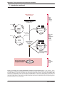
1
Matchmaker™ Library Construction & Screening Kits User Manual PT3529-1 (PR6Z2169) Published 22 December 2006 Matchmaker™ Library Construction & Screening Kits User Manual Table of Contents I. Introduction 4 II. List of Components 7 III. Additional Materials Required 9 IV. Yeast Strains 11 V. Yeast Vectors 14 VI. Protocol Overview: One-Hybrid Library Construction & Screening 16 VI. Protocol Overview: One-Hybrid Library Construction & Screening 17 VII. Constructing a Reporter Vector for One-Hybrid Analysis 18 VIII. Constructing a DNA-BD Fusion Vector for Two-Hybrid Analysis 19 IX. Generating a cDNA Library 21 X. Constructing & Screening One-Hybrid and Two-Hybrid Libraries (overview) 27 XI. Constructing & Screening a One-Hybrid Library 28 XII. Constructing & Screening a Two-Hybrid Library 30 Protocol A: Screen by Yeast Mating 30 Protocol B: Screen by Cotransformation 35 XIII. Analyzing Positive Interactions 38 XIV. Troubleshooting Guide 44 XV. References 47 XVI. Related Products 50 Appendix A: Typical Results of ds cDNA Synthesis 52 Appendix B: Preparation of Competent Yeast Cells—LiAc Method 53 Appendix C: One-Hybrid Control Vector Information 54 Appendix D: Two-Hybrid Control Vector Information 55 List of Tables Table I. Table II. Table III. Table IV. Table V. Table VI. Table VII. Table VIII. Table IX. Table X. Table XI. Table XII. Table XIII. Matchmaker Yeast Strain Genotypes Matchmaker Yeast Strain Phenotypes One-Hybrid System Vectors Two-Hybrid System Vectors Comparison of Matchmaker DNA-BD Vectors Relationship Between Amount of RNA and Optimal Number of Thermal Cycles Set-Up for One-Hybrid Control Cotransformations Control One-Hybrid Cotransformations: Expected Results Set-Up for Control Two-Hybrid Transformations Set-Up for Control Two-Hybrid Matings Set-Up for Control Two-Hybrid Cotransformations Control Two-Hybrid Cotransformations: Expected Results Assembling Master Mixes for PCR Colony Screening Clontech Laboratories, Inc. www.clontech.com 11 11 14 15 19 24 29 29 33 34 36 37 40 Protocol No. PT3529-1 Version No. PR6Z2169 Matchmaker™ Library Construction & Screening Kits User Manual Table of Contents continued List of Figures Figure 1. Figure 2. Figure 3. Figure 4. Figure 5. Figure 6. Figure 7. Figure 8. Figure 9. Figure 10. Figure 11. Figure 12. Figure 13. Figure 14. Figure 15. Figure 16. Figure 17. Figure 18. Figure 19. Screening for protein-DNA interactions with the Matchmaker One-Hybrid System. Screening for protein-protein interactions with the Matchmaker Two-Hybrid System. General steps of yeast one-hybrid and two-hybrid screening. Constructing and screening Matchmaker One-Hybrid and Two-Hybrid libraries. Reporter gene constructs in yeast strains AH109 and Y187. Matchmaker One-Hybrid Library Construction & Screening. Matchmaker Two-Hybrid Library Construction & Screening. Synthesis of high-quality ds cDNA using SMART™ technology. CHROMA SPIN Column and Collection Tubes. Constructing AD fusion libraries by recombination-mediated cloning in yeast. Screening an AD fusion library for two-hybrid interactions. Strategies for analyzing and verifying putative positive one-hybrid and two-hybrid interactions. Yeast mating to verify protein-protein (two-hybrid) interactions. Double-stranded cDNA synthesized from Control Human Placenta Poly A+ RNA. Map of p53HIS2 Control Vector. Map of pGAD-Rec2-53 AD Control Vector. Map of pGADT7-RecT AD Control Vector. Map of pGBKT7-53 DNA-BD Control Vector. Map of pGBKT7-Lam DNA-BD Control Vector. 4 4 5 6 12 16 17 21 26 27 32 39 42 52 54 54 55 55 56 Notice to Purchaser Clontech products are to be used for research purposes only. They may not be used for any other purpose, including, but not limited to, use in drugs, in vitro diagnostic purposes, therapeutics, or in humans. Clontech products may not be transferred to third parties, resold, modified for resale, or used to manufacture commercial products or to provide a service to third parties without written approval of Clontech Laboratories, Inc. Practice of the two-hybrid system is covered by U.S. Patent Nos. 5,283,173, 5,468,614, and 5,667,973 assigned to the Research Foundation of the State University of New York. Purchase of any Clontech two-hybrid reagent does not imply or convey a license to practice the two-hybrid system covered by these patents. Commercial entities purchasing these reagents must obtain a license from the Research Foundation of the State University of New York before using them. Clontech is required by its licensing agreement to submit a report of all purchasers of two-hybrid reagents to SUNY Stony Brook. Please contact the Office of Technology Licensing & Industry Relations at SUNY Stony Brook for license information (Tel: 631.632.9009; Fax: 631.632.1505). SMART™ Technology is covered by U.S. Patent Nos. 5,962,271 and 5,962,272. For-Profit and Not-For-Profit purchasers of SMART™ Products are entitled to use the reagents for internal research. However, the following uses are expressly prohibited: (1) performing services for third parties; (2) identifying nucleic acid sequences to be included on nucleic acid arrays, blots, or in libraries or other cDNA collections which are then sold to third parties. Reproduction, modification, reformulation, or resale of the reagents provided in SMART™ Products is not permitted. For information on licensing SMART™ Technology for commercial purposes, please contact a licensing representative by phone at 650.919.7320 or by e-mail at [email protected]. The pBridge Vector is the property of the Institut National de la Santé et de la Recherche Médicale (INSERM). Inquiries regarding the commercial use or resale of this vector must be directed to INSERM, France. NucleoSpin® and NucleoBond® are a registered trademarks of Macherey-Nagel GmbH & Co. Parafilm® is a registered trademark of the American National Can Co. Clontech, Clontech logo and all other trademarks are the property of Clontech Laboratories, Inc. Clontech is a Takara Bio Company. ©2006 Protocol No. PT3529-1 www.clontech.com Clontech Laboratories, Inc. Version No. PR6Z2169 Matchmaker™ Library Construction & Screening Kits User Manual I. Introduction Matchmaker™ Library Construction & Screening Kits provide a simple method for constructing cDNA libraries for yeast two-hybrid and one-hybrid screening. These kits combine Matchmaker Systems with SMART cDNA Synthesis technology, which allows you to construct cDNA libraries from any tissue source starting with as little as 1 µg of poly A+ RNA or total RNA. Following a routine in vivo cloning step, you can then screen these libraries for one-hybrid and two-hybrid interactions using a sensitive transcriptional assay provided by our Matchmaker Systems. Principle of the one-hybrid assay—a protein-DNA interaction assay One-hybrid assays enable you to identify and characterize proteins that bind to a target, cis-acting DNA sequence—an upstream element that enhances transcription from a downstream minimal promoter. The assay may also be used to map the DNA-binding domain of previously known, or newly identified, DNA-binding proteins. With the Matchmaker One-Hybrid System, you can readily obtain the genes encoding the corresponding DNA-binding protein. In a Matchmaker one-hybrid assay, potential DNA-binding proteins are expressed as fusions to the GAL4 activation domain (AD) by cloning the corresponding cDNA into pGADT7-Rec2, a lowcopy vector designed for one-hybrid screening. One or more tandem copies of the target DNA sequence are cloned into pHIS2, a reporter vector that contains the nutritional reporter gene HIS3. Interaction between a DNA-binding protein and the target sequence stimulates transcription of HIS3 (Figure 1), enabling the yeast host strain, Y187, a His auxotroph, to grow on minimal media lacking histidine. Principle of the two-hybrid assay—a protein-protein interaction assay Two-hybrid assays can be used to identify novel protein-protein interactions, confirm suspected interactions, and define interacting domains. In a Matchmaker Two-Hybrid assay, a bait gene is expressed as a fusion to the GAL4 DNA-binding domain (DNA-BD), while another gene or cDNA is expressed as a fusion to the GAL4 activation domain (AD; Fields & Song, 1989; Chien et al., 1991). When bait and library fusion proteins interact in a yeast reporter strain such as AH109, the DNA-BD and AD are brought into proximity and activate transcription of the reporter genes: ADE2, HIS3, lacZ, and MEL1 (Figure 2). DNA-BD and AD fusions are created by cloning cDNAs into the yeast expression vectors pGBKT7 and pGADT7-Rec. pGBKT7 expresses proteins as fusions to the GAL4 DNA-BD, while pGADT7-Rec expresses proteins as fusions to the GAL4 AD. In yeast, both fusions are expressed at high levels from the constitutive ADH1 promoter (PADH1). Other GAL4-based DNA-BD cloning vectors such as pGBT9, pAS2-1, and pBridge are also compatible with this kit. pBridge Vector can be used to perform three-hybrid assays to identify ternary protein complexes. Biological basis for one-hybrid and twohybrid assays One-hybrid (and two-hybrid) methods are based on the finding that many eukaryotic transcription factors are modular; their transcription activating and DNA-binding domains are structurally and functionally distinct. This allows researchers to construct various gene fusions that, when expressed as fusion proteins in yeast, can simultaneously bind to a target DNA sequence and activate transcription of a downstream reporter (Figures 1 and 2). Matchmaker systems use the transcription activating and DNA-binding domains of GAL4, a well-characterized yeast transcription factor. To learn more about GAL4based yeast hybrid technology, see Zhu, L. & Hannon, G. J., 2000. GAL AD Transcription activator Library protein transcription T T T minimal promoter HIS3 Figure 1. Screening for protein-DNA interactions with the Matchmaker One-Hybrid System. In this construct, three copies of the DNA target (T) have been inserted into the pHIS2 reporter vector. GAL4 AD Bait protein Library protein GAL4 DNA-BD GAL UAS minimal promoter transcription ADE2, HIS3, lacZ, and MEL1 Figure 2. Screening for protein-protein interactions with the Matchmaker Two-Hybrid System. Clontech Laboratories, Inc. www.clontech.com Protocol No. PT3529-1 Version No. PR6Z2169 Matchmaker™ Library Construction & Screening Kits User Manual I. Introduction continued Constructing and screening Matchmaker one-hybrid and two-hybrid libraries Constructing and screening Matchmaker one-hybrid and two-hybrid libraries consists of four main steps (Figure 3). Notice that both procedures follow the same general path. If you intend to screen for two-hybrid interactions, the first step (Step 1) is to construct a DNA-BD fusion vector. If, on the other hand, you intend to screen for one-hybrid interactions, your first step is to construct a DNA target-HIS3 reporter vector. Next (Step 2), use the SMART reagents we provide to generate a cDNA library from the poly A+ or total RNA that you provide. In the case of yeast two-hybrid screening, you may skip RNA isolation, cDNA synthesis, and AD fusion library construction (Step 3) if, instead of preparing your own library, you intend to screen one of our many premade Matchmaker cDNA libraries. Representing a broad range of tissues, these libraries are available as glycerol stocks or pretransformed in yeast strain Y187. We also offer a Matchmaker Custom Library Service. To use this service, send us the tissue or cells you wish to screen, and we will make the AD fusion library for you. Please note, however, that many of our premade and pretransformed Matchmaker cDNA Libraries are constructed in high-copy yeast expression vectors, ideal for two-hybrid work, but less suitable for one-hybrid analysis. We recommend using low copy vectors such as pGADT7-Rec2 and pHIS2 for one-hybrid screening because they generate fewer false positives. Following cDNA synthesis, construct a GAL4 AD fusion library by cloning the cDNA into one of our Matchmaker AD Cloning Vectors: pGADT7-Rec2 if you are constructing a one-hybrid library; pGADT7-Rec if you are constructing a two-hybrid library. The cloning takes place in vivo via homologous recombination (Figure 4).This step takes advantage of the highly efficient recombination system in yeast to fuse ds cDNA with the appropriate GAL4 AD plasmid. With recombinationmediated cloning, library construction and screening (Steps 3 and 4) can be completed in quick succession without the need for any bacterial transformation or amplification steps. Simply transform yeast with the cDNA library and the appropriate Matchmaker vectors; then spread the cells on dropout medium to select for one-hybrid or two-hybrid interactions. One-Hybrid Library Construction & Screening Overview: Section VI 1. Construct a DNA target-reporter vector by cloning your DNA target sequence into pHIS2. Section VII Two-Hybrid Library Construction & Screening Overview: Section VI 1. Construct a DNA-BD fusion vector by cloning your bait gene into pGBKT7 or other GAL4 DNA-BD vector. Section VIII 2. Generate a cDNA library from any tissue or cell source using SMART™ cDNA Synthesis technology. Section IX 3. Construct a GAL AD fusion library using homologous recombination. Sections XI & XII . Screen for one-hybrid or two-hybrid interactions by yeast mating or transformation. Sections XI & XII Figure 3. General steps of yeast one-hybrid and two-hybrid screening. For more detailed flow charts of the one-hybrid and two-hybrid procedures, please refer to Figures 6 and 7. Protocol No. PT3529-1 www.clontech.com Clontech Laboratories, Inc. Version No. PR6Z2169 Matchmaker™ Library Construction & Screening Kits User Manual I. Introduction continued SMART™ cDNA synthesis and amplification by LD-PCR Step 1: cDNA synthesis ~ 5 hours ds cDNA with SMART™ III & CDS III anchors protein bait MCS Kanr pGBKT GAL4 DNA-BD GAL4 AD Two-Hybrid DNA-BD Vector TRP1 pGADT-Rec[2] AD Vector Sma I-linearized LEU2 Amp pGBKT7 or Steps 2 & 3: Library construction (in vivo cloning) ~ 2 hours pHIS2 DNA target HIS3 Kanr pHIS2 One-Hybrid Reporter Vector TRP1 Prepare competent yeast cells and transform GAL4 AD pGADT-Rec[2] AD Vector Sma I-linearized LEU2 Ampr Homologous recombination in yeast mediates GAL AD Vector assembly Plate on selective medium to screen for one-hybrid or two-hybrid interactions Step : Screening 3 days Figure 4. Constructing and screening Matchmaker One-Hybrid and Two-Hybrid libraries. As this diagram shows, recombination-mediated cloning makes library construction and screening quick and efficient. Though not shown here, two-hybrid libraries can also be screened by yeast mating (See Section XII for details). For details about SMART cDNA synthesis and amplification, please refer to Section IX. pGADT7-Rec is used for two-hybrid library construction and screening, while pGADT7-Rec2 is used for one-hybrid library construction and screening.Though related, the two vectors, denoted as pGADT7-Rec[2] in the figure, have different replication elements. See Section X and the corresponding Vector Information Packets for more information. Clontech Laboratories, Inc. www.clontech.com Protocol No. PT3529-1 Version No. PR6Z2169 Matchmaker™ Library Construction & Screening Kits User Manual II. List of Components This kit contains sufficient reagents to make 5 one-hybrid (Cat. No. 630304) or 5 two-hybrid (Cat. No. 630445) libraries. Store Deionized H2O, CHROMA SPIN Columns, NaCl Solution, Dropout (DO) Supplements, NaOAc, LiAc, PEG, TE Buffer, and YPD Plus Medium at room temperature. Store yeast strains, Control Poly A+ RNA, and the SMART III Oligo at –70°C. Store all other reagents at –20°C. First-strand cDNA synthesis • 10µl SMART III Oligo (10 µM; 5'-AAGCAGTGGTATCAACGCAGAGTGGCCATTATGGCCGGG-3') • 10µl CDS III Primer (10 µM; 5'-ATTCTAGAGGCCGAGGCGGCCGACATG-d(T)30VN-3')* • 10µl CDS III/6 Primer (10 µM; 5'-ATTCTAGAGGCCGAGGCGGCCGACATG-NNNNNN-3')* *N = A, G, C, or T; V = A, G, or C • 20µl MMLV (Moloney Murine Leukemia Virus) Reverse Transcriptase • 7µl RNase H • 100µl 5X First-Strand Buffer (250 mM Tris (pH 8.3); 30 mM MgCl2; 375 mM KCl) • 100µl DTT (dithiothreitol; 20 mM) • 5µl Control Poly A+ RNA (Human Placenta; 1 µg/µl) • 50µl dNTP Mix (dATP, dCTP, dGTP, dTTP, 10 mM each) • 500 µl Deionized H2O (Cat. No. 630445 only) cDNA amplification • 50µl 5' PCR Primer (10 µM; 5'-TTCCACCCAAGCAGTGGTATCAACGCAGAGTGG-3') • 50µl 3' PCR Primer (10 µM; 5'-GTATCGATGCCCACCCTCTAGAGGCCGAGGCGGCCGACA-3') • 500µl 10X GC-Melt Solution cDNA purification • 10 CHROMA SPIN+TE-400 Columns • 300µl Sodium Acetate (3 M; pH 4.8) One-Hybrid Library Construction (Cat. No. 630304) • 20µg pHIS2 Reporter Vector (500 ng/µl) • 20µg pGADT7-Rec2 AD Cloning Vector (Sma I-linearized; 500 ng/µl) • 20µg pGAD-Rec2-53 Control Vector (500 ng/µl) • 20µg p53HIS2 Control Vector (500 ng/µl) • 0.5 ml S. cerevisiae strain Y187 • 50ml NaCl Solution (0.9%) • 10g –Leu DO Supplement • 10g –Trp DO Supplement • 10g –Leu/–Trp DO Supplement • 10g –His/–Leu/–Trp DO Supplement Two-Hybrid Library Construction (Cat. No. 630445) • 20µg pGBKT7 DNA-BD Cloning Vector (500 ng/µl) • 25µg pGADT7-Rec AD Cloning Vector (Sma I-linearized; 500 ng/µl) • 20µg pGBKT7-53 Control Vector (500 ng/µl) • 20µg pGBKT7-Lam Control Vector (500 ng/µl) • 20µl SV40 Large-T PCR Fragment (25 ng/µl) • 0.5 ml S. cerevisiae strain AH109 Protocol No. PT3529-1 www.clontech.com Version No. PR6Z2169 Clontech Laboratories, Inc. Matchmaker™ Library Construction & Screening Kits User Manual • • • • • II. List of Components continued 0.5 ml 50ml 10g 10g 10g S. cerevisiae strain Y187 NaCl Solution (0.9%) –Leu DO Supplement –Leu/–Trp DO Supplement –Ade/–His/–Leu/–Trp DO Supplement Yeastmaker Yeast Transformation System 2 (Cat. No. 630439) includes the following: • 50ml 1 M LiAc (10X) • 50ml 10X TE Buffer • 50ml YPD Plus Liquid Medium • 20µl pGBT9 (0.1 µg/µl; control plasmid) • 2 x 1ml Herring Testes Carrier DNA, denatured (10 mg/ml) • 2 x 50ml 50% PEG 3350 Clontech Laboratories, Inc. www.clontech.com Protocol No. PT3529-1 Version No. PR6Z2169 Matchmaker™ Library Construction & Screening Kits User Manual III. Additional Materials Required The following reagents are required but not supplied. Store all reagents and solutions at room temperature (20–22°C) unless specified otherwise. irst-strand cDNA synthesis and SMART™ PCR cDNA amplification F • Advantage® 2 PCR Kit (Cat. Nos. 639206 & 639207) • Sterile, 0.5-ml microcentrifuge tubes • Poly A+ or total RNA • Mineral oil • Thermal Cycler Note: The cycling parameters in this protocol were set using a hot-lid thermal cycler and may not be optimal for non hot-lid cyclers. • DNA size markers (1-kb DNA ladder) • 1.2% Agarose/EtBr gel cDNA size fractionation • 1.5-ml sterile microcentrifuge tubes • Ring-stand with small clamp for holding CHROMA SPIN columns • 95% ethanol (–20°C) • 1% xylene cyanol Constructing bait plasmids • Competent E. coli cells. Use a general purpose strain such as DH5α or Fusion-Blue Competent Cells (Cat. No. 636700) Fusion-Blue Competent Cells are an E. coli K-12 strain that provides high transformation efficiency paired with blue-white screening capability when used with the appropriate plasmids. The strain carries recA and endA mutations that make it a good host for obtaining high yields of plasmid DNA. • T4 DNA ligase • 10X T4 ligation buffer (Sambrook et al., 1989; or the buffer provided with the enzyme) • LB/amp plates • 50 mM NaCl • Materials for purifying plasmid from E. coli transformants Yeast transformation (Prepare reagents in sterile containers) • PEG/LiAc Solution (polyethylene glycol 3350/lithium acetate) Prepare a fresh 10-ml solution just prior to transformation using the stock solutions provided: Mix 8 ml of 50% PEG 3350 with 1 ml of 10X TE Buffer and 1 ml of 1M LiAc (10X). • 1.1X TE/LiAc Solution Should be freshly prepared before each transformation using the stock solutions provided: Combine 1.1 ml of 10X TE with 1.1 ml of 1 M LiAc (10X). Bring the total volume to 10 ml using sterile, deionized H2O. • Dimethyl Sulfoxide (DMSO; Sigma Cat. No. D8418) Protocol No. PT3529-1 www.clontech.com Clontech Laboratories, Inc. Version No. PR6Z2169 Matchmaker™ Library Construction & Screening Kits User Manual III. Additional Materials Required continued Yeast culture & mating • YPD Medium Cat. No. 630409; or prepare your own; see the Yeast Protocols Handbook (PT3024-1) • YPDA medium (YPD medium supplemented with 30 mg/L adenine hemisulfate; see the Yeast Protocols Handbook) • TE buffer or sterile, distilled H2O • Appropriate sterile tubes or flasks for transformations • 100- and 150-mm culture plates • Sterile glass rod, bent pasteur pipette, or 5-mm glass beads for spreading cells on plates • X-α-Gal (Cat. No. 630407) for blue/white screening of yeast two-hybrids expressing MEL1 (α-galactosidase) • Minimal SD Base with and without agar (Cat. Nos. 630412 and 630411) • 3-amino-1,2,4-triazole (3-AT; Sigma Cat. No. A8056; for suppressing background growth on SD minimal media lacking His) • Kanamycin stock solution (50 mg/ml in H2O; 1000X); Store at –20°C. • Ampicillin stock solution (50 mg/ml in H2O; 1000X); Store at –20°C. ong-term library storage L • 100% Glycerol • Freezing Medium: YPD medium with 25% (v/v) glycerol Two-Hybrid Library Construction & Screening The following dropout (DO) supplements are not supplied with the Matchmaker Library Construction & Screening Kit (Cat. No. 630445). You must obtain these supplements separately from a commercial supplier or prepare them yourself using the recipe given in Appendix C of the Yeast Protocols Handbook (PT3024-1). • –Trp DO Supplement Required for selection of Matchmaker DNA-BD cloning vectors in yeast • –His/–Leu/–Trp DO Supplement Optional triple-dropout supplement for low stringency screening of yeast two-hybrid libraries • –His/–Trp DO Supplement Required for testing yeast strains transformed with a DNA-BD plasmid for background growth on SD minimal media lacking His • –Ade/–Trp DO Supplement Required for testing yeast strains transformed with a DNA-BD plasmid for background growth on SD minimal media lacking Ade One-Hybrid Library Construction & Screening The following dropout (DO) supplement is not supplied with the Matchmaker One-Hybrid Library Construction & Screening Kit (Cat. No. 630304). You must obtain this supplement separately from a commercial supplier or prepare it yourself using the recipe given in Appendix C of the Yeast Protocols Handbook (PT3024-1). • –His/–Trp DO Supplement Required for testing yeast strains transformed with a pHIS2 reporter plasmid for background growth on SD minimal media lacking His PCR Colony-Screening • Matchmaker AD LD-Insert Screening Amplimer Set (Cat. No. 630433) Clontech Laboratories, Inc. www.clontech.com 10 Protocol No. PT3529-1 Version No. PR6Z2169 Matchmaker™ Library Construction & Screening Kits User Manual IV. Yeast Strains For additional information on the growth and maintenance of yeast, see theYeast Protocols Handbook (PT3024-1). We also recommend Guthrie & Fink’s Guide to Yeast Genetics and Molecular Biology (1991) and Heslot & Gaillardin’s Molecular Biology and Genetic Engineering of Yeasts (1992). A. Genotypes table i. matchmaker yeast strain genotypes Strain Genotypea AH109b Y187 References MATa, trp1-901, leu2-3, 112, ura3-52, his3-200, gal4Δ, gal80Δ, LYS2 : : GAL1UAS-GAL1TATA-HIS3, GAL2UAS -GAL2TATA-ADE2, URA3 : : MEL1UAS-MEL1TATA-lacZ, MEL1 James et al., 1996; Our unpublished observations MATα, ura3-52, his3-200, ade2-101, trp1-901, Harper et al., 1993 leu2-3, 112, gal4Δ, met –, gal80Δ, URA3 : : GAL1UAS-GAL1TATA-lacZ, MEL1 a The GAL1, GAL2, and MEL1 upstream activating sequences (UASs) are responsive to the GAL4 transcriptional activator. The trp1, his3, gal4, and gal80 mutations are all deletions; leu2-3, 112 is a double mutation. b AH109 is a derivative of strain PJ69-2A and includes the ADE2 and HIS3 nutritional markers and an endogenous MEL1 gene (James et al., 1996). The lacZ reporter gene was introduced into PJ69-2A to create strain AH109. B. Phenotypes It is important to verify the phenotypes of the AH109 and Y187 strains (Table II). 1.To recover strains from frozen stock, scrape a small amount of cells from the surface with a sterile loop or wooden stick and streak them onto YPDA plates. 2.Incubate plates at 30°C for 3–5 days until colonies appear. Propagate additional cultures only from isolated colonies on this working stock plate. Notes: • AH109 (and transformants derived from this strain) should be maintained on adenine-supplemented YPD (i.e.,YPDA) for optimal viability of the strain and to prevent selection of spontaneous ade1 or ade5 mutations (Guthrie & Fink, 1991). • If you cannot recover the strain by scraping the frozen stock, the cells may have settled to the bottom of the tube before the stock was frozen. If this happens, thaw the frozen culture on ice and vortex it before restreaking. • Although nonlibrary stock cultures may be thawed and refrozen several times without significantly decreasing the viability, we recommend that you divide the once-thawed stock into aliquots before you refreeze it. This will keep the viability higher and will reduce the risk of bacterial contamination. 3.Test for the nutritional requirements shown in Table II. a. Using a sterile loop or toothpick, streak 3–4 colonies from the working stock onto separate, appropriate SD selection plates. b. Incubate plates at 30°C for 4–6 days. Yeast grows slower on SD selection medium than on YPDA. c. Compare your results with those shown in Table II. Proceed only if AH109 and Y187 have the expected phenotypes. 4.Use well-isolated colonies from the verified working stock plate to inoculate liquid cultures for mating or for preparing competent cells. Seal the verified working stock plate with Parafilm and store at 4°C.table ii. Matchmaker yeast strain phenotypes table ii. matchmaker yeast strain phenotypes Strain SD/–Ade SD/–Met SD/–Trp SD/–Leu SD/–His SD/–Ura YPDA AH109 – + – – – + + Y187 – – – – – + + Notes: • AH109 and Y187 can grow on SD/–Leu/–Trp if functional TRP1 and LEU2 genes are introduced. • AH109 and AH109/Y187 diploids can grow on SD/–Ade/–His if the ADE2 and HIS3 genes—carried by AH109—are activated (i.e., in the presence of GAL4). Protocol No. PT3529-1 www.clontech.com Version No. PR6Z2169 Clontech Laboratories, Inc. 11 Matchmaker™ Library Construction & Screening Kits User Manual IV. Yeast Strains continued C. Mating type compatibilities • Y187 (MATα) can mate with AH109, HF7c, CG-1945, Y190, or SFY526 (all MATa). D. Colony Color and Size • Y187 carries the ade2-101 mutation and AH109 exhibits the Ade– phenotype in the absence of GAL4. On medium with low amounts of adenine, the colonies will turn pink after a few days and may turn darker as the colony ages.When grown onAde-supplemented medium, the color change may not be noticeable.These colonies grow to >2 mm in diameter. However, small (<1 mm) white colonies will form at a rate of 1–2% due to spontaneous mutations that eliminate mitochondrial function (Holm, 1993). Avoid these white colonies when inoculating cultures. • Y187 grows more slowly and forms noticeably smaller colonies (on average) than AH109. E. Reporter genes AH109 contains four reporters—ADE2, HIS3, MEL1, and lacZ—under the control of three distinct GAL4 upstream activating sequences (UASs) and TATA boxes (Figure 5). The ADE2 reporter alone provides strong nutritional selection. For higher stringency, and to reduce the incidence of false positives, select for ADE2 and HIS3 (James et al., 1996). You also have the option of assaying for MEL1, which encodes α-galactosidase. MEL1 is endogenous to both Y187 and AH109. Because α-galactosidase is a secreted enzyme, its activity can be detected by adding X-α-Gal (Cat. No. 630407) to the selection plate: If MEL1 is active and X-α-Gal is present, the colony will turn blue. lacZ in Y187 exhibits a high level of induced β-galactosidase activity in a positive two-hybrid assay because it is under the control of the intact GAL1 UAS. AH109 Constructs GAL1 UAS GAL1 TATA HIS3 GAL2 UAS GAL2 TATA ADE2 MEL1 UAS MEL1 TATA lacZ MEL1 UAS MEL1 TATA MEL1 lacZ GAL1 TATA lacZ Y1 Constructs GAL1 UAS Figure 5. Reporter gene constructs in yeast strains AH109 andY187. Strain AH109 is a derivative of strain PJ69-2A and includes the ADE2 and HIS3 nutritional markers (James et al., 1996). MEL1 is an endogenous GAL4-responsive gene.The lacZ reporter gene was introduced into PJ69-2A to create strain AH109.The HIS3, ADE2, and MEL1/lacZ reporter genes are under the control of three completely heterologous GAL4-responsive UAS and promoter elements—GAL1, GAL2, and MEL1, respectively. Clontech Laboratories, Inc. www.clontech.com 12 Protocol No. PT3529-1 Version No. PR6Z2169 Matchmaker™ Library Construction & Screening Kits User Manual IV. Yeast Strains continued F. Leaky HIS3 expression • 3-amino-1,2,4-triazole (3-AT) is a competitive inhibitor of the yeast HIS3 protein (His3p). 3-AT is used to inhibit low levels of His3p expressed in a leaky manner and thus to suppress background growth on SD medium lacking histidine (Fields, 1993; Durfee et al., 1993). • Transformants derived from AH109 may show slightly elevated HIS3 expression because of intrinsic DNA-binding properties of the bait protein. A small amount of 3-AT is generally sufficient to suppress background growth on SD/–His. However, if you are selecting for both HIS3 and ADE2 expression, it is generally not necessary to suppress HIS3 leakiness in the initial library screen. • Some yeast strains have relatively high basal levels of His3p. If you useY190 (MATa) as a host strain, 25–45 mM 3-AT will be required in the medium to suppress background growth. • To optimize the 3-AT concentration in your selection medium: Before starting this procedure, note that –His/–Trp Dropout Supplement is not supplied with this kit.You must purchase –His/–Trp dropout supplement separately or prepare your own using the recipe given in Appendix C of the Yeast Protocols Handbook (PT3024-1). 1. Plate yeast transformants on a series of SD/–His/–Trp plates containing different concentrations of 3-AT. • If you are working with AH109 transformants containing DNA-BD plasmids such as pGBKT7, we recommend you start by testing [3-AT] in the range 0 to 15 mM (e.g., 0, 2.5, 5, 7.5, 10, 12.5, and 15 mM). • If you are working with Y187 transformants containing pHIS2 reporter plasmids, we recommend you start by testing [3-AT] in the range 10 to 60 mM. Note:These are recommendations only.The optimal concentration may be slightly higher or lower depending on the construct and strain used. 2. Use the lowest concentration of 3-AT that, after one week, allows only small (<1 mm) colonies to grow. Too much 3-AT in the medium can kill freshly transformed cells. Protocol No. PT3529-1 www.clontech.com Version No. PR6Z2169 Clontech Laboratories, Inc. 13 Matchmaker™ Library Construction & Screening Kits User Manual V. Yeast Vectors A. One-Hybrid System 1.Cloning Vectors • pHIS2 is a one-hybrid reporter vector that contains the HIS3 nutritional reporter gene. It has a multiple cloning site (MCS) upstream of the HIS3 reporter gene so that a cis-acting DNA target sequence can be inserted, and therefore, linked to the minimal promoter of the HIS3 locus (PminHIS3). It also contains a CEN6/ARS4 sequence for stable, low-copy propagation in yeast. • pGADT7-Rec2 is a cloning vector that can be used to express a protein of interest as a fusion with the GAL4 activation domain (AD).This vector is engineered for homologous recombination-mediated cloning in yeast (Figure 4).Thus, you can construct cDNA/AD fusion libraries by transforming yeast with Sma I-linearized pGADT7-Rec2 (provided) and ds cDNA generated with the SMART library construction protocol (Section IX). 2.Control Vectors (Appendix C) • p53HIS2 is a positive control reporter vector that contains three tandem copies of the cis-acting DNA consensus sequence recognized by p53. p53HIS2 was constructed by inserting the DNA targets into the multiple cloning site of pHIS2. As a result, the DNA targets are positioned just upstream of the minimal promoter of the HIS3 locus (PminHIS3) and the HIS3 reporter gene. • pGAD-Rec2-53 is a positive control vector that encodes murine p53 as a fusion with the GAL4 AD. Yeast cells that contain both p53HIS2 and pGAD-Rec2-53 should grow on minimal SD media lacking histidine—i.e., on SD/–His/–Leu/–Trp. table iII. One-hybrid system vectors Use Epitopea Yeast selectionb Bacterial selectionc Cloning vectors pHIS2 pGADT7-Rec2 (Sma I-linearized) Link any DNA target to HIS3 Express potential DNA-binding proteins as AD fusions HA Control vectors p53HIS2b pGAD-Rec2-53 Detect DNA-binding activity of p53 Express p53 as an AD fusion TRP1 kanamycin LEU2 ampicillin TRP1 kanamycin HA LEU2 ampicillin a HA = hemagglutinin;These epitope tags can be used to verify protein-protein interactions in vitro by coimmunoprecipitation (Co-IP) using the antibodies and protocol provided with the Matchmaker Co-IP Kit (Cat. No. 630449).They are not intended to be used for detection, affinity purification, or Co-IP of hybrid proteins expressed in yeast. b The reporter and AD vectors have different nutritional markers, so they can be independently selected when yeast transformants are plated on SD minimal medium lacking specific nutrients. The selection medium you choose depends on which plasmids you are using, whether you are selecting for one or two plasmids, and whether you are selecting for colonies in which one hybrid interactions are occurring. c The vectors carry different antibiotic markers so that they can be independently selected in E. coli. Clontech Laboratories, Inc. www.clontech.com 14 Protocol No. PT3529-1 Version No. PR6Z2169 Matchmaker™ Library Construction & Screening Kits User Manual V. Yeast Vectors continued B Two-Hybrid System 1.Cloning Vectors • pGBKT7: Used to express a protein of interest as a fusion with the GAL4 DNA binding domain (DNA-BD). • pGADT7-Rec: Used to express a protein of interest as a fusion with the GAL4 activation domain (AD). This vector is engineered for homologous recombination-mediated cloning. pGADT7-Rec is provided as Sma I-digested linear DNA. 2.Control Vectors a. Positive Control • pGBKT7-53 encodes a fusion between the GAL4 DNA-BD and murine p53. • SV40 Large T PCR Fragment encodes SV40 largeT-antigen. Use this DNA fragment together with pGADT7-Rec to check the transformation-recombination efficiency in yeast. In vivo, SV40 Large T PCR Fragment and pGADT7-Rec recombine to form pGADT7-RecT, which encodes a fusion between the GAL4 AD and large Tantigen. • p53 and SV40 largeT-antigen interact in a yeast two-hybrid assay (Li & Fields, 1993; Iwabuchi et al., 1993). b. Negative Control • pGBKT7-Lam encodes a fusion of the GAL4 DNA-BD with human lamin C and provides a control for a fortuitous interaction between an unrelated protein and either the pGADT7-RecT control or your AD/library plasmid. Lamin C neither forms complexes nor interacts with most other proteins (Bartel et al., 1993b; S. Fields, pers. comm.; Ye & Worman, 1995). table iV. two-hybrid system vectors Use Epitopea Yeast selectionc Bacterial selectiond Cloning vectors pGBKT7 pGADT7-Rec (Sma I-linearized) Express any protein as a GAL4 DNA-BD fusion Express any protein as a GAL4 AD fusion Control vectors pGADT7-RecT b pGBKT7-53 pGBKT7-Lam Express SV40 large T-antigen HA as a GAL4 AD fusion Express p53 as a GAL4 c-Myc DNA-BD fusion Express lamin C as a GAL4 c-Myc DNA-BD fusion c-Myc TRP1 kanamycin HA LEU2 ampicillin LEU2 ampicillin TRP1 kanamycin TRP1 kanamycin a HA = hemagglutinin;These epitope tags can be used to verify protein-protein interactions in vitro by coimmunoprecipitation (Co-IP) using the antibodies and protocol provided with the Matchmaker Co-IP Kit (Cat. No. 630449).They are not intended to be used for detection, affinity purification, or Co-IP of hybrid proteins expressed in yeast. b Created by homologous recombination in vivo by cotransforming yeast with SV40 Large T PCR Fragment and pGADT7Rec. c The DNA-BD and AD vectors have different nutritional markers, so they can be independently selected when yeast transformants are plated on SD minimal medium lacking specific nutrients. The selection medium you choose depends on which plasmids you are using, whether you are selecting for one or two plasmids, and whether you are selecting for colonies in which two hybrid proteins are interacting. d The vectors carry different antibiotic markers so that they can be independently selected in E. coli.. Protocol No. PT3529-1 www.clontech.com Version No. PR6Z2169 Clontech Laboratories, Inc. 15 Matchmaker™ Library Construction & Screening Kits User Manual VI. Protocol Overview: One-Hybrid Library Construction & Screening Poly A+ RNA 5' poly A 3' CDS III oligo(dT) or random primer First-strand synthesis coupled with (dC) tailing by RT 5' SMART™ III Oligonucleotide 5' GGG CCC poly A Template switching and extension by RT GGG CCC poly A Amplification by LD-PCR ds cDNA with SMART™ III & CDS III anchors Figure 6. Matchmaker One-Hybrid Library Construction & Screening. Clontech Laboratories, Inc. www.clontech.com 16 Protocol No. PT3529-1 Version No. PR6Z2169 Matchmaker™ Library Construction & Screening Kits User Manual VI. Protocol Overview: Two-Hybrid Library Construction & Screening Construct a DNA-BD Fusion Section VII Clone bait gene into pGBKT7 or other Matchmaker GAL4-based DNA-BD vector. Prepare competent yeast cells Generate a cDNA Library Section IX Appendix B 100 ng of total RNA or 25 ng of poly A+ RNA Transform AH109 and Y187 with bait plasmid. Test for autonomous reporter gene activation and cell toxicity. Synthesize first-strand cDNA. or Amplify ds cDNA by LD-PCR. Purify (size-select) ds cDNA. Protocol A • Construct an AD Fusion Library • Screen by Yeast Mating or Section XII Inactive and Nontoxic Active or Toxic Prepare Y187 cells for mating. See Troubleshooting Guide. Protocol B • Construct an AD Fusion Library • Screen by Cotransformation Section XII Prepare competent AH109 yeast cells Appendix B Library Construction [Three-Step] Cotransform yeast strain AH109 with: • ds cDNA • pGADT7-Rec Library Construction [One-Step] Cotransform yeast strain AH109 with: • ds cDNA • pGADT7-Rec • DNA-BD/bait plasmid Select transformants on SD/–Leu. Harvest (Pool) Transformants. Two-Hybrid Screening Mate with Y187 pretransformed with DNA-BD/bait plasmid. Two-Hybrid Screening Select for transformants expressing interacting proteins. Analysis Verify positive interactions. Select for yeast diploids expressing interacting proteins. Analysis Verify positive interactions. Figure 7. Matchmaker Library Construction & Screening Kit. Two-hybrid libraries may be screened by either yeast mating or cotransformation. Protocol No. PT3529-1 www.clontech.com Version No. PR6Z2169 Clontech Laboratories, Inc. 17 Matchmaker™ Library Construction & Screening Kits User Manual VII. Constructing a Reporter Vector for One-Hybrid Analysis A. Background To use the Matchmaker One-Hybrid System to screen a cDNA library for DNA-binding proteins, you must have identified a true or putative target element. It must be precisely defined using, for example, deletion and/or point mutation analysis. A construct composed of one or more tandem copies of your target regulatory element bordered by restriction sites is then prepared and inserted into the multiple cloning site (MCS) of pHIS2. This links the target element to the HIS3 reporter gene. Inserting your target element may alter the level of background HIS3 expression. Therefore, constructs should be tested for background (leaky) HIS3 expression before you start a one-hybrid analysis. Background growth due to leaky HIS3 expression is controlled by adding 3-AT to the selection medium, as described in Section IV.F. B. Synthesize Your Target Element Each target-reporter construct should contain at least one copy of the DNA target element inserted upstream of the reporter gene. Many early studies indicated that the reporter should contain at least three tandem copies of the DNA target. However, as Wei et al. (1999) have demonstrated, a single copy may be sufficient in many cases. For more information about target copy number, see Ghosh et al., 1993. Tandem copies may be generated by various methods, but we have found the most convenient and reliable method for generating them to be oligonucleotide synthesis. It works nicely because well-defined regulatory elements are usually <20 bp. 1.Design two antiparallel oligonucleotides, one representing the sense strand and the other its antisense complement. Note: The sense strand should consist of one or more copies of the target element with a different restriction site on each end. When the two strands are annealed, the resulting double-stranded DNA will have a different overhang at each end for directional cloning into pHIS2. See the pHIS2 Vector Information Packet (PT3705-5) for a diagram of the multiple cloning site. 2.Synthesize both strands without 5' phosphates (according to the protocol of the synthesizer manufacturer). C. Insert Your DNA Target into the Multiple Cloning Site of pHIS2 1.For each construct planned, mix 0.1 µg of sense-strand and 0.1 µg of antisense-strand oligonucleotide in 10 µl of 50 mM NaCl. 2.Anneal the oligonucleotides by heating at 70°C for 5 min and then slowly cooling to room temperature (~30 min). 3.Completely digest 0.1 µg of pHIS2 in a 20-µl double digest using an appropriate pair of restriction enzymes. Incubate at 37°C for 2 hr, or as directed by the enzyme manufacturer. 4.Electrophorese a 2-µl sample of the digest on a 1% agarose gel to confirm that the plasmid has been efficiently linearized. 5.Combine 5 µl of digested plasmid with 1 µl of annealed oligo and 4 µl of H2O. 6. Add 1.2 µl of 10X T4 ligation buffer and 0.8 µl (at least 0.8 units) of T4 DNA ligase, and incubate at room temperature for 4 hr. Note: Since the molar ratio of oligonucleotide to vector is 100:1 or greater, gel purification to remove the stuffer fragment is unnecessary. 7.Separately transform competent E. coli cells with each construct using a standard method (Sambrook et al., 1989). We recommend using a general purpose strain such as DH5α or Fusion-Blue Competent Cells. 8. Plate transformants on LB/kan plates, and incubate at 37°C overnight. 9. Prepare plasmid using any standard method that yields highly pure DNA (Sambrook et al., 1989). Check for inserts by electrophoresing on a 2% agarose gel and sequencing across the junctions. D. Test your Target-Reporter Construct for Background HIS3 Expression 1.Transform Y187 with the target-reporter construct using a small-scale protocol. (For example, see the small-scale protocol used in Section XI.C.) 2.Follow the procedure in Section IV.F to determine the optimum concentration of 3-AT to use in the selection medium. For example, we find that 10 mM 3-AT is sufficient to suppress background growth of Y187 cells transformed with p53HIS2 Control Vector. Clontech Laboratories, Inc. www.clontech.com 18 Protocol No. PT3529-1 Version No. PR6Z2169 Matchmaker™ Library Construction & Screening Kits User Manual VIII.Constructing a DNA-BD Fusion Vector for Two-Hybrid Analysis A. Construct a DNA-BD Fusion • You can generate a GAL4 DNA-BD fusion gene if compatible restriction sites are present in the test gene and the corresponding vector (Table V). If not, generate the gene fragment by PCR using primers that contain the desired restriction site (Scharf, 1990). A restriction site at the end of a gene can often be changed into a different site or put into a different reading frame by using a PCR primer that incorporates the desired mutation. Alternatively, if you have already cloned your gene into a CreatorTM DonorVector, use Cre recombinase to transfer your gene to pLP-GBKT7. Refer to the Creator DNA Cloning Kits User Manual (PT3460-1) for details. • For more detailed information on cloning, see Sambrook et al. (1989). table V. comparison of Matchmaker dna-bd vectors DNA-BD vector Description Size pGBKT7 GAL4(1–147) DNA-BD TRP1, Kanr 7.3 kb High pBridgea GAL4(1–147) DNA-BD TRP1, Ampr 6.5 kb Low pGBT9b GAL4(1–147) DNA-BD, TRP1, Ampr 5.4 kb Low pAS2-1b GAL4(1–147) DNA-BD, TRP1, Ampr, CYH s2 8.4 kb High pLP-GBKT7c GAL4(1–147) DNA-BD, TRP1, Kanr, loxP 7.5 kb High Protein Expression Contains two distinct expression cassettes for investigating ternary protein complexes. DNA-BD vectors used in previous Matchmaker Two-Hybrid Systems. c Creator Acceptor Vector (LP = loxP). Accepts a gene of interest from any Creator Donor Vector and expresses it as a GAL4 DNA-BD fusion. a b 1.Purify the gene fragment. Note: We recommend the NucleoSpin Extraction Kit (Cat. No. 635961) for rapid isolation of DNA fragments. 2.Digest the DNA-BD vector with the appropriate restriction enzyme(s), treat with phosphatase, and purify. 3.Ligate the appropriate vector and insert. Transform ligation mixtures into E. coli. 4.Identify insert-containing plasmids by restriction analysis or PCR. B. Test the DNA-BD Fusion for Transcriptional Activation 1.Transform AH109 and Y187 with the hybrid construct using a small-scale transformation protocol such as the one given in Section XII.A.7. Plate transformants on the following media*: • SD/–Trp/X-α-Gal • SD/–His/–Trp/X-α-Gal • SD/–Ade/–Trp/X-α-Gal Include a negative control. For example, transform cells with an "empty" DNA-BD vector. * Note: The dropout supplements required to make these media are not supplied with the Matchmaker Library Construction & Screening Kit (Cat. No. 630445). You must purchase these supplements separately from a commercial supplier, or prepare them yourself using the recipe given in Appendix C of the Yeast Protocols Handbook (PT3024-1). 2.Analyze results. • Bait protein is inactive if the transformant colonies are white and do not grow on SD/–His/–Trp or SD/–Ade/–Trp. Go to Steps 5–6. • Bait protein is active if transformant colonies are blue and grow on SD/–His/–Trp or SD/–Ade/–Trp. Continue with Steps 3–4. 3.If a bait strain exhibits background growth on –His medium, you may be able to eliminate (or reduce) the background by adding 3-AT to the selection medium (Section IV.F). Alternatively, use –Ade/–His/–Leu/–Trp medium for the library screening. 4.If a bait strain exhibits background growth on –Ade and –His medium, it will be difficult to use this bait protein in a two-hybrid library screening. See Troubleshooting Guide. Protocol No. PT3529-1 www.clontech.com Version No. PR6Z2169 Clontech Laboratories, Inc. 19 Matchmaker™ Library Construction & Screening Kits User Manual VIII.Constructing a DNA-BD Fusion Vector for Two-Hybrid Analysis continued 5.[Optional] If a known protein partner for your bait protein is available, use it to check whether a two-hybrid interaction is detectable with this particular DNA-BD/bait fusion. 6.[Optional] Verify protein expression: • PrepareWestern blots from the lysate of transformants and probe the blots with antibodies to the GAL4 DNA-BD (Cat. No. 630403). Use untransformed yeast lysate as a control. Notes: • Using polyclonal antibodies may result in multiple cross-reacting bands. • The level of expression from pGBT9 or pBridge is too low to permit detection using our GAL4 DNA-BD Monoclonal Antibody. C. Test the DNA-BD Fusion for Toxicity • Compare the growth rate in liquid culture of Y187 cells transformed with the "empty" DNA-BD vector and cells transformed with the DNA-BD/bait plasmid. If the bait strain grows noticeably slower, your DNA-BD/bait protein may be toxic. Note: We also recommend checking for toxicity in strain AH109. • Use a Small-ScaleYeastTransformation Protocol (such as the one given in Section XII.A.7) to prepare transformants. Select transformants on SD/–Trp, then prepare an overnight culture as follows: 1. Inoculate 50 ml of SD/–Trp/Kan (20 µg/ml) with one large (2–3 mm) colony. 2. Incubate at 30°C overnight (16–24 hr) with shaking at 250–270 rpm. 3. Check the OD600 of the culture; it should be ≥0.8. If the OD600 is much less than 0.8, the DNA-BD fusion may be toxic (see the Troubleshooting Guide). If the fusion does not appear to hamper yeast growth—i.e. is nontoxic—and you plan to screen your two-hybrid library by yeast mating, continue with Steps 4–7. If you are planning to screen by cotransformation, you may stop here and proceed to Section IX. 4. Centrifuge at 600 x g for 5 min. 5. Remove supernatant. 6. Resuspend in ~5 ml SD/–Trp liquid medium. Count cells using a hemacytometer. The cell density should be ≥1 x 10⁹ cells/ml. 7. Mate this bait strain with your AD fusion library host strain (Section XII.A.4). Note: Y187 (MATα) can mate with AH109, HF7c, CG-1945, Y190, or SFY526 (all MATa). Clontech Laboratories, Inc. www.clontech.com 20 Protocol No. PT3529-1 Version No. PR6Z2169 Matchmaker™ Library Construction & Screening Kits User Manual IX. Generating a cDNA Library A. Generating a cDNA library with SMART™ technology Messenger RNA transcripts are efficiently copied into ds cDNA using SMART (Switching Mechanism at 5' end of RNA Transcript) technology (Zhu, Y. Y., et al., 2001). This cDNA synthesis and amplification system is particularly well suited for one-hybrid and two-hybrid library construction because it consistently delivers high yields of cDNA while maintaining sequence representation. By maintaining the complexity of the original tissue, the SMART procedure provides you with the best opportunity of detecting rare and potentially novel interactions during yeast one-hybrid and two-hybrid screening. B. How SMART cDNA Synthesis and Amplification Works In the first-strand cDNA synthesis step, MMLV (Moloney Murine Leukemia Virus) Reverse Transcriptase (RT) is used to transcribe RNA into DNA. To prime RNA for cDNA synthesis, you may use either a modified oligo(dT) primer (our CDS III Primer) or a random primer (our CDS III/6 Primer). The composition of the resulting cDNA library may differ depending on which primer you choose. If you use the CDS III Primer, which hybridizes to the 3'-end of poly A+ RNA, sequences close to the 5'-end of the transcript may be slightly under-represented. If instead you use the CDS III/6 Primer, a random primer that can hybridize to many different sequences on the RNA template, your library should contain a variety of 5'- and 3'-end sequences, which are represented in near equal proportions. When MMLV RT encounters a 5'-terminus on the template, the enzyme’s terminal transferase activity adds a few additional nucleotides, primarily deoxycytidine, to the 3' end of the cDNA. The SMART IIITM Oligonucleotide, which has an oligo(G) sequence at its 3' end, base-pairs with the deoxycytidine stretch, creating an extended template (Figure 8). RT then switches templates and continues replicating to the end of the oligonucleotide. In the majority of syntheses, the resulting ss cDNA contains the complete 5' end of the mRNA as well as the sequence complementary to the SMART III Oligo, which then serves as a universal priming site ( SMART anchor) in the subsequent amplification by long-distance PCR (LD PCR; Chenchik et al., 1998). Only those ss cDNAs having a SMART anchor sequence at the 5' end can serve as a template and be exponentially amplified by long-distance PCR (LD PCR). In the second step, ss cDNA is amplified by LD PCR to produce a ds cDNA library. We recommend using the Advantage® 2 PCR Kit (Cat. Nos. 639206 & 639207) to generate and amplify ds cDNA. The Advantage 2 Polymerase Mix consists of TITANIUM Taq DNA Polymerase (a nuclease-deficient N-terminal deletion of Taq DNA polymerase), TaqStart Antibody to provide automatic hot-start PCR (Kellogg et al.,1994), and a minor amount of a proofreading polymerase. This polymerase system lets you amplify cDNA (as large as 20 kb) with a fidelity rate significantly higher than that of conventional PCR (Barnes, 1994). Poly A+ RNA 5' poly A 3' CDS III oligo(dT) or random primer First-strand synthesis coupled with (dC) tailing by RT 5' SMART™ III Oligonucleotide 5' GGG CCC poly A Template switching and extension by RT GGG CCC poly A Amplification by LD-PCR ds cDNA with SMART™ III & CDS III anchors Figure 8. Synthesis of high-quality ds cDNA using SMART technology. Protocol No. PT3529-1 www.clontech.com Version No. PR6Z2169 Clontech Laboratories, Inc. 21 Matchmaker™ Library Construction & Screening Kits User Manual IX. Generating a cDNA Library continued C. Good Laboratory Practices • Wear gloves to protect your RNA and cDNA samples from degradation by nucleases. • When resuspending pellets or mixing reactions, gently pipet the solution up and down or tap the bottom of the tube. Spin briefly to bring contents to the bottom of the tube. Do not vortex samples when resuspending pellets; vortexing may shear your cDNA. • Perform all reactions on ice, unless otherwise indicated. • Add enzymes to reaction mixtures last. Be sure the enzyme is thoroughly blended into the reaction mixture by gently pipetting the mixture up and down. • Do not increase the size (volume) of any of the reactions. All components have been optimized for the volumes specified. • Ethidium bromide is a carcinogen. Use appropriate precautions in handling and disposing this reagent. For more information, see Sambrook et al. (1989). BondEx Ethidium Bromide Detoxification Cartridges are available from Clontech. • Phenol is a corrosive. Always wear gloves and protective clothing when handling solutions containing this reagent. If possible, handle solutions containing phenol and/or chloroform under a chemical fume hood. • In preparing your reactions, use the Deionized H2O supplied. Otherwise, use freshly deionized (e.g., Milli-Q-grade) H2O, without treatment with DEPC (diethyl pyrocarbonate). Avoid using autoclaved H2O because recycled steam in some autoclaves can introduce contaminants that may interfere with PCR. • Rinse all glassware with 0.5 N NaOH, followed by deionized H2O.Then bake the glassware at 160–180°C for 4–9 hr. • Use only single-use plastic pipettes and pipette tips when handling RNA. D. RNA Isolation • Many procedures are available for the isolation of total RNA and poly A+ RNA (Chomczynski & Sacchi, 1987; Farrell, 1993; Sambrook et al., 1989). Clontech offers several kits for the isolation of total RNA and subsequent isolation of poly A+ RNA (see Related Products). • The minimum amount of starting material for cDNA synthesis is 100 ng of total RNA or 25 ng of poly A+ RNA. In general, the more RNA you start with, the fewer PCR cycles will be required for amplification (seeTable VI). Fewer thermal cycles are less likely to generate nonspecific PCR products, and therefore are best for optimal cDNA and library quality. Thus, if your RNA sample is not limiting, use the higher starting amounts of RNA shown in the table. E. RNA Analysis • The sequence complexity of the ds cDNA synthesized, and ultimately of the cDNA library constructed, depends on the quality of the experimental RNA starting material.Therefore, before you use it in a first-strand synthesis, we recommend you estimate the integrity of the RNA by examining a sample on a denaturing formaldehyde/agarose gel.Total RNA from mammalian sources should appear as two bright bands (28S and 18S ribosomal RNA) at approximately 4.5 and 1.9 kb.The ratio of intensities of the 28S and 18S rRNA bands should be 1.5–2.5:1. Intact mammalian poly A+ RNA should appear as a smear (usually 0.5–10 kb [or more]) with faint 28S and 18S rRNA bands.The size distribution may be considerably smaller (0.5–3 kb) for nonmammalian species (e.g., plants, insects, yeast, amphibians, etc.). • If the ratio of intensity of 28S RNA to 18S RNA (for total RNA) is less than 1:1 or if your experimental poly A+ RNA appears significantly smaller than expected (e.g., no larger than 1–5 kb), we suggest you prepare fresh RNA after checking your RNA purification reagents for RNase and other impurities. If problems persist, you may need to find another source of tissue or cells. Clontech Laboratories, Inc. www.clontech.com 22 Protocol No. PT3529-1 Version No. PR6Z2169 Matchmaker™ Library Construction & Screening Kits User Manual IX. Generating a cDNA Library continued please read entire protocol before starting. • We suggest you also perform a positive control cDNA synthesis with Human Placenta Poly A+ RNA. This control lets you verify that all components are working properly and lets you evaluate the yield and sizes of the ds cDNA synthesized from your RNA sample. • In the protocols that follow, you have the option of priming first-strand cDNA synthesis with an oligo (dT) or random primer (Sections F and G, respectively).The reaction conditions vary slightly depending on the primer used. F. Synthesize First-Strand cDNA using an Oligo (dT) Primer 1.Combine the following reagents in a sterile 0.25-ml microcentrifuge tube: 1–2 µl RNA sample (0.025–1.0 µg poly A+ or 0.10–2.0 µg total RNA) (For the control reaction, use 1 µl [1 µg] of the control RNA.) 1.0 µl CDS III Primer 1–2 µl Deionized H2O to bring volume up to 4.0 µl. 4.0 µl Total volume 2.Mix contents and spin briefly. 3.Incubate at 72°C for 2 min. 4.Cool on ice for 2 min. 5.Spin briefly. 6.Add the following to the reaction tube: 2.0 µl 5X First-Strand Buffer 1.0 µl DTT (20 mM) 1.0 µl dNTP Mix (10 mM ) 1.0 µl MMLV Reverse Transcriptase 9.0 µl Total volume 7.Mix gently by tapping. Spin briefly. 8.Incubate at 42°C for 10 min. 9.Add 1.0 µl SMART III Oligonucleotide. 10.Incubate at 42°C for 1 hr in an air incubator or hot-lid thermal cycler. Note: If you use a water bath or non hot-lid thermal cycler for this incubation, cover the reaction mixture with one drop of mineral oil before you close the tube. This will prevent loss of volume due to evaporation. 11.Place the tube at 75°C for 10 min to terminate first-strand synthesis. 12.Cool the tube to room temperature, then add 1.0 µl RNase H. 13.Incubate at 37°C for 20 min. 14.If you plan to proceed directly to the LD-PCR step, take a 2-µl aliquot from the firststrand synthesis and place it in a clean, prechilled, 0.5-ml tube. Place the tube on ice, and proceed to Section H. If you used mineral oil in your first-strand reaction tube, be sure to take the 2-µl sample from the bottom of the tube to avoid the oil. 15.Any first-strand reaction mixture that is not used right away should be placed at –20°C. First-strand cDNA can be stored at –20°C for up to three months. G. Synthesize First-Strand cDNA using a Random Primer 1.Combine the following reagents in a sterile 0.25-ml microcentrifuge tube: 1–2 µl RNA sample (0.025–1.0 µg poly A+ or 0.10–2.0 µg total RNA) (For the control reaction, use 1 µl [1 µg] of the control RNA.) 1.0 µl CDS III/6 Primer 1–2 µl Deionized H2O to bring volume up to 4.0 µl. 4.0 µl Total volume 2.Mix contents and spin briefly. 3.Incubate at 72°C for 2 min. 4.Cool on ice for 2 min. 5.Spin briefly. Protocol No. PT3529-1 www.clontech.com Version No. PR6Z2169 Clontech Laboratories, Inc. 23 Matchmaker™ Library Construction & Screening Kits User Manual IX. Generating a cDNA Library continued 6.Keep the tube at room temperature and add the following: 2.0µl 5X First-Strand Buffer 1.0µl DTT (20 mM) 1.0µl dNTP Mix (10 mM ) 1.0 µl MMLV Reverse Transcriptase 9.0µl Total volume 7.Mix gently by tapping. Spin briefly. 8.Incubate at 25–30°C for 10 min at room temperature. 9.Incubate at 42°C for 10 min. 10.Add 1.0 µl SMART III Oligonucleotide. 11.Incubate at 42°C for 1 hr in an air incubator or hot-lid thermal cycler. Note: If you use a water bath or non hot-lid thermal cycler for this incubation, cover the reaction mixture with one drop of mineral oil before you close the tube. This will prevent loss of volume due to evaporation. 12.Place the tube at 75°C for 10 min to terminate first-strand synthesis. 13.Cool the tube to room temperature, then add 1.0 µl (2 units) RNase H. 14.Incubate at 37°C for 20 min. 15.If you plan to proceed directly to the LD-PCR step, take a 2-µl aliquot from the firststrand synthesis and place it in a clean, prechilled, 0.5-ml tube. Place the tube on ice, and proceed to Section H. If you used mineral oil in your first-strand reaction tube, be sure to take the 2-µl sample from the bottom of the tube to avoid the oil. 16.Any first-strand reaction mixture that is not used right away should be placed at –20°C. First-strand cDNA can be stored at –20°C for up to three months. H. Amplify ds cDNA by Long Distance PCR (LD-PCR) Table VI shows the optimal number of thermal cycles to use based on the amount of RNA used in the first-strand synthesis. Fewer cycles generally mean fewer nonspecific PCR products. The optimal cycling parameters inTable VI were determined using the Control Poly A+ Human Placenta RNA; these parameters may vary with different templates and thermal cyclers. table VI. relationship between amount of rna and optimal number of thermal cycles Total RNA (µg) Poly A+ RNA (µg) Number of Cycles 1.0–2.0 0.5–1.0 15–20 0.5–1.0 0.25–0.5 20–22 0.25–0.5 0.125–0.25 22–24 0.05–0.25 0.025–0.125 24–26 Clontech Laboratories, Inc. www.clontech.com 24 Protocol No. PT3529-1 Version No. PR6Z2169 Matchmaker™ Library Construction & Screening Kits User Manual IX. Generating a cDNA Library continued 1.Preheat the PCR thermal cycler to 95°C. 2.To prepare sufficient ds cDNA for transformation, set up two 100-µl PCR reactions for each experimental sample. Set up one reaction for the Control sample. In each reaction tube, combine the following components: 2 70 10 2 2 2 10 2 µl µl µl µl µl µl µl µl First-Strand cDNA Deionized H2O 10X Advantage 2 PCR Buffer 50X dNTP Mix 5' PCR Primer 3' PCR Primer 10X GC-Melt Solution 50X Advantage 2 Polymerase Mix 100 µl Total volume 3.Mix gently by flicking the tube. Centrifuge briefly. 4.Overlay the reaction mixture with 2 drops of mineral oil if necessary. Cap the tube and place it in a preheated (95°C) thermal cycler. 5.Begin thermal cycling. If you have a hot-lid thermal cycler, use the following program: •95°C 30 sec •x cyclesa: 95°C 10 sec 68°C 6 minb •68°C 5 min a b Refer to Table VI to determine the optimal number of cycles to use. Program the cycler to increase the extension time by 5 sec with each successive cycle. For example, in the second cycle, the extension should last 6 min and 5 sec; in the third, 6 min and 10 sec. And so on. Note: These cycling parameters may not be optimal for non hot-lid thermal cyclers. 6.When the cycling is complete, analyze a 7-µl aliquot of the PCR product from each sample alongside 0.25 µg of a 1-kb DNA size marker on a 1.2% agarose/EtBr gel. Typical results obtained with Human Placenta Poly A+ RNA are shown in Appendix A. If your PCR product does not appear as expected, refer to the Troubleshooting Guide. 7.Proceed with Section I or store ds cDNA at –20°C until use. I. Purify ds cDNA with a CHROMA SPIN™ TE-400 Column CHROMA SPIN Columns are packed with resins that fractionate molecules based on size. Molecules larger than the pore size are excluded from the resin.These molecules quickly move through the gel bed when the column is centrifuged, while molecules smaller than the pore size are held back. In the following protocol, a CHROMA SPINTE-400 Column is used to select for DNA molecules >200 bp. For more information about CHROMA SPIN Columns, please refer to the CHROMA SPIN Columns User Manual (PT1300-1), available at our web site at www.clontech.com. Note: We recommend centrifuging CHROMA SPIN Columns in a swinging bucket or horizontal rotor. Fixed-angle rotors can also be used, but there is a risk that a portion of the sample will slide down the inner side of the column instead of passing through the gel matrix, resulting in reduced or inconsistent purification. Perform the following steps for each experimental and control sample. 1.Remove the CHROMA SPIN Column from the protective plastic bag and invert it several times to resuspend the gel matrix completely. Use one column for each ~95-µl cDNA sample. 2.Holding the CHROMA SPIN Column upright, grasp the break-away end between your thumb and index finger and snap off (Figure 9). Place the end of the spin column into one of the 2-ml microcentrifuge (collection) tubes, and lift off the top cap. Save the top cap and the white-end cap. Protocol No. PT3529-1 www.clontech.com Version No. PR6Z2169 Clontech Laboratories, Inc. 25 Matchmaker™ Library Construction & Screening Kits User Manual IX. Generating a cDNA Library continued Clear top cap CHROMA SPIN Column main body Matrix Break-away end White-end cap Figure 9. CHROMA SPIN Column and Collection Tubes. Note that a conventional, tapered 1.5-ml microcentrifuge tube can be substituted for the 2-ml collection tube. This will allow the sample to be confined to a narrower area for easier handling. 2-ml Collection Tubes 3.Centrifuge at 700 x g for 5 min. After centrifugation, the column matrix will appear semi-dry. This step purges the equilibration buffer from the column and re-establishes the matrix bed. Note: The column fitted in the 2-ml microcentrifuge tube can be placed inside a 17 x 100-mm polypropylene tube during centrifugation in a swinging bucket rotor. 4.Remove the spin column and collection tube from the centrifuge rotor, and discard the collection tube and column equilibration buffer. Note: Always hold both the CHROMA SPIN Column and the 2-ml microcentrifuge tube when removing them from the rotor. 5.Place the spin column into the second 2-ml microcentrifuge tube. Carefully and slowly apply your cDNA sample (~95 µl from Step IX.H.7) to the center of the gel bed’s flat surface. Do not allow any sample to flow along the inner wall of the column. Note: A conventional, tapered 1.5-ml microcentrifuge tube can be substituted for the 2-ml collection tube. This will allow the sample to be confined to a narrower area for easier handling. 6.Centrifuge at 700 x g for 5 min. 7.Remove the spin column and collection tube from the rotor and detach them from each other. The purified sample is at the bottom of the collection tube. Note: Hold the sample collection tube to prevent it from detaching from the spin column. 8.Combine duplicate experimental samples in a single tube. 9.Add the following reagents: 1/10 vol. Sodium Acetate (3 M; pH 4.8) 2.5 vol. 95% ethanol (–20°C) 10.Mix gently by rocking the tube back and forth. 11.Place the tube in a –20°C freezer or a dry-ice/ethanol bath for 1 hr. (Optional: You may incubate at –20°C overnight, which may result in better recovery.) 12.Centrifuge the tube at 14,000 rpm for 20 min at room temperature. 13.Carefully remove the supernatant with a pipette. Do not disturb the pellet. 14.Briefly centrifuge the tube to bring all remaining liquid to the bottom. 15.Carefully remove all liquid and allow the pellet to air dry for ~10 min. 16.Resuspend the pellet in 20 µl of Deionized H2O and mix gently. The cDNA is now ready for in vivo recombination (Library Construction) with pGADT7-Rec or pGADT7-Rec2. Proceed with One-Hybrid orTwo-Hybrid Library Construction, or store the cDNA at –20°C until you are ready. Clontech Laboratories, Inc. www.clontech.com 26 Protocol No. PT3529-1 Version No. PR6Z2169 Matchmaker™ Library Construction & Screening Kits User Manual X. Constructing & Screening One-Hybrid and Two-Hybrid Libraries A. Constructing GAL4 AD Fusion Libraries for One-Hybrid and Two-Hybrid Screening 1.Whether you plan to screen for one-hybrid or two-hybrid interactions, the methods used to construct the library are the same. In both cases, recombination-mediated cloning is used to fuse SMART ds cDNA with the GAL4 AD (Figure 10). While the cloning methods are the same, the GAL4 AD cloning vectors are not— • To construct a one-hybrid library, use pGADT7-Rec2. • To construct a two-hybrid library, use pGADT7-Rec. pGADT7-Rec2 and pGADT7-Rec differ in their mode of replication. pGADT7-Rec is a highcopy plasmid; it contains a 2 µ origin of replication, which enables it to replicate multiple times during the cell cycle. pGADT7-Rec2, on the other hand, is a low-copy plasmid; it contains an autonomous replication sequence, or ARS element, which allows the vector to replicate only once during the cell cycle. pGADT7-Rec2 also contains a centromeric sequence, or CEN element, to ensure stable segregation of the plasmid during mitosis and meiosis. Low-copy plasmids such as pGADT7-Rec2 are preferred for one-hybrid analysis because they generate fewer false positives. 2.A GAL4 AD fusion library is produced by cotransforming yeast with SMART ds cDNA and Sma I-linearized pGADT7-Rec or pGADT7-Rec2, depending on whether you are constructing a two-hybrid or one-hybrid library. SMART ds cDNA recombines with the AD cloning vector in vivo to yield a complete GAL4 AD expression vector (Figure 10). The resulting construct will express the cDNA insert as a GAL4 AD fusion protein. This one-step cloning procedure is possible because the SMART III and CDS III sequences— incorporated into the cDNA by RT and LD-PCR—have been engineered into the pGADT7Rec and pGADT7-Rec2 plasmids. In yeast, the linear plasmid is restored to its circular form by recombination with overlapping sequences at the ends of the SMART cDNA. Successful plasmid assembly results in a positive (Leu2+) transformant. SMART™ III ds cDNA CDS III Recombination pGADT-Rec[2] PADH1 GAL AD PT HA TADH1 LEU2 pGADT-Rec[2] pGADT-Rec or pGADT-Rec2 Vector (Sma I-linearized) Figure 10. Constructing AD fusion libraries by recombination-mediated cloning in yeast. To clone cDNA into pGADT7-Rec or pGADT7-Rec2, cotransform yeast with ds cDNA (generated with our modified SMART procedure) and either pGADT7Rec (two-hybrid screen) or pGADT7-Rec2 (one-hybrid screen). Yeast repair enzymes will restore the Sma I-linearized plasmid to its circular form by recombining sequences at the ends of the cDNA with homologous sequences at the ends of pGADT7-Rec(2). The outcome is a fully functional GAL4 AD/cDNA expression vector. B. Screening GAL4 AD Fusion Libraries 1.One-Hybrid Library Screening (cotransformation) With recombination-mediated cloning (Figure 10), library construction and screening can be carried out in the same host strain on the same day. If you prepare a DNA target/reporter plasmid—e.g., pHIS2/DNA target—in advance, you can include it in the cotransformation reaction together with your cDNA library and pGADT7-Rec2. With a single transformation step, all three DNA components can be introduced into the yeast reporter strain (Figure 4). Screening begins as soon as the pGADT7-Rec2 AD vector is assembled by the host’s recombination processes. Positive one-hybrids can be identified immediately after cotransformation by plating the transformation mixture on medium that selects for the HIS3 nutritional reporter. For protocol details, see Section XI. 2.Two-Hybrid Library Screening (yeast mating or cotransformation) Two-Hybrid libraries can be screened by either yeast mating or cotransformation. As described above, cotransformation allows you to construct and screen your library in a single host strain.The procedure is quick and efficient. For details please review Protocols A and B in Section XII and see theTwo-Hybrid Library Construction & Screening flow chart in Figure 7. Protocol No. PT3529-1 www.clontech.com Version No. PR6Z2169 Clontech Laboratories, Inc. 27 Matchmaker™ Library Construction & Screening Kits User Manual XI. Constructing & Screening a One-Hybrid Library PLEASE READ ENTIRE PROTOCOL BEFORE STARTING. Before starting library construction: • Pour an appropriate number of SD agar plates: – SD/–His/–Leu/–Trp + optimal [3-AT] to select for one-hybrid interactions (150-mm) – SD/–Leu to measure the transformation efficiency of the library plasmid (100-mm) – SD/–Trp to measure the transformation efficiency of the reporter plasmid (100-mm) – SD/–Leu/–Trp to measure the number of clones screened (100-mm plates) Allow SD agar plates to dry at room temperature for 2–3 days or at 30°C for 3 hr. • Prepare PEG/LiAc Solution (Section III). • Be sure to run the necessary controls (Part C) in parallel with your experimental sample. Important: Note that you must prepare competent yeast cells before starting library construction. Please take some time to review the procedure in Appendix B, and plan your work accordingly. A. Cotransform Yeast Strain Y187 with ds cDNA, pGADT7-Rec2, and pHIS2/target DNA. 1.Prepare competent yeast cells (Appendix B). 2.In a sterile, 15-ml tube combine the following: • 20 µl ds cDNA (from Section IX.I, Step 16) • 6 µl pGADT7-Rec2 (0.5 µg/µl) • 5 µgpHIS2/target DNA (prepared in Section VII) • 20 µl Herring Testes Carrier DNA, denatured* Note: The combined volume of these DNA components should not exceed 60 µl, or 1/10 the volume of the competent cells added at Step 3, below. * Transfer ~50 µl of Herring DNA to a microcentrifuge tube and heat at 100°C for 5 min. Then, immediately chill the DNA by placing the tube in an ice bath. Repeat once more before adding the DNA to the 15-ml reaction tube. 3.Add 600 µl of competent cells to the DNA. 4.Gently mix by vortexing. 5.Add 2.5 ml PEG/LiAc Solution. 6.Mix gently by vortexing for 3–5 sec. 7.Incubate at 30°C for 45 min. Mix cells every 15 min. 8.Add 160 µl DMSO, mix, and then place the tube in a 42°C water bath for 20 min. Mix cells every 10 min. 9.Centrifuge at 700 x g for 5 min. 10.Discard the supernatant and resuspend in 3 ml of YPD Plus Liquid Medium. Note: YPD Plus is specially formulated to promote transformation. Do not use standardYPD medium for this step. 11.Incubate at 30°C with shaking at ~265 rpm for 90 min. 12.Centrifuge at 700 x g for 5 min. 13.Discard the supernatant and resuspend in 6 ml of NaCl Solution (0.9%). B. Select for One-Hybrid Interactions 1.To determine the transformation efficiency and to calculate the number of clones screened, spread 100 µl of a 1:10, 1:100, and 1:1,000 dilution onto 100-mm SD/–Leu, SD/–Trp, and SD/–Leu/–Trp agar plates. 2.Spread the remaining mixture on SD/–His/–Leu/–Trp + optimal [3-AT] plates (150 µl cells/ 150-mm plate) to select for one-hybrid interactions. 3.Incubate at 30°C for 3–7 days until colonies appear. 4.Calculate the transformation efficiency and number of clones screened: a. Colonies on SD/–Leu x dilution factor ÷ volume (ml) plated x 6 ml = #transformants per 3 µg pGADT7-Rec2. Expected: ≥1 x 10⁶ transformants / 3 µg pGADT7-Rec2 b. Colonies on SD/–Leu/–Trp x dilution factor ÷ volume (ml) plated x 6 ml = #clones screened Expected: ≥3 x 10⁵ clones / library Clontech Laboratories, Inc. www.clontech.com 28 Protocol No. PT3529-1 Version No. PR6Z2169 Matchmaker™ Library Construction & Screening Kits User Manual XI. Constructing & Screening a One-Hybrid Library continued 5.Restreak the His+ colonies on fresh SD/–His/–Leu/–Trp + optimal [3-AT]. Incubate at 30°C for 2–4 days. Seal the plates with Parafilm and store at 4°C. For long-term storage, prepare glycerol stocks and store at –70°C. 6.Analyze positive interactions as described in Section XIII. C. One-Hybrid Controls One-Hybrid Controls Control Strain[plasmids] Cotransform Y1 with: 1. Positive Control • pGAD-Rec2-53 • p53HIS2 Transform 2. Negative Control • pGAD-Rec2-53 • pHIS2 Transform Y187 Y187 Y187[pGAD-Rec2-53 + p53HIS2] Y187[pGAD-Rec2-53 + pHIS2] table VII. Set-up for One-hybrid Control Cotransformations Component #1 Positive Control (µl) #2 Negative Control (µl) pGAD-Rec2-53 (500 ng/µl) p53HIS2 (500 ng/µl) pHIS2 (500 ng/µl) Herring Testes Carrier DNA (10 mg/ml), denatured* Y187 Competent Yeast Cells PEG/LiAc Solution 1.0 1.0 — 5 50 500 1.0 — 1.0 5 50 500 *Transfer ~50 µl of Herring DNA to a microcentrifuge tube and heat at 100°C for 5 min. Then, immediately chill the DNA by placing the tube in an ice bath. Repeat once more before adding the DNA to the 1.5-ml reaction tube. 1.Prepare competent yeast cells (Appendix B). 2.Set up two 1.5-ml microcentrifuge tubes. Add DNA, competent yeast cells, and PEG/LiAc Solution using the volumes and in the order indicated (Table VII). 3.Mix thoroughly by gently vortexing. 4.Incubate in a water bath at 30°C for 30 min. Vortex gently every 10 min. 5.Add 20 µl of DMSO to each tube, mix, and then place the tube in a 42°C waterbath for 15 min. Vortex gently every 7.5 min. 6.Centrifuge at high speed for 15 sec. 7.Remove supernatant and resuspend in 1 ml of YPD Plus Liquid Medium. 8.Shake at 30°C for 90 min. 9.Centrifuge at high speed for 15 sec. 10.Discard the supernatant and resuspend in 1 ml of NaCl Solution (0.9%) by gently pipetting up and down. 11.Spread 100 µl of a 1:10, 1:100, and 1:1,000 dilution on SD/–Leu, SD/–Trp, and SD/–Leu/–Trp to check the transformation efficiency, and on SD/–His/–Leu/–Trp + 10 mM 3-AT to select for positive one-hybrids. 12.Incubate the plates at 30°C (face down) for 2–7 days, until colonies appear. 13.Compare your results with those shown in Table VIII. table VIII. control one-hybrid Cotransformations: Expected Results Control Strain Plated on SD Minimal Media Phenotype (Growth) Positive Control Y187[pGAD-Rec2-53 + p53HIS2] –His/–Leu/–Trp + 10 mM 3-AT + Negative Control Y187[pGAD-Rec2-53 + pHIS2] –His/–Leu/–Trp + 10 mM 3-AT – Protocol No. PT3529-1 www.clontech.com Version No. PR6Z2169 Clontech Laboratories, Inc. 29 Matchmaker™ Library Construction & Screening Kits User Manual XII. Constructing & Screening a Two-Hybrid Library: Protocol A There are two ways to screen a Matchmaker two-hybrid library: • Yeast mating • Cotransformation The method you choose determines the protocol you will now use to construct (and screen) your library. To screen by yeast mating, construct your library using Protocol A.To screen by cotransformation, construct your library using Protocol B. For a quick comparison of these two protocols, refer back to Figure 7. Important: Note that you must prepare competent yeast cells before starting library construction. Please take some time to review the procedure in Appendix B, and plan your work accordingly. Protocol A: Screen by Yeast Mating PLEASE READ ENTIRE PROTOCOL BEFORE STARTING. Before starting: • Pour SD agar plates. You will need: SD/–Leu ~200 150-mm plates SD/–Leu ~5–10 100-mm plates SD/–Trp (see note below)* ~5–10 100-mm plates SD/–Leu/–Trp ~5–10 100-mm plates SD/–His/–Leu/–Trp (see note below)*~50 150-mm plates SD/–Ade/–His/–Leu/–Trp/X-α-Gal ~50 150-mm plates Allow SD agar plates to dry at room temperature for 2–3 days or at 30°C for 3 hr before plating any transformation mixtures. * Note: As explained in the List of Additional Materials Required (Section III), the dropout supplements required to make these media are not supplied. You must obtain these supplements separately from a commercial supplier or prepare them yourself using the recipe in Appendix C of theYeast Protocols Handbook (PT3024-1). • Plan controls (Steps 7–8) Controls should be done in parallel with experimental work. • Prepare Freezing Medium: YPD medium with 25% (v/v) glycerol. • Prepare PEG/LiAc Solution (Section III). 1.Transform yeast strain AH109 with ds cDNA and pGADT7-Rec. a. Prepare competent yeast cells (Appendix B). b. In a sterile, prechilled, 15-ml tube combine the following: • 20µlds cDNA (from Section IX.I, Step 16) • 6µlpGADT7-Rec (0.5 µg/µl) • 20µlHerring Testes Carrier DNA, denatured* *Transfer ~50 µl of Herring DNA to a microcentrifuge tube and heat at 100°C for 5 min. Then, immediately chill the DNA by placing the tube in an ice bath. Repeat once more before adding the DNA to the 15-ml reaction tube. c. d. e. f. g. h. Add 600 µl of competent cells to the DNA. Gently mix by vortexing. Add 2.5 ml PEG/LiAc Solution. Gently mix by vortexing. Incubate at 30°C for 45 min. Mix cells every 15 min. Add 160 µl DMSO, mix, and then place the tube in a 42°C water bath for 20 min. Mix cells every 10 min. i. Centrifuge at 700 x g for 5 min. j. Discard the supernatant and resuspend in 3 ml of YPD Plus Liquid Medium. Note: YPD Plus is specially formulated to promote transformation. Do not use standard YPD medium for this step. Clontech Laboratories, Inc. www.clontech.com 30 Protocol No. PT3529-1 Version No. PR6Z2169 Matchmaker™ Library Construction & Screening Kits User Manual XII. Constructing & Screening a Two-Hybrid Library: Protocol A continued k. Incubate at 30°C with shaking for 90 min. l. Centrifuge at 700 x g for 5 min. m. Discard the supernatant and resuspend in 30 ml of NaCl Solution (0.9%). 2.Select transformants on SD/–Leu plates. a. Spread 150 µl on each 150-mm plate (~200 plates total). Note: To check the transformation efficiency, spread 100 µl of a 1:10, 1:100, 1:1,000, and 1:10,000 dilution on 100-mm SD/–Leu plates. b. Incubate plates upside down at 30°C until colonies appear (~3–6 days). c. Calculate the transformation efficiency. Expected results: ≥1 x 10⁶ transformants / 3 µg pGADT7-Rec 3.Harvest (pool) transformants. a. Chill plates at 4°C for 3–4 hr. b. Add 5 ml Freezing Medium to each plate. c. Use sterile glass beads and gentle swirling to dislodge the cells into the liquid. d. Combine all liquids in a sterile flask. Mix well. e. Check the cell density using a hemacytometer. If the cell density ≤ 2 x 10⁷ cells/ml, reduce the volume of the suspension by centrifuging. f. Aliquot (1-ml) and store at –80°C (not longer than 1 year). g. To determine the library titer, spread 100 µl of a 1:100, 1:1,000, and 1:10,000 dilution on 100-mm SD/–Leu plates. Incubate at 30°C until colonies appear (~2–3 days). Count the colonies (cfu) and calculate the number of clones in your library. 4.Mate the library host strain with your bait strain. a. Thaw a 1-ml aliquot (≥ 2 x 10⁷ cells) of your AH109[library] in a room temperature water bath. b. Combine the 5-ml Y187[bait] culture from Section VIII.C.7 (≥1 x 10⁹ cells/ml) and the 1-ml aliquot of AH109[library] cells (≥2 x10⁷ cells/ml) in a sterile 2-L flask. Note:The flask size must be at least 2 L to permit sufficient aeration of the mating culture at low-speed swirling. c. Add 45 ml 2X YPDA/Kan (50 µg/ml). Swirl gently. d. Rinse cells from library vial with two 1-ml aliquots of 2X YPDA/Kan (50µg/ml). e. Incubate at 30°C for 20–24 hr with gentle swirling (30–50 rpm). Note: Low-speed swirling is necessary to keep cells from settling to the bottom of the flask. However, shaking the culture at speeds >50 rpm will significantly reduce mating efficiency. f. After 20 hr of mating, check a drop of the mating culture under a phase-contrast microscope (400X). If zygotes are present, allow mating to continue for four more hours. Otherwise, continue to Step g. Note: A zygote typically has a three-lobed shape, the lobes representing the two haploid (parental) cells and the budding diploid cell. g. Transfer the mating mixture to a sterile 100-ml centrifuge bottle. Centrifuge at 1,000 x g for 10 min. Meanwhile, rinse the mating flask twice (50 ml each rinse) with 0.5XYPDA/Kan (50 µg/ml). Combine the rinses and use them to resuspend the pellet. h. Centrifuge at 1,000 x g for 10 min. Resuspend the cell pellet in 10 ml of 0.5XYPDA/Kan (50 µg/ml). Measure the total volume of cells + medium. 5.Select for yeast diploids expressing interacting proteins a. To determine the mating efficiency, spread 100 µl of a 1:10,000, 1:1,000, 1:100, and 1:10 dilution of the mating mixture on three media (100-mm plates): •SD/–Leu •SD/–Trp •SD/–Leu/–Trp b. Spread remaining mating mixture onTDO or QDO plates (200 µl cells / 150-mm plate). •TDO stands for Triple Dropout Medium: SD/–His/–Leu/–Trp •QDO stands for Quadruple Dropout Medium: SD/–Ade/–His/–Leu/–Trp See Figure 11. Protocol No. PT3529-1 www.clontech.com Version No. PR6Z2169 Clontech Laboratories, Inc. 31 Matchmaker™ Library Construction & Screening Kits User Manual XII. Constructing & Screening a Two-Hybrid Library: Protocol A continued Y1 DNA-BD/bait Marker: TRP1 AH109 AD/fusion library Marker: LEU2 Mate Medium-stringency Plate culture on SD/–His/–Leu/–Trp Replica plate to SD/–Ade/–His/–Leu/–Trp High-stringency Plate culture on SD/–Ade/–His/–Leu/–Trp Colony growth indicates an interaction between the two-hybrid proteins Figure 11. Screening an AD fusion library for two-hybrid interactions. Use the stringency of your choice to screen for interacting proteins. Note: high stringency selections result in fewer colonies, and reduce the number of false positives. However, weak interactions may be missed. c. Incubate at 30°C until colonies appear. After 2–3 days, some colonies will be visible, but plates should be incubated for at least 5 days to allow slower growing colonies (i.e., weak positives) to appear. Ignore the small, pale colonies that may appear after 2 days but never grow to >1 mm in diameter.True Ade+, His+ colonies are robust and can grow to >2 mm. Also, Ade+ colonies are white or light pink, whereas Ade– colonies will slowly turn red on adenine-limited medium. d. Score for growth on SD/–Leu, SD/–Trp, and SD/–Leu/–Trp. Calculate Mating Efficiency and Number of Colonies Screened (Part 6). e. For colonies growing on TDO medium: Replica plate colonies onto QDO medium. Incubate at 30°C for 3–8 days. f. Choose Ade+, His+ colonies for further analysis. Not all of the colonies surviving this selection will be true two-hybrid positives. However, the most common class of false positives will be eliminated by screening for expression of ADE2 and HIS3 (James et al., 1996). Other types of false positives can be eliminated as described in Section XIII. g. Streak out Ade+/His+ colonies on fresh SD/–Ade/–His/–Leu/–Trp/X-α-Gal master plates and grow for 2–4 days at 30°C. Having X-α-Gal present in the medium enables you to test Ade+/His+ colonies for the activation of a third reporter: MEL1, which encodes αgalactosidase, a secreted enzyme that hydrolyzes X-α-Gal to produce a blue end product. h. Seal the master plates with Parafilm and store at 4°C. If desired, prepare glycerol stock cultures of interesting clones and freeze at –70°C for long-term storage. 6.Calculate Mating Efficiency & Number of Clones Screened a. Count the colonies (cfu) growing on the SD/–Leu, SD/–Trp, and SD/–Leu/–Trp dilution plates that have 30–300 cfu. b. Calculate the viable cfu/ml on each type of SD medium: cfu = # viable cfu/ml Vol. plated (ml) x dilution factor # cfu/ml on SD/–Leu = viability of Y187 partner # cfu/ml on SD/–Trp = viability of AH109 partner # cfu/ml on SD/–Leu/–Trp = viability of diploids Clontech Laboratories, Inc. www.clontech.com 32 Protocol No. PT3529-1 Version No. PR6Z2169 Matchmaker™ Library Construction & Screening Kits User Manual XII. Constructing & Screening a Two-Hybrid Library: Protocol A continued c. Compare the viable cfu/ml of the two mating partners. The strain with the lower viability is the “limiting” partner. In this library screening protocol, the AH109[library] strain should be the limiting partner to ensure that the maximum number of library cells find a mating partner. In a control cross, either strain could be limiting. d. Calculate the mating efficiency (i.e., % Diploid): # cfu/ml of diploids x 100 = % Diploid # cfu/ml of limiting partner Note: If the mating efficiency was <2%, and if you obtained few (if any) positive clones, you may wish to repeat the library screening with another 1-ml aliquot. But first, see the Troubleshooting Guide for tips on improving the mating efficiency. e. Estimate the number of clones screened: # cfu/ml of diploids x resuspension volume (ml) = # of clones screened 7.Controls for Protocol A: Small-Scale Yeast Transformation Use this small-scale transformation protocol to produce the following three control strains. Two-Hybrid Transformation Controls Control Vectors Control Strain[plasmid] 1. SV40 Large T PCR Fragment + pGADT7-Rec Recombination AH109 in vivo Transform 2. pGBKT7-53 Transform Y187[pGBKT7-Lam] Y187 AH109[pGADT7-RecT] Y187[pGBKT7-53] Y187 3. pGBKT7-Lam Cotransform table IX. Set-up for Control two-hybrid transformations #1 (µl) #2 (µl) #3 (µl) SV40 Large T PCR Fragment (25 ng/µl) 5.0 pGADT7-Rec (Sma I-linearized; 500 ng/µl) 0.5 pGBKT7-53 (500 ng/µl) — pGBKT7-Lam (500 ng/µl) — Herring Testes Carrier DNA (10 mg/ml), denatured* 5 AH109 competent yeast cells 50 Y187 competent yeast cells — PEG/LiAc Solution 500 Select for transformants by plating on: SD/–Leu — — 0.5 — 5 — 50 500 SD/–Trp — — — 0.5 5 — 50 500 SD/–Trp Component *Transfer ~50 µl of Herring DNA to a microcentrifuge tube and heat at 100°C for 5 min. Then, immediately chill the DNA by placing the tube in an ice bath. Repeat once more before adding the DNA to the 1.5-ml reaction tube. a. Prepare competent yeast cells (Appendix B). b. Set up three 1.5-ml microcentrifuge tubes. Add DNA, competent yeast cells, and PEG/LiAc Solution using the volumes and in the order indicated (Table IX). c. Mix thoroughly by gently vortexing. d. Incubate at 30°C for 30 min. Vortex gently every 10 min. e. Add 20 µl of DMSO to each tube, mix, and then place the tube in a 42°C waterbath for 15 min. Vortex gently every 5 min. f. Centrifuge at high speed in a microcentrifuge for 15 sec. Protocol No. PT3529-1 www.clontech.com Version No. PR6Z2169 Clontech Laboratories, Inc. 33 Matchmaker™ Library Construction & Screening Kits User Manual XII. Constructing & Screening a Two-Hybrid Library: Protocol A continued g. h. i. j. Remove supernatant and resuspend in 1 ml of YPD Plus Liquid Medium. Incubate at 30°C for 90 min. Centrifuge at high speed for 15 sec. Discard the supernatant and resuspend in 1 ml of NaCl Solution by gently pipetting up and down. k. Spread 100 µl of a 1:10, 1:100, and 1:1,000 dilution onto 100-mm plates containing SD/–Leu or SD/–Trp, depending on the nutritional marker carried by the plasmid (Table IX). l. Incubate the plates at 30°C (face down) for 2–4 days, until colonies appear. m. Pick the largest colonies and restreak them on the same selection medium. Seal these master plates with Parafilm and store at 4°C (not longer than 1 month). Use these colonies for the control matings in Section 8. 8.Controls for Protocol A: Small-Scale Yeast Mating a. Pick one colony of each type—i.e., AH109[pGBKT7-RecT], Y187[pGBKT7-53], and Y187[pGBKT7-Lam]—to use in the mating. Use only large (2–3-mm), fresh (<2 weeks old) colonies from the master plates. b. Place both colonies in one 1.5-ml microcentrifuge tube containing 0.5 ml of 2XYPDA medium. Vortex the tube for 1 min to completely resuspend the cells. c. Incubate at 30°C overnight (20–24 hr) with shaking. Note: Use the lowest shaking speed possible to prevent cells from settling. Vigorous shaking can reduce the mating efficiency. d. Plate cells on SD minimal media (Table X; 100 µl cells/100-mm plate). Use sterile 5-mm glass beads to promote even spreading of the cells. e. Incubate plates (colony side down) at 30°C for 3–5 days to allow diploid cells to form visible colonies. f. Score for growth. Calculate the mating efficiency. g. Confirm nutritional and reporter phenotypes of diploids (Table X). h. Pick representative colonies from selection plates. Streak onto fresh medium. i. After colonies have grown, seal plates with Parafilm and store at 4°C. For long term storage (>2 weeks), prepare glycerol stock cultures and freeze at –70°C.These diploids are useful as reference strains when you wish to check a new batch of SD selection medium. table X. Set-up for control two-hybrid Matings Cross Plate on SD Minimal Mediaa (100-mm plates) Positive Control AH109[pGADT7-RecT] x Y187[pGBKT7-53] –Leub –Trpb –Leu/–Trpb –Ade/–His/–Leu/–Trp/X-α-Gal Negative Control AH109[pGADT7-RecT] x Y187[pGBKT7-Lam] –Leub –Trpb –Leu/–Trpb –Ade/–His/–Leu/–Trp/X-α-Gal Phenotype Mel1 His/Ade Blue + no colonies – a Spread 100-µl aliquots of 1:10 and 1:100 dilutions of the mating culture. Use these plates to calculate the mating efficiency. b Clontech Laboratories, Inc. www.clontech.com 34 Protocol No. PT3529-1 Version No. PR6Z2169 Matchmaker™ Library Construction & Screening Kits User Manual XII. Constructing & Screening a Two-Hybrid Library: Protocol B Protocol B: Screen by Cotransformation PLEASE READ ENTIRE PROTOCOL BEFORE STARTING. Before starting: • Pour SD agar plates. SD/–Leu ~5–10 100-mm plates SD/–Trp ~5–10 100-mm plates SD/–Leu/–Trp ~5–10 100-mm plates SD/–His/–Leu/–Trp ~50 150-mm plates SD/–Ade/–His/–Leu/–Trp/X−α-Gal ~50 150-mm plates Allow SD agar plates to dry at room temperature for 2–3 days or at 30°C for 3 hr before plating any transformation mixtures. • Plan controls (Step 3) Controls should be done in parallel with experimental work. • Prepare Freezing Medium: YPD medium with 25% (v/v) glycerol. • Prepare PEG/LiAc Solution (Section III). 1.Cotransform yeast strain AH109 with ds cDNA, pGADT7-Rec, and pGBKT7/bait. Important: Note that you must prepare competent yeast cells before starting library construction. Please take some time to review the procedure in Appendix B, and plan your work accordingly. a. Prepare competent yeast cells (Appendix B). b. In a sterile, 15-ml tube combine the following: • 20µl ds cDNA (from Section IX.I, Step 16) • 6µl pGADT7-Rec (0.5 µg/µl) • 5µg pGBKT7/bait plasmid DNA (≤10 µl) • 20µlHerring Testes Carrier DNA, denatured* *Transfer ~50 µl of Herring DNA to a microcentrifuge tube and heat at 100°C for 5 min. Then, immediately chill the DNA by placing the tube in an ice bath. Repeat once more before adding the DNA to the 15-ml reaction tube. c. d. e. f. g. h. Add 600 µl of competent cells to the DNA. Gently mix by vortexing. Add 2.5 ml PEG/LiAc Solution. Gently mix by vortexing. Incubate at 30°C for 45 min. Mix cells every 15 min. Add 160 µl DMSO, mix, and then place the tube in a 42°C water bath for 20 min. Mix cells every 10 min. i. Centrifuge at 700 x g for 5 min. j. Discard the supernatant and resuspend in 3 ml of YPD Plus Liquid Medium. Note: YPD Plus is specially formulated to promote transformation. Do not use standard YPD medium for this step. k. Incubate at 30°C with shaking for 90 min. l. Centrifuge at 700 x g for 5 min. m. Discard the supernatant and resuspend in 6 ml of NaCl Solution (0.9%). 2.Select for transformants expressing interacting proteins a. Spread the cotransformation mixture on TDO or QDO plates (150 µl cells/ 150-mm plate). •TDO stands for Triple Dropout Medium: SD/–His/–Leu/–Trp •QDO stands for Quadruple Dropout Medium: SD/–Ade/–His/–Leu/–Trp See Figure 11. Note: Determine the transformation efficiency as follows: i. Remove a 30-µl aliquot from the 6-ml suspension and dilute with 720 µl of NaCl Solution (for a final volume of 750 µl). ii. Spread 150-µl aliquots of this dilution on two separate 150-mm plates: SD/–Leu/–Trp and SD/–Leu. Protocol No. PT3529-1 www.clontech.com Version No. PR6Z2169 Clontech Laboratories, Inc. 35 Matchmaker™ Library Construction & Screening Kits User Manual XII. Constructing & Screening a Two-Hybrid Library: Protocol B continued b. Incubate at 30°C until colonies appear. After 2–3 days, some colonies will be visible, but plates should be incubated for at least 5 days to allow slower growing colonies to appear. Ignore the small, pale colonies that may appear after 2 days but never grow to >1 mm in diameter. True Ade+, His+ colonies are robust and can grow to >2 mm. Also, Ade+ colonies are white or light pink, whereas Ade– colonies will slowly turn red on adenine-limited medium. c. Score for growth on the SD/–Leu and SD/–Leu/–Trp dilution plates. i.Count the colonies growing on SD/–Leu. #colonies x 1,000 = #transformants / 3 µg pGADT7-Rec. Expected results: ≥1 x 10⁶ transformants / 3 µg pGADT7-Rec ii.Count the colonies growing on SD/–Leu/–Trp. #colonies x 1,000 = #clones screened Expected results: ≥5 x 10⁵ clones / library d. For colonies growing on TDO medium: Replica plate colonies onto QDO medium. Incubate at 30°C for 3–8 days. e. Choose Ade+/His+ colonies for further analysis. Not all of the colonies surviving this selection will be true two-hybrid positives. However, the most common class of false positives will be eliminated by screening for expression of ADE2 and HIS3 (James et al., 1996). Other types of false positives can be eliminated as described in Section XIII. f. Streak out Ade+/His+ colonies on fresh SD/–Ade/–His/–Leu/–Trp/X−α-Gal master plates and grow for 2–4 days at 30°C. Having X-α-Gal present in the medium enables you to test Ade+/His+ colonies for the activation of a third reporter: MEL1, which encodes α-galactosidase, a secreted enzyme that hydrolyzes X-α-Gal to produce a blue end product. 3.Controls for Protocol B: Small-Scale Yeast Cotransformation Use this small-scale cotransformation protocol to produce the following two control strains. Two-Hybrid Cotransformation Controls Control Strain[plasmids] Cotransform AH109 with: 1. Positive Control • SV40 Large T PCR Fragment • pGADT7-Rec • pGBKT7-53 2. Negative Control • SV40 Large T PCR Fragment • pGADT7-Rec • pGBKT7-Lam Recombination in vivo Recombination in vivo AH109[pGADT7-RecT + pGBKT7-53] AH109[pGADT7-RecT + pGBKT7-Lam] table XI. Set-up for Control Two-hybrid Cotransformations Component #1 Positive Control (µl) #2 Negative Control (µl) SV40 Large T PCR Fragment (25 ng/µl pGADT7-Rec (Sma I-linearized; 500 ng/µl) pGBKT7-53 (500 ng/µl) pGBKT7-Lam (500 ng/µl) Herring Testes Carrier DNA (10 mg/ml), denatured* AH109 Competent Yeast Cells PEG/LiAc Solution 5.0 0.5 0.5 — 5 50 500 5.0 0.5 — 0.5 5 50 500 *Transfer ~50 µl of Herring DNA to a microcentrifuge tube and heat at 100°C for 5 min. Then, immediately chill the DNA by placing the tube in an ice bath. Repeat once more before adding the DNA to the 1.5-ml reaction tube. Clontech Laboratories, Inc. www.clontech.com 36 Protocol No. PT3529-1 Version No. PR6Z2169 Matchmaker™ Library Construction & Screening Kits User Manual XII. Constructing & Screening a Two-Hybrid Library: Protocol B continued a. Prepare competent yeast cells (Appendix B). b. Set up two 1.5-ml microcentrifuge tubes. Add DNA, competent yeast cells, and PEG/ LiAc Solution using the volumes and in the order indicated (Table XI). c. Mix thoroughly by gently vortexing. d. Incubate in a water bath at 30°C for 30 min. Vortex gently every 15 min. e. Add 20 µl of DMSO to each tube, mix, and then place the tube in a 42°C waterbath for 20 min. Vortex gently every 5 min. f. Centrifuge at high speed for 15 sec. g. Remove supernatant and resuspend in 1 ml of YPD Plus Liquid Medium. h. Incubate in a water bath at 30°C for 90 min. Mix every 15 min by gently vortexing. i. Centrifuge at high speed for 15 sec. j. Discard the supernatant and resuspend in 1 ml of NaCl Solution by gently pipetting up and down. k. Spread 100 µl of a 1:10, 1:100, and 1:1,000 dilution onto 100-mm SD agar plates: • SD/–Leu/–Trp To check the cotransformation efficiency • SD/–Ade/–His/–Leu/–Trp/X−α-Gal To select for cotransformants expressing interacting proteins l. m. n. o. Incubate the plates at 30°C (face down) for 2–4 days, until colonies appear. Compare your results with those shown in Table XII. Pick the largest colonies and restreak them on the same selection medium. After colonies have grown, seal these master plates with Parafilm and store at 4°C. For long term storage (>2 weeks), prepare glycerol stock cultures and freeze at –70°C. These strains are useful references for checking new batches of SD selection medium. table XII. control two-hybrid Cotransformations: Expected Results Control Strain Positive Control AH109[pGADT7-RecT + pGBKT7-53] Plate on SD Minimal Media Phenotype Mel1 His/Ade –Ade/–His/–Leu/–Trp/X-α-Gal Blue + –Ade/–His/–Leu/–Trp/X-α-Gal no colonies – Negative Control AH109[pGADT7-RecT + pGBKT7-Lam] Protocol No. PT3529-1 www.clontech.com Version No. PR6Z2169 Clontech Laboratories, Inc. 37 Matchmaker™ Library Construction & Screening Kits User Manual XIII. Analyzing Positive Interactions This section presents strategies for verifying and analyzing protein-protein and protein-DNA interactions. Figure 12 provides a detailed overview. A. Retest the Phenotype 1.Library transformants may contain more than one AD/library plasmid, which can complicate the analysis of putative positive clones. Thus, it is a good idea to restreak the positive colonies on SD dropout plates 2–3 times to segregate the AD/library plasmids. You should restreak on an SD dropout medium that selects for both the library and bait plasmids as well as for the one-hybrid or two-hybrid interaction: a. Restreak positive one-hybrid clones on SD/–His/–Leu/–Trp + optimal [3-AT]. Restreak positive two-hybrid clones on either TDO or QDO medium. Keep in mind that QDO is a more stringent selection. Reminder: TDO stands for triple dropout medium = SD/–Ade/–Leu/–Trp or SD/–His/–Leu/–Trp QDO stands for quadruple dropout medium = SD/–Ade/–His/–Leu/–Trp b. Incubate plates at 30°C for 4–6 days. 2.[Optional]Test the phenotype further by assaying for a third reporter gene or by selecting for the interaction under different concentrations of 3-AT. • Two-hybrid colonies: Replica plate or transfer well-isolated colonies to SD/–Ade/–His/– Leu/–Trp plates containing X-α-Gal to verify that they maintain the correct phenotype and to test for the expression of MEL1. • One-hybrid colonies: Replica plate or transfer well-isolated colonies to SD/–His/–Leu/– Trp plates containing different concentrations of 3-AT to verify that they maintain the correct phenotype and to test the strength of the interaction. 3.Collect the restreaked and retested positive colonies in a grid fashion on fresh master plates. 4.Incubate plates at 30°C for 4–6 days. 5.After colonies have grown, seal plates with Parafilm, and store at 4°C for up to 4 weeks. B. Rescue the Library cDNA Insert To identify the gene (and thus protein) responsible for a positive one-hybrid or two-hybrid interaction, first rescue the gene by Plasmid Isolation or by PCR Colony-Screening: Plasmid Isolation: 1. Isolate plasmid DNA from yeast using the Yeastmaker Yeast Plasmid Isolation Kit (Cat. No. 630441) or other suitable method. 2. Because the plasmid DNA isolated from each yeast colony will be a mixture of the bait plasmid and at least one type of AD/library plasmid, you will need to separate the plasmids by selection in E. coli. Note: Bait pasmids used with previous versions of our two-hybrid and one-hybrid systems contained the gene for ampicillin resistance. Bait plasmids used with newer versions of our kits, however, contain the gene for kanamycin resistance. If you used bait and library plasmids that contain different antibiotic selection markers: a. Transform the isolated plasmid extract from yeast into a general purpose E. coli strain. b. Plate the transformants on LB medium containing ampicillin to select for the AD/ library plasmid only. If you used bait and library plasmids that contain the same antibiotic selection marker: a. Transform the yeast-purified plasmid into KC8 E. coli cells. b. Plate the transformants on M9 medium lacking leucine to select for the AD/library plasmid only. Note: KC8 cells have a defect in leuB that can be complemented by yeast LEU2. 3. Purify the AD/library plasmid using any suitable method. 4. Amplify the cDNA insert by PCR using the Matchmaker AD LD-Insert Screening Amplimer Set (Cat. No. 630433) and the Advantage 2 PCR Kit (Cat. No. 639206). 5. Analyze the PCR product as described in Part C. Clontech Laboratories, Inc. www.clontech.com 38 Protocol No. PT3529-1 Version No. PR6Z2169 Matchmaker™ Library Construction & Screening Kits User Manual XIII. Analyzing Positive Interactions continued Retest the phenotype Rescue the library cDNA insert using one of the following procedures: or PCR Colony Screening using Matchmaker AD LD-Insert Screening Amplimer Set (Cat. No. 630433) Plasmid Isolation (Yeastmaker Yeast Plasmid Isolation Kit) Amplify the cDNA insert by PCR using the Matchmaker AD LD-Insert Screening Amplimer Set (Cat. No. 630433) Analyze the PCR product by gel electrophoresis. PCR product consists of one band. Yeast colony contains one AD plasmid. Sequence the PCR product or AD plasmid insert: PCR product consists of more than one band. Yeast colony contains more than one AD plasmid. Use one of the following methods to segregate the AD plasmids: • Restreak the yeast colony on selective medium. • Isolate the plasmids from yeast and transform E. coli. Select on LB/Amp. • Gel purify each PCR fragment. Compare the sequence with that of other proteins in GenBank, EMBL, or other databases. Creator™ Acceptor Vectors Retest the interaction in yeast in vitro transcription and translation Retest the interaction in vitro: Y2H→ Coimmunoprecipitation Matchmaker Co-IP Kit Y1H→ Gel-shift assay Analyze protein function using Matchmaker One-Hybrid and Two-Hybrid Systems Purify the protein using PRO Bacterial Expression Vectors. • Develop antibodies • Determine tissue-specific expression using Protein Medleys • Study the protein's physical properties Localize the protein in vivo in mammalian cells using Living Colors Fluorescent Protein Vectors Express the protein in a variety of mammalian cell types using RevTet-On & RevTet-Off Gene Expression Systems Retest the interaction in mammalian cells: • Matchmaker Mammalian Two-Hybrid Assay Kit • pCMV-Myc & pCMV-HA Vector Set Isolate the full-length cDNA from one of the following SMART cDNA libraries: • SMART™ Libraries in Creator™ Donor Vectors • Large-Insert cDNA Libraries or Gene transfer catalyzed by Cre recombinase, 15 min, RT Your own Acceptor Vector Map the interacting domain(s): • Diversify PCR Random Mutagenesis Kit Figure 12. Strategies for analyzing and verifying putative positive one-hybrid and two-hybrid interactions. Protocol No. PT3529-1 www.clontech.com Version No. PR6Z2169 Clontech Laboratories, Inc. 39 Matchmaker™ Library Construction & Screening Kits User Manual XIII. Analyzing Positive Interactions continued PCR Colony Screening: This procedure uses the Matchmaker AD LD-Insert Screening Amplimer Set (Cat. No. 630433) and Advantage 2 PCR Polymerase Mix. We recommend using the Advantage 2 Polymerase Mix, rather than any other DNA polymerase formulation, because we find that it performs well in yeast cell samples, and because it is optimized for applications that involve longer templates and require high fidelity. 1. Preheat a PCR thermal cycler to 94°C. 2. Place the Advantage 2 PCR Kit components and GAL4 AD LD-Insert Screening Amplimers on ice and allow them to thaw completely. Mix each component thoroughly before use. 3. Prepare a Master Mix by combining the components as specified in Table XIII. TABLE XIII: ASSEMBLING MASTER MIXES for PCR colony screening POLYMERASE MIX (Mg 2+ included in the buffer) Reagent 1 rxn 10 rxns + 1 extra 25 rxns +1 extra PCR-grade deionized H2O 10X Advantage 2 PCR Buffer 5' LD Amplimer (20 µM) 3' LD Amplimer (20 µM) 50X dNTP Mix (10 mM ea.) 50 X Advantage 2 Polymerase Mix 41 µl 5 µl 1 µl 1 µl 1 µl 1 µl 451 55 11 11 11 11 µl µl µl µl µl µl 1066 µl 130 µl 26 µl 26 µl 26 µl 26 µl Total 50 µl 550 µl 1300 µl 4. Using a sterile pipette tip, scrape a few cells from a colony that you wish to analyze. Place the cells in the bottom of a clean 250-µl PCR tube. 5. Add 50 µl of Master Mix to the tube. Gently pipette up and down to disperse the cells. 6. Overlay the reaction mixture with 2 drops of mineral oil if necessary. Cap the tube and place it in a preheated thermal cycler. 7. Begin thermal cycling. If you have a hot-lid thermal cycler, use the following program: Note: These cycling parameters may not be optimal for non hot-lid thermal cyclers. •94°C 3 min •25–30 cycles: 94°C 30 sec 68°C 3 min •68°C 3 min • Soak at 15°C 8. Analyze the PCR product as described in Part C. C. Analyze the cDNA Insert by Agarose/EtBr Gel Electrophoresis Analyze a 5-µl aliquot of the PCR product (from Part B) alongside DNA size markers on a 0.8% 1X TAE agarose/EtBr gel. Tip: To distinguish the PCR product from similar size inserts in other AD/library plasmids, digest the PCR product with a frequent-cutter restriction enzyme such as Alu I or Hae III. Run a small sample on a 2% agarose/EtBr gel. Compare the digestion pattern with that of other inserts. • If a high percentage of the colonies appear to contain the same AD/library insert, expand your PCR analysis to another batch of 50 colonies. Alternatively, eliminate the abundant clones by performing yeast colony hybridization on each master plate. Refer to theYPH for this procedure. Use a vector-free oligonucleotide probe designed from the sequence of the most abundant insert.Transfer a representative of each type of insert to a new master plate. • If the PCR product consists of more than one band, see Part E, below. • If the PCR product consists of a single band: Clontech Laboratories, Inc. www.clontech.com 40 Protocol No. PT3529-1 Version No. PR6Z2169 Matchmaker™ Library Construction & Screening Kits User Manual XIII. Analyzing Positive Interactions continued 1. Prepare a new master plate with a representative clone from each group. 2. If you are satisfied with the number of unique clones, prepare a glycerol stock of each unique type. Store at –80°C. 3. Purify the PCR product using any suitable method. We normally use a NucleoSpin Extraction Kit (Cat. No. 635961) 4. Proceed with Part D. D. Sequence the cDNA Insert AD/library cDNA inserts can be sequenced using the Matchmaker AD LD-Insert Screening Amplimer Set (Cat. No. 630433), a T7 Sequencing Primer, or the 3' AD Sequencing Primer provided with Matchmaker Two-Hybrid System 3 (Cat. No. 630303). Always check the vector sequence against the primer you wish to use. Be aware that some Matchmaker AD plasmids (e.g., pACT2) do not contain a T7 Promoter. Verify the presence of an open reading frame (ORF) fused to the GAL4 AD sequence, and compare the sequence to those in GenBank, EMBL, or other databases. • If your sequencing results reveal a very short peptide (<10 amino acids) fused to the AD—or no fusion peptide at all—keep sequencing beyond the stop codon. You may find another (larger) open reading frame (ORF). Such gaps can occur when a portion of the 5' untranslated region of an mRNA is cloned along with the coding region. A Western blot will reveal the presence and size of an AD fusion protein. • In some cases, two different ORFs may be expressed as a fusion with the AD even though a nontranslated gap comes between them due, for example, to occasional translational read-through. • If your sequencing results fail to reveal any ORF in frame with the AD coding region, it could be that the positive library clone is transcribed in the reverse orientation from a cryptic promoter within the ADH1 terminator (Chien et al., 1991).Yeast also allow translational frameshifts. A large ORF in the wrong reading frame may actually correspond to the expressed protein. E. Segregation of AD/Library Plasmids Use one of the following procedures when a positive one-hybrid or two-hybrid colony contains multiple, non-identical AD/library plasmids. • Segregation in Yeast: 1. Restreak positive colonies on SD dropout plates 2–3 times to segregate the AD/library plasmids, as described in Part A, Step 1. 2. Replica plate or transfer well-isolated colonies to the appropriate SD dropout plates to verify that they maintain the correct phenotype. • Segregation in Bacteria: 1. Isolate the plasmids from yeast using any standard method. Note:TheYeastmakerYeast Plasmid Isolation Kit (Cat. No. 630441) provides a complete system for isolating plasmid DNA from yeast. 2. Transform E. coli DH5α cells with the plasmid preparation and select on LB/amp plates. Pick individual colonies. • PCR Fragment Purification using agarose gel electrophoresis. F. Retest the Interaction in Yeast Retest protein-protein and protein-DNA interactions in yeast by either cotransformation or yeast mating. 1. Cotransformation a. Transform competent yeast cells* with the bait and AD/library plasmids. Alternatively, transform yeast cells* with bait plasmid, pGADT7-Rec(2), and the PCR product of interest, generated in Part B. * For one-hybrid analysis, use yeast strain Y187. For two-hybrid analysis, use yeast strain AH109. Be sure to include the appropriate negative and positive controls. For examples, refer to the controls used in Sections XI and XII. One negative control should consist of yeast cells transformed with your candidate AD/library plasmid and an empty bait plasmid—either pHIS2 or pGBKT7, depending on the system you are using. Protocol No. PT3529-1 www.clontech.com Clontech Laboratories, Inc. Version No. PR6Z216941 Matchmaker™ Library Construction & Screening Kits User Manual XIII. Analyzing Positive Interactions continued If you are retesting a one-hybrid interaction, and a non-binding mutant of your target element is available, consider using it as an additional negative control. Prepare a mutant-type construct otherwise identical to your original target-reporter construct and transform Y187 with the mutant reporter plasmid, the candidate cDNA insert, and pGADT7-Rec2. Colonies should result from transcriptional activation using the wild-type but not the mutant-type target. For an example, see Li & Herskowitz, 1993. b. Plate on the appropriate SD dropout medium. c. Incubate plates at 30°C until colonies appear. 2.Yeast Mating The following procedure describes how yeast mating can be used to retest positive two-hybrid interactions. If you have access to a MATa strain such as AH109, a similar approach could be used for the analysis of one-hybrid interactions, in which case the DNA-BD/bait plasmid is replaced by the pHIS2 reporter plasmid. If you have many positive clones to analyze, it will be more convenient to handle the clones in batches of 10 or so each. a. Transform AH109 with the AD/library plasmid(s), and select on SD/–Leu. b. Transform Y187 (or a suitable MATα strain) with the following three plasmids, and select on SD/–Trp plates: i. DNA-BD ii. DNA-BD/bait iii. pGBKT7-Lam c. For each candidate AD/library plasmid to be tested, set up the yeast matings indicated in Figure 13 using the Trp+ and Leu+ transformants obtained in Steps a & b above. Refer to Section XII.A.8 for a small-scale yeast mating procedure. d. Select for diploids by spreading mating mixtures on SD/–Leu/–Trp plates as directed. e. Streak or replica-plate to SD/–Ade/–His/–Leu/–Trp/X-α-gal.True positives are AD/library clones exhibiting reporter gene expression only when the AD/library plasmid is introduced by mating with the plasmid encoding the DNA-BD/bait protein. Additional Two-Hybrid Tests: • Transfer the library insert from the AD to the DNA-BD vector and vice versa, and then repeat the two-hybrid assay (Chien et al., 1991; van Aelst et al., 1993). You should still be able to detect the interaction. • Create a frameshift mutation just upstream of the library insert in the AD plasmid by cutting at the Mlu I site, filling in the overhangs, and then religating (Bendixen et al., 1994). Repeat the two-hybrid assay; you should not be able to detect the interaction. • Generate site-specific deletion or substitution mutants and repeat the two-hybrid assay. 1. Transform AH109 with AD/library plasmid(s) 2. Plate on SD/–Leu Master plate with candidate clones 3. Inoculate 0.5-ml YPD cultures. 4. Mate to Y187 transformed with: DNA-BD DNA-BD/bait DNA-BD/lamin C 5. Plate on SD/–Leu/–Trp 6. Replica plate or streak onto SD/–Ade/–His/–Leu/–Trp/X-α-Gal Figure 13. Yeast mating to verify protein-protein (two-hybrid) interactions. Clontech Laboratories, Inc. www.clontech.com 42 Protocol No. PT3529-1 Version No. PR6Z2169 Matchmaker™ Library Construction & Screening Kits User Manual XIII. Analyzing Positive Interactions continued G. Retest the Interaction In Vitro • Two-hybrid (Protein-protein) interactions can often be confirmed in vitro using coimmunoprecipitation (Co-IP). The Matchmaker™ Co-IP Kit (Cat. No. 630449) includes all the essential reagents (including antibodies, Protein A Beads, and a detailed step-by step protocol) needed to perform in vitro Co-IP. Following is a general Co-IP protocol suitable for Matchmaker vectors. a. Transcribe and translate the epitope-tagged fusion proteins in vitro using the T7 promoters in the AD and DNA-BD vectors. Note: Matchmaker System 3 vectors—pGADT7, pGADT7-Rec, pGADT7-Rec2, pLP-GADT7, pGBKT7, and pLP-GBKT7—contain a T7 RNA polymerase promoter and either a c-Myc or hemagglutinin (HA) epitope tag so that you can use them directly for in vitro transcription and translation. (For all other GAL4-based vectors, you must incorporate aT7 promoter and epitope tag using PCR and an appropriate pair of primers.) Because the T7 promoter in System 3 vectors is located downstream of the GAL4 coding sequence, the GAL4 domains are not transcribed. Thus, in vitro Co-IP specifically detects interactions between bait and library proteins. b. Coimmunoprecipitate the epitope-tagged fusion proteins using c-Myc and HA antibodies (Durfee et al., 1993; Zhang et al., 1993). Note: If the fusion proteins do not coimmunoprecipitate, use other means to confirm the interaction. Protein interactions with weak affinities may escape detection by coimmunoprecipitation. See Phizichy & Fields (1995) for details on more sensitive detection methods. Furthermore, the AD fusion proteins may potentially not be in-frame with the epitope tag. • One-Hybrid (Protein-DNA) interactions can often be confirmed and studied in vitro using an electrophoretic mobility-shift assay (EMSA; Wu et al., 1994). a. Transcribe and translate the HA epitope-tagged fusion protein in vitro using the T7 promoter in the AD vector pGADT7-Rec2. Note: The T7 promoter in pGADT7-Rec2 is located downstream of the GAL4 coding sequence; thus, the GAL4 activation domain is not transcribed in vitro. b. Perform an EMSA assay with your wild-type and mutant DNA targets to verify and, if desired, to map the DNA-binding activity of the protein. H. Retest the Interaction in Mammalian Cells In mammalian cells, proteins are more likely to be in their native conformations and to have the appropriate post-translational modifications; therefore, results are more likely to represent biologically significant interactions. • Two-hybrid interactions Clontech offers the following products for testing two-hybrid interactions in mammalian cells: – The pCMV-Myc & pCMV-HAVector Set (Cat. No. 631604) for in vivo coimmunoprecipitation in mammalian cells. The CMV promoter in these vectors allows constitutive expression of the bait and library cDNA in a wide variety of mammalian cell types. – The MammalianTwo-Hybrid Assay Kit (Cat. No. 630301) is ideal for confirming protein interactions via two-hybrid interactions in mammalian cells. • One-hybrid interactions One way to test one-hybrid interactions in mammalian cells is to clone your DNA target into a mammalian reporter vector. For example, if your DNA target is part of larger, cisacting regulatory sequence—a promoter or promoter/enhancer element—the element can be cloned into one of our promoterless Living Colors® Fluorescent Protein, pSEAP, or pβgal Reporter Vectors. Similarly, the cDNA encoding the putative DNA-binding protein can be cloned into a constitutive mammalian expression vector such as pIRES2-EGFP. If the putative DNA-binding protein functions as a transcriptional activator, you may be able to detect its activity by cotransfecting cells with these vector constructs. Protocol No. PT3529-1 www.clontech.com Clontech Laboratories, Inc. Version No. PR6Z216943 Matchmaker™ Library Construction & Screening Kits User Manual XIV. Troubleshooting Guide A. Constructing DNA-BD Fusions DNA-BD/bait activates reporter genes • The bait protein has a transcriptional activation domain. This is especially likely if the bait protein is a transcription factor. Acidic amphipathic domains are often responsible for unwanted transcriptional activation (Ruden et al., 1991; Ruden, 1992). Remove the activating domain by creating specific deletions within the gene. Retest the deletion constructs for activation. At the amino acid level, a net negative charge per 10 amino acids is a minimal AD. Note that such deletions may also eliminate a potentially interacting domain. • If two test proteins are being assayed, switch from the DNA-BD to the AD vector and vice versa. Bait protein is toxic to yeast cells • In some cases, strains that do not grow well in liquid culture will grow reasonably well on agar plates. Resuspend the colony in 1 ml of SD/–Trp, then spread the cell suspension on five 100-mm SD/–Trp plates. Incubate the plates at 30°C until the colonies are confluent. Scrape the colonies from each plate, pool them in one tube, and resuspend in a total of 5 ml of 0.5X YPDA. Use the cell suspension in the normal mating procedure. B. Generating cDNA Libraries Low yield of ds cDNA • One or more essential reagents may have been inadvertently omitted from the first-strand or ds cDNA synthesis steps. Repeat both steps, being careful to check off every item as you add it to the reaction. • Low yields of ds cDNA may be due to PCR undercycling. If you suspect undercycling, incubate the PCR reaction mixture for two more cycles and recheck the product. If you already used the maximum recommended number of cycles, increase by three more cycles. If additional cycles do not improve the yield of PCR product, repeat the PCR using a fresh aliquot of the first-strand product. • A low yield of ds cDNA may also be due to a low yield of first-strand cDNA. Possible problems with the first-strand reaction include a mistake in the procedure (such as using a suboptimal incubation temperature or omitting a component) or insufficient RNA in the reaction. It is also possible that the RNA has been partially degraded (by contaminating RNases) before the first-strand synthesis. Problems with the first-strand cDNA synthesis can be more easily diagnosed if you perform parallel reactions using the control RNA provided in the kit. If good results were obtained with the control RNA but not with your experimental RNA, then there may be a problem with your RNA. Size distribution of ds cDNA is less than expected • Your RNA starting material may be degraded, very impure, or too dilute. Check the quality and quantity of your RNA by running a sample on a gel. If the RNA seems too dilute, but otherwise of good quality, restart the experiment using more RNA. If the RNA seems degraded, restart the experiment using a fresh lot or preparation of RNA. Also, check the stability of your RNA by incubating a small sample at 37°C for 2 hr. Run it on a gel, parallel to a fresh (unincubated) sample. If the RNA appears to be unstable, it will yield poor results. If this is the case, reisolate the RNA using a different method (see Related Products). Problems with your RNA are easily diagnosed if you perform parallel reactions using the control RNA provided in this kit. Presence of low molecular weight (<0.1 kb) material in the ds cDNA product • The raw cDNA (e.g., before size fractionation) is expected to contain some low-molecularweight DNA contaminants, including unincorporated primers, SMART oligonucleotides, and very short PCR products. However, these small fragments are generally removed from the ds cDNA preparation in the size fractionation step using the columns provided. Note that a preponderance of low-molecular-weight (<0.1 kb) material in the raw PCR product may be indicative of overcycling. If you suspect overcycling, then the PCR step must be repeated with a fresh sample of first-strand cDNA, using 2–3 fewer cycles. Clontech Laboratories, Inc. www.clontech.com 44 Protocol No. PT3529-1 Version No. PR6Z2169 Matchmaker™ Library Construction & Screening Kits User Manual XIV. Troubleshooting Guide continued Presence of low molecular weight (<0.1 kb) in the size-fractionated ds cDNA • CHROMA SPIN columns are designed to remove low-molecular-weight cDNA fragments, small DNA contaminants, and unincorporated nucleotides from the cDNA. When using these columns to purify ds cDNA, please keep the following points in mind: – The resolving function of the column will be diminished if the gel matrix becomes dry. In drying, the matrix body may shrink away from the inner wall of the column casing. The ds cDNA mixture can then flow down the sides of the column, allowing small contaminants to elute with the cDNA. – The column should be stored and used at room temperature. If it is chilled at 4°C and then warmed to room temperature for use, bubbles may form, which interfere with the proper functioning of the column. – Extreme, uneven deposition of the ds cDNA mixture on the surface of the column can cause inefficient separation of ds cDNA from low-molecular-weight contaminants. C. Constructing & Screening One-Hybrid and Two-Hybrid Libraries Low transformation efficiency • Check the purity of the DNA and, if necessary, repurify by ethanol precipitation. • The fusion protein encoded by the DNA-BD/bait plasmid (Two-Hybrid System) may be toxic. Try using a vector that expresses lower levels of the fusion, such pGBT9. • Improper media preparation. Remake media and test with control transformations. • Check the efficiency using the pGBT9 Control Plasmid. Plate on SD/–Trp.The transformation should yield ≥1 x 10⁵ colonies/µg DNA. Low mating efficiency (Two-Hybrid System) • There may have been an insufficient number of pretransformed bait cells in the mating.When you prepare the overnight liquid culture of the bait strain, be sure to use a large, fresh colony for the inoculum.After centrifuging and resuspending, count the cells using a hemacytometer. The density should be ≥1 x 10⁹/ml, an ~100-fold excess over the pretransformed library cells. • One or both of the fusion proteins is toxic to yeast.You may be able to engineer the fusion in a way that alleviates its toxicity but still allows the interaction to occur. Alternatively, use a DNA-BD or AD vector (e.g., pBridge or pGBT9) that expresses lower levels of the fusion. It may be necessary to perform the mating on agar plates (Bendixen et al., 1994) or on filters (Fromont-Racine et al., 1997). Be sure to set up mating controls for comparison. • Bait proteins may interfere with mating if they share homology to yeast-mating proteins—e.g., pheromone receptors (Shultz et al., 1995; Pringle et al., 1992). If homology is suspected, it may be necessary to screen your library by cotransformation. Excessive background growth on library screening medium • Check to make sure you have prepared the selection medium correctly. Add the appropriate amount of 3-AT. Perform a 3-AT titration with the transformed reporter strain to optimize the concentration. • If your target-pHIS2 reporter (One-Hybrid System) grows on SD/–His medium even in the presence of ≥60mM 3-AT, the inserted target element may be interactingwith yeast endogenous transcriptional activators, or may not require trans-acting factors to activate the HIS3 reporter. It may be necessary to redesign the target element and construct a new reporter vector. • The DNA-BD fusion protein (Two-Hybrid System) may have transcriptional activating properties. Deletion of certain portions of bait proteins may be required to eliminate unwanted activity before the protein can be used in a two-hybrid screen (Bartel et al., 1993b). Failure to detect known protein-protein (Two-Hybrid) or DNA-protein interactions (OneHybrid) interactions • If expression of one or both of the hybrid proteins is toxic to the cell, transformants will not grow or will grow very slowly. Truncation of one of the hybrid proteins may alleviate the toxicity and still allow the interaction to occur. Try using vectors such as pGBT9 or pBridge that express lower levels of the DNA-BD fusion protein. • The cotransformation efficiency is too low.You may not be screening a sufficient number of library cotransformants. This can be critical, especially if the interacting target protein is encoded by a rare transcript in the source tissue. Protocol No. PT3529-1 www.clontech.com Clontech Laboratories, Inc. Version No. PR6Z216945 Matchmaker™ Library Construction & Screening Kits User Manual XIV. Troubleshooting Guide continued • If one of the following situations is occurring, it may interfere with the ability of the two hybrid proteins to interact: (1) the hybrid proteins are not stably expressed in the host cell; (2) the fused GAL4 domains occlude the site of interaction; (3) the hybrid protein folds improperly; or (4) the hybrid protein cannot be localized to the yeast nucleus. In these cases, it may help to construct hybrids containing different domains of the bait or library protein. For example, to study proteins that normally do not localize to the nucleus, it may be necessary to generate mutant forms of the protein that can be transported across the nuclear membrane. • Some types of protein interactions may not be detectable in a GAL4-based system. The conditions in yeast cells may not allow the proper folding or post-translational modifications required for interaction of some mammalian proteins. AD fusion library plasmid activates the reporters independent of the DNA-BD/bait protein (Two-Hybrid System) • A rare category of false positives in which an AD/library hybrid activates transcription inappropriately. Refer to Section XIII for methods to verify protein interactions; see Bartel et al. (1993a) for further discussion of false positives. Clontech Laboratories, Inc. www.clontech.com 46 Protocol No. PT3529-1 Version No. PR6Z2169 Matchmaker™ Library Construction & Screening Kits User Manual XV. References Allen, J. B., Wallberg, M. W., Edwards, M. C. & Elledge, S. J. (1995) Finding prospective partners in the library: the yeast two-hybrid system and phage display find a match. TIBS 20:511–516. Aho, S., Arffman, A., Pummi, T. & Uitto, J. (1997) A novel reporter gene MEL1 for the yeast two-hybrid system. Anal. Biochem. 253:270–272. Arndt, K.T., Styles, C. & Fink, G. R. (1987) Multiple global regulators control HIS4 transcription in yeast. Science 237:874–880. Ausubel, F. M., Brent, R., Kingston, R. E., Moore, D. D., Seidman, J. G., Smith, J. A. & Struhl, K. (1995) Current Protocols in Molecular Biology (John Wiley & Sons). Barnes, W. M. (1994) PCR amplification of up to 35-kb DNA with high fidelity and high yield from l bacteriophage templates. Proc. Natl. Acad. Sci. USA 91:2216–2220. Bartel, P. L., Chien, C.-T., Sternglanz, R. & Fields, S. (1993a) Using the two-hybrid system to detect protein-protein interactions. In Cellular Interactions in Development: A Practical Approach., ed. Hartley, D.A. (Oxford University Press, Oxford) pp 153–179. Bartel, P. L, Chien, C.-T., Sternglanz, R. & Fields, S. (1993b) Elimination of false positives that arise in using the two-hybrid system. BioTechniques 14:920–924. Bendixen, C., Gangloff, S. & Rothstein, R. (1994) A yeast mating-selection scheme for detection of protein-protein interactions. Nucleic Acids Res. 22:1778–1779. Birnboim, H. C. & Doly, J. (1979) A rapid alkaline extraction procedure for screening recombinant plasmid DNA. Nucleic Acids Res. 7:1513. Borson, N. D., Sato, W. L. & Drewes, L. R. (1992) A lock-docking oligo(dT) primer for 5' and 3' RACE PCR. PCR Methods & Appl. 2:144–148. Chen, X., Rubock, M. J. & Whitman, M. (1996) A transcriptional partner for MAD proteins in TGF-b signalling. Nature 383:691–696. Chenchik, A., Diatchenko, L., Chang, C. & Kuchibhatla, S. (January 1994) Great Lengths cDNA Synthesis Kit for high yields of full-length cDNA. Clontechniques IX(1):9–12. Chenchik, A., Zhu, Y. Y., Diatchenko, L., Li, R., Hill, J. & Siebert, P. D. (1998) Generation and use of high-quality cDNA from small amounts of total RNA by SMART PCR. In Gene Cloning and Analysis by RT-PCR (BioTechniques Books, MA), pp. 305–319. Chien, C. T., Bartel, P. L., Sternglanz, R. & Fields, S. (1991) The two-hybrid system: A method to identify and clone genes for proteins that interact with a protein of interest. Proc. Nat. Acad. Sci. USA 88:9578–9582. Chomczynski, P. & Sacchi, N. (1987) Single-step method of RNA isolation by acid guanidinium thiocyanate-phenol-chloroform extraction. Anal. Biochem. 162:156–159. D'Alessio, J. M. & Gerard, G. F. (1988) Second-strand cDNA synthesis with E. coli DNA polymerase I and RNase H: the fate of information at the mRNA 5' terminus and the effect of E. coli DNA ligase. Nucleic Acids Res. 16:1999–2014. Dalton, S. & Treisman, R. (1992) Characterization of SAP-1, a protein recruited by serum response factor to the c-fos serum response element. Cell 68:597–612. Durfee, T., Becherer, K., Chen, P. L., Yeh, S. H., Yang, Y., Kilbburn, A. E., Lee, W. H. & Elledge, S. J. (1993) The retinoblastoma protein associates with the protein phosphatase type 1 catalytic subunit. Genes Devel. 7:555–569. Estojak, J., Brent, R. & Golemis, E. A. (1995) Correlation of two-hybrid affinity data with in vitro measurements. Molecular and Cellular Biology 15:5820–5829. Farrell, Jr., R. E. (1993) RNA Methodologies—A Lab Guide for Isolation and Characterization (Academic Press, San Diego, CA). Feilotter, H. E., Hannon, G. J., Ruddel, C. J. & Beach, D. (1994) Construction of an improved host strain for two hybrid screening. Nucleic Acids Res. 22:1502–1503. Fields, S. (1993)The two-hybrid system to detect protein-protein interactions. METHODS: A Companion to Meth. Enzymol. 5:116–124. Fields, S. & Song, O. (1989) A novel genetic system to detect protein-protein interactions. Nature 340: 245–247. Fields, S. & Sternglanz, R. (1994)The two-hybrid system: an assay for protein-protein interactions. Trends Genet. 10: 286–292. Finley, Jr., R. L. & Brent, R. (1994) Interaction mating reveals binary and ternary connections between Drosophila cell cycle regulators. Proc. Natl. Acad. Sci. USA 91:12980–12984. Fromomt-Racine, M., Bertrand, E., Pictet, R. & Grange, T. (1993) A highly sensitive method for mapping the 5' termini of mRNAs. Nucleic Acids Res. 21:1683–1684. Fromont-Racine, M., Rain, J. C., Legrain, P. (1997) Toward a functional analysis of the yeast genome through exhaustive two-hybrid screens. Nat Genet. 16:216–217. Furuichi, Y. & Miura, K. (1975) A blocked structure at the 5' terminus of mRNA from cytoplasmic polyhedrosis virus. Nature 253:374–375. Ghosh, S., Selby, M. J. & Peterlin, B. M. (1993) Synergism between Tat and VP16 in trans-activation of HIV-1 LTR. J. Mol. Biol. 234:610–619. Gietz, D., St. Jean, A., Woods, R. A., & Schiestl, R. H. (1992) Improved method for high efficiency transformation of intact yeast cells. Nucleic Acids Res. 20:1425. Golemis, E. A., Gyuris, J. & Brent, R. (1996) Analysis of protein interactions; and Interaction trap/two-hybrid systems to identify interacting proteins. In Current Protocols in Molecular Biology (John Wiley & Sons, Inc.), Sections 20.0 and 20.1. Gstaiger, M., Knoepfel, L., Georgiev, O., Schaffner, W. & Hovens, C. M. (1995) A B-cell coactivator of octamer-binding transcription factors. Nature 373:360–362. Protocol No. PT3529-1 www.clontech.com Clontech Laboratories, Inc. Version No. PR6Z216947 Matchmaker™ Library Construction & Screening Kits User Manual XV. References continued Guarente, L. (1993) Strategies for the identification of interacting proteins. Proc. Natl. Acad. Sci. USA 90:1639–1641. Gubler, U. & Hoffman, B. J. (1983) A simple and very efficient method for generating cDNA libraries. Gene 25:263–269. Gubler, U. & Hoffman, B. J. (1983) A simple and very efficient method for generating complementary DNA libraries. Gene 25:263–269. Guthrie, C. & Fink, G. R. (1991) Guide to yeast genetics and molecular biology. Methods in Enzymology (Academic Press, San Diego) 194:1–932. Gyuris, J., Golemis, E., Chertkov, H. & Brent, R. (1993) Cdi1, a human G1 and S phase protein phosphatase that associates with Cdk2. Cell 75:791–803. Hagen, F. S., Gray, C. L. & Kuijper, J. L. (1988) Assaying the quality of cDNA libraries. BioTechniques 6(4):340–345. Harper, J. W., Adami, G. R., Wei, N., Keyomarsi, K. & Elledge, S. J. (1993) The p21 Cdk-interacting protein Cip1 is a potent inhibitor of G1 cyclin-dependent kinases. Cell 75:805–816. Heslot, H. & Gaillardin, C., eds. (1992) Molecular Biology and Genetic Engineering of Yeasts (CRC Press, Inc.). Hill, J., Donald, K. A. & Griffiths, D. E. (1991) DMSO-enhanced whole cell yeast transformation. Nucleic Acids Res. 19:5791. Holm, C. (1993) A functional approach to identifying yeast homologs of genes from other species. In Methods: A Companion to Methods in Enzymology 5:102–109. Hopkin, K. (1996) Yeast two-hybrid systems: more than bait and fish. J. NIH Res. 8:27–29. Huynh, T. V., Young, R. A. & Davis, R. W. (1985) DNA Cloning: A Practical Approach, ed. Glover, D. M. (IRL Press, Oxford). Ito, H., Fukada,Y., Murata, K. & Kimura, A. (1983)Transformation of intact yeast cells treated with alkali cations. J. Bacteriol. 153:163–168. Iwabuchi, K., Li, B., Bartel, P. & Fields, S. (1993) Use of the two-hybrid system to identify the domain of p53 involved in oligomerization. Oncogene 8:1693–1696. James, P., Haliaday, J. & Craig, E. A. (1996) Genomic libraries and a host strain designed for highly efficient two-hybrid selection in yeast. Genetics 144:1425–1436. Kato, S., Sekine, S., Oh, S.-W., Kim, N.-S., Umezawa, Y., Abe, N., Yokoyama-Kobayashi, M. & Aoki, T. (1994) Construction of a human full-length cDNA bank. Gene 150:243–250. Kellogg, D. E., Rybalkin, I., Chen, S., Mukhamedova, N., Vlasik,T., Siebert, P. & Chenchik, A. (1994)TaqStart Antibody: Hot start PCR facilitated by a neutralizing monoclonal antibody directed against Taq DNA polymerase. BioTechniques 16:1134–1137. Kumar, R., Chen, S., Scheurer, D., Wang, Q.-L., Duh, E., Sung, C.-H., Rehemtulla, A., Swaroop, A., Adler, R. & Zack, D. J. (1996) The bZIP transcription factor Nrl stimulates rhodopsin promoter activity in primary retinal cell cultures. J. Biol. Chem. 271(47):29612–29618. Kuo, H. J., Maslen, C. L., Keene, D. R. & Glanville, R. W. (1997) Type VI collagen anchors endothelial basement membranes by interacting with type IV collagen. J. Biol. Chem. 272: 26522–26529. Lehming, N., Thanos, D., Brickman, J. M., Ma, J., Maniatis, T. & Ptashne, M. (1994) An HMG-like protein that can switch a transcriptional activator to a repressor. Nature 371:175–179. Li, B. & Fields, S. (1993) Identification of mutations in p53 that affect its binding to SV40 T antigen by using the yeast two-hybrid system. FASEB J. 7:957–963. Li, J. J. & Herskowitz, I. (1993) Isolation of ORC6, a component of the yeast origin of recognition complex by a one-hybrid system. Science 262:1870–1873. Liaw, G.-J. (1994) Improved protocol for directional multimerization of a DNA fragment. BioTechniques 17:668–670. Liu, J., Wilson,T.E., Milbrandt, J. & Johnston, M. (1993) Identifying DNA-binding sites and analyzing DNA-binding domains using a yeast selection system. In Methods: A Companion to Methods in Enzymology 5:125–137. Louret, O. F., Doignon, F. & Crouzet, M. (1997) Stable DNA binding yeast vector allowing high bait expression for use in the two-hybrid system. BioTechniques 23:816–819. Luban, J., Alin, K. B., Bossolt, K. L., Humaran, T. & Goff, S. P. (1992) Genetic assay for multimerization of retroviral gag polyproteins. J Virol. 66:5157–5160. Luban, J. & Goff, S. P. (1995) The yeast two-hybrid system for studying protein interactions. Curr. Opinion. Biotechnol. 6(1): 59–64. Luo, Y., Vijaychander, S., Stile, J. & Zhu, L. (1996) Cloning and analysis of DNA-binding proteins by yeast one-hybrid and one-two-hybrid systems. Biotechniques 20:564–568. Ma, J. & Ptashne, M. (1987) A new class of yeast transcriptional activators. Cell 51:113–119. Marcil, R. & Higgins, D. R. (1992) Direct transfer of plasmid DNA from yeast to E. coli by electroporation. Nucleic Acids Res. 20:917. Matchmaker Two-Hybrid System 3 (January 1999)Clontechniques XIV(1):12–14. Matchmaker Library Construction & Screen Kit (October 2000)Clontechniques XV(4):5–7. McNabb, D. S. & Guarente, L. (1996) Genetic and biochemical probes for protein-protein interactions. Curr. Opin. Biotechnol. 7(5):554–559. Mendelsohn, A. R. & Brent, R. (1994) Biotechnology applications of interaction traps/two-hybrid systems. Curr. Opinion Biotechnol. 5:482–486. Okayama, H. & Berg, P. (1982) High efficiency cloning of full-length cDNA. Mol. Cell. Biol. 2:161–170. Clontech Laboratories, Inc. www.clontech.com 48 Protocol No. PT3529-1 Version No. PR6Z2169 Matchmaker™ Library Construction & Screening Kits User Manual XV. References continued Ozenberger, B. A. & Young, K. H. (1995) Functional interaction of ligands and receptors of the hematopoietic superfamily in yeast. Mol. Endocrinol. 9:1321–1329. Phizichy, E. M. & Fields, S. (1995) Protein-protein interactions: Methods for detection and analysis. Microbiol. Rev. 59:94–123. Pringle, J. R., Broach, J. R. & Jones, E. W., eds. (1992) The Molecular and Cellular Biology of the Yeast Saccharomyces: Genome Dynamics, Protein Synthesis, and Energetics (Cold Spring Harbor Laboratory Press, Cold Spring Harbor, NY). Ruden, D. M. (1992) Activating regions of yeast transcription factors must have both acidic and hydrophobic amino acids. Chromosoma 101:342–348. Ruden, D. M., Ma, J., Li, Y., Wood, K. & Ptashne, M. (1991) Generating yeast transcriptional activators containing no yeast protein sequences. Nature 350:250–251. Sambrook, J., Fritsch, E. F. & Maniatis, T. (1989) Molecular Cloning: A Laboratory Manual (Cold Spring Harbor Laboratory, Cold Spring Harbor, NY). Scharf, S. J. (1990) Cloning with PCR. In PCR Protocols: A Guide to Methods and Applications. eds. Innis, M. A., Gelfand, D. H., Sninsky, J. J. & White, T. J. (Academic Press, Inc., San Diego), pp. 84–91. Schiestl, R. H. & Gietz, R. D. (1989) High efficiency transformation of intact cells using single stranded nucleic acids as a carrier. Curr. Genet. 16:339–346. Schultz, J., Ferguson, B., Sprague, G. F., Jr. (1995) Signal transduction and growth control in yeast. Curr. Opin. Genet. Dev. 5:31–37. Strubin, M., Newell, J. W. & Matthias, P. (1995) OBF-1, a novel B cell-specific coactivator that stimulates immunoglobin promoter activity through association with octamer-binding proteins. Cell 80:497–506. Thukral, S. K., Blain, G. C., Chang, K. K. & Fields, S. (1994) Distinct residues of human p53 implicated in binding to DNA, simian virus 40 large T antigen, 53BP1, and 53BP2. Mol. Cell. Biol. 14:8315-8321. Tirode, F., Malaguti, C., Romero, F., Attar, R., Camonis, J. & Egly, J. M. (1997) A conditionally expressed third partner stabilizes or prevents the formation of a transcriptional activator in a three-hybrid system. J. Biol. Chem. 272: 22995–22999. van Aelst, L., Barr, M., Marcus, S., Polverino, A. & Wigler, M. (1993) Complex formation between RAS and RAF and other protein kinases. Proc. Natl. Acad. Sci. USA 90:6213–6217. Vojtek, A., Hollenberg, S. & Cooper, J. (1993) Mammalian Ras interacts directly with the serine/threonine kinase Raf. Cell 74:205–214. Wang, M. M. & Reed, R. R. (1993) Molecular cloning of the olfactory neuronal transcription factor Olf-1 by genetic selection in yeast. Nature 364:121–126. Wei, Z., Angerer, R. C. & Angerer, L. M. (1999) Identification of a new sea urchin ets protein, SpEts4, by yeast one-hybrid screening with the hatching enzyme promoter. Mol. Cell. Biol. 19:1271–1278. Wilson, T. E., Fahrner, T. J., Johnston, M. & Milbrandt, J. (1991) Identification of the DNA binding site for NGFI-B by genetic selection in yeast. Science 252:1296–1300. Wu,Y., Liu,Y., Lee, L., Miner, Z. & Kulesz-Martin, M. (1994) Wild-type alternatively spliced p53: binding to DNA and interaction with the major p53 protein in vitro and in cells. EMBO J. 13:4823–4830. Yang, M., Wu, Z. & Fields, S. (1995) Protein-peptide interactions analyzed with the yeast two-hybrid system. Nucleic Acid Res. 23(7):1152–1156. Ye, Q., & Worman, H. J. (1995) Protein-protein interactions between human nuclear lamins expressed in yeast. Experimental Cell Res. 219:292–298. Zhang, X., Settleman, J., Kyriakis, J. M., Takeuchi-Suzuki, E., Elledge, S. J., Marshall, M. S., Bruder, J. T., Rapp, U. R. & Avruch, J. (1993) Normal and oncogenic p21ras proteins bind to the amino-terminal regulatory domain of c-Raf-1. Nature 364:308–313. Zhu, L. & Hannon, G. J. (2000) Yeast Hybrid Technologies. Eds. Zhu, L. & Hannon, G. J. (Eaton Publishing, BioTechniques Books Division, Natick, MA). Zhu,Y.Y., Machleder, E. M., Chenchik, A., Li, R. and Siebert, P. D. (2001) Reverse transcriptase template switching: A SMART™ approach for full-length cDNA library construction. Biotechniques 30:892–897. Protocol No. PT3529-1 www.clontech.com Clontech Laboratories, Inc. Version No. PR6Z216949 Matchmaker™ Library Construction & Screening Kits User Manual XVI. Related Products For the latest and most complete listing of all Clontech products, please visit www.clontech.com Products RNA isolation, cDNA synthesis, PCR purification • NucleoTrap® mRNA Kit (Mini or Midi sizes) • NucleoSpin® RNA II Kit • NucleoSpin® Extraction Kit • Advantage® 2 PCR Kit • Advantage® 2 Polymerase Mix • Poly A+ RNA • Total RNA • Human b-Actin Control Amplimer Set • Mouse b-Actin Control Amplimer Set • Rat b-Actin Control Amplimer Set • CHROMA SPIN™+TE-400 Columns • SMART™ PCR cDNA Synthesis Kit Yeast culture & plasmid isolation • Yeastmaker™ Yeast Transformation System 2 • Yeastmaker™ Yeast Plasmid Isolation Kit • YPD Medium • YPD Agar Medium • Minimal SD Base (contains glucose) • Minimal SD Agar Base (contains glucose) • Dropout (DO) Supplements for use with any SD Base Two-Hybrid screening & analysis • Matchmaker™ Two-Hybrid System 3 • pCMV-Myc & pCMV-HA Vector Set • Mammalian Matchmaker™ Two-Hybrid Assay Kit • Matchmaker™ cDNA & Genomic Libraries • Matchmaker™ Pretransformed Libraries Cat. Nos. 636022 636023 635990 635991 635992 635961 639206 639207 639201 639202 many many 639001 639002 639007 639008 639011 639012 636076 634902 630439 630441 630409 630410 630411 630412 many 630303 631604 630301 many many Note: Clontech offers premade cDNA and genomic AD fusion libraries from a broad range of tissues for use in two-hybrid assays. For added convenience, you can also choose from a variety of Pretransformed Libraries.These libraries are not recommended for use in Matchmaker one-hybrid screens. • Matchmaker™ AD LD-Insert Screening Amplimer Set • GAL4 DNA-BD Monoclonal Antibody • GAL4 AD Monoclonal Antibody • HA-Tag Polyclonal Antibody • c-Myc Monoclonal Antibody • pGBKT7 DNA-BD Vector • pGADT7 AD Vector • pBridge Vector • pLP-GBKT7 DNA-BD Acceptor Vector Clontech Laboratories, Inc. www.clontech.com 50 630433 630403 630402 631207 631206 630443 630442 630404 630406 Protocol No. PT3529-1 Version No. PR6Z2169 Matchmaker™ Library Construction & Screening Kits User Manual XVI. Related Products Two-Hybrid screening & analysis (continued) • pLP-GADT7 AD Acceptor Vector • X-α-gal • Matchmaker™ Co-IP Kit • Luminescent b-gal Detection Kit II General cloning reagents • Creator™ pDNR Cloning Kits • QUICK-Clone™ cDNA • KC8 Electrocompetent Cells • Fusion-Blue™ Competent Cells Cat. Nos. 630405 630407 630449 631712 Protocol No. PT3529-1 www.clontech.com Version No. PR6Z2169 many many 630435 636700 Clontech Laboratories, Inc. 51 Matchmaker™ Library Construction & Screening Kits User Manual Appendix A: Typical Results of ds cDNA Synthesis Double-stranded cDNA synthesized from Control Human Placenta Poly A+ RNA using the protocol in this manual should appear as a moderately strong smear from ≥0.1 kb to 4 kb (or more) on a 1.2% agarose/EtBr gel (Figure 14). Double-stranded cDNA kb M 1 2 3 4 M 10 4 3 2 1.5 1 0.5 Figure 14. Double-stranded cDNA synthesized from Control Human Placenta Poly A+ RNA. 1 µl (1.0 µg) of Control Human Placenta Poly A+ RNA was used as the template for first-strand cDNA synthesis. Two first-strand samples were prepared: One with a random primer (our CDS III/6 Primer; Lanes 1 & 3), and the other with an oligo(dT) primer (our CDS III Primer; Lanes 2 & 4). Next, 2 µl of the single-stranded cDNA was amplified by LD-PCR. Each ds cDNA product was then purified with a CHROMA SPIN+TE-400 Column. The ds cDNA was analyzed on a 1.2% agarose/EtBr gel before (Lanes 1 & 2; 7 µl cDNA per lane) and after (Lanes 3 & 4; 5 µl cDNA per lane) column purification. Lane M was loaded with 250 ng of a 1-kb DNA size marker. Clontech Laboratories, Inc. www.clontech.com 52 Protocol No. PT3529-1 Version No. PR6Z2169 Matchmaker™ Library Construction & Screening Kits User Manual Appendix B: Preparation of Competent Yeast Cells—LiAc Method This protocol will yield 1.2 ml of competent cells. Before starting: • Streak a YPDA agar plate with a small portion of frozen yeast stock—e.g., AH109 or Y187. (If the tube has thawed prior to streaking, vortex to ensure even distribution of the yeast cells.) Incubate the plate upside down at 30°C until colonies appear (~ 3 days). Yeast strains can be stored for up to 1 month at 4°C on YPDA medium in culture plates sealed with Parafilm. • Prepare 1.1X TE/LiAc Solution (Section III) • Prepare YPDA liquid medium (Yeast Protocols Handbook) 1.Inoculate one colony (< 4 weeks old, 2–3 mm in diameter) into 3 ml of YPDA medium in a sterile, 15-ml centrifuge tube. 2.Incubate at 30°C with shaking for 8 hr. 3.Transfer 5 µl of the culture to a 250-ml flask containing 50 ml of YPDA. 4.Incubate at 30°C with shaking at 230–250 rpm for 16–20 hr. The OD600 should reach 0.15–0.3. 5.Centrifuge the cells at 700 x g for 5 min at room temperature. 6. Discard the supernatant and resuspend the cell pellet in 100 ml of YPDA. 7.Incubate at 30°C for 3–5 hr (OD600 = 0.4–0.5). 8.Centrifuge the cells at 700 x g for 5 min at room temperature. 9.Discard the supernatant and resuspend the cell pellet in 60 ml of sterile, deionized H2O. 10.Centrifuge the cells at 700 x g for 5 min at room temperature. 11.Discard the supernatant and resuspend the cells in 3 ml of 1.1X TE/LiAc Solution. 12.Split the resuspension between two 1.5-ml microcentrifuge tubes (1.5 ml per tube). 13.Centrifuge each tube at high speed for 15 sec. 14.Discard the supernatant and resuspend each pellet in 600 µl of 1.1X TE/LiAc Solution. Note: Competent cells should be used for transformation immediately following preparation; however, if necessary they can be stored at room temperature for a few hours without significantly affecting the competency. Protocol No. PT3529-1 www.clontech.com Version No. PR6Z2169 Clontech Laboratories, Inc. 53 Matchmaker™ Library Construction & Screening Kits User Manual Appendix C: One-Hybrid Control Vector Information 3 x p53 DNA elements EcoR I (2) CEN6/ARS4 Kan PminHIS3 HIS3 3' UTR & T p53HIS2 r Xho I (1051) minHIS3 .2 kb Col E1 ori TRP1 Figure 15. Map of p53HIS2 Control Vector. p53HIS2 is a yeast one-hybrid reporter vector that serves as a positive control in the Matchmaker One-Hybrid Library Construction & Screening Kit (Cat N o. 630304). It contains 3 tandem copies of the consensus DNA binding site for p53. The three DNA targets are located upstream of the minimal promoter of the HIS3 locus (PminHIS3) and the HIS3 nutritional reporter gene. p53HIS2 is designed for use with pGAD-Rec2-53, a plasmid that encodes murine p53 as a fusion to the GAL4 AD. Yeast cells that contain both of these plasmids will display the His+ phenotype as a result of the interaction between murine p53 and the DNA binding sites in p53HIS2. When the GAL4 AD-p53 fusion interacts with these sites, it stimulates transcription of HIS3, giving yeast strains such as Y187 and AH109, which are normally auxotrophic for histidine, the ability to grow on histidine dropout medium. p53HIS2 contains an autonomous replication sequence (ARS4) and TRP1 nutritional marker for replication and selection in yeast; the centromeric sequence CEN6 ensures proper segregation of the plasmid during mitosis and meiosis. The vector also contains a Col E1 ori and kanamycin resistance gene (Kanr) for propagation and selection in E. coli. This vector has not been completely sequenced. CEN6/ARS4 Ampr PADH1 Hind III (1480) SV40 NLS GAL4 AD pGAD-Rec2-53 . kb murine p53 pUC ori TADH1 LEU2 Not I (5385) Hind III (3146) Figure 16. Map of pGAD-Rec2-53 AD Control Vector. pGAD-Rec2-53 encodes a fusion of the GAL4 AD and murine p53, a known DNA-binding protein (Thukral, S. K., et al., 1994). The vector is derived from pGADT7-Rec2 and was constructed at Clontech by homologous recombination in E. coli. Specifically, the vector was produced by transforming competent E. coli cells with EcoR I/BamH I-linearized pGADT7-Rec2 and ds cDNA encoding murine p53 (a.a. 73-391). As a result, this vector does not contain the T7 RNA polymerase promoter or hemagglutinin (HA) epitope tag, nor does it share any homology with the SMART III or CDS III oligonucleotides. pGAD-Rec2-53 is designed for use as a positive control vector in Matchmaker yeast one-hybrid assays. It is not intended to serve as a cloning vector, nor is it intended to be used as a source of murine p53 cDNA. Instead, use pGAD-Rec2-53 with p53HIS2 to produce a positive control yeast strain. Yeast strains AH109 and Y187, which are normally unable to grow on histidinedeficient media, will grow on medium lacking histidine when transformed with pGAD-Rec2-53 and p53HIS2.Transformants acquire the ability to synthesize histidine as a result of the interaction between the GAL4 AD-p53 fusion, expressed by pGAD-Rec2-53, and the p53 consensus DNA-binding sequence in p53HIS2. Upon binding the consensus sequence, the GAL4 AD-p53 fusion stimulates transcription of the HIS3 reporter gene in p53HIS2 and confers the His+ phenotype to the host. pGAD-Rec2-53 contains an autonomous replication sequence (ARS4) and LEU2 nutritional marker for replication and selection in yeast; the centromeric sequence CEN6 ensures proper segregation of the plasmid during mitosis and meiosis. The vector also contains a pUC ori and ampicillin resistance gene (Ampr) for propagation and selection in E. coli. This vector has not been completely sequenced. Clontech Laboratories, Inc. www.clontech.com 54 Protocol No. PT3529-1 Version No. PR6Z2169 Matchmaker™ Library Construction & Screening Kits User Manual Appendix D: Two-Hybrid Control Vector Information 2 micron ori Ampr P ADH1 SV40 NLS GAL4 AD pGADT-RecT P T7 10.0 kb pUC ori LEU2 T Large T Antigen HA epitope tag SMART™ III sequence ADH1 CDS III sequence Figure 17. Map of pGADT7-RecT AD Control Vector. pGADT7-RecT is a product of recombination that encodes a fusion of the SV40 largeT antigen and the GAL4 AD.To generate this control, cotransform yeast with the SV40 LargeT PCR Fragment and pGADT7-Rec Cloning Vector (Sma I-linearized). Because the linearized vector shares sequence homology with the ends of the Large T PCR Fragment, these two components recombine via a double-crossover mechanism to produce the circular control plasmid pGADT7-RecT. The SV40 Large T DNA (GenBank Locus SV4CG) was derived from a plasmid referenced in Li & Fields (1993). PCR amplification was performed at Clontech. TRP1 f1 ori PADH1 GAL4 DNA-BD PT7 pGBKT-53 .3 kb 2 ori MCS Nde I Nco I Sfi I EcoR I Sma I Xma I BamH I Sal I Pst I Δ TT7 & ADH1 pUC ori (a.a. 72) Murine p53 insert (a.a. 390) Kanr Δ = c-Myc epitope tag Figure 18. Map of pGBKT7-53 DNA-BD Control Vector. pGBKT7-53 is a positive control plasmid that encodes a fusion of the murine p53 protein (a.a. 72–390) and the GAL4 DNA-BD (a.a. 1–147). The murine p53 cDNA (GenBank Accession Cat. No. K01700) was cloned into pGBKT7 at the EcoR I and BamH I sites. The p53 insert was derived from the plasmid described in Iwabuchi et al. (1993); plasmid modification was performed at Clontech. pGBKT7-53 has not been sequenced. Protocol No. PT3529-1 www.clontech.com Version No. PR6Z2169 Clontech Laboratories, Inc. 55 Matchmaker™ Library Construction & Screening Kits User Manual Appendix D: Two-Hybrid Control Vector Information continued TRP1 f1 ori PADH1 GAL4 DNA-BD PT7 MCS Nde I Nco I Sfi I EcoR I Sma I Xma I BamH I Sal I Pst I Δ TT7 & ADH1 pGBKT-Lam .9 kb 2 ori Kanr pUC ori Δ (a.a. 66) Human lamin C (a.a. 230) = c-Myc epitope tag Figure 19. Map of pGBKT7-Lam DNA-BD Control Vector. pGBKT7-Lam is a negative control plasmid that encodes a fusion of the human lamin C protein (a.a. 66–230) and the GAL4 DNA-BD (a.a. 1–147). The lamin C cDNA insert (GenBank Accession Cat. No. M13451) was derived from the plasmid referenced in Bartel et al. (1993a). Plasmid modification was performed at Clontech. Yeast cotransformed with pGBKT7-Lam and pGADT7-RecT, provide a measure of the background that is due to false-positive two-hybrid interactions. pGBKT7-Lam has not been sequenced. Clontech Laboratories, Inc. www.clontech.com 56 Protocol No. PT3529-1 Version No. PR6Z2169 Matchmaker™ Library Construction & Screening Kits User Manual Notes Protocol No. PT3529-1 www.clontech.com Version No. PR6Z2169 Clontech Laboratories, Inc. 57 Matchmaker™ Library Construction & Screening Kits User Manual Notes Clontech Laboratories, Inc. www.clontech.com 58 Protocol No. PT3529-1 Version No. PR6Z2169 Matchmaker™ Library Construction & Screening Kits User Manual Notes Protocol No. PT3529-1 www.clontech.com Version No. PR6Z2169 Clontech Laboratories, Inc. 59