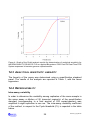
1
User manual REALQUALITY RS-MYCO P code RQ-S47 Kit for the identification and quantification of Mycoplasma pneumoniae RQ-S47_48_EN.doc 1 PRODUCT INFORMATION 3 1.1 3 Intended use 2 KIT CONTENT 4 3 STORAGE AND STABILITY OF REAGENTS 5 4 PRECAUTIONS FOR USE 6 5 SAFETY RULES 7 5.1 General safety rules 7 5.2 Safety rules concerning the kit 7 MATERIAL REQUIRED, BUT NOT PROVIDED 8 6.1 Reagents 8 6.2 Instruments 8 6.3 Disposables 8 6 7 INTRODUCTION 9 8 TEST PRINCIPLE 10 9 PRODUCT DESCRIPTION 12 10 SAMPLE COLLECTION, MANIPULATION AND PRETREATMENT 13 11 PROTOCOL 14 11.1 DNA Extraction 14 11.2 Internal control 14 11.3 Instrument programming 15 11.4 PROTOCOL FOR QUALITATIVE ANALYSIS 16 11.5 PROTOCOL FOR QUANTITATIVE ANALYSIS 17 11.6 Qualitative analysis and interpretation of results 18 11.7 Quantitative analysis and interpretation of results 20 1 RQ-S47_48_EN.doc 12 TROUBLESHOOTING 22 13 DEVICE LIMITATIONS 24 14 DEVICE PERFORMANCES 24 14.1 Analytical specificity 24 14.2 Analytical sensitivity: detection limit 24 14.3 Analytical sensitivity: linearity 25 14.4 Reproducibility 25 14.5 Diagnostic specificity 26 14.6 Diagnostic sensitivity 26 14.7 Accuracy 27 15 REFERENCES 27 16 RELATED PRODUCTS 28 RQ-S47_48_EN.doc 2 1 PRODUCT INFORMATION 1.1 INTENDED USE The REALQUALITY RS-MYCO P is an IVD device for the identification of Mycoplasma pneumoniae (MYCO P) by amplification of the gene coding cytoadhesion P1. If used together with the REALQUALITY RS-MYCO P STANDARD, code RQ-48-ST, it allows the quantification of the number of bacterial genome copies present in the sample. This kit uses Real-Time PCR amplification, starting from DNA extracted from human clinical samples. This in vitro diagnostic test allows the detection and quantification of M. pneumoniae, constituting an auxiliary device for diagnosis and monitoring of M. pneumoniae infections. It is recommended to use this kit as indicated in the instructions herein. This manual refers to the following product: REALQUALITY RS-MYCO P Kit for the identification and quantification of Mycoplasma pneumoniae . This product is in accordance with 98/79/CE Directive (Annex III) regarding the in vitro medical diagnostic devices (CE mark). Contains all the reagents needed for Real-Time amplification Code RQ-S47-48 RQ-S47-96 Product REALQUALITY RS-MYCO P REALQUALITY RS-MYCO P 3 PKG 48 test 96 test RQ-S47_48_EN.doc 2 KIT CONTENT BOX F* STORE AT –30°C/–20°C DESCRIPTION 2X Mastermix Primer and probe Mix for amplification of MYCO P and the β-globin gene (BG) TUBE (T) OR LID COLOR LABEL 2X EV Real Time Mix Oligomix MYCO P Purple 24 test 48 test 96 test 1 × 340 L 2 × 340 L 4 × 340 L 1 × 27 L 2 × 27 L 4 × 27 L BOX F STORE AT +2°C/+8°C DESCRIPTION LABEL TUBE (T) OR LID COLOR 24 test 48 test 96 test DNA containing a part of the Mycoplasma pneumoniae genome (positive control) POSITIVE CONTROL MYCO P Purple 1 × 30 L 1 × 60 L 1 × 110 L DNA containing a part of the β-globin gene (positive control) POSITIVE CONTROL BG Blue 1 × 30 L 1 × 60 L 1 × 110 L DNA containing a part of the β-globin gene (internal control) INTERNAL CONTROL 2 × 125 L 4 × 125 L 8 × 125 L RQ-S47_48_EN.doc 4 3 STORAGE AND STABILITY OF REAGENTS Each component of the kit must be stored according to the directions indicated on the label of each box. In particular: Box F* Store at –30°C/–20°C Box F Store at +2°C/+8°C If stored at the recommended temperature, all test reagents are stable until their expiration date. In order to avoid degradation of the reagents the 2X EV Real Time Mix and the Oligomix should not undergo more than two freeze/thaw cycles. The 2X EV Real Time Mix contains fluorescent molecules, therefore it is recommended to store it protected from direct light. If performing runs with low numbers of samples, it is recommended to aliquot the reagents beforehand. 5 RQ-S47_48_EN.doc 4 PRECAUTIONS FOR USE The kit must be used only as an IVD and handled by qualified technicians who are trained in molecular quantitative biology techniques applied to diagnostics; Before starting the kit procedure, read the user manual carefully and completely; Keep the kit away from heat sources; Please pay particular attention to the expiration date on the label of each box: do not use any part of the kit past the expiration date; The reagents present in the kit must be considered an undividable unit. Do not use them separately or in combination with reagents from other kits or lots; All frozen reagents must be thawed at room temperature before use. Do not vortex! Mix the solutions by inverting the tubes several times, then centrifuge briefly. Work swiftly, particularly if preparing the reactions at room temperature. If possible work on ice or on a cooling block. In case of any doubt about storage conditions, box integrity or application of the method, please contact AB ANALITICA’s technical support at: [email protected] During nucleic acid amplification, the technician has to take the following special precautions: Use filter-tips; In order to avoid contamination store the biological samples, the extracted DNA, amplified DNA and the positive controls included in the kit separated from the Mastermix and the Oligomix(es); Set up pre- and post-PCR work areas; do not share instruments or consumables (pipettes, tips, tubes, etc.) between those areas; Change gloves frequently; Wash the bench surfaces with 5% Sodium Hypochlorite. RQ-S47_48_EN.doc 6 5 SAFETY RULES 5.1 GENERAL SAFETY RULES Wear disposable gloves when handling reagents and clinical samples. Wash hands after the procedure; Do not pipette by mouth; No known diagnostic method can assure the absence of infective agents. Therefore, consider every clinical sample to be potentially infectious and handle it accordingly; All devices that come into contact with clinical samples must be considered contaminated and disposed of as such. In case of accidental spilling of the samples, clean up with 10% Sodium Hypochloride. The materials you use to clean must be disposed of in special containers for contaminated products; Clinical samples, materials and decontaminated before disposal. contaminated products must be We recommend to use one of the following decontamination methods: immerse for 30 minutes in a solution of 5% Sodium Hypochlorite (1 volume of 5% Sodium Hypochlorite solution on 10 volumes of contaminated fluid) OR autoclave at 121°C for at least 2 hours (ATTENTION: Do not autoclave solutions containing Sodium Hypochlorite!!). 5.2 SAFETY RULES CONCERNING THE KIT The risks for the use of this kit are related to the single components. Dangerous components: none. The Material Safety Data Sheet (MSDS) of this device is available upon request. 7 RQ-S47_48_EN.doc 6 MATERIAL REQUIRED, BUT NOT PROVIDED 6.1 REAGENTS Reagents for DNA extraction; DNAse- and RNAse-free sterile water; REALQUALITY RQ-MYCO quantitative analysis) P STANDARD, code RQ-48-ST (for 6.2 INSTRUMENTS Laminar flow cabinet (its use is recommended while preparing the amplification mix to avoid contamination; it is recommended to use another similar laminar flow cabinet to add the extracted DNA, positive controls and/or standards); Micropipettes (range: 0.5-10 µL; 2-20 µL; 10-100 µL; 20-200 µL; 100-1000 µL); Microcentrifuge (max. 12-14,000 rpm); Plate centrifuge (optional); Real-time amplification instrument. This kit was standardized on Applied Biosystems 7500 Fast Dx, 7300 and StepOnePlus™ Real-time PCR Systems (Applied Biosystems). The kit can be used on instruments that work with 25 μL of reaction volume and can detect the FAM and JOE fluorophores. The JOE fluorophore, depending on the instrument, can also be detected in CY3 and HEX channels, etc. For further information on instrument compatibility of the kit, please contact AB ANALITICA’s technical support. 6.3 DISPOSABLES Talc-free disposable gloves; Disposable sterile filter-tips (range: 0.5-10 µL; 2-20 µL; 10-100 µL; 20-200 µL; 100-1000 µL); 96-well plates for Real-time PCR and optical adhesive film or 0.1-0.2 mL tubes with optical caps. RQ-S47_48_EN.doc 8 7 INTRODUCTION Mycoplasma pneumoniae is the most common etiological agents of primary atypical pneumonia, which mainly occurs in children and in youths before 30 years of age. M. pneumoniae is an obligate aerobe bacterium, which adheres to epithelium ciliatum by particular adhesins, causing ciliatosis (block of cilium), and afterwards can cause epithelium destructions and therefore irritation and coughing. The infection, in the majority of cases, is asymptomatic, however there have been reported cases of severe types pneumonia with haematological and neurological involvement in immunocompromised patients. Respiratory tract infections, caused by this bacterium, are however a major cause of other pathologies, in addition to pneumonia, as tracheobronchitis, pharyngitis and asthma. When this bacterium moves to other parts of the body, it can be associated to various manifestation non pulmonary pathology, involving central nervous system, liver, pancreas, blood, skin and joints. M. pneumoniae is transmitted by coughing saliva droplets. People with an active infection of M. pneumoniae can carry the bacterium in the nose, throat and sputum, indicating a general involvement. The traditional diagnosis of M. pneumoniae infections is difficult, since serological and cultural methods require long times, making it impossible to prescribe an effective treatment. It is difficult to be culture Micoplasma in laboratory and frequently, for this reason, it cannot be identified as pathological cause of the diseases. Today, the most commonly reliable diagnostic method is he polymerase chain reaction (PCR). With Real time PCR, it allows for a rapid and reliable quali/quantitative diagnosis. 9 RQ-S47_48_EN.doc 8 TEST PRINCIPLE The PCR method (Polymerase Chain Reaction) was the first method of DNA amplification method described in literature (Saiki RK et al., 1985). It is can be defined as an in vitro amplification reaction of a specific part of DNA (target sequence) by a thermostable DNA polymerase. This technique was shown to be a valuable and versatile instrument of molecular biology: its application contributed to a more efficient study of new genes and their expression and has revolutionized the fields of laboratory diagnostics and forensic medicine. The Real-time PCR represents an advancement of a basic research technology, providing the possibility to determine the number of amplified DNA molecules (amplicons) during the polymerase chain reactions. The monitoring of amplicons is based on primers or probes labeled with fluorescent molecules (molecular beacon, scorpion primer, etc.). These primers or probes usually contain a fluorophore (reporter) and a molecule that blocks the reporter’s specific fluorescence (quencher). Fluorescent emission is determined by the relative proximity of the reporter molecule to the quencher. While a primer or probe are not bound to a target sequence their reporter and quencher are in close proximity and the reporter’s fluorescence is blocked. Upon binding to a target sequence the quencher and reporter become separated and the light emitted by the reporter can be detected. Typically, the main part of a Real-time PCR run consists of several (30 – 50) amplification cycles. The higher the initial concentration of an amplified sequence the earlier the PCR produces an amplicon concentration that displays a fluorescence clearly distinguishable from the background. Thus, the initial concentration of a target sequence can be determined. Specific for each reaction is the so-called Ct value or threshold cycle. It is defined as the point or cycle at which the fluorescence signal becomes clearly distinguishable from the background while the PCR is still in the exponential amplification phase. The latter condition makes sure the number of amplicons is proportional to the number of reaction cycles passed. Using a standard curve the initial concentration of a target sequence can be calculated. The standard curve is established by amplifying standard samples with known concentrations of the target sequence. A thermocycler equipped with a corresponding detector can record the fluorescence events and thus monitor the reaction in “real time”. RQ-S47_48_EN.doc 10 The initial concentration of the target in the samples is determined by comparing the Ct value of each sample with a standard curve that was created by amplifying standards with known concentrations (Figure 1). Figure 1: Creating a standard curve using standards with known concentrations. The main advantage of Real-time PCR compared to conventional techniques of amplification is the possibility to perform a semi-automated amplification. This means the extra steps necessary to visualize the amplification result can be avoided and the risk of contamination by post-PCR manipulation is reduced. 11 RQ-S47_48_EN.doc 9 PRODUCT DESCRIPTION The REALQUALITY RS-MYCO P kit, code RQ-S45, is an IVD device for the identification of Mycoplasma pneumoniae by amplification of the gene coding cytoadhesion P1. If used together with REALQUALITY RQ-MYCO P STANDARD, code RQ-48ST, it allows the quantification of the number of bacterial genome copies present in the sample. The respective standard curve consists of 4 points (from 102 to 105 genome copies per reaction). The positive controls supplied in this kit contain DNA fragments that correspond to the genetic region of interest, and as such, these controls are not dangerous for the user. The kit allows to detect the presence of reaction inhibitors and to monitor the extraction process by amplification of the β-globin gene (amplification control) in multiplex with the target pathogen. This is a valuable tool for identifying false-negative results. In cellular samples the endogenous gene is amplified. For acellular specimens an internal control is added which consists of recombinant DNA containing the β-globin gene. The kit includes a ready-to-use Mastermix and an Oligomix. The Oligomix contains the specific primers and probes, whereas the Mastermix contains all reagents necessary for the amplification reaction as well as the following components: ROX™ is an inert colorant that exhibits stable fluorescent properties throughout all amplification cycles. On some Real-time PCR instruments it is used for normalization in order to compensate for differences between wells caused by pipetting errors or instrument limitations. The dUTP/UNG system prevents contamination from previous amplification runs. The dUTPs are used to incorporate uracil residues into amplicons during amplification sessions. Before each new run the UNG enzyme degrades any single or double stranded DNA containing uracil. This way any amplification products from former sessions are eliminated. RQ-S47_48_EN.doc 12 10 SAMPLE COLLECTION, MANIPULATION AND PRETREATMENT Commonly Mycoplasma pneumoniae is identified from samples from oroand nasopharyngeal swabs, spit, bronchoalveolar lavage (BAL), bronchial aspirate, sputum, but also pulmonary biopsies and PBMC (peripheral blood mononuclear cells). The device was tested on DNA extracted from nasopharyngeal swabs, spit, saliva, sputum, PBMC and FFPE tissue. Respiratory samples can be liquefied by adding 1.5% N-acetyl-L-cysteine (NAC) or 0.1% dithiothreitol (DTT). Use sterile, disposable sample collectors with a screw cap. Samples must be stored at +2°C/+8°C (not longer than 48 hours), or at –30°C/–20°C for a longer period (some months). FFPE samples as well as fresh or frozen histological samples must be disrupted mechanically (e.g. using a sterile scalpel) and then treated with lysis buffer and proteinase K. FFPE histological samples (formalin fixed paraffin embedded) have to be deparaffinized before digestion. PBMC have to be separated from whole peripheral blood by density gradient centrifugation (e.g. Ficoll-Hypaque). Sample collection should follow the common sterility precautions. The samples have to be transported in sterile boxes without transport medium. Blood must be treated with EDTA. Other anticoagulation agents, as heparin, are strong inhibitors of TAQ polymerase and can impair the Real-Time PCR. If processed within 4h after the collection, fresh blood can be stored at +2°C/+8°C. Otherwise, it must be frozen at –30°C/–20°C. 13 RQ-S47_48_EN.doc 11 PROTOCOL 11.1 DNA EXTRACTION For DNA extraction we recommend to use the QIAamp DNA Mini Kit (QIAGEN, Hilden, Germany). For use, refer to the manufacturer’s instructions. Follow for each application use the most appropriate protocol, as recommended by the manufacturer. This IVD device can be used with DNA extracted with the most common manual and automated extraction methods. For further information regarding the compatibility of the device with different extraction methods, please contact AB ANALITICA’s technical support. 11.2 INTERNAL CONTROL This kit includes an internal control (IC) consisting of recombinant DNA containing part of the β-globin gene (BG). The use of this control is recommended for analysis of acellular samples and provides a means of verifying the extraction procedure as well as detecting inhibition of the amplification reaction. For use of the internal control follow to the instructions below: The internal control has to be added at the beginning of DNA extraction procedure (e.g. during the lysis step). This is important for verifying the extraction procedure properly. Do NOT add the control to the untreated sample, this may result in degradation of the DNA in contained in the control. If possible, use the internal control (IC) in a ratio of 1:6 in respect to the final DNA elution volume. For example, use 10 µL of internal control if the final elution volume is 60 µl. Also, refer to the instructions provided by the manufacturer of your extraction system. The sample is suitable for analysis if the resulting Ct value of the internal control is ≤ 35. This threshold is only valid if the internal control was as added as described above (ratio of 1:6). (Applied Biosystems 7500 Fast Dx Real time PCR System; Threshold 0.05). For further information, please contact AB ANALITICA’s technical support. RQ-S47_48_EN.doc 14 11.3 INSTRUMENT PROGRAMMING 11.3.1 Create a thermal profile Use the following thermal profile: UNG Activation Taq Activation Amplification cycles Cycle 1 2 Repeats 1 1 3 45 Step 1 1 1 3* Time 02:00 10:00 00:15 01:00 (°C) 50.0 95.0 95.0 60.0 * Fluorescence detection step 11.3.2 Plate Setup On the grid of the new plate set up the positions for the negative control (NTC), positive control or standards (STD) and samples (Unknown). Quantitative Analysis: For Quantitative Analysis enter the corresponding concentrations of the MYCO P standards (102, 103, 104 and 105 bacteria gene copies/reaction). Note: Make sure the position is the same as on the plate and mark each sample with its name. Select the following the detectors for MYCO P and BG as follows Name MYCO P β-globin Reporter dye FAM JOE Quencher dye none none ATTENTION! Some instruments require to also select the fluorophore ROX™ (see 9 PRODUCT DESCRIPTION on page 12) for all positions/wells in use. Enter, where required, 25 μl as final reaction volume. 15 RQ-S47_48_EN.doc 11.4 PROTOCOL FOR QUALITATIVE ANALYSIS Once thawed, mix the reagents by inverting the tubes several times (do not vortex!), then centrifuge briefly. Prepare the reaction mix rapidly at room temperature or work on ice or on a cooling block. Try, when possible, to work in an area away from direct light. Prepare, as described below, a mix sufficient for all the samples to be tested, counting also the positive and negative control, in the latter H2O is added instead of DNA, and when calculating the volume, consider an excess of at least one reaction volume. Reagent 2X EV Real Time mix Oligo Mix MYCO P H2O Total volume 1 Rxn 12.5 μL 1 μL 6.5 μL 20 μL Mix by inverting the tubes several times, in which the mix was prepared in and then centrifuge briefly. Pipette 20 μL of the mix in each well on the plate. Add 5 μL of extracted DNA to each well or 5 μL of positive control DNA, in the correct position on the plate. Always amplify a negative control together with the samples to be analyzed (add sterile water instead of extracted DNA to the corresponding well). Hermetically seal the plate by using an optical adhesive film or the appropriate sealer. Make sure that there are no air bubbles in the bottom of the wells and/or centrifuge the plate at 4000 rpm for about 1 minute. Load the plate on the instrument making sure to position it correctly and start the amplification cycle. RQ-S47_48_EN.doc 16 11.5 PROTOCOL FOR QUANTITATIVE ANALYSIS The quantitative analysis can be performed by using REALQUALITY RQMYCO P STANDARD code RQ-48-ST. Follow the instructions reported in the previous paragraph to prepare a reaction mix sufficient for the standard curve. A negative amplification control must be included on the plate, in which H2O is added instead of DNA. Aliquot 20 μL of the mix in each well on the plate. Add 5 μL of extracted DNA to each well or 5 μL of each quantification standard dilution, in the corresponding positions on the plate. Hermetically seal the plate by using an optical adhesive film or the appropriate sealer. Make sure that there are no air bubbles in the bottom of the wells and/or centrifuge the plate at 4000 rpm for about 1 minute. Load the plate on the instrument making sure to position it correctly and start the amplification cycle. 17 RQ-S47_48_EN.doc 11.6 QUALITATIVE ANALYSIS AND INTERPRETATION OF RESULTS At the end of the reaction, view the graph in logarithmic scale. Analyze the MYCO P and β-globin amplification results separately by selecting the correct detector and use the following instructions for interpretation. Before considering the sample results, make sure that the positive and negative controls have the expected results β-globin positive control β-globin negative control MYCO P positive control MYCO P negative control RQ-S47_48_EN.doc RESULT INTERPRETATION Amplification signal present Correct β-globin amplification No amplification signal Amplification problems, repeat the analysis No amplification signal No contamination Amplification signal Contamination, repeat the analysis RESULT Amplification signal present INTERPRETATION Correct MYCO P amplification No amplification signal Amplification problems, repeat the analysis No amplification signal No contamination Amplification signal Contamination, repeat the analysis 18 β-globin detector Amplification signal No amplification signal MYCO P detector INTERPRETATION Amplification signal Sample positive for MYCO P No amplification signal Sample negative for MYCO P Amplification signal Sample positive for MYCO P* No amplification signal Sample not suitable Repeat the DNA extraction *ATTENTION: The assay was standardized in order to favour the target pathogen amplification reaction. Therefore, the amplification signal of the βglobin gene (fluorescence in JOE) can have a delayed or absent Ct in MYCO P positive samples. 19 RQ-S47_48_EN.doc 11.7 QUANTITATIVE ANALYSIS AND INTERPRETATION OF RESULTS When the PCR run is finished analyze the amplification results separately for control gene (β-globin) and MYCO P. View the analysis graph in logarithmic scale (see Figure 2) Set the threshold so you obtain a correlation coefficient (R2) and slope of the standard curve (Fig. 3) as close as possible to the optimum (R2: 1.00; slope: 3.33). Values of R2 ≥ 0.990 and a slope in the range of -3.75 and -3.10 (amplification efficiency 85 - 110%) are acceptable values. Figure 2: Post run data analysis (amplification graph in logarithmic scale) RQ-S47_48_EN.doc 20 Figure 3: Post run data analysis: standard curve. 21 RQ-S47_48_EN.doc 12 TROUBLESHOOTING Absence of amplification signals for positive controls, standards and samples The instrument was not programmed correctly – Repeat the amplification taking care of the instrument programming. Pay particular attention to the thermal profile, the fluorophores selected and that the position of the samples in the plate protocol corresponds to the positions on the actual plate. The amplification mix was not prepared correctly – Prepare a new amplification mix making sure to follow the instructions given in paragraph 11.3. The kit was not stored properly or it was used past the expiration date – Check both the storage conditions and the expiration date reported on the label. Use a new kit if needed. Weak amplification signal for positive controls / standards Positive controls/standard solutions were stored incorrectly and have degraded – Store the positive controls/standard solutions correctly at +2°/+8°C, and make sure that they do not undergo any freeze/thaw cycle as well; – Do not use the positive controls/standards past the expiration date. The reaction mix does not function correctly – Make sure to store the 2X EV Real Time Mix and Oligomix correctly at –20°C/–30°C. Avoid unnecessary freeze/thaw cycles. RQ-S47_48_EN.doc 22 Amplification signal of β-globin very delayed or absent in the extracted sample (MYCO P negative) The extracted DNA was not suitable for amplification and the amplification reaction was inhibited – Make sure to extract the nucleic acids correctly; – If an extraction method uses wash steps with solutions containing Ethanol, make sure no Ethanol residue remains in the DNA sample; – Use the extraction methods suggested in paragraph 11.1. For any further problems, please contact AB ANALITICA’s technical support at: [email protected], fax (+39) 049-8709510, or tel. (+39) 049761698). 23 RQ-S47_48_EN.doc 13 DEVICE LIMITATIONS The kit can have reduced performance if: The clinical sample is not suitable for this analysis (sampling and/or storage error, i.e. blood treated with anticoagulants other than EDTA, like heparin, etc.); DNA is not suitable for amplification (due to the presence of amplification reaction inhibitors or to the use of inappropriate extraction method); The kit was not stored correctly. 14 DEVICE PERFORMANCES 14.1 ANALYTICAL SPECIFICITY The specificity of the REALQUALITY RS-MYCO P, code RQ-S45 kit is guaranteed by an accurate and specific selection of primers and probe and also by the use of stringent amplification conditions. Alignment of primers and probes in the most important databanks showed no non-specific pairing. In order to determine the cross-reactivity of this device, samples positive for other potentially cross-reactive pathogens or pathogens from the anatomical region as Mycoplasma pneumoniae was identified in were amplified with this device. None of the tested pathogens gave a positive signal with the IVD device. 14.2 ANALYTICAL SENSITIVITY: DETECTION LIMIT Serial dilutions of quantification standard, ranging from 1 to 0.05 copies of viral genome copies/μL, were tested in three consecutive experiments in order to determine the analytical sensitivity. For each dilution, 5 μL were amplified in eight replicates per run, in multiplex with the internal control. The results were analyzed by Probit analysis, as illustrated in the graph reported in Figure 4. The limit of the analytical sensitivity for the REALQUALITY RS-MYCO P (p = 0.05) kit is reported in Table 1. RQ-S47_48_EN.doc 24 Figure 4: Graph of the Probit analysis results for determination of analytical sensitivity for the REALQUALITY RS-MYCO P kit on Applied Biosystems 7500 Fast DX Real-Time PCR System expressed in bacteria genome copies/reaction. 14.3 ANALYTICAL SENSITIVITY: LINEARITY The linearity of the assay was determined using a quantification standard panel. The results of the analysis are reported in Table 1, with the linear regression. 14.4 REPRODUCIBILITY Intra-assay variability In order to determine the variability among replicates of the same sample in the same assay a dilution of 50 transcript copies/µL of the quantification standard (corresponding to a final amount of 250 copies/reaction) was amplified in eight replicates in one run. The intra-assay variability coefficient of the method, in respect to the Cycle threshold (Ct), is reported in the table below. 25 RQ-S47_48_EN.doc Inter-assay variability In order to determine the variability of replicates of the same sample in different runs the least concentrated quantification standard (20 transcript copies/µL, corresponding to a final amount of 100 copies/reaction) was amplified in duplicates in three consecutive runs. For each run, the variability coefficient was calculated from the Ct of the samples. The inter-assay variability coefficient was calculated from the average of the variable coefficients of each run and is reported in the table below. Table 1 ABI 7500 Fast Dx ABI 7300 StepOnePlus™ 1 1.5 0.9 5 – 107 6 – 107 5 – 107 Intra-assay variability (%) 0.12 0.12 0.44 Inter-assay variability (%) 0.33 0.287 0.711 Detection limit (bacterial genome copies/µL) probit p = 0.05 Range of linearity (bacterial genome copies/reaction) 14.5 DIAGNOSTIC SPECIFICITY A significant number of samples negative for MYCO P were tested simultaneously with the REALQUALITY RS-MYCO P kit and another CE IVD device or reference method. From the obtained results the diagnostic specificity of this device was calculated to be 100%. 14.6 DIAGNOSTIC SENSITIVITY A significant number of samples positive for Mycoplasma pneumoniae were tested simultaneously with the REALQUALITY RS-MYCO P kit and another CE IVD device or reference method. From the obtained results the diagnostic specificity of this device was calculated to be 100%. RQ-S47_48_EN.doc 26 14.7 ACCURACY The accuracy was calculated as the number of correct amplifications over the total number of executed amplifications. The REALQUALITY RS-MYCO P kit has an accuracy of 100%. 15 REFERENCES Balin BJ et al. Med Microbiol Immunol (Berlin) 187, 23-42, 1998. Dowell SF et al. Clin Infect Dis 33, 492-503, 2001. Gieffers JE et al. J clin Microbiol 38, 881-882, 2000. Grayston JT. Clin Infect Dis 155, 757-761, 1992. Gupta S et al. Circulation 96, 404-407, 1997. Hammerschlag MR. Infect Dis Clin Practice 8, 232-240, 1999. Hammerschlag MR. Curr Infect Dis Rep 2, 115-120, 2000. Hammerschlag MR et al. J Clin Microbiol 38, 4274-4276, 2000. Kuo CC et al. Clin Microbiol Rev 8, 451-461. Kuo CC et al. Arterioscler Thromb 10, 1501-1504, 1993. Maurin M et al. J clin Microbiol 35, 2283-2287, 1997. Muhlestein JB et al. Circulation 97, 633-636, 1998. Normann E et al. Pediatr Infect Dis J 18, 72-73, 1999. Peeling RW et al. J Infect Dis 181 (Suppl 3), S426-S429, 2000. Ramirez JA. Ann Intern Med 125, 979-982, 1996. Saiki RK et al. Science 230, 1350-1354, 1985. Schrag SJ et al. Pediatr Infect Dis J 19, 17-22, 2000. Sriram et al. Ann Neurol 46, 6-14, 1999. Tuuminen T et al. Clin Diagn Lab Immunol 7, 734-738, 2000 27 RQ-S47_48_EN.doc 16 RELATED PRODUCTS REALQUALITY RQ-MYCO P STANDARD: Quantification standard for Mycoplasma pneumoniae ; quantified and readyto-use. This product is in accordance with 98/79/CE Directive (Annex III) regarding the in vitro medical diagnostic devices (CE mark). Code RQ-S46-ST RQ-S47_48_EN.doc Product REALQUALITY RQ-MYCO P STANDARD 28 PKG 4 × 60 µL AB ANALITICA srl Via Svizzera 16 - 35127s PADOVA, (ITALY) Tel +39 049 761698 - Fax +39 049 8709510 e-mail: [email protected]