1
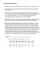
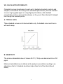
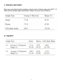
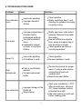
RayBio® Human GLP-1 (7-36a)
ELISA Kit
Catalog #: ELH-GLP736A
User Manual
Last revised December 7, 2015
Caution:
Extraordinarily useful information enclosed
ISO 13485 Certified
3607 Parkway Lane, Suite 100
Norcross, GA 30092
Tel: 1-888-494-8555 (Toll Free) or 770-729-2992, Fax:770-206-2393
Web: www.RayBiotech.com, Email: [email protected]
1
RayBiotech, Inc.
________________________________________
RayBio® Human GLP-1 (7-36a) ELISA Kit Protocol
Table of Contents
Section
Page #
I.
Introduction
3
II.
Storage
4
III.
Reagents
4
IV.
Additional Materials Required
4
V.
Reagent Preparation
5
VI.
Assay Procedure
6
VII.
Assay Procedure Summary
7
VIII.
Calculation of Results
A. Typical Data
B. Sensitivity
C. Spiking & Recovery
D. Linearity
E. Reproducibility
8
8
8
9
9
10
IX.
Specificity
10
X.
Troubleshooting Guide
11
Please read the entire manual carefully before starting your experiment
2
I. INTRODUCTION
The RayBio® Human GLP-1 (7-36a) ELISA kit is an in vitro enzyme-linked
immunosorbent assay for the quantitative measurement of human GLP-1 (7-36a) in
serum (human GLP-1 concentration is low in normal serum/plasma and may not be
detectable in this assay), plasma (collect plasma using EDTA as an anticoagulant;
Heparin and Citrate are not recommended as anticoagulants for use in this assay),
and cell culture supernatants. This assay employs an antibody specific for human
GLP-1 (7-36a) coated on a 96-well plate. Standards and samples are pipetted into
the wells and GLP-1 (7-36a) present in a sample is bound to the wells by the
immobilized antibody. The wells are washed and biotinylated anti-human GLP-1 (736a) antibody is added. After washing away unbound biotinylated antibody, HRPconjugated streptavidin is pipetted to the wells. The wells are again washed, a TMB
substrate solution is added to the wells and color develops in proportion to the
amount of GLP-1 (7-36a) bound. The Stop Solution changes the color from blue to
yellow, and the intensity of the color is measured at 450 nm.
3
II. STORAGE
The entire kit may be stored at -20°C for up to 1 year from the date of shipment.
Avoid repeated freeze-thaw cycles. The kit may be stored at 4°C for up to 6
months. For extended storage, it is recommended to store at -80°C. For prepared
reagent storage, see table below.
III. REAGENTS
Component
Size / Description
Storage / Stability
After Preparation
GLP-1 (7-36a) Microplate
(Item A)
96 wells (12 strips x 8 wells) coated with antiHuman GLP-1 (7-36a) .
1 month at 4°C*
Wash Buffer Concentrate
(20X) (Item B)
25 ml of 20X concentrated solution.
1 month at 4°C
Standard Protein (Item C)
2 vials of Human GLP-1 (7-36a) . 1 vial is enough
to run each standard in duplicate.
1 week at -80°C
Detection Antibody GLP-1 (736a) (Item F)
2 vials of biotinylated anti-Human GLP-1 (7-36a) .
Each vial is enough to assay half the microplate.
5 days at 4°C
HRP-Streptavidin
Concentrate (Item G)
200 µl 200X concentrated HRP-conjugated
streptavidin.
Do not store and
reuse.
TMB One-Step Substrate
Reagent (Item H)
12 ml of 3,3,5,5'-tetramethylbenzidine (TMB) in
buffer solution.
N/A
Stop Solution (Item I)
8 ml of 0.2 M sulfuric acid.
N/A
Assay Diluent (Item E2)
15 ml of 5x concentrated buffer.
1 month at 4°C
*Return unused wells to the pouch containing desiccant pack, reseal along entire edge.
IV. ADDITIONAL MATERIALS REQUIRED
1.
2.
3.
4.
5.
6.
7.
8.
Microplate reader capable of measuring absorbance at 450 nm.
Precision pipettes to deliver 2 µl to 1 ml volumes.
Adjustable 1-25 ml pipettes for reagent preparation.
100 ml and 1 liter graduated cylinders.
Absorbent paper.
Distilled or deionized water.
Log-log graph paper or computer and software for ELISA data analysis.
Tubes to prepare standard or sample dilutions.
4
V. REAGENT PREPARATION
1. Bring all reagents and samples to room temperature (18 - 25ºC) before use.
2. Assay Diluent (Item E2) should be diluted 5-fold with deionized or distilled
water before use.
3. Sample dilution: 1X Assay Diluent (Item E2) should be used for dilution of
serum, plasma, and cell culture supernatant samples. The suggested dilution
for normal serum/plasma is 2 fold. Collect plasma using EDTA as an
anticoagulant. Heparin and citrate are not recommended.
Note: Levels of GLP-1 (7-36a) may vary between different samples. Optimal
dilution factors for each sample must be determined by the investigator.
4. Preparation of standard: Briefly spin a vial of Item C. Add 400 µl 1X Assay
Diluent (Item E2; Assay Diluent should be diluted 5-fold with deionized or
distilled water before use) into Item C vial to prepare a 100 ng/ml standard
solution. Dissolve the powder thoroughly by a gentle mix. Add 100 µl of the
100 ng/ml standard solution from the vial of Item C, into a tube with 400 µl 1X
Assay Diluent to prepare a 20,000 pg/ml standard solution. Pipette 300 µl 1X
Assay Diluent into each tube. Use the 20,000 pg/ml standard solution to
produce a dilution series (shown below). Mix each tube thoroughly before the
next transfer. 1X Assay Diluent serves as the zero standard (0 pg/ml).
5
5. If the Wash Concentrate (20X) (Item B) contains visible crystals, warm to room
temperature and mix gently until dissolved. Dilute 20 ml of Wash Buffer
Concentrate into deionized or distilled water to yield 400 ml of 1X Wash Buffer.
6. Briefly spin the Detection Antibody vial (Item F) before use. Add 100 µl of 1X
Assay Diluent (Item E2) into the vial to prepare a detection antibody
concentrate. Pipette up and down to mix gently (the concentrate can be stored
at 4ºC for 5 days). The detection antibody concentrate should be diluted 65fold with 1X Assay Diluent (Item E2) and used in step 5 of Part VI Assay
Procedure.
7. Briefly spin the HRP-Streptavidin concentrate vial (Item G) and pipette up and
down to mix gently before use, as precipitates may form during storage. HRPStreptavidin concentrate should be diluted 200-fold with 1X Assay Diluent
(Item E2).
For example: Briefly spin the vial (Item G) and pipette up and down to mix
gently. Add 50 µl of HRP-Streptavidin concentrate into a tube with 10 ml 1X
Assay Diluent to prepare a 400-fold diluted HRP-Streptavidin solution (don't
store the diluted solution for next day use). Mix well.
VI. ASSAY PROCEDURE
1. Bring all reagents and samples to room temperature (18 - 25ºC) before use. It
is recommended that all standards and samples be run at least in duplicate.
2. Label removable 8-well strips as appropriate for your experiment.
3. Add 100 µl of each standard (see Reagent Preparation step 3) and sample into
appropriate wells. Cover well and incubate for 2.5 hours at room temperature
or over night at 4ºC with gentle shaking.
4. Discard the solution and wash 4 times with 1X Wash Solution. Wash by filling
each well with Wash Buffer (300 µl) using a multi-channel Pipette or
autowasher. Complete removal of liquid at each step is essential to good
performance. After the last wash, remove any remaining Wash Buffer by
aspirating or decanting. Invert the plate and blot it against clean paper towels.
6
5. Add 100 µl of 1X prepared biotinylated antibody (Reagent Preparation step 6)
to each well. Incubate for 1 hour at room temperature with gentle shaking.
6. Discard the solution. Repeat the wash as in step 4.
7. Add 100 µl of prepared Streptavidin solution (see Reagent Preparation step 7)
to each well. Incubate for 45 minutes at room temperature with gentle shaking.
8. Discard the solution. Repeat the wash as in step 4.
9. Add 100 µl of TMB One-Step Substrate Reagent (Item H) to each well.
Incubate for 30 minutes at room temperature in the dark with gentle shaking.
10. Add 50 µl of Stop Solution (Item I) to each well. Read at 450 nm immediately.
VII. ASSAY PROCEDURE SUMMARY
1. Prepare all reagents, samples and standards as instructed.
2. Add 100 µl standard or sample to each well. Incubate 2.5 hours at room
temperature or over night at 4ºC.
3. Add 100 µl prepared biotin antibody to each well. Incubate 1 hour at room
temperature.
4. Add 100 µl prepared Streptavidin solution. Incubate 45 minutes at room
temperature.
5. Add 100 µl TMB One-Step Substrate Reagent to each well. Incubate 30
minutes at room temperature.
6. Add 50 µl Stop Solution to each well. Read at 450 nm immediately.
7
VIII. CALCULATION OF RESULTS
Calculate the mean absorbance for each set of duplicate standards, controls and
samples, and subtract the average zero standard optical density. Plot the standard
curve on log-log graph paper or using Sigma plot software, with standard
concentration on the x-axis and absorbance on the y-axis. Draw the best-fit straight
line through the standard points.
A. TYPICAL DATA
These standard curves are for demonstration only. A standard curve must be run
with each assay.
B. SENSITIVITY
The minimum detectable dose of Human GLP-1 (7-36a) was determined to be 150
pg/ml.
Minimum detectable dose is defined as the analyte concentration resulting in an
absorbance that is 2 standard deviations higher than that of the blank (diluent
buffer).
8
C. SPIKING & RECOVERY
Recovery was determined by spiking various levels of Human php echo GLP-1 (736a) into the sample types listed below. Mean recoveries are as follows:
D. LINEARITY
9
E. REPRODUCIBILITY
Intra-Assay CV%: <10%
Inter-Assay CV%: <12%
IX. SPECIFICITY
This ELISA antibody pair detects human GLP-1 (7-36a). Other species not
determined
10
X. TROUBLESHOOTING GUIDE
Problem
Cause
Solution
Poor standard
curve
Inaccurate pipetting
Improper standard
dilution
Check pipettes
Briefly centrifuge Item C and
dissolve the powder thoroughly
by gently mixing
Low signal
Improper preparation of
standard and/or
biotinylated antibody
Too brief incubation
times
Inadequate reagent
volumes or improper
dilution
Briefly spin down vials before
opening. Dissolve the powder
thoroughly.
Ensure sufficient incubation
time; assay procedure step 2
may be done overnight
Check pipettes and ensure
correct preparation
Large CV
Inaccurate pipetting
Air bubbles in wells
Check pipettes
Remove bubbles in wells
High
background
Plate is insufficiently
washed
Contaminated wash
buffer
Review the manual for proper
wash. If using a plate washer,
ensure that all ports are
unobstructed.
Make fresh wash buffer
Improper storage of the
ELISA kit
Stop solution
Store your standard at <-70ºC
after reconstitution, others at
4ºC. Keep substrate solution
protected from light.
Add stop solution to each well
before reading plate
Low sensitivity
11
RayBio® ELISA Kits
Over 2,000 ELISA kits available, visit www.RayBiotech.com/ELISA-Kits.html for
details.
This product is for research use only.
©2015 RayBiotech, Inc
12