1
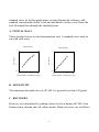
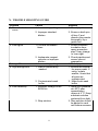
Human sICAM-1ELISA Kit(KT20353)
User Manual
For research use only. Not intended for diagnostic testing.
www.abgent.com
TABLE OF CONTENTS
I.
Introduction……..……………………………….2
II.
Reagents……………………………..…………..2
III.
Storage.…………………………………………. 3
IV. Additional Materials Required…………………..3
V.
Reagent Preparation……………………………..4
VI. Assay Procedure………………………………...6
VII. Assay Procedure Summary……………………...7
VIII. Calculation of Results
A.
Typical Data…..……………………….…….…..8
B.
Sensitivity……………………………….….…… 8
C.
Recovery…………………………..…………….8
D.
Linearity………………………………….…..... .9
E.
Reproducibility…………………………………..9
IX. Specificity…………………………………....…. 9
X.
Troubleshooting Guide.……………………....…11
1
I. INTRODUCTION
sICAM-1 (soluble Intercellular Adhesion Molecular-1) has been
reported in serum, cerebrospinal fluid and bronchoalveolar lavage. ICAM-1
expression is weak on leukocytes, epithelial and resting endothelial cells.
ICAM-1 is a ligand for LFA-1 (CD11a/CD18) and Mac-1 (CD11b/CD18).
Its expression is up-regulated upon stimulation by inflammatory mediators
such as cytokines and LPS.
The Human sICAM-1 ELISA (Enzyme-Linked
Immunosorbent Assay) kit is an in vitro enzyme-linked immunosorbent
assay for the quantitative measurement of human soluble ICAM-1 in serum,
plasma, cell culture supernatants and urine. This assay employs an antibody
specific for human sICAM-1 coated on a 96-well plate. Standards and
samples are pipetted into the wells and sICAM-1 present in a sample is
bound to the wells by the immobilized antibody. The wells are washed and
biotinylated anti-human sICAM-1 antibody is added. After washing away
unbound biotinylated antibody, HRP-conjugated streptavidin is pipetted to
the wells. The wells are again washed, a TMB substrate solution is added to
the wells and color develops in proportion to the amount of sICAM-1 bound.
The Stop Solution changes the color from blue to yellow, and the intensity of
the color is measured at 450 nm.
II. REAGENTS
1. sICAM-1 Microplate (Item A): 96 wells (12 strips x 8 wells) coated
with anti-human sICAM-1.
2. Wash Buffer Concentrate (20x) (Item B): 25 ml of 20x concentrated
solution.
3. Standards (Item C): 2 vials, recombinant human sICAM-1.
4. Assay Diluent A (Item D): 30 ml, 0.09% sodium azide as preservative.
For Standard/Sample (serum/plasma) diluent.
5. Assay Diluent B (Item E): 15 ml of 5x concentrated buffer. For
Standard/Sample (cell culture medium/urine) diluent.
2
6. Detection Antibody sICAM-1 (Item F): 2 vial of biotinylated antihuman sICAM-1 (each vial is enough to assay half microplate).
7. HRP-Streptavidin Concentrate (Item G): 200 µl of 340x concentrated
HRP-conjugated streptavidin.
8. TMB One-Step Substrate Reagent (Item H): 12 ml of 3,3’,5,5’tetramethylbenzidine (TMB) in buffered solution.
9. Stop Solution (Item I): 8 ml of 0.2 M sulfuric acid.
III. STORAGE
May be stored for up to 6 months at 2o to 8oC from the date of shipment.
Standard (recombinant protein) should be stored at -20 oC or -80 oC
(recommended at –80 oC) after reconstitution. Opened Microplate Wells or
reagents may be store for up to 1 month at 2o to 8oC. Return unused wells to
the pouch containing desiccant pack, reseal along entire edge.
Note: the kit can be used within one year if the whole kit is stored at -20 oC.
Avoid repeated freeze-thaw cycles.
IV. ADDITIONAL MATERIALS REQUIRED
1
2
3
4
5
6
7
8
Microplate reader capable of measuring absorbance at 450 nm.
Precision pipettes to deliver 2 µl to 1 ml volumes.
Adjustable 1-25 ml pipettes for reagent preparation.
100 ml and 1 liter graduated cylinders.
Absorbent paper.
Distilled or deionized water.
Log-log graph paper or computer and software for ELISA data analysis.
Tubes to prepare standard or sample dilutions.
3
V. REAGENT PREPARATION
1. Bring all reagents and samples to room temperature (18 - 25°C) before
use.
2. Sample dilution: If your samples need to be diluted, Assay Diluent A
(Item D) should be used for dilution of serum/plasma samples. Assay
Diluent B (Item E) should be used for dilution of culture supernatants
and urine.
Suggested dilution for normal serum/plasma: 5-200 fold*.
* Please note that levels of the target protein may vary between different
specimens. Optimal dilution factors for each sample must be
determined by the investigator.
3. Assay Diluent B should be diluted 5-fold with deionized or distilled
water.
4. Preparation of standard: Briefly spin the vial of Item C and then add
750 µl Assay Diluent A (for serum/plasma samples) or 1x Assay Diluent
B (for cell culture medium and urine) into Item C vial to prepare a 20
ng/ml standard. Dissolve the powder thoroughly by a gentle mix.
Pipette 250 µl Assay Diluent A or 1x Assay Diluent B into each tube.
Use the 20 ng/ml standard solution to produce a dilution series (shown
below). Mix each tube thoroughly before the next transfer. Assay
Diluent A or 1x Assay Diluent B serves as the zero standard (0 ng/ml).
The 20 ng/ml standard in Assay Diluent B may be saturated, we
recommend 10 ng/ml serves as starting point (the highest standard point)
for Assay Diluent B.
4
Standard, Item C +
250µl
750 µl
20
ng/ml
250 µl
10
ng/ml
250 µl
5
ng/ml
250 µl
2.5
ng/ml
1.25
ng/ml
250 µl
0.625
ng/ml
250 µl
0.313
ng/ml
0
ng/ml
5. If the Wash Concentrate (20x) (Item B) contains visible crystals, warm
to room temperature and mix gently until dissolved. Dilute 20 ml of
Wash Buffer Concentrate into deionized or distilled water to yield 400
ml of 1x Wash Buffer.
6. Briefly spin the Detection Antibody vial (Item F) before use. Add 100 µl
of 1x Assay Diluent B into the vial to prepare a detection antibody
concentrate. Pipette up and down to mix gently (the concentrate can be
stored at 4°C for 5 days). The detection antibody concentrate should be
diluted 80-fold with 1x Assay Diluent B and used in step 4 of Part VI
Assay Procedure.
7. Briefly spin the HRP-Streptavidin concentrate vial (Item G) and pipette
up and down to mix gently before use. HRP-Streptavidin concentrate
should be diluted 340-fold with 1x Assay Diluent B.
For example: Briefly spin the vial (Item G) and pipette up and down
to mix gently . Add 25 µl of HRP-Streptavidin concentrate into a tube
with 8.5 ml 1x Assay Diluent B to prepare a 340-fold diluted
HRP-Streptavidin solution (don’t store the diluted solution for next
day use). Mix well.
5
VI. ASSAY PROCEDURE:
1. Bring all reagents and samples to room temperature (18 - 25°C) before
use. It is recommended that all standards and samples be run at least in
duplicate.
2. Add 100 µl of each standard (see Reagent Preparation step 2) and
sample into appropriate wells. Cover well and incubate for 2.5 hours at
room temperature or over night at 4°C with gentle shaking.
3. Discard the solution and wash 4 times with 1x Wash Solution. Wash by
filling each well with Wash Buffer (300 µl) using a multi-channel
Pipette or autowasher. Complete removal of liquid at each step is
essential to good performance. After the last wash, remove any
remaining Wash Buffer by aspirating or decanting. Invert the plate and
blot it against clean paper towels.
4. Add 100 µl of 1x prepared biotinylated antibody (Reagent Preparation
step 6) to each well. Incubate for 1 hour at room temperature with
gentle shaking.
5. Discard the solution. Repeat the wash as in step 3.
6. Add 100 µl of prepared Streptavidin solution (see Reagent Preparation
step 7) to each well. Incubate for 45 minutes at room temperature with
gentle shaking.
7. Discard the solution. Repeat the wash as in step 3.
8. Add 100 µl of TMB One-Step Substrate Reagent (Item H) to each well.
Incubate for 30 minutes at room temperature in the dark with gentle
shaking.
6
9. Add 50 µl of Stop Solution (Item I) to each well. Read at 450 nm
immediately.
VII. ASSAY PROCEDURE SUMMARY
1. Prepare all reagents, samples and standards as instructed.
2. Add 100 µl standard or sample to each well.
Incubate 2.5 hours at room temperature or over night at 4oC.
3. Add 100 µl prepared biotin antibody to each well.
Incubate 1 hour at room temperature.
4. Add 100 µl prepared Streptavidin solution.
Incubate 45 minutes at room temperature.
5. Add 100 µl TMB One-Step Substrate Reagent to each well.
Incubate 30 minutes at room temperature.
6. Add 50 µl Stop Solution to each well.
Read at 450 nm immediately.
VIII. CALCULATION OF RESULTS
Calculate the mean absorbance for each set of duplicate standards, controls
and samples, and subtract the average zero standard optical density. Plot the
7
standard curve on log-log graph paper or using Sigma plot software, with
standard concentration on the x-axis and absorbance on the y-axis. Draw the
best-fit straight line through the standard points.
A. TYPICAL DATA
These standard curves are for demonstration only. A standard curve must be
run with each assay.
Assay Diluent B
10
10
1
1
OD=450 nm
OD=450 nm
Assay Diluent A
0.1
0.01
0.1
1
10
0.1
0.01
0.1
100
Human sICAM-1 concentration (ng/ml)
1
10
100
Human sICAM-1 concentration (ng/ml)
B. SENSITIVITY
The minimum detectable dose of sICAM-1 is typically less than 150 pg/ml.
C. RECOVERY
Recovery was determined by spiking various levels of human sICAM-1 into
human serum, plasma and cell culture media. Mean recoveries are as follows:
8
Sample Type
Average % Recovery
Range (%)
Serum
97.48
88-106
Plasma
98.67
90-107
Cell culture media
97.37
89-107
D. LINEARITY
Sample Type
Serum
Plasma
Cell Culture Media
1:2
Average % of Expected
Range (%)
98
91-106
97
89-105
96
89-106
1:4
Average % of Expected
Range (%)
97
89-106
95
88-105
96
90-107
E. REPRODUCIBILITY
Intra-Assay: CV<10%
Inter-Assay: CV<12%
IX. SPECIFICITY
Cross Reactivity: This ELISA kit shows no cross-reactivity with any of the
cytokines tested (e.g., human Angiogenin, BDNF, BLC, ENA-78, FGF-4,
IL-1α, IL-1β, IL-2, IL-3, IL-4, IL-5, IL-7, IL-8, IL-9, IL-10, IL-11, IL-12
p70, IL-12 p40, IL-13, IL-15, IL-309, IP-10, G-CSF, GM-CSF, IFN-γ,
Leptin, MCP-1, MCP-2, MCP-3, MDC, MIP-1α, MIP-1 β, MIP-1δ, PARC,
9
PDGF, RANTES, SCF, TARC, TGF-β, TIMP-1, TIMP-2, TNF-α, TNF-β,
TPO, VEGF).
10
X. TROUBLE SHOOTING GUIDE
Problem
1. Poor standard
curve
2. Low signal
3. Large CV
4. High background
5. Low sensitivity
Cause
1. Inaccurate pipetting
Solution
1. Check pipettes
2. Improper standard
dilution
2. Ensure a brief spin
of Item C and
dissolve the powder
thoroughly by a
gentle mix.
1. Ensure sufficient
incubation time;
assay procedure
step 2 may change
to over night
2. Check pipettes and
ensure correct
preparation
1. Check pipettes
1. Review the manual
for proper wash. If
using a plate
washer, check that
all ports are
unobstructed.
2. Make fresh wash
buffer
1. Store your standard
at<-20oC after
reconstitution,
others at 4 oC. Keep
substrate solution
protected from light
2. Stop solution should
be added to each
well before measure
1.Too brief incubation
times
2. Inadequate reagent
volumes or improper
dilution
1. Inaccurate pipetting
1. Plate is insufficiently
washed
2. Contaminated wash
buffer
1. Improper storage of the
ELISA kit
2. Stop solution
11
Note:
This product is for research use only.
12
USA
Abgent,Inc.
Toll Free (888)735-7227
Or (858)875-1900
[email protected]
CHINA
Abgent Suzhou
+86 512 69369088
[email protected]
EUROPE
Abgent Europe
+44(0) 1235 854042
[email protected]
For other countries
www.abgent.com