1
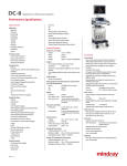
DC-T6
Color Doppler Ultrasound System
Datasheet
2012-08
1
System Overview
1.1 Application
Abdomen
Obstetrics
Gynecology
Cardiology
Small parts
Urology
Vascular
Pediatrics
EM (Emergency)
Others
1.2 Transducer types
Curved array transducer
Linear array transducer
Phased array transducer
1.3 Imaging modes
B-Mode
THI (Tissue Harmonic Imaging)
PSHI (Phase Shift Harmonic Imaging)
M-Mode/Color M-mode
Free Xros M (Anatomical M-mode)
Free Xros CM (Curved Anatomical
M-mode)
Color Doppler Imaging
Power Doppler Imaging/Directional PDI
Pulsed Wave Doppler
Continuous Wave Doppler
TDI
Smart 3D (Freehand 3D)
Static 3D
4D
Stress Echo
iScape View (Panoramic Imaging)
1.4 Standard features
B-Mode
THI and PSH
M-Mode
Color Doppler Imaging
Power Doppler Imaging and Directional
PDI
Pulsed Wave Doppler
iBeam (Spatial Compounding Imaging)
iClear (Speckle Suppression Imaging)
iTouch (Auto Optimization)
Zoom/iZoom (Full Screen Zoom)
FCI (Frequency Compounding
Imaging)
B steer
ExFOV Imaging: Extend imaging for
convex transducer
Trapezoid imaging for linear transducer
4 active probe ports
320G hard drive
DVD R/W driver
4-USB
1.5 Optional features
Continuous Wave Doppler
Free Xros M
Free Xros CM
iScape View
Smart 3D
4D
- iPage
- STIC
IMT
Stress Echo
TDI (Include TVI, TVD, TVM, TEI)
TDI QA (TDI Quantitative Analysis)
iNeedle(7L4A, L14-6N, L14-6 support)
DICOM
Clinical Measurement Package
1.6 Language support
Software: English, Chinese, German,
Spanish, French, Italian, Portuguese,
Russian, Czech, Polish
Keyboard input: English, Chinese,
German, Spanish, French, Italian,
Portuguese, Russian, Czech, Polish,
Icelandic, Norwegian, Swedish, Finnish,
Turkish, Danish
Control panel overlay: Chinese, Italian,
Portuguese, Spanish, German,
Russian, French, Czech, Polish
User manual: English, Chinese,
German, Spanish, French, Italian,
Portuguese, Russian
2 Physical Specification
2.1 Dimension and weight
Height: 1290-1570mm
Width: 460mm
2012-08
Depth: 730mm
Weight: approx. 95kg
2.2 Monitor
15-inch high resolution color LCD
monitor(17-inch LCD optional)
Resolution: 1024×768
Viewing angle: 170 degree
Digital on-screen display of brightness
and contrast controls
Auto-calibrate brightness after system
boot-up each time
Independent tilt of 30 degrees up, 30
degrees down and swivel of -90 to +90
degrees.
2.3 Audio speakers
Stereo audio speakers
2.4 Multi-directional articulating monitor arm
for better user-friendly experience
Upper arm:
- Rotate: ±90 degrees (from center)
- Up: 140mm
Lower arm:
- Rotate: ±90 degrees (from center)
2.5 Wheels
Diameter: 125mm
Front castor (2 ea): total lock and break
Rear castor (2 ea): total lock and break
2.6 Probe port and holder
Probe ports: 4 active ports, plus 1
pencil probe port
Probe holder: 4
2.7 Electrical power
Voltage: 100-127V~, or 220-240V~
Frequency: 50/60 Hz
Power consumption: Max. 600 VA
Circuit breaker: 240V~, 13A
2.8 Operating Environment
Ambient temperature: 0-40 °C
Relative humidity: 30%-95% (no
condensation)
Atmospheric pressure:
700hPa-1060hPa
2.9 Storage & Transportation Environment
Ambient temperature: -20~55 °C
Relative humidity: 30%-95% (no
condensation)
Atmospheric pressure:
700hPa-1060hPa
3 User Interface
3.1 Control panel
User-centric control panel with
home-based layout favors easy access
to keys
Backlit keys ensure accurate work in
the dark room
Programmable keys available for
user-defined functions
8-segment TGC control
Full-sized, backlit QWERTY keyboard
for text input, function keys and system
programming
Adjustable key volume and trackball
speed meet different needs
Dedicated palm rest design to help
reduce user repetitive stress injury
Independent rotation and up/down of
control panel facilitates optimal
positioning
-rotate: ±45 degrees (from center)
-down/up: 780-970mm (190 mm range)
3.2 System boot-up
Boot-up from complete shut-down in
less than 61-68 sec(Depends on the
Configuration)
Boot-up from standby mode in less
than 15 sec
Shut-down in less than 33 sec
3.3 Comments
Supports text input and arrow
Adjustable text size and arrow size
Supports home position
Covers various application
User customizable
3.4 Bodymark
More than 134 bodymarks for versatile
application
User customizable
3.5 Screen information*
Common info:
3
-
-
Mindray logo
Hospital name
Exam date
Exam time
Acoustic output indices
Mechanical index
ID, Last name, First Name, Middle
initial, Gender, Age, DOB(Date of
birth)
Probe model
ECG icon (when ECG connected),
Operator
TGC Curve
Focus position
Thumbnail
Machine model
Imaging parameters
Help guidance
*Not all items are listed in this part, detail info
please refer to user manual
4 Imaging Parameters
4.1 Overview
Digital Multi-stage beamformer
Up to 2560 channels
Octal-beam forming
4.2 B-mode
Display formats: Single(B), Dual(B+B),
Quad(4B)
iClear: Off; 4 steps
iBeam: On/Off; max. 5 angles
iTouch: Auto optimization for B/PW/
Color mode
FCI: Frequency compounded imaging
Dual Live: side by side live display
B steer: available on linear transducers
ExFOV: extended FOV available on
convex and volume transducers
Trapezoid for linear transducer
Depth: 1.8-38.8cm ,
0.924-1.848cm/step (depend on
transducer )
Frame rate (max): 643 f/s
2012-08
Acoustic output power: 7%-100%
TGC: 8 pods on control panel
Dynamic range: 30-160, 5/step
Gain: 0-100, 1/step
Focus number: 1-4 (depend on
transducer)
Focus position: Max 16 steps,
adjustable
FOV: 4 steps, W/M1/M2/N
Line density: L/M/H/UH
Persistence: 7 steps
Horizontal Scale: on/off
L/R flip and U/D flip: on/off
Rotation: 0, 90, 180, 270
TSI: General/Muscle/Fluid/Fat
Gray Map: 8 types
Colorize: On/Off
Colorize map: off; 10 types
4.3 THI and PSHI
Available on all types of transducer
Patent PSH technology, obtains purer
harmonic, better contrast resolution,
higher SNR, exceptional high
frequency harmonic
iClear available
4.4 M-mode
Display formats: V1:1, V1:2, FULL ,
L/R(V: vertical)
Acoustic output power: 7%-100%
Dynamic range: 30-160, 5/step
Gain: 0-100, 2/step
M sweep speeds: 6 steps
M soften: 5 steps, 0-4
Colorize: On/Off
Colorize map: off; 10 types
Gray Map: 8 types
Edge enhancement: 4 steps, 0-3
4.5 Color M-mode
Display formats: V1:1, V1:2, FULL ,
L/R(V: vertical)
Acoustic output power: 7%-100%
Dynamic range: 30-160, 5/step
Gain: 0-100, 2/step
M sweep speeds: 6 steps
M soften: 5 steps, 0-4
4
2012-08
Colorize: On/Off
Colorize map: off; 10 types
Gray Map: 8 types
Edge enhancement: 4 steps, 0-3
Acoustic output power: 7%-100%
Gain: 0-100, 2/step
Scale: 30 steps
Baseline: 9 steps
Wall filter: 8 steps, 0-7
Smooth: 5 steps, 0-4
Priority: 0%-100%, 10%/step
Color map: 21 types
Persistence: 5 steps, 0-4
4.6 Free Xros M (option)
Display formats: V1:1, V1:2, FULL , L/R
(V: vertical)
Up to 3 lines
Display all lines
Sweep speeds: 6 steps
Colorize: On/Off
Colorize map: off, 10 types
Gray Map: 8 types
4.7 Color Free Xros M(included in FreeXrosM
option)
Display formats: V1:1, V1:2, FULL , L/R
(V: vertical)
Up to 3 lines
Display all lines
Sweep speeds: 6 steps
Colorize: On/Off
Colorize map: off, 10 types
Gray Map: 8 types
Gain: 0-100, 2/step
Scale: 30 steps
Baseline: 9 steps
Wall filter: 8 steps, 0-7
Smooth: 5 steps, 0-4
Priority: 0%-100%, 10%/step
Color map: 21 types
Persistence: 5 steps, 0-4
4.8 Free Xros CM (option)
Display formats: V1:1, V1:2, FULL , L/R
(V: vertical)
Gain: 0-100, 2/step
Sweep speeds: 6 steps
Colorize: On/Off
Colorize map: off; 10 types
Gray Map: 8 types
Edit, undo, delete function for curved
line
4.9 Color Doppler Imaging
Dual live
Max velocity: 226cm/s
Steer: max. 20 degrees (linear
transducer)
Max frame rate: 368 f/s
Acoustic output power: 7%-100%
Gain: 0-100, 2/step
ROI size/position: adjustable
Scale: 30 steps
Baseline: 9 steps
Wall filter: 8 steps, 0-7
PRF: max. 14.3kHz
Packet size: 4 steps, 0-3
Flow state: L/M/H
Smooth: 5 steps, 0-4
B/C Align: on/off
Priority: 0%-100%, 10%/step
Color map: 21 types
Invert: on/off
Persistence: 5 steps, 0-4
Line density: L/M/H/UH
Focus Position: 0%-100%, 10%/step
4.10 Power Doppler Imaging/ Directional
power doppler
Dual live
Acoustic output power: 7%-100%
Dynamic range: 10-70, 5/step
Gain: 0-100, 2/step
ROI size/position: adjustable
Steer: max. 20 degrees (linear
transducers)
Scale: 30 steps
Wall filter: 8 steps, 0-7
PRF: Max. 14.3kHz
Packet size: 4 steps, 0-3
Flow state: L/M/H
Smooth: 5 steps, 0-4
B/C Align: on/off
Priority: 0%-100%, 10%/step
5
2012-08
Color map: 4 types
Directional color map: 4 types
Persistence: 5 steps, 0-4
Line density: L/M/H/UH
4.11 PW/CW-Mode
Display formats: V1:1, V1:2, FULL,
L/R(V: vertical)
PW velocity: max. 924cm/s
PW velocity: min. 4.1cm/s
CW velocity: max. 56.0m/s
Sample volume size: 0.5-20mm (PW
only), 12 steps
Sample gate depth: adjustable
Scale: max. 4.62m/s(PW), 28m/s(CW)
Baseline: 9 steps
PW Steer: max. 20 degrees (linear
transducer)
Audio: 0%-100%, 2%/step
PW PRF: max. 24kHz
Gain: 0%-100%
Dynamic range: 24-72, 2/step
Sweep speed: 6 steps
Wall filter: 7 steps, 0-6
Invert: on/off
Angle: -80~80 degrees, 1/step
Quick angle: 0, -60, 60 degrees
Gray map: 8 types
Colorize map: Off; 10 types
Time/frequency resolution: 4 steps,0-3
Auto calc: on/off
Auto Spectrum Calculation: Max.19
parameters
Auto calc cycle: 1, 2, 3, 4, 5
Trace area: Above, Below, All
Duplex/Triplex: On/Off
4.12 Tissue Velocity Imaging/Tissue Energy
Imaging (included in TDI option)
Available on phased array transducer
Dual live: side by side displays B and
B+TVI
Max frame rate: 428 f/s
PRF: max. 12.2kHz
Acoustic output power: 7%-100%
Gain: 0-100
Dynamic range: 10-70, 5/step (TEI
only)
ROI size/position: adjustable
Scale: max. 30 steps
Baseline: 17steps (TVI only)
Wall filter: 8 steps, 0-7
Packet size: 4 steps, 0-3
Tissue state: L/M/H
Smooth: 5 steps, 0-4
B/C Align: on/off
Priority: 0%-100%, 10%/step
Color map: 11 types
Invert: on/off (TVI only)
Persistence: 5 steps,0-4
Line density: L/M/H/UH
4.13 Tissue Velocity Doppler(included in TDI
option)
Available on phased array transducer
Display formats: V1:1, V1:2, FULL,
L/R(V: vertical)
Sample volume size: 0.5-20mm , 12
steps
Sample gate depth: adjustable
Scale: max. 4.62m/s
Baseline: 9 steps
Volume: 0%-100%, 2%/step
PRF: max. 24kHz
Gain: 0%-100%
Dynamic range: 24-72, 2/step
Sweep speed: 6 steps
Wall filter: 7 steps,0-6
Invert: on/off
Angle: -80-80 degrees, 1/step
Quick angle: 0, -60, 60 degrees
Gray map: 8 types
Colorize: On/Off
Colorize map: Off; 8 types
Time/frequency resolution: 4 steps,0-3
Duplex/Triplex: On/Off
4.14 Tissue Velocity Motion (included in TDI
option)
Display formats: V1:1, V1:2, FULL, L/R
(V: vertical)
Acoustic output power: 7%-100%
Dynamic range: 30-160, 5/step
Gain: 0-100
6
2012-08
M sweep speeds: 6 steps
M soften: 5 steps, 0-4
Gray Map: 8 types
Edge enhancement: 4 steps, 0-3
4.15 Smart 3D (option)
Smart 3D
- Disp Format: Quad,Dual,Single
- Render : Surface, Min, Max, X-ray
- Inversion: on/off
- Reset ROI
- Reset Ori: reset orientation
- Reset All
- Auto Rot. : Auto rotation
- Edit
- Direction: D/U, U/D, B/F, F/B, L/R, R/L
(U:up, D:down, B:back, F:front)
- MPR Line: Entire, Partial, Off
- Slice: Move Slice
- Accept VOI: On/Off
- Quick Rot.: 0, 90, 180, 270
- Colorize: On/Off
- Colorize map: Off, 5 types
- Angle: 10-80 degrees, 2-degree/step
- Contrast: 0%-100%, 2%/step
- Brightness: 0%-100%, 2%/step(only
on VR)
- Current Window: A, B, C, 3D
- Method: Fan, Linear
- Threshold: 0%-100%, 1%/step
- Smooth: 21 steps, 0-20
- Transparency: 0%-100%, 5%/step
- Reset Curve
- iPage: On/Off
Auto Rot.
- Direction: Left/Right, Up/Down
- Set Start Pos
- Set End Pos
- Repeat Mode: SE/ES, SE/SE(SE:
Start to End, ES: End to Start)
- L/R Rot. : -360~360
- U/D Rot: -360~360
- Step: 1°,2°,3°,5°,7°,10°,15°
- Speed: 6 types
- Auto Rot. : On/Off
- Save AVI to USB
Edit:
- Disp Format: Single, Dual, Quad
- X Rotation
- Y Rotation
- Z Rotation
- Edit Depth: 0%~100%
- Full Depth: On/Off
- Tool: Polygon.Out, Polygon.In,
Contour.In, Contour.Out, Rect.In,
Rect.Out
- Undo
- Undo all
- Redo
- Done
4.16 4D (option)
Available on all volume transducers
Static 3D, STIC and 4D
- 4D frame rate: max. 31.5 vps
- X Rotation
- Y Rotation
- Z Rotation
- Disp Format: Single, Dual, Quad
- Accept VOI: on/off
- Direction: D/U, U/D, B/F, F/B, L/R, R/L
(U:up, D:down, B:back, F:front)
- Render : Surface, Min, Max, X-ray
- Inversion: on/off
- MPR Line: Entire, Partial, Off
- Current Window: A, B, C, 3D
- Quick Rot.: 0, 90, 180, 270
- Threshold: 0%-100%, 1%/step
- Contrast: 0%-100%, 2%/step
- Brightness: 0%-100%, 2%/step(only
on VR)
- Smooth: 21 steps, 0-20
- Transparency: 0%-100%, 5%/step
- Colorize: On/Off
- Colorize map: Off, 5 types
- Angle: 10-70 degrees, 5/step
- Quality: low1, low2, mid, high1, high2
- Reset Ori
- Reset Curve
- Reset All
- iPage: On/Off
- Slice: Move Slice
7
2012-08
- Auto Rot.
- Edit
Auto rotation
- Direction: Left/Right, Up/Down
- Set Start Pos
- Set End Pos
- Repeat Mode: SE/ES, SE/SE(SE:
Start to End, ES: End to Start)
- L/R Rot. : -360~360
- U/D Rot: -360~360
- Step: 1°,2°,3°,5°,7°,10°,15°
- Speed: 6 types
- Auto Rot. : On/Off
- Save AVI to USB
Edit:
- Disp Format: Single, Dual, Quad
- X Rotation
- Y Rotation
- Z Rotation
- Edit Depth: 0%~100%
- Full Depth: On/Off
- Tool: Polygon.Out, Polygon.In,
Contour.In, Contour.Out, Rect.In,
Rect.Out
- Undo
- Undo all
- Redo
- Done
iPage:
- X Rotation
- Y Rotation
- Z Rotation
- Spacing: 0.5-10mm, 0.1mm/Step
- Slices Number: 3, 5, 7, 9
- Disp. Format: 1*1, 2*2, 3*3, 4*4, 5*5
- Line Direction: V, H
- Ref. Plane: A Plane, B Plane, C Plane
- Select Slice: -1, 0, 1
- Adjust Slice
- Reset Ori
- Gray Map
- Colorize Map: Off, 1-10
- Ref. Img: On/Off
4.17 iScape View (option)
Available on all transducers
4.18
4.19
4.20
4.21
4.22
Acquisition method: B
Supports speed indicator
Actual size
Fit size
Ruler: On/Off
Colorize: On/Off
Colorize map: 11 types, 0-10
Rotation: 0~355 degrees, 5/step
Review Cine
Save Cine
iNeedle(option)
iNeedle: On/Off
B/iNeedle: On/Off
Needle Steer: 20°, 30°, 40°, 50°, -50°,
-40°, -30°, -20°, 2°/step
Stress Echo
Fully integrated stress echo module
14 fixed protocols
Up to 12-stage and 6-view
Save up to 20 loops or 2 minutes each
stage
ASE16 and ASE 17 wall motion scoring
User-defined protocols
Suspend/resume
Zoom
Zoom:
Spot zoom (write zoom) 1.0-10.0x,
Pan zoom (read zoom) 1.0-10.0x
iZoom: convertible 3 steps ;normal
image, zoom standard area, zoom only
image area
QSave
In Probe & Exam dialogue
Quick save image, measurement,
comment and bodymark parameters
after adjustment done
Support Save, Save as, Restore
TDI QA (option)
Dedicated quantification tool for TDI
velocity analysis
Freehand ROI: manually deploy ROI
on the image
Up to 8 ROIs
Delete all
Delete current
8
2012-08
ROI tracking: reduce influence caused
by myocardial movement
Std.Height: 1.5-50 mm
Std.Width: 1.5-50 mm
Std.Angle: -89-90 degrees
Export: export current data as CSV
format file
5 Cine Review
5.1 Cine review
Available in all modes
Frame by frame manual cineloop
review or auto playback with variable
speed
Independent cine review in 2D Dual
and Quad mode one by one
Maximum cine memory is up to 3407
frames or 131.1 s(depends on the
mode)
Maximum 4D cine memory is around
3956 volumes
Retrospective and prospective storage
are available and length is pre-settable
Frame compare: displays one cine in
dual format and allows frame by frame
compare side by side
Cine compare: compare cines which
are saved in same imaging mode
Jump to first and jump to last: one
stroke go to first or last frame in the
cine
6 Measurement/Analysis and
Report*
6.1 Generic measurements
2D-mode
- Depth
- Distance
- Angle
- Area
- Volume
- Cross
- Parallel
- T Length
- Ration (D)
- Ratio (A)
- B-Hist
- B-Profile
- Color Vel
- VF Area
M-mode
- HR
- Slope
- Distance
- Time
- Velocity
Doppler mode
- D Velocity
- HR
- Time
- Acceleration
- D Trace
- PS/ED
- Vas Area
Automatic Doppler Spectrum Analysis
- Heart cycle pre-settable (1, 2, 3, 4, 5)
- Automatic real-time and retrospective
tracing
- User configurable display of items
- Support PI, RI, TAMAX, TAMEAN,
Volume Flow calculations
- Appropriate factory setting according
to applications
6.2 Clinical option measurement package
Abdominal
- Liver
- Renal L (Renal Length)
- Renal H (Renal Height)
- Renal W (Renal Width)
- Cortex (Renal Cortical Thickness)
- Adrenal L (Adrenal Length)
- Adrenal H (Adrenal Height)
- Adrenal W (Adrenal Width)
- CBD (Common bile duct)
- Portal V Diam (Portal Vein Diameter)
- CHD (Common hepatic duct)
- GB L (Gallbladder Length)
- GB H (Gallbladder Height)
- GB wall th (Gallbladder wall
9
2012-08
thickness)
- Panc duct (Pancreatic duct)
- Panc head (Pancreatic head)
- Panc body (Pancreatic body)
- Panc tail (Pancreatic tail)
- Spleen
- Aorta Diam (Aorta Diameter)
- Aorta Bif
- Iliac Diam
- Pre-BL L (Pre-void Bladder Length)
- Pre-BL H (Pre-void Bladder Height)
- Pre-BL W (Pre-void Bladder Width)
- Post-BL L (Post-void Bladder Length)
- Post-BL H (Post-void Bladder Height)
- Post-BL W (Post-void Bladder Width)
- Renal Vol (Renal Volume)
- Pre-BL Vol (Pre-void Bladder Volume)
- Post-BL Vol (Post-void Bladder
Volume)
- Mictur.Vol (Micturated Volume)
- Ren A Org (Renal Artery Origin)
- Arcuate A (Arcuate Artery)
- Segment A (Segmental Artery)
- Interlobar A (Interlobar Artery)
- Renal A (Renal Artery)
- M Renal A (Main Renal Artery)
- Renal V (Renal Vein)
- Aorta
- Celiac Axis
- SMA (Superior Mesenteric Artery)
- C Hepatic A (Common Hepatic
Artery)
- Hepatic A (Hepatic Artery)
- Splenic A (Splenic Artery)
- IVC (Inferior Vena Cava)
- Portal V (Portal Vein)
- M Portal V (M Portal Vein)
- Hepatic V (Hepatic Vein)
- M Hepatic V (Middle Hepatic Vein)
- Splenic V (Splenic Vein)
- SMV (Superior Mesenteric Vein)
Gynecology
- UT L
- UT H
- UT W
Cervix L
Cervix H
Cervix W
Endo
Ovary L
Ovary H
Ovary W
Follicle1-16 L
Follicle1-16 W
Follicle1-16 H
Ovary Vol
UT Vol
Uterus Body
UT-L/ CX-L
Uterus (Length, height and width of
uterus, endometrium thickness)
- Uterine Cervix (Length, height and
width of uterine cervix)
- Ovary (Length, height and width of
ovary)
- Follicle 1-16 (Length and width of
follicle 1-16)
Obstetrics
- GS (Gestational Sac Diameter)
- YS (Yolk Sac)
- CRL (Crown Rump Length)
- NT (Nuchal Translucency)
- BPD (Biparietal Diameter)
- OFD (Occipital Frontal Diameter)
- HC (Head Circumference)
- AC (Abdominal Circumference)
- FL (Femur Length)
- TAD (Abdominal Transversal
Diameter)
- APAD (Anteroposterior Abdominal
Diameter)
- TCD (Cerebellum Diameter)
- Cist Magna (Cist Magna)
- LVW (Lateral Ventricle Width)
- HW (Hemisphere Width)
- OOD (Outer Orbital Diameter)
- IOD (Inter Orbital Diameter)
- HUM (Humerus Length)
- Ulna (Ulna Length)
- RAD (Radius Length)
-
10
2012-08
-
Tibia (Tibia Length)
FIB (Fibula Length)
CLAV (Clavicle Length)
Vertebrae (Length of Vertebrae)
MP (Middle Phalanx Length)
Foot (Foot Length)
Ear (Ear Length)
APTD (Anteroposterior trunk
diameter)
TTD (Transverse trunk diameter)
FTA (Fetal Trunk Cross-sectional
Area)
THD (Thoracic Diameter)
HrtC (Heart Circumference)
TC (Thoracic circumference)
Umb VD (Umbilical Vein Diameter)
F-kidney (Fetal kidney Length)
Mat Kidney (Matrix Kidney Length)
Cervix L (Cervical Length)
AF (Amniotic Fluid)
NF (Nuchal Fold)
Orbit (Orbit)
PL Thickness (Placental Thickness)
Sac Diam1 (Gestational Sac
Diameter 1)
Sac Diam2 (Gestational Sac
Diameter 2)
Sac Diam3 (Gestational Sac
Diameter 3)
AF1 (Amniotic Fluid 1)
AF2 (Amniotic Fluid 2)
AF3 (Amniotic Fluid 3)
AF4 (Amniotic Fluid 4)
LVIDd (Left Ventricular Internal
Diameter at End-diastole)
LVIDs (Left Ventricular Internal
Diameter at End-systole)
LV Diam (Left Ventricular Diameter)
LA Diam (Left Atrium Diameter)
RVIDd (Right Ventricular Internal
Diameter at End-diastole)
RVIDs (Right Ventricular Internal
Diameter at End-systole)
RV Diam (Right Ventricular Diameter)
RA Diam (Right Atrium Diameter)
- IVSd (Interventricular Septal
Thickness at End-diastole)
- IVSs (Interventricular Septal
Thickness at End-systole)
- IVS (Interventricular Septal
Thickness)
- LV Area (Left Ventricular Area)
- LA Area (Left Atrium Area)
- RV Area (Right Ventricular Area)
- RA Area (Right Atrium Area)
- Ao Diam (Aorta Diameter)
- MPA Diam (Main Pulmonary Artery
Diameter)
- LVOT Diam (Left Ventricular Outflow
Tract Diameter)
- RVOT Diam (Right Ventricular
Outflow Tract Diameter)
- Mean Sac Diam (Mean Gestational
Sac Diameter)
- AFI
- EFW (Estimated Fetal Weight )
- EFW2 (Estimated Fetal Weight 2)
- HC/AC
- FL/AC
- FL/BPD
- AXT
- CI
- FL/HC
- HC(c)
- HrtC/TC
- TCD/AC
- LVW/HW
- LVD/RVD
- LAD/RAD
- AoD/MPAD
- LAD/AoD
- FHR (Fetal Heart Rate)
- LVIDd (Left ventricular diameter at
end diastole)
- LVIDs (Left ventricular diameter at
end systole)
- RVIDd (Right ventricular diameter at
end diastole)
- RVIDs (Right ventricular diameter at
end systole)
11
- IVSd (interventricular septal thickness
at en diastole)
- IVSs (interventricular septal thickness
at en systole)
- Umb A (Umbilical Artery)
- Duct Veno (Ductus Veno)
- Placenta A (Placenta Artery)
- MCA (Middle Cerebral Artery)
- Fetal Ao (Fetal Aorta)
- Desc Aorta (Descending Aorta)
- Ut A (Uterine Artery)
- Ovarian A (Ovarian Artery)
- FHR (Fetal Heart Rate)
Cardiology
- LA Diam (Left Atrium Diameter)
- LA Major (Left Atrium major
Diameter)
- LA Minor (Left Atrium minor
Diameter)
- RA Major (Right Atrium major
Diameter)
- RA Minor (Right Atrium minor
Diameter)
- LV Major (Left Ventricular major
Diameter)
- LV Minor (Left Ventricular minor
Diameter)
- RV Major (Right Ventricular major
Diameter)
- RV Minor (Right Ventricular minor
Diameter)
- LA Area (Left Atrium area)
- RA Area (Right Atrium area)
- LV Area (d) (Left Ventricular area at
end-diastole)
- LV Area (s) (Left Ventricular area at
end-systole)
- RV Area (d) (Right Ventricular area at
end-diastole)
- RV Area (s) (Right Ventricular area at
end-systole)
- LVIDd (Left Ventricular Internal
Diameter at end-diastole)
- LVIDs (Left Ventricular Internal
Diameter at end-systole)
-
2012-08
RVDd (Right Ventricular Diameter at
end-diastole)
RVDs (Right Ventricular Diameter at
end-systole)
LVPWd (Left Ventricular Posterior
wall thickness at end-diastole)
LVPWs (Left Ventricular Posterior
wall thickness at end-systole)
RVAWd (Right Ventricular Anterior
wall thickness at end-diastole)
RVAWs (Right Ventricular Anterior
wall thickness at end-systole)
IVSd (Interventricular Septal
thickness at end-diastole)
IVSs (Interventricular Septal
thickness at end-systole)
Ao Diam (Aorta Diameter)
Ao Arch Diam (Aorta arch Diameter)
Ao Asc Diam (Ascending Aorta
Diameter)
Ao Desc Diam (Descending Aorta
Diameter)
Ao Isthmus (Aorta Isthmus Diameter)
Ao st junct (Aorta ST junct Diameter)
Ao Sinus Diam (Aorta Sinus
Diameter)
Duct Art Diam (Ductus Arteriosus
Diameter)
Pre Ductal (Previous ductal
Diameter)
Post Ductal (Posterior ductal
Diameter)
ACS (Aortic Valve Cusp Separation)
LVOT Diam (Left Ventricular Outflow
Tract Diameter)
AV Diam (Aorta Valve Diameter)
AVA (Aortic Valve Area)
PV Diam (Pulmonary valve Diameter)
LPA Diam (Left pulmonary Artery
Diameter)
RPA Diam (Right pulmonary Artery
Diameter)
MPA Diam (Main pulmonary Artery
Diameter)
RVOT Diam (Right Ventricular
12
2012-08
-
-
Outflow Tract Diameter)
MV Diam (Mitral Valve Diameter)
MVA (Mitral Valve area)
MCS (Mitral Valve Cusp Separation)
EPSS (Distance between point E and
Interventricular Septum when mitral
valve is fully open)
TV Diam (Tricuspid valve Diameter)
TVA (Tricuspid Valve Area)
IVC Diam (Insp) (Inferior vena cava
inspiration Diameter)
IVC Diam (Expir) (Inferior vena cava
expiration Diameter)
SVC Diam (Insp) (Superior vena cava
inspiration Diameter)
SVC Diam (Expir) (Superior vena
cava expiration Diameter)
LCA (Left Coronary Artery)
RCA (Right Coronary Artery)
VSD Diam (Ventricular Septal defect
Diameter)
ASD Diam (Atrial Septal defect
Diameter)
PDA Diam (Patent ductus Arteriosus
Diameter)
PFO Diam (Patent Oval Foramen
Diameter)
PEd (Pericardial Effusion at diastole)
PEs (Pericardial Effusion at systole)
LA/Ao (Left Atrium Diameter/Aorta
Diamete)
Ao/LA (Aorta Diameter/Left Atrium
Diameter)
LA Diam (Left Atrium Diameter)
LVIDd (Left Ventricular Internal
Diameter at end-diastole)
LVIDs (Left Ventricular Internal
Diameter at end-systole)
RVDd (Right Ventricular Diameter at
end-diastole)
RVDs (Right Ventricular Diameter at
end-systole)
LVPWd (Left Ventricular Posterior
wall thickness at end-diastole)
LVPWs (Left Ventricular Posterior
-
wall thickness at end-systole)
RVAWd (Right Ventricular Anterior
wall thickness at end-diastole)
RVAWs (Right Ventricular Anterior
wall thickness at end-systole)
IVSd (Interventricular Septal
thickness at end-diastole)
IVSs (Interventricular Septal
thickness at end-systole)
Ao Diam (Aorta Diameter)
Ao Arch Diam (Aorta arch Diameter)
Ao Asc Diam (Ascending Aorta
Diameter)
Ao Desc Diam (Descending Aorta
Diameter)
Ao Isthmus (Aorta Isthmus Diameter)
Ao st junct (Aorta ST junct Diameter)
Ao Sinus Diam (Aorta Sinus
Diameter)
LVOT Diam (Left Ventricular outflow
tract Diameter)
ACS (Aortic valve Cusp Separation)
LPA Diam (Left pulmonary Artery
Diameter)
RPA Diam (Right pulmonary Artery
Diameter)
MPA Diam (Main pulmonary Artery
Diameter)
RVOT Diam (Right Ventricular outflow
tract Diameter)
MV E Amp (Amplitude of the Mitral
Valve E wave)
MV A Amp (Amplitude of the Mitral
Valve A wave)
MV E-F Slope (Mitral Valve E-F
slope)
MV D-E Slope (Mitral Valve D-E
slope)
MV DE (Amplitude of the Mitral Valve
DE wave)
MCS (Mitral Valve Cusp Separation)
EPSS (Distance between point E and
the interventricular septum)
PEd (Pericardial effusion at diastole)
PEs (Pericardial effusion at systole)
13
2012-08
- LVPEP (Left Ventricular pre-ejection
period)
- LVET (Left Ventricular ejection time)
- RVPEP (Right Ventricular
pre-ejection period)
- RVET (Right Ventricular ejection
time)
- HR (Heart Rate)
- LA/Ao (Left Atrium Diameter/Aorta
Diamete)
- Ao/LA (Aorta Diameter/Left Atrium
Diameter)
- MV Vmax (Mitral Valve Maximum
Velocity)
- MV E Vel (Mitral Valve E-wave
Velocity)
- MV A Vel (Mitral Valve A-wave
Velocity)
- MV E VTI (Mitral Valve E-wave
Velocity)-Time Integral
- MV A VTI (Mitral Valve A-wave
Velocity)-Time Integral
- MV VTI (Mitral Valve Velocity)-Time
Integral
- MV AccT (Mitral Valve Acceleration
Time)
- MV DecT (Mitral Valve Deceleration
Time)
- IVRT (IsoVelocity Relaxation Time)
- IVCT (IsoVelocity Compression Time)
- MV E Dur (Mitral Valve E-wave
Duration)
- MV A Dur (Mitral Valve A-wave
Duration)
- LVOT Vmax (Left Ventricular Outflow
Tract Velocity)
- LVOT VTI (Left Ventricular Outflow
Tract Velocity)-Time Integral)
- LVOT AccT (Left Ventricular Outflow
Tract Acceleration Time)
- AAo Vmax (Ascending Aorta
Maximum Velocity)
- DAo Vmax (Descending Aorta
Maximum Velocity)
- AV Vmax (Aorta Valve Maximum
-
Velocity)
AV VTI (Aorta Valve Velocity)-Time
Integral)
LVPEP (Left Ventricular Pre-ejection
Period)
LVET (Left Ventricular Ejection Time)
AV AccT (Aorta Valve Acceleration
Time)
AV DecT (Aorta Valve Deceleration
Time)
RVET (Right Ventricular Ejection
Time)
RVPEP (Right Ventricular
Pre-ejection Period)
TV Vmax (Tricuspid Valve Maximum
Velocity)
TV E Vel (Tricuspid Valve E-wave
Flow Velocity)
TV A Vel (Tricuspid Valve A-wave
Flow Velocity)
TV VTI (Tricuspid Valve
Velocity)-Time Integral)
TV AccT (Tricuspid Valve
Acceleration Time)
TV DecT (Tricuspid Valve
Deceleration Time)
TV A Dur (Tricuspid Valve A-wave
Duration)
RVOT Vmax (Right Ventricular
Outflow Tract Maximum Velocity)
RVOT VTI (Right Ventricular Outflow
Tract Velocity)-Time Integral)
PV Vmax (Pulmonary Valve
Maximum Velocity)
PV VTI (Pulmonary Valve
Velocity)-Time Integral)
PV AccT (Pulmonary Valve
Acceleration Time)
MPA Vmax (Main Pulmonary Artery
Maximum Velocity)
RPA Vmax (Right Pulmonary Artery
Maximum Velocity)
LPA Vmax (Left Pulmonary Artery
Maximum Velocity)
PVein S Vel (Pulmonary Vein S-wave
14
-
Flow Velocity)
PVein D Vel (Pulmonary Vein D-wave
Flow Velocity)
PVein A Vel (Pulmonary Vein A-wave
Flow Velocity)
PVein A Dur (Pulmonary Vein A-wave
Duration)
PVein S VTI (Pulmonary Vein S-wave
Velocity)-time Integral)
PVein D VTI (Pulmonary Vein D-wave
Velocity)-time Integral)
PVein DecT (Pulmonary Vein
Deceleration Time)
IVC Vel (Insp) (Inferior Vena Cava
Inspiration Maximum Velocity)
IVC Vel (Expir) (Inferior Vena Cava
Expiration Maximum Velocity)
SVC Vel (Insp) (Superior Vena Cava
Inspiration Maximum Velocity)
SVC Vel (Expir) (Superior Vena Cava
Expiration Maximum Velocity)
MR Vmax (Mitral Valve Regurgitation
Maximum Velocity)
MR VTI (Mitral Valve Regurgitation
Velocity)-Time Integral)
MS Vmax (Mitral Valve Stenosis
Maximum Velocity)
dP/dt (Rate of Pressure Change)
AR Vmax (Aortic Valve Regurgitation
Maximum Velocity)
AR VTI (Aortic Valve Regurgitation
Velocity)-Time Integral)
AR DecT (Aortic Valve Regurgitation
Deceleration Time)
AR PHT (Aortic Valve Regurgitation
Pressure Half Time)
AR Ved (Aortic Valve Regurgitation
Velocity) at end-Diastole)
TR Vmax (Tricuspid Valve
Regurgitation Maximum Velocity)
TR VTI (Tricuspid Valve Regurgitation
Velocity)-Time Integral
PR Vmax (Pulmonary Valve
Regurgitation Maximum Velocity)
PR VTI (Pulmonary Valve
-
-
2012-08
Regurgitation Velocity)-Time Integral)
PR PHT (Pulmonary Valve
Regurgitation Pressure Half Time)
PR Ved (Pulmonary Valve
Regurgitation Velocity) at
end-Diastole)
VSD Vmax (Ventricular Septal Defect
Maximum Velocity)
ASD Vmax (Atrial Septal Defect
Maximum Velocity)
PDA Vel (d) (Patent Ductus
Arteriosus Velocity at End-diastole)
PDA Vel (s) (Patent Ductus Arteriosus
Velocity at End-systole)
Coarc Pre-Duct (Coarctation of
Pre-Ductus)
Coarc Post-Duct (Coarctation of
Post-Ductus)
HR (Heart Rate)
RAP (Right Atrium Pressure)
MV E/A (Mitral Valve E-Vel/A-Vel)
MVA(PHT) (Mitral Valve Orifice Area
(PHT))
TV E/A (Tricuspid Valve E-Vel/A-Vel)
TVA(PHT) (Tricuspid Valve Orifice
Area (PHT))
Ea(medial) (Mitral Valve medial Early
diastolic motion)
Aa(medial) (Mitral Valve medial Late
diastolic motion)
Sa(medial) (Mitral Valve medial
Systolic motion)
ARa(medial) (Mitral Valve medial
Acceleration Rate)
DRa(medial) (Mitral Valve medial
Deceleration Rate)
Ea(lateral) (Mitral Valve lateral Early
diastolic motion)
Aa(lateral) (Mitral Valve lateral Late
diastolic motion)
Sa(lateral) (Mitral Valve lateral
Systolic motion)
ARa(lateral) (Mitral Valve lateral
Acceleration Rate)
DRa(lateral) (Mitral Valve lateral
15
2012-08
Deceleration Rate)
Urology
- Renal L
- Renal H
- Renal W
- Cortex
- Adrenal L
- Adrenal H
- Adrenal W
- Prostate L
- Prostate H
- Prostate W
- Seminal L
- Seminal H
- Seminal W
- Testis L
- Testis H
- Testis W
- Pre-BL L
- Pre-BL H
- Pre-BL W
- Post-BL L
- Post-BL H
- Post-BL W
- Renal Vol
- Prostate Vol
- Testis Vol
- Pre-BL Vol
- Post-BL Vol
- Mictur.Vol
Vascular
- Normal (D) (Vessel Diameter)
- Resid (D) (Residual Diameter)
- Normal (A) (Vessel Area)
- Resid (A) (Residual Area)
- CCA IMT (Common Carotid Artery
IMT)
- Bulb IMT (Bulbillate IMT)
- ICA IMT (Internal Carotid Artery IMT)
- ECA IMT (External Carotid Artery
IMT)
- 2D-mode Calculation
- Stenosis D (Stenosis Diameter)
- Stenosis A (Stenosis Area)
- 2D-mode Study
-
Volume Flow
Stenosis
IMT (Intima-Media Thickness)
Doppler-mode Measure
CCA (Common Carotid Artery)
Bulb (Bulbillate)
ICA (Internal Carotid Artery)
ECA (External Carotid Artery)
Vert A (Vertebral Artery)
Innom A (Innominate Artery)
Subclav A (Subclavian Artery)
Axill A (Axillary Artery)
Brachial A (Brachial Artery)
Ulnar A (Ulnar Artery)
Radial A (Radial Artery)
Subclav V (Subclavian Vain)
Axill V (Axillary Vein)
Cephalic V (Cephalic Vein)
Basilic V (Basilic Vein)
Ulnar V (Ulnar Vein)
Radial V (Radial Vein)
C.Iliac A (Common Iliac Artery)
Ex.Iliac A (External Iliac Artery)
CFA (Common Femoral Artery)
SFA (Superficial Femoral Artery)
Pop A (Popliteal Artery)
TP Trunk A (Tibial Peroneal Trunk
Artery)
Peroneal A (Peroneal Artery)
P.Tib A (Posterior Tibial Artery)
A.Tib A (Anterior Tibial Artery)
Dors.Ped A (Dorsalis Pedis Artery)
C.Iliac V (Common Iliac Vein)
Ex.Iliac V (External Iliac Vein)
Femoral V (Femoral Vein)
Saph V (Great Saphenous Vein)
Pop V (Popliteal Vein)
TP Trunk V (Tibial Peroneal Trunk
Vein)
Sural V (Sural Vein)
Soleal V (Soleal Vein)
Peroneal V (Peroneal Vein)
P.Tib V (Posterior Tibial Vein)
A.Tib V (Anterior Tibial Vein)
ACA (Anterior Cerebral Artery)
16
2012-08
- MCA (Middle Cerebral Artery)
- PCA (Posterior Cerebral Artery)
- AComA (Ant.communicating br.)
- PComA (Post.communicating br.)
- BA (Basilar Artery)
- IIA (Internal Iliac Artery)
- PFA (Deep Femoral Artery)
- Ba V (Basilar Vein)
- Brachial V (Brachial Vein)
- IIV (Internal Iliac Vein)
- CFV (Common Femoral Vein)
- SFV (Superficial Femoral Vein)
- PFV (Deep Femoral Vein)
- SSV (Small Saphenous Vein)
- ASP (Ankle Systolic Pressure)
- BSP (Brachial Systolic Pressure)
Small Parts:
- Thyroid L
- Thyroid H
- Thyroid W
- Isthmus H
- Testis L (Testicular Length)
- Testis H (Testicular Height)
- Testis W (Testicular Width)
- Mass1 D1-3
- Mass2 D1-3
- Mass3 D1-3
- Thyroid Vol
- STA (Superior Thyroid Artery)
- ITA (Inferior Thyroid Artery)
Orthopedics
- HIP-α
- HIP-β
- HIP-Graf
- d/D
Emergency
- Renal L
- Renal H
- Renal W
- CBD
- Portal V Diam
- CHD
- GB wall th
- Aorta Diam
- Aorta Bif
- Ureter
- Pre-BL L
- Pre-BL H
- Pre-BL W
- Post-BL L
- Post-BL H
- Post-BL W
- GS
- YS
- BPD
- CRL
- UT L
- UT H
- UT W
- Endo
- Ovary L
- Ovary H
- Ovary W
- Renal Vol
- Pre-BL Vol
- Post-BL Vol
- Mictur.Vol
- Ovary Vol
- UT Vol
6.3 Uterus BodyIMT
Intima-Media Thickness measurement
Automatic detection of IMT when ROI
is set
Support CCA, ICA, ECA, Bulb IMT
Near wall and far wall detection
Angle selectable
6.4 Report
Specific report template to the
application
Editable value in report
Images are selectable
Support anatomical graphics in
vascular reports
Titles are pre-settable in setup
Export as PDF/RTF format
* Not all measurements are listed in this part;
For more detailed information please refer
to User Manual
17
2012-08
7 Exam Storage and Management
7.1 Exam storage
320GB hard drive. More than 280GB
internal hard drive reserved for patient
data storage
Capable of storage up to approximately
130,891 single frames
Direct digital storage of single frame
and cine 2D, color and Doppler.
7.2 Exam management
iStation dedicated for patient exam
management
Patient exam query/retrieve
Support review of current and past
exam
New exam, Active exam, Continue
exam functions, End exam are
available
Support measurements and
calculations on archived exam and
images
Export images as
BMP/JPG/TIFF/DCM/AVI/Origin format
Support backup/send to USB devices,
DVD-RW media
8 Connectivity
8.1 Ethernet Network Connection
8.2 USB to serial data output (need a
converter cable)
8.3 DICOM 3.0 basic (option)
-Verify (SCU, SCP)
-Print
-Store
-Storage Commitment
-Media Exchange
8.4 DICOM Worklist (option)
8.5 DICOM Query/Retrieve (option)
8.6 DICOM Modality Performed Procedure
Step - MPPS (option)
8.7 DICOM OB/GYN structure report (option)
8.8 DICOM Cardiac structure report (option)
8.9 DICOM Vascular structure report (option)
9 Transducers
9.1 Curved array
C5-2
- Application: Abdomen, OB/GYN
- Bandwidth: 1.5 – 5.5 MHz
- Number of Elements: 128
- Field of View (max): 75°
- ExFov: 95°
- Convex Radius: 50 mm
- Physical Footprint: 76.5 mm ×
25.6mm
- Footprint: 64mm × 16.2mm
- B-mode imaging Frequencies: 2.5,
3.5, 5 MHz
- THI Frequencies: 5, 6 MHz
- Color Doppler Frequencies: 2.5, 3
MHz
- Biopsy Guide: Available,
multi-angle, reusable
6C2 (Micro-convex)
- Application: Abdomen, Neonatal
Abdomen, Neonatal Head,
Pediatrics
- Bandwidth: 3.0 – 12.0 MHz
- Number of Elements: 128
- Field of View (max): 100°
- ExFov: 120°
- Convex Radius: 15mm
- Physical Footprint: 33.5 mm×
24.8mm
- Footprint: 29mm × 10mm
- B-mode imaging Frequencies: 5,
6.5, 8 MHz
- THI Frequencies: 8 MHz
- Color Doppler Frequencies: 4.4, 5
MHz
- Biopsy Guide: Available,
multi-angle, reusable
V10-4 (Endocavity)
- Application: OB/GYN, Urology
- Bandwidth: 3.6 – 10.0 MHz
- Number of Elements: 128
- Field of View (max): 160°
- ExFov: 180°
18
2012-08
- Convex Radius: 10mm
- Footprint: 22.1 mm × 9.1mm
- B-mode imaging Frequencies: 5,
6.5, 8 MHz
- THI Frequencies: 8, 9 MHz
- Color Doppler Frequencies: 4, 5
MHz
- Biopsy Guide: Available,
single-angle, reusable
V10-4B (Endocavity)
- Application: OB/GYN, Urology
- Bandwidth: 3.6 – 10.0 MHz
- Number of Elements: 128
- Field of View (max): 160°
- ExFov: 180°
- Convex Radius: 10mm
- Footprint: 22.1 mm × 9.1mm
- B-mode imaging Frequencies: 5,
6.5, 8 MHz
- THI Frequencies: 8, 9 MHz
- Color Doppler Frequencies: 4, 5
MHz
- Biopsy Guide: Available,
single-angle, reusable
CB10-4 (Biplane)
- Application: OB/GYN, Urology
- Bandwidth: 3.0 – 11.0 MHz
- Number of Elements: 128
- Field of View (max): 165°
- ExFov: 180°
- Convex Radius: 10.96mm
- Footprint: 20.1 mm × 9.0mm
- B-mode imaging Frequencies: 5,
6.5, 8 MHz
- THI Frequencies: 8, 9 MHz
- Color Doppler Frequencies: 4, 5
MHz
- Biopsy Guide: Available,
single-angle, reusable
9.2 Volume curved array
4CD4
- Application: OB/GYN, Abdomen
- Bandwidth: 1.5 – 7.0 MHz
- Number of Elements: 128
- Field of View (max): 71°(B) ×
70°(sweep)
- ExFov: 91°(B)
- Convex Radius: 40mm
- Physical Footprint: 73mm × 51.6mm
- Footprint: 51mm × 15.5mm
- B-mode imaging Frequencies: 2.5,
4.5, 6 MHz
- THI Frequencies: 5, 6 MHz
- Color Doppler Frequencies: 2.5, 3
MHz
- Biopsy Guide: NA
9.3 Linear array
7L4A
- Application: Breast, Thyroid,
Peripheral Vascular, Musculoskeletal
- Bandwidth: 3.5 – 13.0 MHz
- Number of Elements: 128
- Field of View (max): 38mm
- Physical Footprint: 45.7mm ×
10.9mm
- Footprint: 43mm × 10mm
- Steered Angle: +/-6°(B), +/-12°(Color)
- B-mode imaging Frequencies: 5, 7.5,
10 MHz
- THI Frequencies: 8, 10 MHz
- Color Doppler Frequencies: 5, 5.7
MHz
- Biopsy Guide: Available, multi-angle,
reusable
L12-4
- Application: Breast, Thyroid,
Peripheral Vascular, Musculoskeletal
- Bandwidth: 3.0 – 13.0 MHz
- Number of Elements: 192
- Field of View (max): 38mm
- Physical Footprint: 45.7mm ×
10.9mm
- Footprint: 43mm × 10mm
- Steered Angle: +/-6°(B), +/-20°(Color)
- B-mode imaging Frequencies: 6, 7.5,
10 MHz
- THI Frequencies: 10, 11 MHz
- Color Doppler Frequencies: 5, 5.7
MHz
- Biopsy Guide: Available, multi-angle,
19
2012-08
reusable
L14-6
- Application: Musculoskeletal, Thyroid,
Breast Peripheral Vascular
- Bandwidth: 3.5 – 16.0 MHz
- Number of Elements: 128
- Field of View (max): 24mm
- Physical Footprint: 31.6mm ×
22.8mm
- Footprint: 30mm × 8mm
- Steered Angle: +/-6°(B), +/20°(Color)
- B-mode imaging Frequencies: 8, 10,
12 MHz
- THI Frequencies: 10, 11 MHz
- Color Doppler Frequencies: 5.7, 6.6
MHz
- Biopsy Guide: Available, multi-angle,
reusable
L14-6N
- Application: Small organ,
Musculo-skeletal, Nerve,Vascular
Orthopedics, Pediatric
- Bandwidth: 3.5 – 16.0 MHz
- Number of Elements: 192
- Field of View (max): 38mm
- Physical Footprint: 61.0mm ×
26.0mm
- Footprint: 44.2mm × 8.5mm
- Steered Angle: +/-6°(B), +/20°(Color)
- B-mode imaging Frequencies: 8, 10,
12 MHz
- THI Frequencies: 10, 11 MHz
- Color Doppler Frequencies: 5.7, 6.6
MHz
- Biopsy Guide: Available, multi-angle,
reusable
L7-3
- Application: Peripheral Vascular,
Thyroid
- Bandwidth: 2.0 – 8.0 MHz
- Number of Elements: 128
- Field of View (max): 38mm
- Physical Footprint: 45.7mm ×
10.9mm
- Footprint: 43mm × 10mm
- Steered Angle: +/-6°(B), +/-12°(Color)
- B-mode imaging Frequencies: 4, 5, 6
MHz
- THI Frequencies: 6, 7 MHz
- Color Doppler Frequencies: 3.8, 5
MHz
- Biopsy Guide: Available, multi-angle,
reusable
7L5
- Application: Peripheral Vascular,
Breast
- Bandwidth: 3.0 – 12 MHz
- Number of Elements: 128
- Field of View (max): 49mm
- Physical Footprint: 59.1mm × 12mm
- Footprint: 56mm × 10mm
- Steered Angle: +/-5°(B), +/-10°(Color)
- B-mode imaging Frequencies: 5, 7.5,
10 MHz
- THI Frequencies: 8, 10 MHz
- Color Doppler Frequencies: 5, 5.7
MHz
- Biopsy Guide: Available, multi-angle,
reusable
7LT4 (Intraoperation)
- Application: Intraoperative, Small
organ, Vascular, Musculo-skeletal,
Abdominal,Pediatric
- Bandwidth: 3.5 – 13.5 MHz
- Number of Elements: 128
- Field of View (max): 37.8mm
- Physical Footprint: 49.5mm ×
14.4mm
- Footprint: 44.9mm × 8.7mm
- Steered Angle: +/-6°(B), +/-12°(Color)
- B-mode imaging Frequencies: 5, 7.5,
10 MHz
- THI Frequencies: 8, 10 MHz
- Color Doppler Frequencies: 5, 5.7
MHz
- Biopsy Guide: Available, multi-angle,
reusable
9.4 Phased array
20
P4-2
- Application: Adult Cardiac, TCD
- Bandwidth: 1.3 – 4.5 MHz
- Number of Elements: 64
- Field of View (max): 90°
- Physical Footprint: 25.2mm ×
20.6mm
- Footprint: 23 mm × 15mm
- B-mode Imaging Frequencies: 2, 2.5,
3 MHz
- THI Frequencies: 3.2, 3.6 MHz
- Color Doppler Frequencies: 2, 2.3
MHz
- Biopsy Guide: Available, multi-angle,
reusable
P7-3
- Application: Pediatric cardiac,
Pediatric abdominal, Cephalic
- Bandwidth: 2.2 – 8 MHz
- Number of Elements: 96
- Field of View (max): 90°
- Physical Footprint: 34mm × 24.5mm
- Footprint: 17.4 mm × 12.5mm
- B-mode Imaging Frequencies: 3.6, 5,
6.6 MHz
- THI Frequencies: 6, 7 MHz
- Color Doppler Frequencies: 3.3, 4
MHz
- Biopsy Guide: N/A
9.5 Pencil Probe
CW5s
- Application: Vascular, TCD
- Bandwidth: 5.0 MHz
- Number of Elements: 2
- Biopsy Guide: N/A
10 Peripheral Devices and
Accessories (Option)
10.1 Black/white digital video printer
SONY UP-D897, MITSUBISHI P93DC
10.2 Black/white analog video printer
SONY UP-897MD, SONY UP-896CN,
MITSUBISHI P93W
10.3 Color digital printer
2012-08
SONY UP-D23MD, SONY UP-D25MD
10.4 Color analog printer
SONY UP-20, MITSUBISHI CP910E
10.5 Graph/text printer
HP OfficeJet J3600 (HP Officejet J3608
All-in-One), HP Color LaserJet
CM1015 , HP Deskjet1280, HP
OfficeJet Pro K5300, HP Photosmart
D5368
10.6 VCR recorder: Sony SVO-9500MD
10.7 DVR recorder: DVO-1000MD
10.8 Footswitch
USB port: 971-SWNOM (2-pedal)
USB port: SP-997-350 (3-pedal)
Support User-definable functions
(Freeze, Save, Print)
10.9 ECG
6-pin, AHA/IEC, for 3-lead wires
ECG wave display: on/off
Gain: 0-30, 1/step
Sweep speed: 1-6, 1/step
Support external 12-lead ECG(DC IN)
10.10 Barcode reader
Laser barcode scanner
Model:
- SYMBOL LS2208
- SYMBOL DS6707
10.11 Wireless adapter
Model: D-Link DWA-125 Wireless150
USB adapter
Interface: USB 2.0
Encryption: WPA, WPA2, WPA2-PSK,
WPA-PSK
Max transfer speed: 300Mbps
Protocols:
802.11g: 54,48,36,24,18,12,9,6 Mbps;
802.11n: up to 300Mbps
10.12 Built-in Battery
Model: LI34I002A
Replaceable and rechargeable lithium
battery
Fully charged battery: normal working
condition of more than 2 hours in the
default operation mode of the system.
Support switching into standby mode
21
when exterior power is intermitted
Full battery lasts more than 24h in
standby mode
Light indicator for standby mode
Empty battery recharged to full in less
than 8h
11 System Inputs and Outputs
11.1 Video/Audio input
Video In: 1 port, PAL/NTSC
S-VideoIn: 1 port, PAL/NTSC
VGA In: 1 port
Audio in: 2 port
Microphone: 1 port
11.2 Video/Audio output
Video out: 1 ports, PAL/NTSC
S-Video out: 1 port, PAL/NTSC
VGA Out: 1 port
DVI: 1 port
Audio Out: 2 port
11.3 Physio input
Support ECG signal
ECG: 1 port
11.4 Other input/output
USB: 4 ports
Ethernet: 1 port
Remote: 1 port
RS-232 port: 1 port
12 Safety and Conformance
12.1 Quality standards
ISO 9001
ISO 13485
12.2 Design standards
UL 60601-1
CSA C22.2 No. 601-1
EN 60601-1 and IEC 60601-1
EN 60601-1-1 and IEC 60601-1-1
EN 60601-1-2 and IEC 60601-1-2
EN 60601-2-37 and IEC60601-2-37
EN60601-1-4 and IEC60601-1-4
EN60601-1-6 and IEC60601-1-6
12.3 CE declaration
DC-T6 system is fully in conformance
2012-08
with the Council Directive 93/42/EEC
Concerning Medical Devices, as amended
by 2007/47/EC. The number adjacent to the
CE marking (0123) is the number of the
EU-notified body that certified meeting the
requirements of Annex II of the Directive.
NOTICE:
Not all features or specifications
described in this document may be
available in all probes and/or modes.
Mindray reserves the right to make
changes in specifications and features
shown herein, or discontinue the product
at any time without notice or obligation.
Contact Mindray Representative for the
most current information
Shenzhen Mindray Bio-Medical
Electronics Co.,LTD Headquarter
Mindray Building, Keji 12th Road South, High-tech
Industrial Park, Nanshan,
Shenzhen 518057,
P.R. China
Tel: +86 755 86119580
www.mindray.com/ultrasound
Local contact information
Mindray North America
U.S., Canada
800 MacArthur Blvd.,
Mahwah, NJ 07430-0619,
USA
Tel: (1-800) 288 2121
Mindray Medical Germany GmbH
Zwischen den Bächen 4,
64625 Bensheim
Tel: (49-6251) 17524 0
Email: [email protected]
22