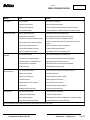
1
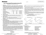
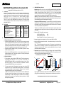
BioVision Cap Code 50 ml 15 ml 0.2 ml 1 vial 1.2 ml 166.7 µg NM WM Green Purple Red Yellow Part No. K347-100-1 K347-100-2 K347-100-3 K347-100-4 K347-100-5 K347-100-6 Decompose of NADP from extraction: To detect NADPH only, aliquot 200 µl samples into eppendorf tubes. Heat samples to 60°C for 30 min in a water bath or a heating block. Under the conditions, all NADP will be decomposed while NADPH will still be intact. Cool samples on ice. Quick spin samples if precipitates occur. Transfer 50 µl of NADPH samples into labeled 96-well plate in duplicates (Note: several sample dilutions should be performed to ensure the reading can be within the standard curve range). 2. Prepare a NADP Cycling Mix for each reaction: NADP Cycling Buffer Mix: NADP Cycling Enzyme Mix: Mix well and add 100 l of the mix into each well, mix well. NADP/NADPH Assay Protocol: A. Reagent Reconstitution and General Consideration: Reconstitute NADPH developer with 1.2 ml of ddH2O. Pipet up and down several times to completely dissolve the pellet into solution. Aliquot enough NADP Cycling Enzyme mix (2 µl per assay) for the number of assays to be performed in each experiment and aliquot and freeze the stock solution immediately at –70 ° C for future use. The reconstituted enzymes are stable for up to 2 months at –70°C. Reconstitute NADPH standard with 200 µl pure DMSO to generate 1 nmol/l NADPH standard stock solution. Ensure that the NADP Cycling Buffer is at room temperature before use. The optimal temperature is 22 ° C. Keep other enzymes on ice during the assay and protect from light as much as possible. B. Sample Preparation: 1. For cell samples*, wash cells with cold PBS. Pellet 4x10⁶ cells for each assay in a microcentrifuge tube (2000 rpm for 5 min). Lyse the cells with 800 µl of NADP/NADPH Extraction Buffer in a microfuge tube and keep on ice for 10 min. Spin down at 10,000 x g for 10 min, and collect the supernatant. Transfer the extracted NADP/NADPH solution into a new labeled tube. 2. For tissue samples*, weight ~50 mg tissue for each assay, wash with cold PBS, homogenize with 500 µl of NADP/NADPH Extraction Buffer in a microcentrifuge tube. Keep on ice for 10 min. Spin the sample at 10000xg for 10 min. Transfer the extracted NADP/NADPH solution into a new labeled tube. * Note: Cell or tissue lysates may contain enzymes that consume NADPH rapidly. We suggest removing these enzymes from the sample either by filtering the samples through 10 kDa molecular weight cut off filters (Cat # 1997-25) or deproteinizing the sample using Deproteinizing Sample preparation Kit (K808) before performing the assays. BioVision Incorporated 155 S. Milpitas Boulevard, Milpitas, CA 95035 USA 98 µl 2 µl 3. Incubate the plate at room temperature for 5 min to convert NADP to NADPH. 4. Add 10 µl NADPH developer into each well. Let the reaction develop for 1 to 4 hours. Read the plate at OD450 nm. Note: The signal increases as the reaction time. The plate can be read multiple times while the color is in developing. The reaction can be stopped by addition of 10 µl Stop Solution each well and mix well. The color should be stable within 48 hours in a sealed plate, after the reactions are stopped. 5. Calculation: Subtract 0 Standard reading from all readings. Apply the sample OD450 nm reading to standard curve. The amount of NADPt or NADPH can be expressed in pmol/10 6 cells or ng/mg protein (NADPH molecular weight 745.4). NADP/NADPH Ratio is calculated as: A. B. 1.6 NADH 1.2 NADPH 1000 0.8 0.4 0 0 50 800 600 400 200 0 NADPt - NADPH NADPH C. 1000 800 600 400 200 0 250 250 200 200 150 150 100 100 50 50 0 0 100 Doses of NADH, NADPH (pmol/well) Figure: A) NADH Standard Curve. Measurement of NADPt and NADPH in rat liver lysate (20 µg) (B) HeLa cell lysate (80 µg) (C). Assays were performed following the kit protocol. FOR RESEARCH USE ONLY! Not to be used on humans. Tel: 408-493-1800 | Fax: 408-493-1801 www.biovision.com | [email protected] Page 1 of 2 NADPH (pmol/mg) K347-100 NADP/NADPH Extraction Buffer NADP Cycling Buffer NADP Cycling Enzyme Mix NADPH Developer Stop Solution NADPH Standard (MW:833.36) Samples: To detect total NADP/NADPH (NADPt), transfer 50 µl of extracted samples into labeled 96-well plate in duplicates. (Note: several sample dilutions should be performed to ensure the reading can be within the standard curve range.) NADPt (pmol/mg) Components 1. Standard Curve: Dilute 10 µl of the 1 nmol/l NADPH standard with 990 l NADP/NADPH Extraction Buffer to generate 10 pmol/l standard NADPH (Note: diluted NADPH solution is unstable, must be used within 4 hours). Add 0, 2, 4, 6, 8, 10 µl of the diluted NADPH standard into labeled 96-well plate in duplicate to generate 0, 20, 40, 60, 80, 100 pmol/well standard. Make the final volume to 50 µl with NADP/NADPH extraction buffer. NADPH (pmol/mg) III. Kit Contents: NADP/NADPH Assay Protocol: OD 450 nm II. (Catalog #K347-100; 100 assays; Store kit at –70°C) Introduction: Assays of nicotinamide nucleotides are of continual interest in the studies of energy transforming and redox state of cells or tissue. The NADP/NADPH Quantification Kit provides a convenient tool for sensitive detection of the intracellular nucleotides: NADP, NADPH and their ratio. The enzymes in the system specifically recognize NADP/NADPH in an enzyme cycling reaction (It does not recognize NAD+/NADH). There is no need to purify NADP/NADPH from sample mix. The enzyme cycling reaction significantly increases detection sensitivity. Results can be quantified using plate reader at OD450 nm. C. NADPt (pmol/mg) NADP/NADPH Quantification Colorimetric Kit I. For research use only rev. 02/15 BioVision For research use only rev. 02/15 GENERAL TROUBLESHOOTING GUIDE: Problems Cause Solution Assay not working • Use of ice-cold buffer • Buffer must be at room temperature • Omission of a step in the protocol • Refer and follow the data sheet precisely • Plate read at incorrect wavelength • Check the wavelength in the data sheet and the filter settings of the instrument • Use of a different 96-well plate • Fluorescence: Black plates ; Luminescence: White plates; Colorimeters: Clear plates • Use of an incompatible sample type • Refer data sheet for details about incompatible samples • Samples prepared in a different buffer • Use the buffer provided in the kit or refer data sheet for instructions • Samples were not deproteinized (if indicated in datasheet) • Use the 10 kDa spin cut-off filter or PCA precipitation as indicated • Cell/ tissue samples were not completely homogenized • Use Dounce homogenizer (increase the number of strokes); observe for lysis under microscope • Samples used after multiple free-thaw cycles • Aliquot and freeze samples if needed to use multiple times • Presence of interfering substance in the sample • Troubleshoot if needed, deproteinize samples • Use of old or inappropriately stored samples • Use fresh samples or store at correct temperatures till use • Improperly thawed components • Thaw all components completely and mix gently before use • Use of expired kit or improperly stored reagents • Always check the expiry date and store the components appropriately • Allowing the reagents to sit for extended times on ice • Always thaw and prepare fresh reaction mix before use • Incorrect incubation times or temperatures • Refer datasheet & verify correct incubation times and temperatures • Incorrect volumes used • Use calibrated pipettes and aliquot correctly • Use of partially thawed components • Thaw and resuspend all components before preparing the reaction mix • Pipetting errors in the standard • Avoid pipetting small volumes • Pipetting errors in the reaction mix • Prepare a master reaction mix whenever possible • Air bubbles formed in well • Pipette gently against the wall of the tubes • Standard stock is at an incorrect concentration • Always refer the dilutions in the data sheet • Calculation errors • Recheck calculations after referring the data sheet • Substituting reagents from older kits/ lots • Use fresh components from the same kit • Measured at incorrect wavelength • Check the equipment and the filter setting • Samples contain interfering substances • Troubleshoot if it interferes with the kit • Use of incompatible sample type • Refer data sheet to check if sample is compatible with the kit or optimization is needed • Sample readings above/below the linear range • Concentrate/ Dilute sample so as to be in the linear range Samples with erratic readings Lower/ Higher readings in Samples and Standards Readings do not follow a linear pattern for Standard curve Unanticipated results Note: The most probable list of causes is under each problem section. Causes/ Solutions may overlap with other problems. BioVision Incorporated 155 S. Milpitas Boulevard, Milpitas, CA 95035 USA Tel: 408-493-1800 | Fax: 408-493-1801 www.biovision.com | [email protected] Page 2 of 2