1
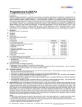
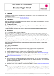
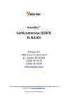
20-HETE ELISA kit Catalog Number: 20H1/20H11/20H21/20H101 Store at -20°C. FOR RESEARCH USE ONLY Introduction This competitive ELISA kit is for determination of 20-HETE (also known as 20-OH-AA) levels in biological samples. The specificity of the 20-HETE ELISA was investigated using authentic 20-HETE and a panel of fatty acids which, based on their structure, might be anticipated to compete with 20-HETE for binding to antibodies for 20-HETE. Anti-20-HETE did not cross-react with 14,15- and 11,12-DHETs, PGE2 and showed almost no crossreactivity even with structurally extremely similar arachidonic acid (AA), linoleic acid and linolenic acid as shown in the competitive ELISA analysis. Considering the only difference between 20-HETE and AA is an oxygen molecule, the specificity of the Detroit R&D 20-HETE ELISA is a surprise. Human essential and salt-sensitive hypertensions were related to differential AA metabolism by cytochrome P450 (CYP) 4A which has AA-ω-hydroxylase (20-HETE synthesis) activity. Increased circulating insulin inhibits 20HETE synthesis in obese hypertensive subjects. Recently, CYP4F2 genetic variants, which increased urinary 20-HETE secretion, were found to be correlated with the risk for hypertension in a Chinese population. This kit can be used for the determination of 20-HETE in serum, plasma, cells, and tissues following proper isolation and purification. Instructions are provided as to the proper isolation and purification in the following pages. Storage and Stability This kit will obtain optimal results if all of the components are stored at the proper temperature prior to use. Items should be stored at the designated temperatures upon receipt of this kit. All components are stored below -20°C and should not be re-frozen and thawed more than necessary. Materials Provided Part Number 1 2 3 4 5 6 7 Item Description Quantity 20-HETE ELISA Plate 20-HETE Standard (2 μL) 20-HETE HRP Conjugates (12 μL) Sample Dilution Buffer (25 mL) HRP Buffer (15 mL) Wash Buffer Solution (25 mL) TMB Substrate (24 mL) Solid 96-well plate coated with anti 20-HETE antibody in each well 1 Stock standard at a concentration of 1 mg/mL 1000 X concentrated solution 1 1 10 X solution of Tris-buffered saline with preservatives 1 1 X solution of Tris-buffered saline with preservatives 1 10 X solution of Tris-buffered saline with detergents and preservatives 1 A solution of TMB (tetra methyl benzadine) 1 Rev. 09/17/2012 2727 Second Ave. Suite 4113 – Detroit, MI 48201 – tel. 313.961.1606 – fax. 313.963.7130 – [email protected] – www.detroitRandD.com Page 2 of 9 Additional Required Materials (Not Provided) Plate reader with a 450 nm filter An 8-channel adjustable pipetter and an adjustable pipetter Storage bottles Costar® cluster tubes (1.2 mL) and microcentrifuge tubes Deionized water Precautions Please read all instructions carefully before beginning the assay. The reagents in this kit have been tested and formulated to perform optimally. This kit may not perform correctly if any of the reagents are replaced or any of the procedures are modified. This kit is intended for research use only and is not to be used as a diagnostic. Procedural Notes Remove all of the reagents required, including the TMB, and allow them to equilibrate to room temperature before proceeding with the assay. It is necessary to thoroughly mix the concentrated buffer solutions. A stir bar is contained within each buffer solution. Sample Preparations There are different protocols for isolating and purifying 20-HETE depending on the medium in which it is in. Listed below are the different protocols. For optimal results follow the appropriate protocol based on the biological sample present. 20-HETE measurement in cells expressing Cytochrome P450 4A 1. Collect and homogenize and/or sonicate the cells using a solution containing a final concentration of ~0.1 mM TPP (triphenylphosphine, 0.03-0.05 mg/mL). TPP is an antioxidant, which looks like a precipitate in samples because it does not easily dissolve. Before using the stored samples containing TPP, spin samples to separate the precipitated TPP from sample solution. 2. Acidify the whole homogenized cells with acetic acid to a pH of approximately 3-4. Measure using standard pH paper. 3. Extraction with ethyl acetate. Add an equal volume of ethyl acetate to the homogenized cells and vortex very well. Place the upper organic phase into a fresh clean tube after centrifugation. Then add another equal volume of ethyl acetate to the homogenized cells to start the second-time extraction. It is strongly recommended that extraction is performed three times. 4. Evaporate the pooled ethyl acetate from the extractions until all has dried up under argon or nitrogen gas. 5. Saponification if needed (see below) 6. Add 20 μL N, N-dimethyl-formamide (DMF), to dissolve the dried-up residue for reconstitution. Add 0.5 mL of 1x Sample Dilution Buffer (provided in kit) to make a solution. Load 100 μL in each well, in triplicates, on the ELISA plate. (Note: We recommend measuring a different dilution of sample in attempt to fit the results to the standard curve. e.g., load 3 wells with 50 μL of the rest of sample plus 50 μL of 1x Sample Dilution Buffer, and 3 wells with 10 μL of the rest of sample plus 90 μL of 1x Sample Dilution Buffer.) 7. Perform the ELISA for 20-HETE (according to the instructions of the manufacturer). 2727 Second Ave. Suite 4113 – Detroit, MI 48201 – tel. 313.961.1606 – fax. 313.963.7130 – [email protected] – www.detroitRandD.com Page 3 of 9 Saponification (to cleave fatty acid from glycerol backbone): 1. Dissolve dried fatty acids (obtained from 3X ethyl acetate extractions) in 2 mL of 20% KOH solution (make working solution: 1 mL of 2 M KOH + 4 mL methanol so that the final conc. of KOH = 0.4 N). 2. Vortex and incubate for 1 h at 50°C. 3. Add 1.5X H2O to the solution and adjust pH with 20% formic acid to pH ~5. 4. Re-extract the solution with ethyl acetate (1 part aqueous solution + 1 part ethyl acetate) and dry. 20-HETE measurement in tissues 1. Homogenize 1 g of tissue, 4 mL of H2O, and 0.01 mg TPP. 2. Acidify the homogenate by adding 8 μL of acetic acid to each homogenate. 3. Extract with an equal amount of ethyl acetate, vortex thoroughly, spin down, and collect the organic phase. Repeat this extraction twice more and combine all of the organic phases. 4. Dry the organic phase with argon or nitrogen gas. 5. Saponification if needed (see 20-HETE measurement in cells) 6. Dissolve the dried residue from above step # 4 with N,N-dimethyl-formamide ( DMF). (Add approximately 20 μL of DMF to reconstitute the dried-up residue.) 7. Dilute further with 1x Sample Dilution Buffer: Add approximately 0.5 mL of 1x Sample Dilution Buffer and centrifuge at 10,000 rpm for five minutes at room temperature. The supernatant will be used for ELISA. 8. Perform the ELISA for 20-HETE (according to the instructions of the manufacturer). 20-HETE measurement in plasma or serum 1. Combine 1.8 mL of plasma (adjusted with approximately 20 μL of acetic acid to pH 4) and 1.8 mL of ethyl acetate. Vortex thoroughly. Centrifuge at 2000 rpm for ten minutes at 22°C. Three phases should result: i. Upper organic phase – ethyl acetate phase (lipoproteins) ii. Interphase – proteins iii. Lower phase – aqueous phase 2. Collect the upper organic phase (a) and set aside. 3. Discard the interphase. Transfer the lower phase with a glass pipette to a new tube, and repeat the ethyl acetate extraction step 2 more times. 4. Evaporation of pooled organic phase: There should be approximately 5-6 mL of the ethyl acetate phase (a). Dry the pooled organic phase in a Speedvac to get the extracted sediment (b). 5. Saponification (to cleave fatty acid from glycerol backbone): Dissolve the dried-up residues (b) in 2 mL of 20% KOH solution (for preparation see 20-HETE in cells). Vortex thoroughly and incubate for 1 h at 50°C. This will yield an aqueous solution (c). 6. Dilute 2 mL of the aqueous solution (c) with 3 mL of H2O. Adjust the pH using 20% formic acid (132 μL) to pH ~5.5. Add ethyl acetate (1 part aqueous solution (c) + 1 part ethyl acetate), vortex thoroughly, and centrifuge at 2000 rpm for ten minutes at 22°C. Repeat the procedure twice more using an equal volume of ethyl acetate per sample. Collect the upper phase with saponified lipids. 7. Dry the pooled ethyl acetate upper phase (d) in a Speedvac, yielding the dried sample-sediment (e). Store the sediment (e) at -20°C. For ELISA assay, dissolve the dried sample-residue (e) in 20 μL N,Ndimethyl-formamide (DMF), then add 130 μL of 1x Sample Dilution Buffer. 8. For the competitive 20-HETE ELISA, the above 150 μL sample needs to be further diluted: Dilute 1:4 (e.g., 80 μL sample + 320 μL 1x Sample Dilution Buffer). Check the final pH (should be pH 7.4). When calculating the concentration, consider the dilution factor. In this case, 150 μL total sample volume from 1.8 mL plasma (12-fold concentration) and then, 80 sample in 400 μL SDB (5-fold dilution). Since, the samples are concentrated 2.4-fold; to get the actual concentration, you must divide by 2.4. 9. Perform the ELISA for 20-HETE (according to the instructions of the manufacturer). 2727 Second Ave. Suite 4113 – Detroit, MI 48201 – tel. 313.961.1606 – fax. 313.963.7130 – [email protected] – www.detroitRandD.com Page 4 of 9 Assay Preparations The solid 96-well plate and TMB solution are provided ready to use. The preparations of other assay reagents are detailed below. Wash Buffer: Mix the solution with a stir bar, applying low heat until a clear colorless solution is obtained. Dilute the entire contents of the Wash Buffer Concentrate (25 mL) with 225 mL of deionized water to yield a final volume of 250 mL of 1 X Wash Buffer. This can then be refrigerated for the entire life of the kit. HRP Conjugate: Dilute 1 vial of the 20-HETE-HRP conjugate (0.012 mL) with 12.00 mL of 1 X HRP buffer. One vial makes enough conjugate for one plate. The conjugate must be used the same day and should not be stored for later use. Standards: Label 5 microtubes as Standard 1 through Standard 5. Dilute the entire contents of Sample Dilution Stock buffer (25 mL) with 225 mL deionized water to yield a final volume of 250 mL of 1 X Sample Dilution Buffer. Add 0.9 mL of the Sample Dilution Buffer to the microtubes 1 to 5. Spin down the enclosed 20-HETE standard vial (2 μL, filled with inert gas) and add 1.998 mL of Sample Dilution Buffer to obtain 2 mL of solution. Label this Standard 6. Add 0.1 mL of the Standard 6 to the microtube labeled Standard 5 and mix thoroughly. Next, add 0.1 mL of Standard 5 into the microtube labeled Standard 4 and mix thoroughly. Continue to serially dilute the standards using 1:10 dilutions for the remaining standards. Samples: Samples can be directly diluted into the 1 X Sample Dilution Buffer if it is in solution. For extracted and dried samples, it is recommended to dissolve the dried-up samples with a minimal amount of ethanol of N, Ndimethyl-formanmide (DMF, 10 μL to 20 μL) and vortex well. Before ELISA assay, add 100 μL of 1 X Sample Dilution Buffer to make the stock sample solution ready for quantification with ELISA. The stock sample solution can be further diluted to a proper range of concentration for ELISA test. Performing the Assay Plate Setup: Each plate must contain a minimum of three blank wells (BL), three maximum binding wells (BO), and a six point standard curve (S1-S6). Each sample should be assayed in triplicate. A suggested plate format is shown below: BL BL BL B0 B0 B0 1 1 1 2 2 2 3 3 3 4 4 4 5 5 5 6 6 6 =BL =BO =S1—S6 =Samples 2727 Second Ave. Suite 4113 – Detroit, MI 48201 – tel. 313.961.1606 – fax. 313.963.7130 – [email protected] – www.detroitRandD.com Page 5 of 9 Standard Dilutions Table Standards No. 6 No. 5 No. 4 No. 3 No. 2 No. 1 Final Concentration (pg/mL) 1,000,000 100,000 10,000 1,000 100 10 Add Sample Dilution Buffer (mL) 1.998 0.9 0.9 0.9 0.9 0.9 Serial Dilutions Procedure 2 μL of stock solution. Add 0.1 mL of No. 6 Add 0.1 mL of No. 5 Add 0.1 mL of No. 4 Add 0.1 mL of No. 3 Add 0.1 mL of No. 2 Assay Procedure Step 1: Load 200 microliters of Sample Dilution Buffer into the blank (BL) wells and 100 microliters of Sample Dilution Buffer into the maximum binding (BO) wells. Step 2: Load 100 microliters of each of the standards into the appropriate wells. Step 3: Load 100 microliters of each of the samples into the appropriate wells. Step 4: Load 100 microliters of the diluted 20-HETE-HRP conjugate in the BO wells, the standard wells, and the sample wells. Do NOT add HRP conjugate into the BL wells. Step 5: Incubate the plate at room temperature for two hours. Step 6: Wash the plate three times with 400 microliters of the diluted Wash Buffer per well. Step 7: After the last of the three wash cycles pat the plate dry onto some paper toweling. Step 8: Add 200 microliters of the TMB substrate to all of the wells (including BL wells). Step 9: Incubate the plate at room temperature for 15-30 minutes. Step 10: Add 50 micoliters of 2 N sulfuric acid to all of the wells. Step 11: Read the plate at 450 nm. Calculating the Results Most plate readers provide data reduction software that can be used to plot the standard curve and determine the sample concentrations. If your plate reader does not have this option, then a data reduction program can be used (4 parameter of log-log curve fit). If you do not have these options, the results can be obtained manually as follows: 1. 2. 3. 4. 5. 6. Average the absorbance readings from the blanks and subtract that value from each well of the plate to obtain the corrected readings. (Note: Some plate readers do this automatically. Consult the user manual of your plate reader.) Average the corrected absorbance readings from the BO wells. This is your maximum binding. Calculate the %B/BO for Standard 1 by averaging the corrected absorbance of the two S1 wells, divide the average by the maximum binding, then multiply by 100. Repeat this formula for the remaining standards. Plot the %B/BO versus the concentration of 20-HETE from the standards using semi-log paper. Calculate the %B/BO for the samples and determine the concentrations, utilizing the standard curve. Multiply the concentrations obtained for each of the samples by their corresponding dilution factor. 2727 Second Ave. Suite 4113 – Detroit, MI 48201 – tel. 313.961.1606 – fax. 313.963.7130 – [email protected] – www.detroitRandD.com Page 6 of 9 Typical Results QC on 12/10/08 Net OD at 450 nm of Bo=1.667 30 min color development 110 . 0 10 0 . 0 90.0 80.0 %B/B0 70.0 60.0 50.0 40.0 30.0 20.0 10 . 0 0.0 1 10 1 00 10 00 1 00 00 1 00 00 0 1 00 00 0 0 20 HETE [pg/m l] The data shown here is an example of typical results obtained using the Detroit R & D 20-HETE ELISA kit. These results are only a guideline, and should not be used to determine values from your samples. The user must run their own standard curve every time. BL wells BO wells = 0.063 = 1.667 Standard No. 1 No. 2 No. 3 No. 4 No. 5 No. 6 Concentration 10 pg/mL 100 pg/mL 1,000 pg/mL 10,000 pg/mL 100,000 pg/mL 1,000,000 pg/mL O.D. 1.630 1.597 1.319 0.687 0.428 0.356 %B/BO 97.8 95.8 79.1 41.2 25.7 21.3 Specificity of anti-20-HETE Anti-20-HETE did not cross-react with 14,15- and 11,12-DHETs, PGE2 and showed almost no cross-reactivity even with structurally extremely similar arachidonic acid, linoleic acid and linolenic acid as shown in the competitive ELISA analysis (shown below). 2727 Second Ave. Suite 4113 – Detroit, MI 48201 – tel. 313.961.1606 – fax. 313.963.7130 – [email protected] – www.detroitRandD.com Page 7 of 9 Troubleshooting No color present in standard wells. The HRP conjugate was not added. Redo the assay and add the conjugate at the proper step. The HRP conjugate was not incubated for the proper time. Redo the assay and incubate for the proper time. No color in any wells, including the TA wells. The TMB substrate was not added. Add substrate. The TMB substrate was not incubated for the proper time. Continue incubation until desired color is reached. The color is faint. One or all of the incubation times were cut short. Redo the assay with the proper incubation times. The TMB substrate was not warmed up to room temperature. Redo the assay making sure all reagents are at room temperature. The lab is too cold. Be sure the lab temperature is between 21-27°C and redo the assay. The background color is very high. The TMB substrate has been contaminated. Redo the assay with a fresh bottle of substrate. Scattered O.D. obtained from the sample. Redo assay using an 8-channel pipetman making sure that 8 channels are equal volume while loading. References 1. Liu, H., Zhao,Y., Nie, D., Shi, J., Fu, L., Li, Y., Yu, D., and Lu, J. Association of a Functional Cytochrome P450 4F2 Haplotype with Urinary 20-HETE and Hypertension. J Am Soc Nephrol 19: 714-721, 2008 2. Meseguer et al. Kidney Androgen-Regulated Protein Transgenic Mice Show Hypertension and Renal Alterations Mediated by Oxidative Stress. Circulation 119, 1908-1917, 2009. 3. Dolegowska, B., Blogowski, W., Domanski, L.. Is it possible to predict the early post-transplant allograft function using 20-HETE measurements? A preliminary report. Transpl Int. 2009 22:546-553. 4. Liu et al. Overexpression of cytochrome P450 4F2 in mice increases 20-hydroxyeicosatetraenoic acid production and arterial blood pressure. Kidney International 75, 1288-1296, 2009. 5. Wang et al. Selective Inhibitors of CYP2J2 Related to Terfenadine Exhibit Activity Strongly against Human Cancers in vitro and in vivo. JPET, 152017, 2009. 6. Cuez, Malik, Tunctan et al. A synthetic analogue of 20-HETE, 5,14-HEDGE, reverses endotoxin-Induced hypotension via increased 20-HETE levels associated with decreased iNOS protein expression and vasodilator prostanoid production in rats. Basic Clin. Pharmacol. Toxicol. 106, 378-388, 2010. 2727 Second Ave. Suite 4113 – Detroit, MI 48201 – tel. 313.961.1606 – fax. 313.963.7130 – [email protected] – www.detroitRandD.com Page 8 of 9 7. Malik et al. 2,3′,4,5′-Tetramethoxystilbene prevents deoxycorticosterone-salt-induced hypertension: contribution of cytochrome P-450 1B1. Am. J. Physiol. Heart Circ. Physiol. 299, H1891-H1901, 2010. 8. Malik et al. Cytochrome P450 1B1 contributes to angiotensin II-induced hypertension and associated pathophysiology. Hypertension 56, 667, 2010. 9. Tunctan et al. Contribution of vasoactive eicosanoids and nitric oxide production to the effect of selective cyclooxygenase-2 inhibitor, NS-398, on endotoxin-induced hypotension in rats. Basic Clin. Pharmacol. Toxicol. 107, 877-882, 2010. 10. Imaizumi et al. L-4F differentially alters plasma levels of oxidized fatty acids resulting in more antiinflammatory HDL in mice. Drug Metab. Letters, 4, 139-148, 2010. 11. Cervenka, Kramer, Falck, Imig, Hammock et al. Combined inhibition of 20-HETE formation and of EET degradation attenuates hypertension and hypertension-induced end-organ damage in Ren-2 transgenic rats. Clinical Science 118, 617-632. 2010. 12. Cervenka, Kramer, Falck, Imig et al. Intrarenal CYP-450 metabolites of arachidonic acid in the regulation of the nonclipped kidney function in two-kidney, one-clip Goldblatt hypertensive rats. J Hypertens 28, 582-593, 2010. 13. Hu, Wang et al. Peripheral and central augmentation indexes in relation to the CYP4F2 polymorphisms in Chinese. J Hypertens 29, 501-508, 2011. 14. Buharalioglu et al. Piroxicam reverses endotoxin-induced hypotension in rats: contribution of vasoactive eicosanoids and nitric oxide. Basic Clin Pharmacol Toxicol 186–194, 2011. 15. Na-Bangchang et al. Study on the association between environmental cadmium exposure, cytochrome P450mediated 20-HETE, heme-oxygenase-1 polymorphism and hypertension in Thai population residing in a malaria endemic areas with cadmium pollution. Environ Toxicol Pharmacol 31, 416-426, 2011. 16. Onoe et al. Increase of 20-HETE synthase after brain ischemia in rats revealed by PET study with (11)C-labeled 20-HETE synthase-specific inhibitor. J Cereb Blood Flow Metab 32, 1737-1746, 2012. 17. Dong H. Metabolomic profiling of lipids for biomarker discovery. Biochem Anal Biochem 1, 5, 2012. 18. Fordsmann JC, Ko RW, Choi HB, Thomsen K, Witgen BM, Mathiesen C, Lønstrup M, Piilgaard H, MacVicar BA, Lauritzen M. Increased 20-HETE synthesis explains reduced cerebral blood flow but not impaired neurovascular coupling after cortical spreading depression in rat cerebral cortex. J Neurosci 33, 2562-2570, 2013. 19. Stobart JL, Lu L, Anderson HD, Mori H, Anderson CM. Astrocyte-induced cortical vasodilation is mediated by D-serine and endothelial nitric oxide synthase. PNAS USA. 110, 3149-3154, 2013. 20. Tunctan 20-HETE mimetics or inhibitors in the treatment of cancer patients with sepsis and septic shock. International Journal of Cancer Studies & Research (IJCR) 2:101, 2013. 21. Tunctan et al. NS-398 reverses hypotension in endotoxemic rats: Contribution of eicosanoids, NO, and peroxynitrite. Prostaglandins & other Lipid Mediators 104–105 93–108, 2013 . Warranty Detroit R&D, Inc., makes no warranty of any kind expressed, or implied, including, but not limited to the warranties of fitness for a particular purpose and merchantability. 2727 Second Ave. Suite 4113 – Detroit, MI 48201 – tel. 313.961.1606 – fax. 313.963.7130 – [email protected] – www.detroitRandD.com Page 9 of 9 Detroit R&D, Inc. Metro Center For High Technology Bldg. (MCHT) 2727 Second Ave. Suite 4113 Detroit, MI 48201 Phone: 313.961.1606 Fax: 313.963.7130 E-mail: [email protected] www.DetroitRandD.com 2727 Second Ave. Suite 4113 – Detroit, MI 48201 – tel. 313.961.1606 – fax. 313.963.7130 – [email protected] – www.detroitRandD.com