1
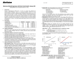
Product Manual OxiSelect™ Glutathione Reductase Assay Kit Catalog Number STA-812 100 assays FOR RESEARCH USE ONLY Not for use in diagnostic procedures Introduction Glutathione reductase is a homodimeric enzyme that is a member of the flavoprotein disulfide oxidoreductases. It has an indirect impact in the prevention of oxidative damage in cells by helping to maintain intracellular reduced glutathione (GSH). Thus, measuring the activity of the enzyme is an indicator of oxidative stress. It is a ubiquitous enzyme that catalyzes the NADPH dependent reduction reaction of oxidized glutathione (GSSG) to reduced glutathione (GSH). Oxidized glutathione is reduced through a multi-step reaction in which glutathione reductase is reduced by NADPH, which in turn reacts with a GSSG molecule. This creates a disulfide interchange reaction that creates two GSH molecules and restores glutathione reductase to its oxidized form. Regenerated GSH is available to detoxify hydrogen peroxide. Maintenance of GSH is vital in oxidation-reduction processes as well as detoxification of hydrogen peroxide and organic peroxides brought on by inflammation in cells. Cell Biolabs’ OxiSelect™ Glutathione Reductase Assay Kit is a quantitative assay for measuring glutathione reductase activity within plasma, erythrocytes, tissues, and cell lysates. Glutathione reductase activity is defined as 1 unit of enzyme reducing 1 µmole oxidized glutathione (GSSG) per minute at pH 7.6 and 25ºC. The kit employs a simple enzymatic recycling reaction for glutathione quantification where the reduction of a chromagen is correlated to glutathione reductase enzymatic activity. The kit has a detection sensitivity limit of approximately 0.6 mU/mL. Each kit provides sufficient reagents to perform up to 100 assays, including standard curve and unknown samples. Assay Principle The OxiSelect™ Glutathione Reductase Assay Kit is a quantitative assay for measuring the glutathione reductase activity within a sample. Glutathione Reductase reduces oxidized glutathione (GSSG) to reduced glutathione (GSH) in the presence of NADPH. Subsequently, the chromogen reacts with the thiol group of GSH to produce a colored compound that absorbs at 405 nm (Figure 1). The glutathione reductase content in unknown samples is determined by comparison with the predetermined glutathione reductase standard curve. The rate of chromophore production is proportional to the concentration of glutathione reductase activity within the sample. The rate can be determined from the absorbance change over time. Figure 1. Assay Principle 2 Related Products 1. STA-310: OxiSelect™ Protein Carbonyl ELISA Kit 2. STA-312: OxiSelect™ Total Glutathione Assay Kit 3. STA-330: OxiSelect™ TBARS Assay Kit (MDA Quantitation) 4. STA-340: OxiSelect™ Superoxide Dismutase Activity Assay 5. STA-341: OxiSelect™ Catalase Activity Assay Kit 6. STA-342: OxiSelect™ Intracellular ROS Assay Kit (Green Fluorescence) 7. STA-347: OxiSelect™ In Vitro ROS/RNS Assay Kit (Green Fluorescence) 8. STA-345: OxiSelect™ ORAC Activity Assay 9. STA-832: OxiSelect™ MDA Competitive ELISA Kit 10. STA-838: OxiSelect™ HNE Adduct Competitive ELISA Kit Kit Components Box 1 (shipped at room temperature) 1. Glutathione Reductase Standard (Part No. 281201): One 20 µL amber tube. 2. Glutathione Disulfide (GSSG) (Part No. 281202): One 5 mL bottle of a 10 mM solution. 3. Chromogen (Part No. 281203): One 0.5 mL amber tube. 4. Assay Buffer (5X) (Part No. 281204): Two 25 mL bottles. Box 2 (shipped on blue ice packs) 1. NADPH (50X) (Part No. 231203): One 50 µL amber tube. Materials Not Supplied 1. 96-well microtiter plate 2. Distilled or deionized water 3. 1X PBS 4. 10 μL to 1000 μL adjustable single channel micropipettes with disposable tips 5. 50 μL to 300 μL adjustable multichannel micropipette with disposable tips 6. Conical tubes and bottles for sample and buffer preparation 7. Centrifuge and/or microfuge 8. Sonicator or tissue homogenizer 9. Multichannel micropipette reservoirs 10. Ethanol 11. Microplate reader capable of reading 405 nm 3 Storage Upon receipt, store the NADPH at -80ºC. Prepare single use aliquots and avoid multiple freeze/thaw cycles. Store the remaining kit components at 4ºC. Preparation of Reagents • 1X Assay Buffer: Prepare 1X Assay Buffer by adding 200 mL of deionized water to 50 mL of the 5X Assay Buffer. Mix thoroughly until homogeneous. Use this buffer for preparing kit reagents. Store at 4ºC when not in use. • Chromogen: Prepare the Chromogen just before use and prepare only enough for immediate applications. Dilute the Chromogen stock 1:15 with 1X Assay Buffer (eg. Add 200 μL of Chromogen stock to 2.8 mL of 1X Assay Buffer. Vortex thoroughly. • 1X NADPH: Prepare 1X NADPH by diluting the stock solution 1:50 with 1X Assay Buffer. Vortex the stock tube thoroughly prior to preparing. Prepare only enough for immediate applications (eg. Add 25 μL of NADPH stock to 1.225 mL Assay Buffer). Preparation of Samples These preparation protocols are intended as a guide for preparing known samples. The user may need to adjust the sample treatment accordingly. All samples should be assayed immediately or stored for up to 1-2 months at -80°C. A trial assay with a representative test sample should be assayed to determine the samples compatibility with the dynamic range of the standard. The assay can be used on cell culture supernatants, plasma, erythrocytes, tissues, and cell lysates. High levels of interfering substances may cause variations in results. Run proper controls as necessary. Always run a standard curve with samples. Notes: • Thiol compounds, such as cysteine, dithiothreitol (DTT), or β-mercaptoethanol can interfere with the assay by competing with GSH for binding to the chromogen. In addition, Nethylmaleimide or other thiol alkylating reagents should also be avoided because they will interfere with glutathione reductase and GSH. • Make serial dilutions of samples as necessary to obtain a quantifiable change in absorbance readings over time. • Samples containing reduced glutathione (GSH) should be accounted for by running the sample without Glutathione Disulfide (GSSG) and subtracting these background values from a treated sample. A kinetic assay is recommended because it is more precise than an end point assay. • Plasma: Add blood sample to a blood collection tube with an anticoagulant such as heparin, EDTA, or sodium citrate. Centrifuge at 3,000 rpm for 10-15 minutes at 4ºC. Remove the upper yellow plasma supernatant layer without disturbing the white buffy coat (leukocytes). Store on ice if assaying immediately or freeze at -80ºC for up to 1-2 months. Dilute the plasma with 1X Assay Buffer before testing. • Erythrocytes: Add blood sample to a blood collection tube with an anticoagulant such as heparin, EDTA, or sodium citrate. Centrifuge the blood at 3,000 rpm for 10-15 minutes at 4ºC. Remove the white interface buffy coat (leukocytes) and yellow plasma layers and discard. Wash the erythrocytes in cold PBS or saline. Determine the packed cell volume and add 4 cell volumes of 4 cold deionized water to lyse the cells. Centrifuge at 10,000-12,000 rpm for 10 minutes at 4ºC. Collect the supernatant. Store on ice if assaying immediately or freeze at -80ºC for up to 1-2 months. Dilute with 1X Assay Buffer before testing. • Cell Lysates: Wash adherent cells at 2-6 x 10⁶ cells with 1X PBS. Harvest adherent cells with a rubber policeman, or gentle trypsinization. Centrifuge adherent or suspension cells at 500-1000 rpm for 5 minutes at 4ºC. Remove supernatant and wash cells in cold PBS. Repeat centrifugation and remove solution. Immediately resuspend the cell pellet with 200-500 µL ice-cold 1X Assay Buffer for a cell number of 1-5 x 10⁶ cells. Mix thoroughly. Homogenize or sonicate cell suspension and store on ice until use. Transfer the suspension to a microfuge tube and centrifuge at 12,000 rpm for 5 minutes at 4ºC. Collect the supernatant. Store on ice if used immediately or freeze at -80ºC for up to 1-2 months. Dilute with 1X Assay Buffer as necessary before testing. • Tissue Lysate: Perfuse or wash the tissue to remove blood cells and clots with a cold PBS/1 mM EDTA. Homogenize the tissue thoroughly with cold isotonic saline solution of 1X PBS with 0.16 mg/mL heparin to prevent coagulation. Blot the tissue dry and weigh. Add ice-cold PBS/1 mM EDTA (~1 mL/100 mg tissue) and homogenize using a glass pestle. Centrifuge the homogenate at 12,000 rpm for 15 minutes at 4ºC. Collect the supernatant. Store on ice if used immediately or freeze at -80ºC for up to 1-2 months. Dilute with 1X Assay Buffer as necessary before testing. Preparation of Standard Curve 1. To prepare glutathione reductase standards, first perform a 1:500 dilution of the stock Glutathione Reductase in 1X Assay Buffer. Use only enough for immediate applications (eg. Add 5 μL of Glutathione Reductase to 2495 μL 1X Assay Buffer). This solution has a concentration of 1 Unit/mL or 1000 mU/mL. 2. Use microfuge tubes to prepare a series of standards according to Table 1 below. Prepare standards fresh for each assay performed. Vortex tubes thoroughly when preparing as the glutathione reductase solution is a suspension, and therefore will settle upon standing. Do not store or reuse standard preparations. Standard Tubes 1 2 3 4 5 6 7 8 1000 mU/mL Glutathione Reductase Standard (µL) 80 500 of Tube #1 500 of Tube #2 500 of Tube #3 500 of Tube #4 500 of Tube #5 500 of Tube #6 0 1X Assay Buffer (µL) 920 500 500 500 500 500 500 500 Table 1. Preparation of Glutathione Reductase Standards. 5 Glutathione Reductase (mU/mL) 80 40 20 10 5 2.5 1.25 0 Assay Protocol 1. Prepare and mix all reagents thoroughly before use. Prepare the glutathione reductase standards simultaneously with the samples so they may be assayed together. Each sample, including unknown and standard, should be assayed in duplicate or triplicate. 2. In a 96-well plate, add 25 µL of the 1X NADPH solution to each well to be tested. 3. Add 100 µL of the prepared glutathione reductase standards or samples to each well to be tested. 4. Add 50 µL of the 1X Chromogen and mix briefly. 5. Ensure that the plate reader is prepared for a kinetic assay and is set to read at 405 nm. 6. Add 25 µL of the Glutathione Disulfide (GSSG) solution and mix briefly. Immediately begin recording the absorbance at 405 nm at 1 minute intervals for 10 minutes. The reaction may be run longer if necessary. If using all the wells within the plate at one time, then it may be necessary to record the absorbance at 2 minute intervals. 7. Calculate the concentration of standards and samples. See Calculation of Results below. Calculation of Results 1. First, determine the average of the replicate absorbance readings for each glutathione reductase standard, sample, and negative control for every time point taken. 2. Graph the average of each standard, sample, and background absorbance at 405nm against incubation time. Determine the slope for each value from the linear portion of each curve. See Figure 2 under Example of Results below. 3. Next, subtract the background slope from the slope of the standards and samples. 4. Plot the net slopes of the glutathione reductase standards against the microunits/mL concentration of glutathione reductase. See Figure 3 under Example of Results below. 5. Compare the net slopes of the samples with the standard curve from Figure 3 and determine the microunits/mL concentration of glutathione reductase for each sample. Note: Remember that the standard and samples are diluted 1:2 dilution within the final assay reaction protocol. Example of Results The following figures demonstrate typical Glutathione Reductase Assay results at 405 nm. One should use the data below for reference only. This data should not be used to interpret actual results. 6 Figure 2. Glutathione Reductase Standard Curve (OD 405nm versus incubation time as a function of Glutathione Reductase concentration). Figure 3: Glutathione Reductase Standard Curve (Net slope versus Glutathione Reductase concentration). 7 References 1. Anderson, M. Glutathione in Free radicals, A Practical Approach. Oxford University Press, New York; 1996. 2. Halliwell, B, Gutteridge, J.M.C. Free Radicals in Biology and Medicine. Oxford University Press, New York; 1999. 3. Julius, M., et al. J. Clin. Epidemiol. (1994) 47: 1021-1026. 4. Mytilineou, C., et al. Parkinsonism Relat. Disord. (2002) 8: 385-387. Warranty These products are warranted to perform as described in their labeling and in Cell Biolabs literature when used in accordance with their instructions. THERE ARE NO WARRANTIES THAT EXTEND BEYOND THIS EXPRESSED WARRANTY AND CELL BIOLABS DISCLAIMS ANY IMPLIED WARRANTY OF MERCHANTABILITY OR WARRANTY OF FITNESS FOR PARTICULAR PURPOSE. CELL BIOLABS’ sole obligation and purchaser’s exclusive remedy for breach of this warranty shall be, at the option of CELL BIOLABS, to repair or replace the products. In no event shall CELL BIOLABS be liable for any proximate, incidental or consequential damages in connection with the products. Contact Information Cell Biolabs, Inc. 7758 Arjons Drive San Diego, CA 92126 Worldwide: +1 858-271-6500 USA Toll-Free: 1-888-CBL-0505 E-mail: [email protected] www.cellbiolabs.com 2014: Cell Biolabs, Inc. - All rights reserved. No part of these works may be reproduced in any form without permissions in writing. 8