1
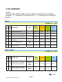
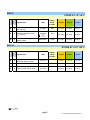
USER MANUAL
Genequality
BCR-ABL
cod. 04-55A
cod. 04-55R
Kit for the detection of the translocation t(9;22)
(q34;q11), involving the abl protooncogene
on chromosome 9 and the bcr gene
on chromosome 22.
04-55A-25(8033622780823)-EN.doc
1 PRODUCT INFORMATION
3
2 KIT CONTENT
4
3 STORAGE AND STABILITY OF THE REAGENTS
6
4 PRECAUTIONS FOR USE
6
5 SAFETY RULES
8
5.1
General safety rules
8
5.2
Safety rules about the kit
8
6 MATERIALS REQUIRED, BUT NOT PROVIDED
10
6.1
Reagents
10
6.2
Instruments
10
6.3
Materials
10
7 PREPARATION OF THE REAGENTS
11
8 INTRODUCTION
12
9 TEST PRINCIPLE
13
10 PRODUCT DESCRIPTION
14
11 COLLECTION, MANIPULATION AND PRE-TREATMENT OF THE
SAMPLES
15
12 PROCEDURE
16
12.1 RNA extraction
16
12.2 Retrotranscription (RT) for cDNA synthesis
16
12.3 Direct amplification (first ampification)
17
12.4 Nested Amplification (second amplification)
18
12.5 Visualization of the amplification products
19
12.5.1 Agarose gel electrophporesis
19
12.5.2 Sample loading on the gel
19
13 INTERPRETATION OF THE RESULTS
21
14 TROUBLESHOOTING
23
15 DEVICE LIMITS
25
pag.1
04-55A-25(8033622780823)-EN.doc
16 DEVICE PERFORMANCES
25
16.1 Specificity
25
16.2 Sensitivity
25
17 BIBLIOGRAPHIC REFERENCES
26
17.1 Useful websites
Hata! Yer işareti tanımlanmamış.
pag.2
04-55A-25(8033622780823)-EN.doc
1 PRODUCT INFORMATION
This user manual describes the instructions for use of the following products:
BCR-ABL
(cod 04-55A)
Kit for the detection of the translocation t(9;22) (q34;q11), involving the abl
protooncogene on chromosome 9 and the bcr gene on chromosome 22 by
DNA amplification (nested RT-PCR) in bcr and abl genes.
The kit includes all the reagents for retrotranscription for nested DNA
amplification and for agarose gel electrophoresis, the positive and negative
controls (cDNA positive and negative for the translocation).
Code
04-55A-25
04-55A-50
Product
BCR-ABL
BCR-ABL
Pkg
25 test
50 test
BCR-ABL- amplification reagents
(cod 04-55R)
Kit for the detection of the translocation t(9;22) (q34;q11), involving the abl
protooncogene on chromosome 9 and the bcr gene on chromosome 22 by
DNA amplification (nested RT-PCR) in bcr and abl genes.
The kit includes all the reagents for retrotranscription for nested DNA
amplification, the positive and negative controls (cDNA positive and negative
for the translocation).
Code
04-55R-25
04-55R-50
Product
BCR-ABL - amplification reagents
BCR- ABL - amplification reagents
Pkg
25 test
50 test
pag.3
04-55A-25(8033622780823)-EN.doc
2 KIT CONTENT
NOTE:
In the kits with different codes (A or R) different components are included.
(legenda: X = component included in the kit; 0 = component not included in
the kit)
BOX P
cod.
04-55A
cod.
04-55R
STORE AT – 20°C
DESCRIPTION
LABEL
TUBE (T)
OR LID
COLOUR
25 test
50 test
8 test
green (T)
25
50
8
X
X
Single-dose RT tubes
X
X
Single-dose premix tubes for
BCR-ABL direct amplification
red (T)
25
50
8
X
X
Single-dose premix tubes for
BCR-ABL nested amplification
yellow (T)
25
50
8
X
X
Thermostable Taq DNA
polymerase
AB TAQ
5 U/μL
red
1X 30μL
1X 60μL
1X 10μL
X
X
Reverse transcriptase
RT
enzyme
purple
1X 30μL
1X 56μL
1X 10μL
X
X
Reagent mix for
retrotranscription
RT MIX
green
1X 240μL
1X 448μL
1X 80μL
SMALL BAG
cod.
04-55A
cod.
04-55R
STORE AT – 20°C
DESCRIPTION
X
X
cDNA positive for the BCRABL p210 b3a2 traslocation
X
X
cDNA negative for the BCRABL p210 b3a2 traslocation
LABEL
TUBE (T)
OR LID
COLOUR
25 test
50 test
8 test
BCR-ABL
positive
(b3a2)
cDNA
Blue
1X 6 μL
1X 10 μL
1X 4 μL
BCR-ABL
negative
cDNA
Blue
1X 6 μL
1X 10 μL
1X 4 μL
pag.4
04-55A-25(8033622780823)-EN.doc
BOX F
cod.
04-55A
cod.
04-55R
STORE AT +2°/ +8°C
DESCRIPTION
0
Electrophoresis loading buffer
(6X solution)
X
0
Ethidium Bromide solution
(2,5 mg/mL)
X
0
DNA Molecular Weight
Marker (MW)
X
LABEL
TUBE (T)
OR LID
COLOUR
25 test
50 test
8 test
6X Blue
Blue
1X 100μL
1X 200μL
1X 5 μL
Ethidium
Bromide
Red
1X 100μL
1X 150μL
1X 50μL
Yellow
1X 100μL
1X 150μL
1X 50μL
TOXIC
R 23 68
S 36/37 45
MW Marker
BOX A
cod.
04-55A
cod.
04-55R
STORE AT +15°/ +25°C
DESCRIPTION
LABEL
TUBE (T)
OR LID
COLOUR
25 test
50 test
8 test
X
0
Agarose
Molecular Biology grade
AGAROSE
1X 8 g
1X 14 g
1X 4 g
X
X
Mineral oil
Mineral oil
1X 600μL
1X 1,2mL
1X 300μL
X
0
Electrophoresis buffer
TRIS-Acetate -EDTA pH 8,0
50X TAE
1X 40 mL
1X 60 mL
1X 20 mL
pag.5
04-55A-25(8033622780823)-EN.doc
3 STORAGE AND STABILITY OF THE REAGENTS
Each component of the kit should be stored according to the directions
indicated on the label of the single boxes.
In particular:
Box P
Small bag
Box F
Box A
store at -20°C
store at -20°C
store at +2/+8°C
store at +15/+25°C
(room temperature)
When stored at the recommended temperature, all test reagents are stable
until their expiration date, indicated on the labels.
4 PRECAUTIONS FOR USE
• This product is for in vitro use only
• The kit should be handled by investigator qualified through education and
training in molecular biology techniques applied to diagnostics.
• Before starting the kit procedure, read carefully and completely the
instruction manual.
• Keep the product out of heating sources;
• Do not use any part of the kit if over the expiration date;
• In case of any doubt about the storage conditions, box integrity or method
application,
contact
AB
ANALITICA
technical
support
at:
[email protected] before using the kit.
In the amplification of nucleic acids, the investigator has to take the
following special precautions:
• Use filter-tips;
pag.6
04-55A-25(8033622780823)-EN.doc
• Store the biologic samples, the purified RNAs, the positive controls
included in the kit and all the amplification products in different places from
where amplification reagents are stored.
• Organise the space in different pre- and post-PCR (RT-PCR) units; do not
share consumables (pipets, tips, tubes,…) between them.
• Change frequently the gloves;
• Wash the bench surfaces with 5% sodium hypochloride;
• Keep the extracted RNA in an ice-bath during reaction setup. Take care to
store RNA samples at -20°C or at -80°C for long term storage.
• Thaw the PCR premixes at room temperature before use. Add Taq DNA
polymerase and cDNA very quickly at room temperature, better if in an icebath
pag.7
04-55A-25(8033622780823)-EN.doc
5 SAFETY RULES
5.1 General safety rules
• Wear disposable gloves to handle reagents and clinical samples, wash
your hands at the end of work.
• Do not pipet with mouth.
• Since no known diagnostic method can assure the absence of infective
agents, it is a good rule to consider every clinical sample as potentially
infectious and handle it as such.
• All the devices that get directly in touch with clinical samples should be
considered as contaminated and disposed as such. In case of accidental
spilling of the samples, clean up with 10% Sodium Hypochloride. The
materials used to clean up should be disposed in special containers for
contaminated products
• Clinical samples, materials and contaminated products should be disposed
after decontamination by:
immersion in a solution of 5% Sodium Hypochloride (1 volume of Sodium
Hypochloride solution every 10 volumes of contaminated fluid) for 30 minutes
OR
autoclaving at 121°C at least for 2 hours (NOTE: do not autoclave solutions
containing Sodium Hypochloride!!)
5.2 Safety rules about the kit
The risks for the use of this kit are related to the single components:
Dangerous components:
ETHIDIUM BROMIDE
(included in the kit cod 04-55A only)
3,8-diamino-1-ethyl-6-phenylphenantridiumbromide <2%
Description of risk:
T (Toxic)
pag.8
04-55A-25(8033622780823)-EN.doc
RISK SENTENCES AND S SENTENCES
R 23 and R 68
S 36/37 45
Toxic for inhalation.
Risk of irreversible effects.
Wear laboratory coat and disposable gloves.
In case of accident or discomfort, seek for medical
assistance and show the container or label.
R and S sentences refer to the concentrated product, as provided in the kit.
In particular for Ethidium Bromide, until the dilution in the agarose gel.
In manipulating concentrated Ethidium Bromide, use a chemical dispensing
fume cabinet. Always wear disposable gloves and laboratory coat in
manipulating the diluted Ethidium solution as well.
The product can not be disposed with the common waste. It must not reach
the drainer system. For the disposal, follow the local law.
In case of accidental spilling of Ethidium Bromide, clean with Sodium
hypochloride and water.
Safety data sheet (MSDS) of the product is available upon request.
pag.9
04-55A-25(8033622780823)-EN.doc
6 MATERIALS REQUIRED, BUT NOT PROVIDED
6.1 Reagents
•
•
•
•
Reagents for RNA extraction;
Sterile DNase and RNase free water;
Distilled water;
Reagents for agarose gel electrophoresis (necessary for cod. 04-55R)
6.2 Instruments
• Laminar flow cabinet (use is recommended while adding TAQ polymerase
to the amplification premix to avoid contamination; it would be
recommended to use another laminar flow cabinet to add the extracted RNA
to the RT tubes and to add cDNA or the product of the first amplification to
the premix tubes.
• Micropipettes (range: 0,2-2 µL; 0,5-10 µL; 2-20 µL);
• Thermal cycler;
• Thermoblock or thermal bath
• Microcentrifuge (max 12-14.000 rpm);
• Balance;
• Magnetic heating stirrer or microwave.
• Chemical cabinet (its use is recommended in handling Ethidium Bromide);
• Horizontal electrophoresis chamber for agarose minigel;
• Power supply (50-150 V);
• UV Transilluminator;
• Photo camera or image analyzer.
6.3 Materials
•
•
•
•
•
Disposable gloves;
Disposable sterile filter-tips (range: 0,2-2 µL; 0,5-10 µL; 2-20 µL)
Graduate cylinders (1 L) for of buffer dilution;
Pyrex bottle or Becker for agarose gel preparation;
Parafilm.
pag.10
04-55A-25(8033622780823)-EN.doc
7 PREPARATION OF THE REAGENTS
Preparation of 1 L of 1X TAE buffer:
Mix 20 mL of 50X TAE (included in the kit cod 04-55A only) with 980 mL of
distilled water.
pag.11
04-55A-25(8033622780823)-EN.doc
8 INTRODUCTION
The study of leukemias and lymphomas allowed the understanding of the
cellular and molecular mechanisms at the base of many neoplastic
pathologies.
The molecular rearrangement known as Philadelphia (Ph) chromosome was
the first clonal marker identified in a neoplastic pathology (Nowell and
Hunderford, 1960). The molecular events at the base of the translocation of
the Philadelphia chromosome are one of the first found of a translocated
oncogene in an human cancer (Groffen et al., 1984).
Ph derives from the translocation t(9;22)(q34;q11) and it is a marker in more
than 95% of the cases of Chronic Mieloid Leukemias and it is found also in
more than 10-25% of patients affected by Acute Limphoblastic Leukemia,
where it is a negative prognostic factor, both in adult and children.
At the genetic level, the translocation opposes the c-abl proto-oncogene,
usually located in chromosome 9, to the bcr gene specific region
encompassing the exons 12, 13, 14, 15 and called major breakpoint cluster
region (M-BCR) on chromosome 22. The bcr-abl fusion gene is transcripted
in a hybrid mRNA and translated into a fusion protein (p210BCR-ABL) of 210
kDa that acquires transforming activity that stimulates uncontrolled cellular
proliferation (Melo, 1996; Verfaillie, 1998).
The translocation can be detected at the molecular level by mean of the RTPCR technique, that consists in total RNA extraction from the sample,
retrotranscription in cDNA and subsequent amplification of the regions of
interest.
This determination provide useful information for diagnosis and prognosis of
this type of Leukemias but, moreover, allows the monitoring of the minimal
residual disease (MRD) with repercussions on the therapy.
For Minimal Residual Disease (MRD) is intended the amount of neoplastic
cells present in an individual during the different phases of chemiotherapy,
that are below the detectable level with normal cytomorphologic techniques.
Even if an aggressive chemiotherapy contributed to the progresses obtained
in the treatment of Leukemias, a significant percentage of cases show
recidives at variable time from the beginning of the treatment.
The recidive is the expression of the persistence of a percentage of cells
resistant to the therapy, whose characteristics were unknown for long, due to
the limited sensitivity of the analytical techniques available.
pag.12
04-55A-25(8033622780823)-EN.doc
The PCR technique opened new perspectives for more efficient and extended
applications for the minimal residual disease (MRD) monitoring (van Dongen
et al., 1998; Hochhaus et al., 2000).
9 TEST PRINCIPLE
PCR method (Polymerase Chain Reaction) has been the first method of DNA
amplification described in literature. (Saiki RK et al., 1985). It can be defined
as an in vitro amplification reaction of a specific part of DNA (target
sequence) by a thermo-stable DNA polymerase.
Three nucleic acid segments are involved in the reaction: double stranded
DNA template to be amplified (target DNA) and two single-stranded
oligonucleotides “primers” that are designed in order to anneal specifically to
the template DNA.
The DNA polymerase begins the synthesis process at the region marked by
the primers and synthesizes new double stranded DNA molecules, identical
to the original double stranded target DNA region, by facilitating the binding
and joining of the complementary nucleotides that are free in solution
(dNTPs). After several cycles, one can get millions of DNA molecules which
correspond to the target sequence.
The sensitivity of this test makes it particularly suitable for the application in
laboratory diagnostics.
Moreover, the amplification reaction can be executed from a wide range of
biological samples and since it allows to amplify very small DNA fragments,
the starting DNA can be also partially degraded.
By the association between the retrotranscription technique with PCR, it is
possible to study expressed sequences.
pag.13
04-55A-25(8033622780823)-EN.doc
10 PRODUCT DESCRIPTION
The molecular approach of this method for the study of the translocation of
the bcr and abl genes, coding for the p210 BCR-ABL protein, consists in a first
step of retrotranscription of a previously extracted RNA, followed by nested
amplification of the regions of interest.
The nested PCR technique guarantees the necessary sensitivity for its
application in the minimal residual disease (MRD) monitoring.
With this method the translocations b3a2, b2a2, b2a3 e b3a3 can be
detected, that all give origin to the p210 BCR-ABL protein.
The co-amplification of a genetic region coding for the ß2-microglobulin
ubiquitously expressed, constitutes a control of the starting sample integrity
and of a successful retrotranscription reaction.
The primers for ß2-microglobulin amplification are specific for cDNA: in
absence of the cDNA target they do not give any amplification product.
A negative result in the amplification of the ß2-microglobulin gene indicates
that there are very few intact RNA molecules in the sample, not sufficient for
the detection of BCR-ABL translocation or that something in the
retrotranscription didn’t work. This valuable tool helps the operator to assess
false negative results and doesn’t require extra-time because the
amplification of the ß2-microglobulin gene is in multiplex with BCR-ABL
amplification.
The kit includes as amplification controls a cDNA positive (b3a2) and a cDNA
negative for the BCR-ABL translocation. The amplification of the reference
cDNA guarantees the correct course of the reaction (DNA band of 353 bp for
DNA positive for the translocation p210 b3a2 and a DNA band of 535 bp for
DNA negative for the translocation p210) .
The kit is in premix format: all the reagents for the amplification are pre-mixed
and aliquoted in monodose test tubes to which Taq polymerase and the
cDNA will be added.
This premix format allows the reduction of the manipulation steps in preamplification, with a considerable time saving for the operator, the repeated
freezing/thawing of reagents (that could alter the product performances) is
avoided and, above all, this form reduces at minimum the risk of
contamination, so the risk to get false positive results
Nevertheless, it is always recommended to use all the proper amplification
controls.
pag.14
04-55A-25(8033622780823)-EN.doc
11 COLLECTION, MANIPULATION AND PRETREATMENT OF THE SAMPLES
The starting sample for the detection of BCR-ABL translocation is peripheral
whole blood or medullar blood.
Collect the whole blood following the routine procedures and all the usual
sterility precautions.
Blood should be treated with EDTA. Other coagulating agents, as heparin,
are strong inhibitors of TAQ polymerase and so they could alter the efficiency
of the RT-PCR reaction.
Fresh blood can be stored at +2 / +8°C for a maximum of 4 hours from
collection, after that time it is necessary to proceed with RNA extraction or
lymphocyte isolation. The lymphocyte pellet must be stored dry at -80°C until
RNA extraction.
pag.15
04-55A-25(8033622780823)-EN.doc
12 PROCEDURE
12.1 RNA extraction
Any RNA extraction method can be used, provided that it allows to obtain a
sufficiently pure and not fragmented RNA.
Please follow the instructions below for the amount of RNA to use in the
retrotranscription (1-5 µg).
For any problem in method application you can contact AB ANALITICA
technical support at: [email protected] .
12.2 Retrotranscription (RT) for cDNA synthesis
To each RT tube add:
extracted RNA
mineral oil
9 μL*
20 μL
*NOTE: 9 μL is the volume available for the reaction. Remember that the
ideal amount of RNA to be transcripted is about 1-5 μg. Do not use less than
250 ng. If RNA volume is less than 9 μL, adjust the volume with sterile DEPCwater.
Put the microtubes into the thermalcycler programmed as below:
1 cycle
70°C
10 min
4°C
1 min
25°C
10 min
Centrifuge briefly, then add 8 μL of RT-mix and mix, centrifuge for few
seconds and incubate in the thermalcycler at:
1 cycle
42°C
pag.16
04-55A-25(8033622780823)-EN.doc
2 min
Add 1 μL of RT Enzyme, mix and centrifuge briefly, incubate in the
thermalcycler:
1 cycle
42°C
50 min
70°C
15 min
4°C
1 min
At the end of the cycle the retrotranscripted samples can be stored in the
fridge (+4°C) until the subsequent amplification.
12.3 Direct amplification (first ampification)
For each sample add to a red tube:
AB Taq
cDNA
0,25 µL
2 µL
It is important to include in each experiment a negative control to monitor the
contamination (add to the mix distilled water instead of cDNA) and 2 μL of the
positive control and of the negative control for the translocation included in
the kit
Centrifuge shortly, then incubate the tubes in a thermalcycler programmed as
follow:
1 cycle
42 cycles
1 cycle
95°C
1 min
94°C
30 sec
55°C
60 sec
72°C
60 sec
72°C
7 min
Proceed directly with the second amplification.
pag.17
04-55A-25(8033622780823)-EN.doc
12.4 Nested Amplification (second amplification)
For each sample add to a yellow tube:
AB Taq
1st amplification product
0,25 µL
1 µL
Amplify again the negative control and the reference controls used in the
direct PCR. If considered necessary, it is possible to prepare also a negative
control of the nested amplification, by adding to the nested mix 1 μL of sterile
water instead of the direct amplification product.
Centrifuge shortly, then incubate the tubes in a thermalcycler programmed as
follow:
1 cycle
95°C
1 min
94°C
30 sec
60°C
60 sec
72°C
60 sec
94°C
30 sec
26 cycles 55°C
60 sec
72°C
60 sec
72°C
7 min
5 cycles
1 cycle
Amplification products length:
translocated BCR-ABL:
353 bp (b3a2)
278 bp (b2a2)
179 bp (b2a3)
104 bp (b3a3)
β2 microglobulin:
535 bp
pag.18
04-55A-25(8033622780823)-EN.doc
12.5 Visualization of the amplification products
12.5.1 Agarose gel electrophoresis
Prepare a 3% agarose gel: weight 1,5 g of Agarose and pour it into 50 mL of
1X TAE.
Leave the solution on a magnetic stirring heater or in a microwave until the
solution becomes clear. Allow the gel to cool to “hand warm” (3-5 min), then
add 10 µL of Ethidium Bromide solution (2.5 mg/mL)
NOTICE: Ethidium Bromide is a strong mutagenic agent: Always wear
gloves and preferably work under a chemical safety cabinet during the
handling of this reagent or gels containing it.
Place the gel into the appropriate gel casting tray, with the comb placed in
and allow the gel to cool at room temperature or in a fridge until the gel
becomes solid.
When the gel is solidified, remove carefully the comb (pay attention to not
damage the gel wells) transfer the tray into an electrophoresis chamber and
pour the appropriate amount of TAE 1X buffer so that it covers completely the
gel (about 1-2 mm over the gel surface).
12.5.2 Sample loading on the gel
For the visualization of the amplification products, mix into a tube or directly
on a parafilm layer:
2 µL
10 µL
6X Blue*
amplification product
2 µL
10 µL
6X Blue*
DNA Molecular Weight Marker*
and
* NOTE:
6X Blue and DNA Molecular Weight Marker are included in cod. 04-55A only;
if other loading buffers or DNA molecular weight markers are used, refer to
the manufacturer’s instructions.
pag.19
04-55A-25(8033622780823)-EN.doc
Load the mixture in the gel wells; switch on the power supply and set the
voltage between 80-100 V.
Run the gel for about 40 min, then place the gel on an UV transilluminator
and analyze the results by comparing the size of the amplification products
with the reference Molecular Weight Marker.
* DNA Molecular Weight Marker (Marker MW):
DNA fragments: 501-489, 404, 353, 242, 190, 147, 110, 89, 67, 34, 26 bp.
(NOTE: In a 3% agarose gel the 501-489 bp bands usually are not clearly
resolved and appear as an unique band; the 26 and 34 bp bands are
sometimes too small to be visible in a 3% agarose gel (because of their low
molecular weight).
NOTICE:
UV rays are dangerous for skin and, above all, eyes: always wear
gloves and safety glass or make use of the protection screen of the UV
transilluminator.
pag.20
04-55A-25(8033622780823)-EN.doc
13 INTERPRETATION OF THE RESULTS
The included controls should show the following results:
CONTROL
cDNA positive for
ABL (b3a2)
translocation
cDNA negative for
BCR-ABL p210
Traslocation
Negative Control
RESULT
INTERPRETATION
BCR- 353 bp band
The amplification reaction for the
identification of BCR-ABL translocation
works correctly.
The amplification of the ß2-microglobulin
gene works correctly.
535 bp band
Absence of
bands
Nested
amplification Absence of
negative control
bands
No contaminations
in the first amplification.
No contaminations
in the second amplification.
Then the interpretation of the bands on agarose gel follows the table below:
DNA BAND
RESULT
INTERPRETATION
ß2-microglobulin band
Absent
degraded RNA; errors in the
retrotranscription.
(repeat the RT-PCR: if bands are
not visible again, repeat the RNA
extraction).
Suitable sample
and
negative for BCR-ABL traslocation.
BCR-ABL
traslocation
(b3a2, b2a2, b2a3, b3a3)
ß2-microglobulin band.
band Absent
Present
traslocation
BCR-ABL
(b3a2, b2a2, b2a3, b3a3)
ß2-microglobulin band.
band Absent
traslocation
BCR-ABL
(b3a2, b2a2, b2a3, b3a3).
band Present
Present or absent*
Suitable sample
and
positive for BCR-ABL traslocation.
*In all the samples that are negative for the BCR-ABL traslocation, the ß2microglobulin band at 535 bp must be always present.
In translocated samples the signal can be very low or not visible, due to the
fact that this is a multiplex amplification, studied for enhancing the
identification of the translocation even if present in a low percentage of cells
only.
pag.21
04-55A-25(8033622780823)-EN.doc
1
2
3
4 5
535 bp (ß2-microglobulin)
353 bp (b3a2)
Fig 1.
3% agarose gel electrophoresis of nested PCR products.
1.
2.
3.
4.
5.
DNA Molecular Weight Marker
Negative sample for BCR/ABL translocation
Negative sample for BCR/ABL translocation
Positive sample for BCR/ABL translocation (b3a2)
Negative control.
pag.22
04-55A-25(8033622780823)-EN.doc
14 TROUBLESHOOTING
1. After the nested PCR reaction neither sample bands, nor control
bands.
TAQ polymerase was not correctly added to the premix
-
Use pipets and tips of suitable volumes (pipet range 0,2 - 2 μL)
Check visually that TAQ polymerase diffuses in the premix: this is easy
because the enzyme is dissolved in glycerol that has higher density.
Alternatively, put the TAQ polymerase on the tube wall, then centrifuge
briefly.
The thermalcycler was not correctly programmed.
– Check the conformity of the thermalcycler program and the temperature
profile in the instruction manual.
The kit doesn’t work properly
– Store the premixes, the TAQ polymerase and reference DNA at -20°C;
– Avoid repeated freezing/thawing of the reagents.
2. Neither amplification bands for ß2-microglobulin nor for the tested
sample, but good signals for the controls.
Problems may be occurred during the extraction step, verify the following:
– be sure that the extraction method is adequate and that you followed all
the instructions correctly;
– consult the troubleshooting section of the extraction kit’s user manual;
– repeat the RNA extraction starting from a new sample.
Problems may be occurred during the retrotranscription step, verify the
following:
– The thermalcycler was not programmed correctly: check the conformity
between the thermalcycler program and the temperature profile in the
instruction manual, then repeat the reaction with the correct program;
– The Reverse Transcriptase was not added correctly: use pipets and tips
of suitable volumes (pipet range 0,2 - 2 μL with proper tips);
pag.23
04-55A-25(8033622780823)-EN.doc
– Check visually that Reverse Transcriptase diffuses in the premix: this is
easy because the enzyme is dissolved in glycerol that has a higher
density. Alternatively, put the drop of Reverse Transcriptase on the tube
wall, then centrifuge briefly.
3. Presence of aspecific products or extra-bands after agarose gel
electrophoresis.
The thermalcycler do temperature changes itoo slowly.
– Program a thermalcycler revision.
The setup of the amplification reaction at room temperature took too long.
– Speed up the work time at room temperature
– Put the reagents in an ice-bath during reaction setup.
The starting sample contained partially degraded RNA.
– Repeat the extraction using another clinical sample.
For any further problem you can contact AB ANALITICA technical support:
e-mail: [email protected]
pag.24
04-55A-25(8033622780823)-EN.doc
15 DEVICE LIMITS
The kit can have reduced performances if:
•
•
The clinical sample is not suitable for the analysis (blood sample not
properly stored or treated with heparin as anti-coagulant)
The kit was not stored in the proper conditions, as indicated on the kit’s
labels.
16 DEVICE PERFORMANCES
16.1 Specificity
Primer sequence alignment in the most important databanks shows the
absence of unspecific alignment. Cross-reaction with genomic DNA was not
detected.
16.2 Sensitivity
Several bibliographic evidences indicate that the nested RT-PCR for the
identification of the translocated BCR-ABL mRNA is the most sensitive
method for the analysis of the minimal residual disease (MRD), allowing the
detection of one translocated cell in 106 healthy cells (Hochhaus et al., 2000;
van Dongen et al., 1999).
Laboratory tests gave almost the same results (one translocated cell in 3-5 x
106 healthy cells): the reported values depend on the number of mRNA
molecules present in a neoplastic cell, that can be variable for many reasons.
pag.25
04-55A-25(8033622780823)-EN.doc
17 BIBLIOGRAPHIC REFERENCES
Nowell PC, Hunderford DA. Science 132, 1497, 1960.
Groffen J, Stevenson JR, Heisterkamp N et al. Cell 36, 93-99, 1984.
Saiki RK, S Scharf, F Faloona, KB Mullis, GT Horn, HA Erlich and N Arnheim, Science
230, 1350-1354, 1985.
Melo JV. Leukemia 10, 751-756, 1996.
Verfaillie CM. Biology and therapy of chronic myelogenous leukaemia vol 12, num 1, 1998.
van Dongen JJ et al. Lancet 352, 1731-1738, 1998.
Hochhaus A, Weisser A, La Rosèe P et al. Leukemia 14, 998-1005, 2000.
Van Dongen JJ, Maclntyre EA, Gabert JA et al. Leukemia12, 1901-1928, 1999.
17.1 Useful websites
www.hematology.org
www.bloodjournal.org
www.bloodline.net
www.haematologica.it
www.il-st-acad-sci.org/data6.html
http://medocs.ucdavis.edu/IMD/420A/dib/index.htm
http://web.tiscali.it/ematologia
www.ematologia-italia.net/frame_b.htm
www.simti.it
http://stemcells.alphamedpress.org
www.blacksci.co.uk/uk/society/bsh
pag.26
04-55A-25(8033622780823)-EN.doc
pag.27
04-55A-25(8033622780823)-EN.doc
pag.28
04-55A-25(8033622780823)-EN.doc
t
AB ANALITICA srl
Via Svizzera 16 - 35127 PADOVA, (ITALY)
Tel +39 049 761698 - Fax +39 049 8709510
e-mail: [email protected]