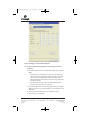
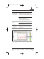
1
tm307vfinal.qxp
11/13/2008
1:03 PM
Page a
Technical Manual
StemElite™ ID System
INSTRUCTIONS FOR USE OF PRODUCT G9530.
PRINTED IN USA.
10/08
Part# TM307
A F 9 T M3 0 7
1 0 0 8 T M3 0 7
tm307vfinal.qxp
11/13/2008
1:03 PM
Page 1
StemElite™ ID System
All technical literature is available on the Internet at: www.promega.com/tbs/
Please visit the web site to verify that you are using the most current version of this
Technical Manual. Please contact Promega Technical Services if you have questions on use
of this system. E-mail: [email protected]
1. Description..........................................................................................................2
2. Product Components and Storage Conditions ............................................5
3. Instrumentation Requirements,
Software and Accessory Products...................................................................5
4. Matrix Standardization or Spectral Calibration..........................................6
5. Precautions ..........................................................................................................6
6. DNA Sample Preparation ................................................................................7
A. DNA Purification..................................................................................................7
B. DNA Quantitation................................................................................................7
7. Protocols for DNA Amplification Using the StemElite™ ID System ....8
A. Amplification Setup .............................................................................................8
B. Amplification Thermal Cycling........................................................................10
8. Detection of Amplified Fragments Using the Applied
Biosystems 3130 or 3130xl Genetic Analyzer and
ABI PRISM® 3100 or 3100-Avant Genetic Analyzer
with Data Collection Software, Versions 2.0 and 3.0 ...............................11
A. Capillary Electrophoresis Sample Preparation..............................................11
B. Capillary Electrophoresis Instrument Preparation .......................................12
9. Detection of Amplified Fragments Using the ABI PRISM® 310
Genetic Analyzer .............................................................................................14
A. Capillary Electrophoresis Sample Preparation..............................................14
B. Capillary Electrophoresis Instrument Preparation .......................................15
10. Data Analysis....................................................................................................17
A. Downloading Panel and Bin Files ...................................................................17
B. Importing Panel and Bin Files for GeneMapper® ID and
GeneMapper® Software.....................................................................................17
C. Creating a Method with GeneMapper® ID and
GeneMapper® Software.....................................................................................18
D. Creating a Size Standard ...................................................................................21
E. Processing Data...................................................................................................22
F. Obtaining a Genotype........................................................................................22
G. Controls................................................................................................................23
H. Results ..................................................................................................................23
Promega Corporation · 2800 Woods Hollow Road · Madison, WI 53711-5399 USA
Toll Free in USA 800-356-9526 · Phone 608-274-4330 · Fax 608-277-2516 · www.promega.com
Printed in USA.
10/08
Part# TM307
Page 1
tm307vfinal.qxp
11/13/2008
1:03 PM
Page 2
11. Troubleshooting...............................................................................................27
A. Amplification and Fragment Detection ..........................................................27
B. GeneMapper® and GeneMapper® ID Analysis Software.............................30
12. Appendix ...........................................................................................................33
A.
B.
C.
D.
E.
F.
1.
Additional STR Locus Information .................................................................33
The Internal Lane Standard 600 .......................................................................34
Preparing the StemElite™ ID System Master Mix ........................................35
Composition of Buffers and Solutions ............................................................36
References ............................................................................................................36
Related Products.................................................................................................38
Description
Cell line misidentification and cross-contamination have become important
concerns for researchers. For example, stem cell lines can become contaminated
by the mouse cell line making up the feeder cell layers on which stem cells are
propagated. It has been shown that laboratories have invested substantial time
and effort researching cell lines that were revealed to be misidentified (1). The
situation has prompted the National Institutes of Health to issue a notice to
researchers strongly recommending authentication procedures when using
cultured cells (2). Genetic profiling can be used as a tool for stem cell line
quality assurance, and human cell lines can be authenticated using short
tandem repeat (STR) loci (3–5). STR loci consist of short, repetitive sequence
elements 3–7 base pairs in length. These repeats are well distributed throughout
the human genome and are a rich source of highly polymorphic markers,
which can be detected using the polymerase chain reaction. Alleles of STR loci
are differentiated by the number of copies of the repeat sequence contained
within the amplified region and are distinguished from one another using
fluorescence detection, following electrophoretic separation.
Early efforts toward genetic characterization of human cell lines used the
PowerPlex® 1.2 System. Substantial databases derived using the eight STR loci
and Amelogenin locus of the PowerPlex® 1.2 System currently exist (6,7). To
support those efforts, we included the PowerPlex® 1.2 System loci in the new
StemElite™ ID System(a–d) plus additional loci. The D21S11 locus was included
for additional discrimination power.
To provide a powerful and complete system for cell line identification we
developed the StemElite™ ID System, which contains all reagents required for
successful identification and authentication of human stem cell lines and
detection of cell line contaminants in a research laboratory. Additionally, we
have incorporated a sensitive marker that specifically detects the presence of
DNA from the mouse (Mus musculus). This locus has been developed to coamplify with the existing human loci in the primer set provided with this
Promega Corporation · 2800 Woods Hollow Road · Madison, WI 53711-5399 USA
Toll Free in USA 800-356-9526 · Phone 608-274-4330 · Fax 608-277-2516 · www.promega.com
Part# TM307
Page 2
Printed in USA.
10/08
tm307vfinal.qxp
11/13/2008
1:03 PM
Page 3
system. The StemElite™ ID System offers the convenience of room-temperature
reaction assembly by including a hot-start Taq DNA polymerase system, which
is included in a 5X enzyme mix. This allows a simple, one-step addition of Taq
DNA polymerase, dNTPs, MgCl2 and reaction buffer necessary for DNA
amplification.
The StemElite™ ID System allows co-amplification and three-color detection of
ten human loci (nine STR loci, Amelogenin for gender identification and one
mouse locus), including D21S11, TH01, TPOX, vWA, Amelogenin, CSF1PO,
D16S539, D7S820, D13S317, D5S818 and the mouse locus. These loci collectively
provide a genetic profile with a random match probability of 1 in 2.92 × 10⁹
(while simultaneously providing detection of a 1% fraction of mouse
contaminant in a human cell line). One primer for each of the D21S11, TH01
and mouse loci is labeled with fluorescein (FL); one primer for each of the
TPOX, vWA and Amelogenin loci is labeled with carboxytetramethylrhodamine (TMR); and one primer for each of the CSF1PO, D16S539,
D7S820, D13S317 and D5S818 loci is labeled with 6-carboxy-4´,5´-dichloro-2´,7´dimethoxy-fluorescein (JOE). The eleven loci are amplified simultaneously in a
single tube and analyzed by capillary electrophoresis in a single injection.
Figure 1 shows the allele ranges for each locus in the StemElite™ ID System.
In addition the StemElite™ ID System contains various controls to provide
increased confidence in the genotypes obtained. An internal lane standard (ILS)
and allelic ladder are provided for standardization, and a control cell line DNA
(9947A) is supplied as a positive control. The ILS is added to every sample after
amplification and used within each capillary electrophoresis run to determine
TH01
M
JOE:
TMR:
CXR:
Size (bp)
D5S818
A
100
D21S11
D13S317
vWA
D7S820
D16S539
CSF1PO
TPOX
200
300
400
A = Amelogenin
M = Mouse
7815MA
Fluorescein:
Figure 1. Allele ranges for the StemElite™ ID System. STR fragments amplified by
the StemElite™ ID System are labeled with different dyes and are separated by
capillary electrophoresis based on size. A size standard is included in each sample to
determine the size of the StemElite™ ID amplified fragments. Fluorescein-labeled
loci are shown in dark gray, JOE-labeled loci are shown in gray and TMR-labeled
loci are shown in white. The CXR-labeled Internal Lane Standard 600 fragments are
represented by black bars.
Promega Corporation · 2800 Woods Hollow Road · Madison, WI 53711-5399 USA
Toll Free in USA 800-356-9526 · Phone 608-274-4330 · Fax 608-277-2516 · www.promega.com
Printed in USA.
10/08
Part# TM307
Page 3
tm307vfinal.qxp
1.
11/13/2008
1:03 PM
Page 4
Description (continued)
the size of each amplified product. The allelic ladder consists of all major alleles
at a particular locus and is used as a standard to positively identify each allele.
It is included in each analysis run to control for run-to-run variation.
StemElite™ ID allelic ladder information, indicating the size range and repeat
numbers for each allele, can be found in Section 12.A. The control 9947A DNA
has a known genotype and can be used to verify genotyping accuracy.
Figure 2 outlines the StemElite™ ID System protocols in this manual. Briefly,
DNA is isolated from cells, quantitated and added to a master mix containing
the StemElite™ ID 10X Primer Pair Mix, StemElite™ ID 5X Enzyme Mix and
Water, Amplification Grade. PCR is performed, and ILS is added to the
amplified product. The alleles are resolved using capillary electrophoresis (CE),
and CE data are analyzed using genotyping software and the parameters given
on the Promega web site at: www.promega.com/stemeliteid/. This site provides
instructions and applications to set the report parameters in the GeneMapper®
or GeneMapper® ID software to make genotyping easier and more accurate.
This manual contains separate protocols for amplification of STR loci and
detection of amplified products. These protocols were tested at Promega. For
more information about required instrumentation and software, see Section 3.
DNA Sample Preparation
Section 6
Amplification Setup
Section 7.A
Thermal Cycling
Section 7.B
GeneAmp® PCR System 9700
GeneAmp® PCR System 9600
Instrument Setup and Sample Preparation
Applied Biosystems
3130 or 3130xl Genetic
Analyzer
Section 8
ABI PRISM® 3100 or
3100-Avant Genetic
Analyzer with Data
Collection Software,
Version 2.0
Section 8
ABI PRISM® 310
Genetic Analyzer
Section 9
Data Analysis
Section 10
GeneMapper® ID Software,
Version 3.2
GeneMapper® Software,
Version 4.0
Figure 2. An overview of the StemElite™ ID System protocol.
Promega Corporation · 2800 Woods Hollow Road · Madison, WI 53711-5399 USA
Toll Free in USA 800-356-9526 · Phone 608-274-4330 · Fax 608-277-2516 · www.promega.com
Part# TM307
Page 4
Printed in USA.
10/08
tm307vfinal.qxp
2.
11/13/2008
1:03 PM
Page 5
Product Components and Storage Conditions
Product
Size
StemElite™ ID System
50 reactions
G9530 contains sufficient reagents for 50 reactions of 25μl each. Includes:
Cat.#
G9530
Pre-amplification Components Box (Blue Label)
250μl
125μl
1.25ml
25μl
StemElite™ ID 5X Enzyme Mix
StemElite™ ID 10X Primer Pair Mix
Water, Amplification Grade
9947A DNA (10ng/μl)
Postamplification Components Box (Beige Label)
25μl
150μl
StemElite™ ID Allelic Ladder
Internal Lane Standard (ILS) 600
Storage Conditions: Store all components at –20°C in a nonfrost-free freezer.
The StemElite™ ID 10X Primer Pair Mix, StemElite™ ID Allelic Ladder and
Internal Lane Standard 600 are light-sensitive and must be stored protected
from light. We strongly recommend that pre-amplification and
postamplification reagents be stored and used separately with different
pipettes, tube racks, etc. See the expiration date on the product label.
3.
Instrumentation Requirements, Software and Accessory Products
This manual contains separate protocols for use of the StemElite™ ID System
with GeneAmp® PCR system 9700 and 9600 thermal cyclers in addition to
protocols to separate amplified products and detect separated material using the
capillary electrophoresis instruments: ABI PRISM® 310, 3100 and 3100-Avant
Genetic Analyzers and Applied Biosystems 3130 and 3130xl Genetic Analyzers.
These protocols were tested at Promega Corporation. Amplification and
detection instrumentation may vary. You may need to optimize protocols
including cycle number and injection time for each laboratory instrument.
Protocols for operation of the fluorescence detection instruments should be
obtained from the instrument manufacturer.
Matrix standards are required for initial setup of the color separation matrix
(Section 4). The matrix standards are sold separately and are available for the
ABI PRISM® 310 Genetic Analyzer (PowerPlex® Matrix Standards, 310, Cat.#
DG4640) and the ABI PRISM® 3100 and 3100-Avant Genetic Analyzers and
Applied Biosystems 3130 and 3130xl Genetic Analyzers (PowerPlex® Matrix
Standards, 3100/3130, Cat.# DG4650).
Promega Corporation · 2800 Woods Hollow Road · Madison, WI 53711-5399 USA
Toll Free in USA 800-356-9526 · Phone 608-274-4330 · Fax 608-277-2516 · www.promega.com
Printed in USA.
10/08
Part# TM307
Page 5
tm307vfinal.qxp
4.
11/13/2008
1:03 PM
Page 6
Matrix Standardization or Spectral Calibration
Proper generation of a matrix file is critical to evaluate multicolor systems with
the ABI PRISM® 310, 3100 and 3100-Avant Genetic Analyzers and Applied
Biosystems 3130 and 3130xl Genetic Analyzers. A matrix must be generated for
each individual instrument. Very high peak heights may not be perfectly
separated spectrally, and an allele peak in one color channel can bleed into
another color channel. A poor or incorrect matrix will allow this as well.
The PowerPlex® Matrix Standards, 310 (Cat.# DG4640), is required for matrix
standardization for the ABI PRISM® 310 Genetic Analyzer. For best results, the
PowerPlex® Matrix Standards, 3100/3130 (Cat.# DG4650), should not be used
to generate a matrix on the ABI PRISM® 310 Genetic Analyzer.
The PowerPlex® Matrix Standards, 3100/3130 (Cat.# DG4650), is required for
spectral calibration on the ABI PRISM® 3100 and 3100-Avant Genetic Analyzers
and Applied Biosystems 3130 and 3130xl Genetic Analyzers. The PowerPlex®
Matrix Standards, 310 (Cat.# DG4640), cannot be used to generate a matrix on
these instruments.
For protocols and additional information on matrix standardization, see the
PowerPlex ® Matrix Standards, 310, Technical Bulletin #TBD021, which is supplied
with Cat.# DG4640. For protocols and additional information about spectral
calibration, see the PowerPlex ® Matrix Standards, 3100/3130, Technical Bulletin
#TBD022, which is supplied with Cat.# DG4650. These manuals are available
upon request from Promega or online at: www.promega.com/tbs/
5.
Precautions
The quality of the purified DNA sample, as well as small changes in buffers,
ionic strength, primer concentrations, choice of thermal cycler and thermal
cycling conditions, can affect PCR success. We suggest strict adherence to
recommended procedures for amplification, as well as electrophoresis and
fluorescence detection.
PCR-based STR analysis is subject to contamination by very small amounts of
nontemplate human DNA. Extreme care should be taken to avoid crosscontamination when preparing sample DNA, handling primer pairs, assembling
amplification reactions and analyzing amplification products. Reagents and
materials used pre-amplification (StemElite™ ID 5X Enzyme Mix, StemElite™
ID 10X Primer Pair Mix, Amplification Grade Water and 9947A DNA) are
provided in a separate box and should be stored separately from those used
post-amplification (StemElite™ ID Allelic Ladder and Internal Lane Standard
600). Always include a negative control reaction (i.e., no template) to detect
reagent contamination. We highly recommend the use of gloves and aerosolresistant pipette tips (e.g., ART® tips).
Promega Corporation · 2800 Woods Hollow Road · Madison, WI 53711-5399 USA
Toll Free in USA 800-356-9526 · Phone 608-274-4330 · Fax 608-277-2516 · www.promega.com
Part# TM307
Page 6
Printed in USA.
10/08
tm307vfinal.qxp
11/13/2008
1:03 PM
Page 7
Some of the reagents used in the analysis of STR products are potentially
hazardous and should be handled accordingly. Formamide is an irritant and a
teratogen; avoid inhalation and contact with skin. Read the warning label, and
take appropriate precautions when handling this substance. Always wear
gloves and safety glasses when working with formamide.
6.
DNA Sample Preparation
6.A. DNA Purification
DNA concentration, purity and integrity are important considerations to
ensure success with the StemElite™ ID System. DNA should not be sheared
and should be free of contaminating protein and salts. Poor-quality DNA may
result in increased background or amplification failure. Too much or too little
DNA in the reaction can cause amplification failure, which can be manifested
in several ways: complete lack of amplification of all loci or dropout of all or
subsets of the alleles.
Commercially available DNA purification systems such as the MagneSil®
Genomic, Fixed Tissue DNA Purification System (Cat.# MD1490), Wizard® SV
Genomic DNA Purification System (Cat.# A2360), Wizard® Genomic DNA
Purification System (Cat.# A1620) and Maxwell® 16 Cell LEV DNA
Purification Kit (Cat.# AS1140) produce DNA of sufficient quality for the
StemElite™ ID System. These systems yield clean DNA for STR analysis easily
and efficiently. The MagneSil® Resin eliminates PCR inhibitors and
contaminants frequently encountered in DNA samples.
DNA should be stored in a low-EDTA TE buffer (pH 8.0). We recommend
TE–4 buffer (10mM Tris-HCl, 0.1mM EDTA [pH 8.0]). High salt concentration
or altered pH can affect amplification. If the DNA template is stored in TE
buffer that is not pH 8.0 or contains a higher EDTA concentration, the DNA
volume should not exceed 20% of the total reaction volume. Carryover of K+,
Na+, Mg2+ or EDTA can negatively affect PCR.
6.B. DNA Quantitation
We recommend quantitating the DNA prior to use in the StemElite™ ID
System, as either too much or too little DNA can cause reactions to fail. Use
the recommended amount of template DNA. Stochastic effects, which can
cause imbalance, can occur when amplifying low amounts of template.
Amplifying high amounts of template can result in less amplification of larger
STR loci. Absorbance readings at 260nm can be used to estimate DNA
concentration, where 1AU = 50μg of double-stranded DNA/ml. The
Quant-iT™ PicoGreen® dsDNA quantitation assay (Invitrogen) can also be
used. Use 2ng of DNA in each StemElite™ ID System reaction.
Note: As little as 20pg of mouse DNA is detectable in the StemElite™ ID
System.
Promega Corporation · 2800 Woods Hollow Road · Madison, WI 53711-5399 USA
Toll Free in USA 800-356-9526 · Phone 608-274-4330 · Fax 608-277-2516 · www.promega.com
Printed in USA.
10/08
Part# TM307
Page 7
tm307vfinal.qxp
11/13/2008
1:03 PM
Page 8
6.B. DNA Quantitation (continued)
!
If you are not using one of these kits for DNA purification, we recommend
measuring absorbance of the DNA sample at 260nm and 280nm to confirm that
the DNA is sufficiently free of impurities. High-quality DNA has a typical
A260/A280 ratio of 1.8. The presence of impurities in the DNA sample can cause
amplification failure. Note that DNA concentration can be overestimated by
spectrophotometry if the A260/A280 ratio is low.
!
Cross-contamination with another template or previously amplified DNA can
lead to extra peaks in the sample. Use aerosol-resistant pipette tips, and
change gloves regularly when working with the StemElite™ ID System.
7.
Protocols for DNA Amplification Using the StemElite™ ID System
Materials to Be Supplied by the User
• GeneAmp® PCR System 9600 or 9700 thermal cyclers (Applied Biosystems)
• 0.2ml thin-walled microcentrifuge tubes, MicroAmp® optical 96-well
reaction plate or 0.2ml MicroAmp® 8-strip reaction tubes (Applied
Biosystems)
• 1.5ml amber-colored microcentrifuge tubes (Fisher Cat.# 05-402-26)
• aerosol-resistant pipette tips
The StemElite™ ID System has been optimized to amplify 2ng of template
DNA in a 25μl reaction using the protocols detailed below. Preferential
amplification of smaller loci can occur. Expect to see high peak heights for the
smaller loci and relatively lower peak heights for the larger loci if more than the
recommended amount of template is used. Reduce the amount of template
DNA or number of PCR cycles to correct this.
The StemElite™ ID System is optimized for the GeneAmp® PCR system 9700
thermal cycler. An amplification protocol for the GeneAmp® PCR system 9600
thermal cycler is also provided.
7.A. Amplification Setup
!
Keep all pre-amplification and postamplification reagents in separate rooms.
Prepare amplification reactions in a room dedicated to reaction setup. Use
equipment and supplies dedicated to amplification setup.
!
Cross-contamination with another template or previously amplified DNA can
lead to extra peaks in the sample. Use aerosol-resistant pipette tips, and
change gloves regularly.
1. Thaw the StemElite™ ID 5X Enzyme Mix and StemElite™ ID 10X Primer
Pair Mix.
Note: Vortex reagents for 15 seconds before each use. Do not centrifuge the
10X Primer Pair Mix, as this may cause the primers to be concentrated at
the bottom of the tube.
Promega Corporation · 2800 Woods Hollow Road · Madison, WI 53711-5399 USA
Toll Free in USA 800-356-9526 · Phone 608-274-4330 · Fax 608-277-2516 · www.promega.com
Part# TM307
Page 8
Printed in USA.
10/08
tm307vfinal.qxp
11/13/2008
1:03 PM
Page 9
2. Determine the number of reactions to be set up. This should include
positive and negative control reactions. Add 1 or 2 reactions to this number
to compensate for pipetting error. While this approach does waste a small
amount of each reagent, it ensures that you will have enough PCR master
mix for all samples. It also ensures that each reaction contains the same
master mix.
3. Place one clean, 0.2ml reaction tube for each reaction into a rack, and label
appropriately. Alternatively, use a MicroAmp® plate or 0.2ml MicroAmp®
8-strip reaction tubes, and label appropriately.
4. Add the final volume of each reagent listed in Table 1 into a sterile, 1.5ml
amber-colored tube. Mix gently. A worksheet to calculate reagent volumes
for a multiple-reaction StemElite™ ID master mix can be found in Section
12.C.
Table 1. PCR Master Mix for the StemElite™ ID System.
PCR Master Mix Component
StemElite™ ID 5X Enzyme Mix
StemElite™ ID 10X Primer Pair Mix
Water, Amplification Grade
template DNA (2ng)1
1The
Volume Per Reaction
5.0μl
2.5μl
to a final volume of 25.0μl
up to 17.5μl
template DNA will be added at Step 7.
Amplification of >2ng of DNA template results in an imbalance in peak
heights from locus to locus in the resulting data. The smaller loci show
greater amplification yield than the larger loci. Reducing the number of
cycles in the amplification program by 2–4 cycles (i.e., 10/20 or 10/18
cycling) can improve locus-to-locus balance.
5. Vortex the PCR master mix for 5–10 seconds.
6. Pipet PCR master mix into each reaction tube.
7. Pipet the template DNA (2ng) for each sample into the respective tube
containing PCR master mix.
8. For the positive amplification control, dilute the 9947A DNA to 2ng in the
desired template DNA volume. Pipet 2ng of the diluted DNA into a
reaction tube containing the PCR master mix.
9. Optional: If including a positive amplification control for mouse DNA,
dilute the Mouse Genomic DNA (Cat.# G3091; must be purchased
separately) to 100pg in the desired template DNA volume. Pipet 100pg of
the diluted DNA into a reaction tube containing the PCR master mix.
10. For the negative amplification control, pipet Water, Amplification Grade,
(instead of template DNA) into a reaction tube containing the PCR master
mix.
Promega Corporation · 2800 Woods Hollow Road · Madison, WI 53711-5399 USA
Toll Free in USA 800-356-9526 · Phone 608-274-4330 · Fax 608-277-2516 · www.promega.com
Printed in USA.
10/08
Part# TM307
Page 9
tm307vfinal.qxp
11/13/2008
1:03 PM
Page 10
7.B. Amplification Thermal Cycling
This manual contains protocols for use of the StemElite™ ID System with the
GeneAmp® PCR system 9600 and 9700 thermal cyclers. We have not tested
other reaction tubes, plates or thermal cyclers. For information about other
thermal cyclers, please contact Promega Technical Services by e-mail:
[email protected]
Amplification and detection instrumentation may vary. You may need to
optimize protocols including cycle number and injection time (or loading
volume) for each laboratory instrument. Testing at Promega Corporation shows
that 10/22 cycles work well for 2ng of purified DNA templates. For higher
amounts of input DNA or to decrease sensitivity, fewer cycles, such as 10/16,
10/18 or 10/20, should be evaluated. In-house validation should be performed.
1. Place the tubes or MicroAmp® plate in the thermal cycler.
2. Select and run a recommended protocol. The preferred protocols for use
with the GeneAmp® PCR system 9700 and 9600 thermal cyclers are
provided below.
3. After completion of the thermal cycling protocol, store the samples at
–20°C in a light-protected box.
Note: Storage of amplified samples at 4°C or higher may produce
degradation products.
Protocol for the GeneAmp® PCR
System 9700 Thermal Cycler
Protocol for the GeneAmp® PCR
System 9600 Thermal Cycler
96°C for 2 minutes, then:
96°C for 2 minutes, then:
ramp 100% to 94°C for 30 seconds
ramp 29% to 60°C for 30 seconds
ramp 23% to 70°C for 45 seconds
for 10 cycles, then:
94°C for 30 seconds, then:
ramp 100% to 90°C for 30 seconds
ramp 29% to 60°C for 30 seconds
ramp 23% to 70°C for 45 seconds
for 22 cycles, then:
ramp 60 seconds to 60°C (hold for 30 seconds)
ramp 50 seconds to 70°C (hold for 45 seconds)
for 10 cycles, then:
90°C for 30 seconds, then:
ramp 60 seconds to 60°C (hold for 30 seconds)
ramp 50 seconds to 70°C (hold for 45 seconds)
60°C for 30 minutes
for 22 cycles, then:
4°C soak
60°C for 30 minutes
4°C soak
Promega Corporation · 2800 Woods Hollow Road · Madison, WI 53711-5399 USA
Toll Free in USA 800-356-9526 · Phone 608-274-4330 · Fax 608-277-2516 · www.promega.com
Part# TM307
Page 10
Printed in USA.
10/08
tm307vfinal.qxp
8.
11/13/2008
1:03 PM
Page 11
Detection of Amplified Fragments Using the Applied Biosystems 3130
or 3130xl Genetic Analyzer and ABI PRISM® 3100 or 3100-Avant Genetic
Analyzer with Data Collection Software, Versions 2.0 and 3.0
Materials to Be Supplied by the User
• 95°C dry heating block, water bath or thermal cycler
• crushed ice or ice-water bath
• aerosol-resistant pipette tips
• 3100 or 3130 capillary array, 36cm
• performance optimized polymer 4 (POP-4™) for the 3100 or 3130
• 10X Genetic Analyzer buffer with EDTA
• MicroAmp® optical 96-well plate and septa
• Hi-Di™ formamide (Applied Biosystems Cat.# 4311320)
• PowerPlex® Matrix Standards, 3100/3130 (Cat.# DG4650)
!
The quality of the formamide is critical. Use Hi-Di™ formamide with a
conductivity less than 100μS/cm. Freeze formamide in aliquots at –20°C.
Multiple freeze-thaw cycles or long-term storage at 4°C may cause a breakdown
of the formamide. Formamide with a conductivity greater than 100μS/cm may
contain ions that compete with DNA during injection. This results in lower peak
heights in the resulting data and reduced sensitivity. A longer injection time
may not increase the signal.
!
Caution: Formamide is an irritant and a teratogen; avoid inhalation and contact
with skin. Read the warning label, and take appropriate precautions when
handling this substance. Always wear gloves and safety glasses when working
with formamide.
8.A. Capillary Electrophoresis Sample Preparation
1. Prepare a loading cocktail by combining and mixing the internal lane
standard and Hi-Di™ formamide as follows:
[(0.5μl ILS 600) × (# injections)] + [(9.5μl Hi-Di™ formamide) × (# injections)]
Note: The volume of internal lane standard used in the loading cocktail can
be increased or decreased to adjust the intensity of the size standard peaks
in the resulting data. The optimal peak height for the 100-base fragment of
the internal lane standard is 500–1,000RFU. If the peak heights are too low,
we recommend altering the formamide/internal lane standard mix to
contain 1.0μl of ILS 600 and 9.0μl of Hi-Di™ formamide. If the peak heights
are too high, we recommend altering the loading cocktail to contain 0.25μl
of ILS 600 and 9.75μl of formamide.
2. Vortex for 10–15 seconds.
3. Pipet 10μl of formamide/internal lane standard mix into each well.
Promega Corporation · 2800 Woods Hollow Road · Madison, WI 53711-5399 USA
Toll Free in USA 800-356-9526 · Phone 608-274-4330 · Fax 608-277-2516 · www.promega.com
Printed in USA.
10/08
Part# TM307
Page 11
tm307vfinal.qxp
11/13/2008
1:03 PM
Page 12
8.A. Capillary Electrophoresis Sample Preparation (continued)
4. Add 1μl of amplified sample (or 1μl of StemElite™ ID Allelic Ladder).
Cover wells with appropriate septa.
!
Note: Instrument detection limits vary; therefore, injection time or the
amount of product mixed with loading cocktail may need to be increased
or decreased. Use the “Module Manager” in the data collection software to
modify the injection time or voltage in the run module. Alternatively, use
less DNA template in the amplification reactions or reduce the number of
cycles in the amplification program by 2–4 cycles to achieve the desired
signal intensity.
5. Centrifuge the plate briefly to remove air bubbles from the wells if necessary.
6. Denature samples at 95°C for 3 minutes, then immediately chill on crushed
ice or in an ice-water bath for 3 minutes.
!
Note: Denatured-sample plates should be run within 24 hours of setup.
Long-term storage of amplified sample in formamide can result in DNA
degradation. Sample preparation can be repeated with fresh formamide by
repeating Steps 1–6.
8.B. Capillary Electrophoresis Instrument Preparation
Refer to the instrument user’s manual for instructions on cleaning, installing
the capillary array, performing a spatial calibration and adding polymer.
Follow the manufacturer’s recommendations for polymer storage and shelf
life. Polymer stored at room temperature for more than 1 week can result in
broad or split peaks or extra peaks visible in one or all of the color channels.
Maintain the instrumentation on a daily or weekly basis as recommended by
the manufacturer for optimal results and fewer instrument-related artifacts.
Contaminants in the water used with the instrument or to dilute the 10X
Genetic Analyzer buffer may generate peaks in the blue and green dye colors.
Use autoclaved water.
Analyze the samples as described in the user’s manual for the ABI PRISM®
3100 or 3100-Avant Genetic Analyzer with data collection software, version 2.0,
and the Applied Biosystems 3130 or 3130xl Genetic Analyzer with the
following exceptions.
1. In the Module Manager, select “New”. Select “Regular” in the Type dropdown list, and select “HIDFragmentAnalysis36_POP4” in the Template
drop-down list. Confirm that the injection time is 5 seconds and the
injection voltage is 3kV. Lengthen the run time to 2,000 seconds. Give a
descriptive name to your run module, and select “OK”.
Note: Sensitivities of instruments may vary. The injection time and voltage
may be adjusted in the Module Manager. A suggested range for the
injection time is 3–22 seconds and for the injection voltage is 1–3kV.
2. In the Protocol Manager, select “New”. Type a name for your protocol.
Promega Corporation · 2800 Woods Hollow Road · Madison, WI 53711-5399 USA
Toll Free in USA 800-356-9526 · Phone 608-274-4330 · Fax 608-277-2516 · www.promega.com
Part# TM307
Page 12
Printed in USA.
10/08
tm307vfinal.qxp
11/13/2008
1:03 PM
Page 13
Select “Regular” in the Type drop-down list, and select the run module you
created in the previous step in the Run Module drop-down list. Lastly,
select “F” in the Dye-Set drop-down list. Select “OK”.
3. In the Plate Manager, create a new plate record as described in the
instrument user’s manual. In the dialog box that appears, select
“GeneMapper-Generic” in the Application drop-down list, and select the
appropriate plate type (96-well). Add entries in the owner and operator
windows, and select “OK”.
Note: If autoanalysis of sample data is desired, refer to the instrument
user’s manual for instructions.
4. In the GeneMapper® plate record, enter sample names in the appropriate
cells. Scroll to the right. In the “Results group 1” column, select the desired
results group. In the “Instrument Protocol 1” column, select the protocol
you created in Step 2. Be sure this information is present for each row that
contains a sample name. Select “OK”.
Note: To create a new results group, select “New” in the drop-down menu
in the results group column. Select the General tab, and enter a name.
Select the Analysis tab, and select “GeneMapper-Generic” in the Analysis
type drop-down list.
5. Place samples in the instrument, and close the instrument doors.
6. In the spectral viewer, confirm that dye set F is active, and set the correct
active calibration for dye set F.
7. In the run scheduler, locate the plate record that you just created in Steps 3
and 4, and click once on the name to highlight it. Once the plate record is
highlighted, click the plate graphic that corresponds to the plate on the
autosampler that contains your amplified samples. When the plate record
is linked to the plate, the plate graphic will change from yellow to green,
and the green Run Instrument arrow becomes enabled.
8. Click on the green Run Instrument arrow on the toolbar to start the sample
run.
9. Monitor electrophoresis by observing the run, view, array or capillaries
viewer window in the data collection software. Each injection will take
approximately 45 minutes.
Note: If peaks are low or absent, the sample can be reinjected with
increased injection time and/or voltage. If the ILS 600 is also affected,
check the laser power.
Promega Corporation · 2800 Woods Hollow Road · Madison, WI 53711-5399 USA
Toll Free in USA 800-356-9526 · Phone 608-274-4330 · Fax 608-277-2516 · www.promega.com
Printed in USA.
10/08
Part# TM307
Page 13
tm307vfinal.qxp
9.
11/13/2008
1:03 PM
Page 14
Detection of Amplified Fragments Using the ABI PRISM® 310 Genetic
Analyzer
Materials to Be Supplied by the User
• 95°C dry heating block, water bath or thermal cycler
• 310 capillaries, 47cm × 50μm
• performance optimized polymer 4 (POP-4™)
• glass syringe (1ml)
• 10X Genetic Analyzer buffer with EDTA
• sample tubes and septa
• aerosol-resistant pipette tips
• Hi-Di™ formamide (Applied Biosystems Cat.# 4311320)
• PowerPlex® Matrix Standards, 310 (Cat.# DG4640)
• crushed ice or ice-water bath
!
The quality of the formamide is critical. Use Hi-Di™ formamide with a
conductivity less than 100μS/cm. Freeze formamide in aliquots at –20°C.
Multiple freeze-thaw cycles or long-term storage at 4°C may cause a breakdown
of the formamide. Formamide with a conductivity greater than 100μS/cm may
contain ions that compete with DNA during injection. This results in lower
peak heights in the resulting data and reduced sensitivity. A longer injection
time may not increase the signal.
!
Caution: Formamide is an irritant and a teratogen; avoid inhalation and contact
with skin. Read the warning label, and take appropriate precautions when
handling this substance. Always wear gloves and safety glasses when working
with formamide.
9.A. Capillary Electrophoresis Sample Preparation
1. Prepare a loading cocktail by combining the Internal Lane Standard 600
(ILS 600) and Hi-Di™ formamide as follows:
[(1.0μl ILS 600) × (# injections)] + [(24.0μl Hi-Di™ formamide) × (# injections)]
Note: The volume of internal lane standard used in the loading cocktail can
be increased or decreased to adjust the intensity of the size standard peaks.
The optimal peak height for the 100-base fragment of the internal lane
standard is 500–1,000RFU. If the peak heights are too low we recommend
altering the ILS/formamide mix to 1.5μl of ILS 600 and 24μl of Hi-Di™
formamide. If the peak heights are too high, we recommend altering the
loading cocktail to contain 0.5μl of ILS 600 and 24.5μl of Hi-Di™ formamide.
2. Vortex for 10–15 seconds.
Promega Corporation · 2800 Woods Hollow Road · Madison, WI 53711-5399 USA
Toll Free in USA 800-356-9526 · Phone 608-274-4330 · Fax 608-277-2516 · www.promega.com
Part# TM307
Page 14
Printed in USA.
10/08
tm307vfinal.qxp
11/13/2008
1:03 PM
Page 15
3. Combine 25μl of the prepared loading cocktail and 1μl of amplified sample
(or 1μl of StemElite™ ID Allelic Ladder).
Note: Instrument detection limits vary; therefore, injection time or the
amount of product mixed with loading cocktail may need to be increased
or decreased. Alternatively, use less DNA template in the amplification
reactions or reduce the number of cycles in the amplification program by
2–4 cycles to achieve the desired signal intensity.
4. Centrifuge the sample tubes briefly to remove air bubbles from the wells if
necessary.
5. Denature the samples and ladder by heating at 95°C for 3 minutes, and
immediately chill on crushed ice or in an ice-water bath for 3 minutes.
Note: Improper denaturation can result in extra peaks.
6. Assemble the tubes in the appropriate autosampler tray (48- or 96-tube).
7. Place the autosampler tray in the instrument, and close the instrument
doors.
!
Note: Denatured sample plates should be run within 24 hours of setup.
Long-term storage of amplified sample in formamide can result in DNA
degradation. Sample preparation can be repeated with fresh formamide.
9.B. Capillary Electrophoresis Instrument Preparation
Refer to the instrument user’s manual for instructions on cleaning the pump
block, installing the capillary, calibrating the autosampler and adding polymer
to the syringe. Follow the manufacturer’s recommendations for polymer
storage and shelf life. Polymer stored at room temperature for more than
1 week can result in broad or split peaks or extra peaks visible in one or all of
the color channels. Maintain the instrumentation on a daily or weekly basis as
recommended by the manufacturer for optimal results and fewer instrumentrelated artifacts. Contaminants in the water used with the instrument or to
dilute the 10X Genetic Analyzer buffer may generate peaks in the blue and
green dye colors. Use autoclaved water.
1. Open the ABI PRISM® 310 data collection software.
2. Prepare a GeneScan® sample sheet as described in the ABI PRISM® 310
Genetic Analyzer user’s manual. Enter the appropriate sample information
in the “sample info” column.
3. Create a new GeneScan® injection list. Select the appropriate sample sheet
by using the pull-down menu.
Promega Corporation · 2800 Woods Hollow Road · Madison, WI 53711-5399 USA
Toll Free in USA 800-356-9526 · Phone 608-274-4330 · Fax 608-277-2516 · www.promega.com
Printed in USA.
10/08
Part# TM307
Page 15
tm307vfinal.qxp
11/13/2008
1:03 PM
Page 16
9.B. Capillary Electrophoresis Instrument Preparation (continued)
4. Select the “GS STR POP4 (1ml) A” Module using the pull-down menu.
Change the injection time to 3 seconds and the run time to 30 minutes.
Keep the settings for the remaining parameters as shown below:
Inj. Secs: 3
Inj. kV: 15.0
Run kV: 15.0
Run °C: 60
Run Time: 30
!
You may need to optimize the injection time for individual instruments.
Injection times of 2–5 seconds are suggested for reactions that contain 2ng
of template DNA.
Note: Migration of fragments can vary slightly over the course of a long
ABI PRISM® 310 Genetic Analyzer run. This may be due to changes in
temperature or changes in the column. When analyzing many samples,
injections of the allelic ladder at different times throughout the run can aid
in accurately genotyping the samples.
5. Select the appropriate matrix file.
6. To analyze the data automatically, select the auto analyze checkbox and the
appropriate analysis parameters and size standard. Refer to the ABI PRISM®
310 Genetic Analyzer user’s manual for specific information on these options.
7. After loading the sample tray and closing the doors, select “Run” to start
the capillary electrophoresis system.
8. Monitor the electrophoresis by observing the raw data and status windows.
Each sample will take approximately 40 minutes for syringe pumping,
sample injection and sample electrophoresis.
Promega Corporation · 2800 Woods Hollow Road · Madison, WI 53711-5399 USA
Toll Free in USA 800-356-9526 · Phone 608-274-4330 · Fax 608-277-2516 · www.promega.com
Part# TM307
Page 16
Printed in USA.
10/08
tm307vfinal.qxp
11/13/2008
1:03 PM
Page 17
10. Data Analysis
To interpret data generated using the StemElite™ ID System, you will need
GeneMapper® or GeneMapper® ID software. To facilitate analysis of data
generated with the StemElite™ ID System, we have created panel and bin files
to allow automatic assignment of genotypes using GeneMapper® ID software,
version 3.2, and GeneMapper® software, version 4.0. We recommend that users
of GeneMapper® ID software, version 3.2, complete the Applied Biosystems
GeneMapper® ID software human identification analysis tutorial to familiarize
themselves with the proper operation of the software. For GeneMapper® ID
software, version 3.1 users, we recommend upgrading to version 3.2.
10.A. Downloading Panel and Bin Files
1. Download the panel and bin files for use with GeneMapper® ID and
GeneMapper® software from the Promega web site:
www.promega.com/stemeliteid/
2. Enter your contact information, and select the appropriate analysis
software. Select “Submit”.
3. Select the “StemElite ID Panels and Bins Set” link, and save the zipped file
to your computer.
4. Open the files using the Windows® WinZip program, and save the
unzipped files to a known location on your computer.
10.B. Importing Panel and Bin Files for GeneMapper® ID and GeneMapper®
Software
These instructions loosely follow the Applied Biosystems GeneMapper® ID
software tutorial, pages 1–4, and the GeneMapper ® Software Version 4.0
Microsatellite Analysis Getting Started Guide.
1. Open the GeneMapper® ID software, version 3.2, or GeneMapper®
software, version 4.0.
2. Select ”Tools”, then “Panel Manager”.
3. Highlight the Panel Manager icon in the upper left tile (navigation pane).
4. Select “File”, then “Import Panels”.
5. Navigate to the panel and bin files saved in Section 10.A. Select
“StemElite_ID_Panels_1.0.txt”. Select “Import”.
6. In the navigation pane, highlight the StemElite_ID_Panels_1.0.txt folder
that you just imported.
7. Select “File”, then “Import Bin Set”.
8. Navigate to the panel and bin files. Select “StemElite_ID_Bins_1.0.txt”, then
“Import”.
Promega Corporation · 2800 Woods Hollow Road · Madison, WI 53711-5399 USA
Toll Free in USA 800-356-9526 · Phone 608-274-4330 · Fax 608-277-2516 · www.promega.com
Printed in USA.
10/08
Part# TM307
Page 17
tm307vfinal.qxp
11/13/2008
1:03 PM
Page 18
10.B. Importing Panel and Bin Files for GeneMapper® ID and GeneMapper®
Software (continued)
9. At the bottom of the Panel Manager window, select “Apply”, then “OK”.
The panel manager window will close automatically.
Note: GeneMapper® ID software was created specifically for forensic
laboratories. HID analysis settings analyze data using panel and bin files,
which supply information regarding the alleles expected within a sample
set. An allelic ladder is used by the HID analysis algorithms to calculate
offsets, or variations, in the migration of alleles on a particular instrument,
allowing the software to correct for subtle differences in sample migration
caused by temperature, voltage, polymer and other factors. Other versions
of GeneMapper® analysis software do not contain these options.
!
Other versions of GeneMapper® analysis software can be used to analyze
StemElite™ ID System data. However, laboratories using other versions
must verify that all allelic ladders and positive controls are called correctly.
Panel and bin files must be created specifically for each CE instrument and
are not interchangeable. The GeneMapper® software does not calculate
offsets, and subsequently the panel and bin files need to be customized to
reflect the actual migration of fragments on a particular instrument.
A custom bin generation tool is available from Promega specifically to
generate panel and bin files customized to reflect the migration of fragments
on a particular instrument for the StemElite™ ID System. This tool requires
data from at least three analyzed runs of the StemElite™ ID Allelic Ladder,
which can be exported as a table as described on the Promega web site.
These instructions and the custom bin generation tool are available at:
www.promega.com/stemeliteid/
10.C. Creating a Method with GeneMapper® ID and GeneMapper® Software
1. Select “Tools”, then “GeneMapper Manager”.
2. Select the Analysis Methods tab.
3. Select “New”, and a new analysis method dialog box will open.
4. Select “HID”, if available, or “Microsatellite”. Select “OK”.
5. Enter a descriptive name for the analysis method, such as “StemElite_ID”.
6. Select the Allele tab.
7. Select the bin set corresponding to the StemElite™ ID System
“StemElite_ID_Bins_1.0”.
Note: If using GeneMapper® analysis software instead of GeneMapper® ID
software, use the custom bin generation tool in Section 10.B, Step 9 (above)
to generate a custom bin file.
8. Ensure that the “Use marker-specific stutter ratio if available” box is checked.
Promega Corporation · 2800 Woods Hollow Road · Madison, WI 53711-5399 USA
Toll Free in USA 800-356-9526 · Phone 608-274-4330 · Fax 608-277-2516 · www.promega.com
Part# TM307
Page 18
Printed in USA.
10/08
tm307vfinal.qxp
11/13/2008
1:04 PM
Page 19
9. For proper filtering of peaks when using the StemElite™ ID System, enter
the values shown in Figure 3 when using GeneMapper® ID and HID
analysis methods or Figure 4 when using GeneMapper® and Microsatellite
(MS) analysis methods.
For an explanation of the proper usage and effect of these settings, refer to
the Applied Biosystems user bulletin titled “Installation Procedures and
New Features for GeneMapper ID Software 3.2”.
7816TA
Note: Some of these settings have been optimized and are different from
the recommended settings in the user bulletin.
Figure 3. GeneMapper® ID Analysis Method Allele tab.
Promega Corporation · 2800 Woods Hollow Road · Madison, WI 53711-5399 USA
Toll Free in USA 800-356-9526 · Phone 608-274-4330 · Fax 608-277-2516 · www.promega.com
Printed in USA.
10/08
Part# TM307
Page 19
11/13/2008
1:04 PM
Page 20
7817TA
tm307vfinal.qxp
Figure 4. GeneMapper® Analysis Method Allele tab.
10.C. Creating a Method with GeneMapper® ID and GeneMapper® Software
(continued)
10. Select the Peak Detector tab. We recommend the settings shown in Figure 5.
Notes:
1. Select full range or partial range for the analysis range. When using a
partial range, choose an appropriate analysis range based on the data.
Choose a start point after the primer peak and just before the first
defined internal lane standard peak to help ensure proper sizing of the
internal lane standard.
2. The StemElite™ ID System contains only tetranucleotide markers.
Settings for other repeat types can be ignored.
11. Select the Peak Quality tab. You may change the settings for peak quality.
Note: For Steps 10 and 11, see the GeneMapper® ID user’s manual for more
information.
12. Select the Quality Flags tab. You may also change these settings.
13. Select “OK” to save your settings.
Promega Corporation · 2800 Woods Hollow Road · Madison, WI 53711-5399 USA
Toll Free in USA 800-356-9526 · Phone 608-274-4330 · Fax 608-277-2516 · www.promega.com
Part# TM307
Page 20
Printed in USA.
10/08
11/13/2008
1:04 PM
Page 21
7264TA
tm307vfinal.qxp
Figure 5. GeneMapper® ID and GeneMapper® Analysis Method Peak Detector tab.
10.D. Creating a Size Standard
1. Select “Tools”, then “GeneMapper Manager”.
2. Select the Size Standard tab.
3. Select “New”.
4. Select “Basic or Advanced” (Figure 5). The type of analysis method selected
must match the type of analysis method created earlier. Select “OK”.
5. Enter a detailed name, such as “ILS 600 advanced”, in the Size Standard
Editor. Choose red as the color for the size standard dye.
6. Enter the sizes of the internal lane standard fragments.
7. Select “OK”.
Promega Corporation · 2800 Woods Hollow Road · Madison, WI 53711-5399 USA
Toll Free in USA 800-356-9526 · Phone 608-274-4330 · Fax 608-277-2516 · www.promega.com
Printed in USA.
10/08
Part# TM307
Page 21
tm307vfinal.qxp
11/13/2008
1:04 PM
Page 22
10.E. Processing Data
1. Import sample files into a new project as described in the Applied
Biosystems GeneMapper® ID software human identification analysis
tutorial or GeneMapper ® Software Version 4.0 Microsatellite Analysis Getting
Started Guide.
2. In the “Sample Type” column, use the drop-down menu to select
“Ladder”, “Sample”, “Positive Control” or “Negative Control”. Every
folder in the project must contain at least one ladder that is designated as
such for proper genotyping.
3. In the “Analysis Method” column, select the analysis method created in
Section 10.C.
4. In the Panel column, select “StemElite_ID_Panels_1.0”. This is the panel set
that was imported in Section 10.B.
5. In the Size Standard column, select the size standard that was created in
Section 10.D.
6. If analyzing data from an ABI PRISM® 310 Genetic Analyzer, ensure that
the appropriate matrix file is selected in the “Matrix” column.
7. Select “Analyze” (green arrow button) to start the data analysis.
10.F. Obtaining a Genotype
Sample genotypes have been generated during data processing (Section 10.E).
1. Select “View”, then “Genotypes”. Genotypes for all samples in the project
will be shown.
Alternatively, highlight a sample or a subset of samples in the project,
select “Analysis”, then select “Display Plots”. Within the Sample Plot
window, select “Genotype”. Genotypes for the selected samples will be
shown.
2. If you are using GeneMapper® software and the sample alleles are being
called as “OL” (off-ladder), the bins may not be appropriate for your
instrument. GeneMapper® software does not offset the bins to match the
allelic ladder like the GeneMapper® ID software does. Visit the Promega
web site at: www.promega.com/stemeliteidapps/ to generate custom bins
specific for your instrument.
Promega Corporation · 2800 Woods Hollow Road · Madison, WI 53711-5399 USA
Toll Free in USA 800-356-9526 · Phone 608-274-4330 · Fax 608-277-2516 · www.promega.com
Part# TM307
Page 22
Printed in USA.
10/08
tm307vfinal.qxp
11/13/2008
1:04 PM
Page 23
10.G. Controls
1. Examine the results for the negative control. The negative control should be
devoid of amplification products.
2. Examine the results for the 9947A positive control DNA. Compare the
control DNA allelic repeat sizes with the locus-specific allelic ladder. The
expected 9947A DNA allele designations for each locus are listed in
Table 2.
Table 2. Expected Allele Designations for the 9947A DNA.
STR Locus
Alleles
D21S11
30, 30
TH01
8, 9.3
TPOX
8, 8
vWA
17, 18
Amelogenin
X, X
CSF1PO
10, 12
D16S539
11, 12
D7S820
10, 11
D13S317
11, 11
D5S818
11, 11
10.H. Results
Representative results of the StemElite™ ID System are shown in Figure 6. The
StemElite™ ID Allelic Ladder is shown in Figure 7.
Locus-to-locus peak height imbalance will likely occur with cell line DNA.
Normal genomic DNA has equal copies of the loci, and amplification will
result in relatively even locus-to-locus balance. Cell line DNA can have
mutations that affect the locus-to-locus allele peak height balance. The STR
genotype of a cell line can evolve over multiple passages. Users can genotype
cell line DNA regularly with the StemElite™ ID System to monitor any change
in the STR genotype. Additionally, cell lines have occasionally been observed
to have tri-allelic patterns at a locus.
Promega Corporation · 2800 Woods Hollow Road · Madison, WI 53711-5399 USA
Toll Free in USA 800-356-9526 · Phone 608-274-4330 · Fax 608-277-2516 · www.promega.com
Printed in USA.
10/08
Part# TM307
Page 23
tm307vfinal.qxp
11/13/2008
1:04 PM
Page 24
7818TA
10.H. Results (continued)
Figure 6. Representative data for the StemElite™ ID System. Genomic DNA from
9947A cells (2.0ng) combined with 20pg of mouse genomic DNA prior to
amplification was amplified using the StemElite™ ID System. Amplification
products were mixed with Internal Lane Standard 600 and analyzed with an
Applied Biosystems 3130xl Genetic Analyzer using a 1.5 kV, 5-second injection. The
results were analyzed using GeneMapper® ID software, version 3.2, and the
appropriate panel and bin files.
Artifacts and Stutter
Stutter bands are a common amplification artifact associated with STR analysis.
Stutter products are often observed one repeat unit below the true allele peak and,
occasionally, two repeat units smaller or one repeat unit larger than the true allele
peak. Frequently, alleles with a greater number of repeat units will exhibit a higher
percent stutter. The pattern and intensity of stutter can differ slightly between
primer sets for the same loci. The degree of stutter for the STR loci used in
StemElite™ ID System was determined and published as part of the PowerPlex® 16
System validation (8).
Promega Corporation · 2800 Woods Hollow Road · Madison, WI 53711-5399 USA
Toll Free in USA 800-356-9526 · Phone 608-274-4330 · Fax 608-277-2516 · www.promega.com
Part# TM307
Page 24
Printed in USA.
10/08
11/13/2008
1:04 PM
Page 25
7819TA
tm307vfinal.qxp
Figure 7. The StemElite™ ID Allelic Ladder. The StemElite™ ID Allelic Ladder
was analyzed with an Applied Biosystems 3130xl Genetic Analyzer using a 3kV,
5-second injection. The sample file was analyzed with the GeneMapper® ID
software, version 3.2, and the StemElite™ ID panel and bin files.
In addition to stutter peaks, other artifact peaks can be observed at some
StemElite™ ID System loci. Low-level products can be seen in the n–2 and n+2
positions (two bases below and above the true allele peak, respectively) with some
loci, such as D21S11. Samples may show low-level artifacts in the noncalling regions
between the D7S820 and D13S317 allele ranges and between the Mouse and TH01
allele ranges. Occasionally an off-ladder artifact can be observed in the 270–271bp
position in the JOE dye channel. One or more extra peaks that are not directly
related to amplification may be observed at positions 8–26 bases smaller than TPOX
alleles and 6–21 bases smaller than vWA alleles. These extra peaks occur when the
amplified peaks are particularly intense (high signal level or template amount), the
formamide, polymer or capillary was of poor quality, or denaturation was ineffective.
One or more extra peaks that are not directly related to amplification may be
observed at 73bp in the fluorescein channel and at 72–77bp in the JOE channel. See
Section 11. Troubleshooting, for more information on how to minimize these
artifacts.
Promega Corporation · 2800 Woods Hollow Road · Madison, WI 53711-5399 USA
Toll Free in USA 800-356-9526 · Phone 608-274-4330 · Fax 608-277-2516 · www.promega.com
Printed in USA.
10/08
Part# TM307
Page 25
tm307vfinal.qxp
11/13/2008
1:04 PM
Page 26
10.H. Results (continued)
We have carefully selected STR loci and primers to avoid or minimize artifacts,
including those associated with Taq DNA polymerase, such as repeat slippage and
terminal nucleotide addition. Repeat slippage (9,10), sometimes called n–4 peaks,
stutter or shadow bands, is due to the loss of a repeat unit during DNA
amplification, somatic variation within the DNA sample material, or both. The
amount of this artifact observed depends primarily on the locus and the DNA
sequence being amplified. Terminal nucleotide addition (11,12) occurs when Taq
DNA polymerase adds a nucleotide, generally adenine, to the 3´ ends of amplified
DNA fragments in a template-independent manner. The efficiency with which this
occurs varies with different primer sequences. Thus, an artifact band one base
shorter than expected (i.e., missing the terminal addition) is sometimes seen. We
have modified primer sequences and added a final extension step of 60°C for 30
minutes (13) to the amplification protocols to provide conditions for essentially
complete terminal nucleotide addition when recommended amounts of template
DNA are used. The presence of microvariant alleles (alleles differing from one
another by lengths other than the repeat length) complicates interpretation and
assignment of alleles. There appears to be a correlation between a high degree of
polymorphism, a tendency for microvariants and increased mutation rate (14,15).
Thus, D21S11 displays numerous, relatively common microvariants.
CE-related artifacts “spikes” are occasionally seen in one or all of the color channels.
Minor voltage changes or urea crystals passing by the laser can cause “spikes” or
unexpected peaks. Spikes sometimes appear in one color, but often are easily
identified by their presence in more than one color. Reinject the samples to confirm.
Promega Corporation · 2800 Woods Hollow Road · Madison, WI 53711-5399 USA
Toll Free in USA 800-356-9526 · Phone 608-274-4330 · Fax 608-277-2516 · www.promega.com
Part# TM307
Page 26
Printed in USA.
10/08
tm307vfinal.qxp
11/13/2008
1:04 PM
Page 27
11. Troubleshooting
For questions not addressed here, please contact your local Promega Branch Office
or Distributor. Contact information available at: www.promega.com
E-mail: [email protected]
11.A. Amplification and Fragment Detection
Symptoms
Causes and Comments
Faint or absent allele peaks Impure template DNA. Depending on the DNA extraction
procedure used and the sample source, inhibitors may be
present in the DNA sample.
Insufficient template. Use the recommended amount of
template DNA.
Incorrect amplification program. Confirm the amplification
program.
High salt concentration or altered pH. If the DNA template
is stored in TE buffer that is not pH 8.0 or contains a higher
EDTA concentration, the DNA volume should not exceed
20% of the total reaction volume. Store and dilute DNA in
TE–4 buffer (10mM Tris HCl [pH 8.0], 0.1mM EDTA) or
nuclease-free water.
Thermal cycler, plate or tube problems. Review the thermal
cycling protocols in Section 7.B. We have not tested other
reaction tubes, plates or thermal cyclers. Calibrate the
thermal cycler heating block, if necessary.
Primer concentration was too low. Use the recommended
primer concentration. Vortex the StemElite™ ID 10X
Primer Pair Mix for 15 seconds before use. Do not
centrifuge the 10X Primer Pair after mixing.
Poor CE injection (ILS 600 peaks also affected). Reinject the
sample. Check the system for leakage. Check the laser
power.
Poor-quality formamide was used. Use only Hi-Di™
formamide when running samples.
If DNA template consists of >50% mouse DNA content,
human DNA peaks may be reduced. If a genotype is still
required from this sample, consider the use of a cell line
STR system without a mouse marker, such as the Cell ID™
System (Cat.# G9500).
Promega Corporation · 2800 Woods Hollow Road · Madison, WI 53711-5399 USA
Toll Free in USA 800-356-9526 · Phone 608-274-4330 · Fax 608-277-2516 · www.promega.com
Printed in USA.
10/08
Part# TM307
Page 27
tm307vfinal.qxp
11/13/2008
1:04 PM
Page 28
11.A. Amplification and Fragment Detection (continued)
Symptoms
Causes and Comments
Extra peaks visible in one
or all of the color channels
Contamination with another template DNA or of the color
channel of previously amplified DNA. Cross-contamination
can be a problem. Use aerosol-resistant pipette tips, and
change gloves regularly.
Samples were not completely denatured. Heat denature the
samples for the recommended time, and cool on crushed ice
or in an ice-water bath immediately prior to loading the
capillary.
Artifacts of STR amplification. PCR amplification of STR
systems sometimes generates artifacts that appear as peaks
that are one repeat unit smaller than the allele and have
low peak heights. These stutter peaks can be high if the
samples are overloaded.
CE-related artifacts (“spikes”). Minor voltage changes or
urea crystals passing by the laser can cause “spikes” or
unexpected peaks. Spikes sometimes appear in one color
but often are easily identified by their presence in more
than one color. Reinject the samples to confirm.
CE-related artifacts (contaminants). Contaminants in the
water used with the instrument or to dilute the 10X Genetic
Analyzer buffer may generate peaks in the blue and green
dye colors. Use autoclaved water, change vials and wash
buffer reservoir.
High background caused by excessive amount of DNA.
Use less template DNA, or reduce the number of cycles in
the amplification program by 2–4 cycles (10/20 or 10/18
cycling).
Heights of pull-up or bleedthrough peaks are too high or a
poor or incorrect matrix has been applied to the samples.
• Generate a new matrix or spectral calibration.
• Sensitivities of instruments may vary.
Optimize the injection conditions. See Section 8.B or 9.B.
Long-term storage of amplified sample in formamide can
result in degradation. Repeat preparation of samples using
fresh formamide.
The CE polymer was beyond its expiration date, or
polymer was stored at room temperature for more than
one week.
Maintain instrumentation on a daily or weekly basis, as
recommended by the manufacturer.
Promega Corporation · 2800 Woods Hollow Road · Madison, WI 53711-5399 USA
Toll Free in USA 800-356-9526 · Phone 608-274-4330 · Fax 608-277-2516 · www.promega.com
Part# TM307
Page 28
Printed in USA.
10/08
tm307vfinal.qxp
11/13/2008
Symptoms
1:04 PM
Page 29
Causes and Comments
Precipitate observed in
A precipitate may form as a result of thermodenaturation
samples after amplification of the protein associated with hot start. This precipitate
does not affect downstream amplification or capillary
performance.
Allelic ladder not running
the same as the sample
Be sure the allelic ladder and samples are from the same
instrument run.
Poor injection of allelic ladder. Include more than one
ladder per instrument run.
Peak height imbalance
Many cell lines will exhibit peak height imbalance after
many passages. This may be normal for some cell lines.
Excessive amount of DNA. Amplification of too much
template can result in an imbalance with yields with
smaller loci showing more product than the larger loci. Use
less template, or reduce the number of cycles in the
amplification program by 2–4 cycles.
Degraded DNA sample. DNA template is degraded, and
the larger loci show diminished yield. Repurify the
template DNA.
Miscellaneous balance problems. Thaw the StemElite™ ID
10X Primer Pair Mix completely, and vortex for 15 seconds
before using. Do not centrifuge the 10X Primer Pair Mix
after mixing.
Calibrate thermal cyclers and pipettes routinely.
Impure template DNA. Inhibitors can lead to allele
dropout or imbalance.
Promega Corporation · 2800 Woods Hollow Road · Madison, WI 53711-5399 USA
Toll Free in USA 800-356-9526 · Phone 608-274-4330 · Fax 608-277-2516 · www.promega.com
Printed in USA.
10/08
Part# TM307
Page 29
tm307vfinal.qxp
11/13/2008
1:04 PM
Page 30
11.B. GeneMapper® and GeneMapper® ID Analysis Software
Symptoms
Causes and Comments
Alleles not called
To analyze samples with GeneMapper® ID software, the
analysis parameters and size standard must both have
“Basic or Advanced” as the analysis type. If they are
different, an error is obtained.
An insufficient number of ILS 600 fragments was defined.
Be sure to define at least one ILS 600 fragment smaller than
the smallest sample peak and at least one ILS 600 fragment
larger than the largest sample peak.
The internal lane standard was not properly identified in
the sample. Manually redefine the sizes of the size
standard fragments in the sample.
Off-ladder alleles
An allelic ladder from a different run than the samples was
used. Reanalyze the samples with an allelic ladder from
the same run.
The GeneMapper® ID software requires that the allelic
ladder be imported from the same folder as the sample. Be
sure that the allelic ladder is in the same folder as the
sample. Create a new project and reanalyze, as described in
Section 10.
Panel file selected for analysis was incorrect. Assign correct
panel file that corresponds to the StemElite™ ID System.
The allelic ladder was not identified as an allelic ladder in
the sample type column when using GeneMapper® ID
software.
The internal lane standard was not properly identified in
the sample. Manually redefine the sizes of the size
standard fragments in the sample.
When using the GeneMapper® software, the bins may need
to be adjusted for the unique migration characteristics of
your instrument. Use the custom bin generator.
Promega Corporation · 2800 Woods Hollow Road · Madison, WI 53711-5399 USA
Toll Free in USA 800-356-9526 · Phone 608-274-4330 · Fax 608-277-2516 · www.promega.com
Part# TM307
Page 30
Printed in USA.
10/08
tm307vfinal.qxp
11/13/2008
1:04 PM
Page 31
Symptoms
Causes and Comments
Size standard not called
correctly (Figure 8)
Starting data point was incorrect for the partial range
chosen in Section 10.C. Adjust the starting data point in the
analysis method. Alternatively, use a full range for the
analysis.
Run was too short, and larger peaks in ILS were not
detected. Not all of the ILS 600 peaks defined in the size
standard were detected during the run.
• Create a new size standard using the internal lane
standard fragments present in the sample.
• Rerun the samples using a longer run time.
Peaks in size standard
missing
If peaks are below threshold, reinject the sample.
Error message:
“Either panel, size
standard, or analysis
method is invalid”
The size standard and analysis method were not in the
same mode (“Classic” vs. “Basic or Advanced”). Be sure
both files are set to the same mode, either Classic or Basic
or Advanced mode.
5686TA
If peaks are low-quality, redefine the size standard for the
sample to skip these peaks.
Figure 8. An example showing improper assignment of size standard fragments in
the GeneMapper® ID software.
Promega Corporation · 2800 Woods Hollow Road · Madison, WI 53711-5399 USA
Toll Free in USA 800-356-9526 · Phone 608-274-4330 · Fax 608-277-2516 · www.promega.com
Printed in USA.
10/08
Part# TM307
Page 31
tm307vfinal.qxp
11/13/2008
1:04 PM
Page 32
11.B. GeneMapper® and GeneMapper® ID Analysis Software (continued)
Symptoms
Causes and Comments
No alleles called, but no
error message appears
Panel or size standard was not selected for sample. Select
the appropriate options and reanalyze.
Size standard was not correctly defined or size peaks were
missing. Check the size standard.
Error message:
The wrong bin set or a deleted bin set was chosen in the
“Both the Bin Set used in
analysis method Allele tab. Be sure to choose the
the Analysis Method and
appropriate bin set, as shown in Figure 3 or 4.
the Panel must belong to the
same Chemistry Kit”.
Significantly raised baseline • Poor spectral calibration for the ABI PRISM® 3100 and
3100-Avant Genetic Analyzers and Applied Biosystems
3130 and 3130xl genetic analyzers. Perform a new
spectral calibration and rerun the samples.
• Poor matrix for the ABI PRISM® 310 genetic analyzer.
Rerun and optimize the matrix.
Use of Classic mode analysis method can result in
baselines with more noise than those analyzed using the
Basic or Advanced mode analysis method. Advanced
mode analysis methods and size standards are
recommended.
Red bar appears during
analysis of samples, and the
following error message
appears when data are
displayed: “Some selected
sample(s) do not contain
analysis data. Those
sample(s) will not be
shown”.
If none of the samples had matrices applied when run on
the ABI PRISM® 310 genetic analyzer, no data will be
displayed. Apply a matrix file during analysis in the
GeneMapper® ID software and reanalyze.
Error message after
There was a conflict between different sets of panel and bin
attempting to import panel files. Delete all panel and bin sets, and reimport files in a
and bin files “Unable to
different order.
save panel data:
java.SQLEException: ORA00001: unique constraint
(IFA.CKP_NNN) violated”.
Promega Corporation · 2800 Woods Hollow Road · Madison, WI 53711-5399 USA
Toll Free in USA 800-356-9526 · Phone 608-274-4330 · Fax 608-277-2516 · www.promega.com
Part# TM307
Page 32
Printed in USA.
10/08
tm307vfinal.qxp
11/13/2008
1:05 PM
Page 33
12. Appendix
12.A. Additional STR Locus Information
Additional information about the human STR loci amplified by the
StemElite™ ID System can be found in Table 3. StemElite™ ID System allelic
ladder information can be found in Table 4.
Table 3. The StemElite™ ID System Locus-Specific Information.
Label
Chromosomal
Location
GenBank® Locus and
Locus Definition
Repeat Sequence1
→ 3´
5´→
D21S11
FL
21q11–21q21
HUMD21LOC
TCTA Complex
(16)
TH01
FL
11p15.5
HUMTH01, human
tyrosine hydroxylase gene
AATG (16)
Mouse2
FL
Mouse
chromosome 4
Mouse locus near
D4Mit113
NA
TPOX
TMR
2p23–2pter
HUMTPOX, human
thyroid peroxidase gene
AATG
vWA
TMR
12p12–pter
HUMVWFA31, human von
Willebrand factor gene
TCTA
Complex (16)
Amelogenin3
TMR
Xp22.1–22.3
and Y
HUMAMEL, human Y
chromosomal gene for
Amelogenin-like protein
NA
CSF1PO
JOE
5q33.3–34
HUMCSF1PO, human
c-fms proto-oncogene for
CSF-1 receptor gene
AGAT
D16S539
JOE
16q24–qter
NA
GATA
D7S820
JOE
7q11.21–22
NA
GATA
D13S317
JOE
13q22–q31
NA
TATC
D5S818
JOE
5q23.3–32
NA
AGAT
STR Locus
1The
August 1997 report (17,18) of the DNA Commission of the International Society
for Forensic Haemogenetics (ISFH) states, “1) for STR loci within coding genes, the
coding strand shall be used and the repeat sequence motif defined using the first
possible 5´ nucleotide of a repeat motif; and 2) for STR loci not associated with a
coding gene, the first database entry or original literature description shall be used”.
2Mouse is not an STR but displays a mouse-specific band.
3Amelogenin is not an STR but displays a 106-base, X-specific band and a 112-base,
Y-specific band.
TMR = carboxy-tetramethylrhodamine
FL = fluorescein
JOE = 6-carboxy-4´,5´-dichloro-2´,7´-dimethoxyfluorescein
NA = not applicable
Promega Corporation · 2800 Woods Hollow Road · Madison, WI 53711-5399 USA
Toll Free in USA 800-356-9526 · Phone 608-274-4330 · Fax 608-277-2516 · www.promega.com
Printed in USA.
10/08
Part# TM307
Page 33
tm307vfinal.qxp
11/13/2008
1:05 PM
Page 34
Table 4. The StemElite™ ID System Allelic Ladder Information.
Label
Size Range of Allelic
Ladder Components1,2
(bases)
D21S11
FL
203–259
24, 24.2, 25, 25.2, 26–28, 28.2, 29,
29.2, 30, 30.2, 31, 31.2, 32, 32.2,
33, 33.2, 34, 34.2, 35, 35.2, 36–38
TH01
FL
156–195
4–9, 9.3, 10–11, 13.3
Mouse
FL
90, 91
(Mouse)
TPOX
TMR
262–290
6–13
vWA
TMR
123–171
10–22
Amelogenin
TMR
106, 112
X, Y
CSF1PO
JOE
321–357
6–15
D16S539
JOE
264–304
5, 8–15
D7S820
JOE
215–247
6–14
D13S317
JOE
176–208
7–15
D5S818
JOE
119–155
7–16
STR Locus
Repeat Numbers of Allelic
Ladder Components
1The
length of each allele in the allelic ladder has been confirmed by sequence
analysis.
2When
using an internal lane standard, such as the Internal Lane Standard 600, the
calculated sizes of allelic ladder components may differ from those listed. This
occurs because different sequences in allelic ladder and ILS components may cause
differences in migration. The dye label also affects migration of alleles.
12.B. The Internal Lane Standard 600
The Internal Lane Standard (ILS) 600 contains 22 DNA fragments of 60, 80,
100, 120, 140, 160, 180, 200, 225, 250, 275, 300, 325, 350, 375, 400, 425, 450, 475,
500, 550 and 600 bases in length (Figure 9). Each fragment is labeled with
carboxy-X-rhodamine (CXR) and may be detected separately (as a fourth color)
in the presence of StemElite™ ID-amplified material. The ILS 600 is designed
for use in each CE injection to increase precision in analyses when using the
StemElite™ ID System. Protocols to prepare and use this internal lane
standard are provided in Sections 8 and 9.
Promega Corporation · 2800 Woods Hollow Road · Madison, WI 53711-5399 USA
Toll Free in USA 800-356-9526 · Phone 608-274-4330 · Fax 608-277-2516 · www.promega.com
Part# TM307
Page 34
Printed in USA.
10/08
tm307vfinal.qxp
11/13/2008
1:05 PM
Page 35
1,200
100
200
400
300
600
500
1,000
800
600
60 80
120 140 160 180
225 250 275
325 350 375
425 450 475
550
400
200
0
Figure 9. An electropherogram showing the fragments of the Internal Lane
Standard 600.
12.C. Preparing the StemElite™ ID System Master Mix
A worksheet to calculate the required amount of each PCR master mix
component is provided in Table 5. Multiply the volume (μl) per reaction by
the total number of reactions to obtain the final master mix volume (μl).
Table 5. Master Mix for the StemElite™ ID System.
PCR Master
Mix Component
Volume Per
Reaction
×
StemElite™ ID 5X Enzyme
Mix
5.0μl
×
=
StemElite™ ID 10X Primer
Pair Mix
2.5μl
×
=
μl
×
=
×
=
up to 17.5μl
×
=
25μl
×
=
Water, Amplification Grade1
Per tube
template DNA volume1
(1–4ng)
total reaction volume
Number of
Final Volume
=
Reactions
(μl)
1The
master mix volume and template DNA volume should total 25μl. Consider
the volume of template DNA, and add Water, Amplification Grade, to the
master mix to bring the final volume of the final reaction to 25μl.
Promega Corporation · 2800 Woods Hollow Road · Madison, WI 53711-5399 USA
Toll Free in USA 800-356-9526 · Phone 608-274-4330 · Fax 608-277-2516 · www.promega.com
Printed in USA.
10/08
Part# TM307
Page 35
tm307vfinal.qxp
11/13/2008
1:05 PM
Page 36
12.D. Composition of Buffers and Solutions
TE–4 buffer (10mM Tris-HCl, 0.1mM
EDTA [pH 8.0])
2.21g Tris base
0.037g EDTA (Na2EDTA • 2H2O)
Dissolve Tris base and EDTA in 900ml
of deionized water. Adjust to pH 8.0
with HCl. Bring the final volume to
1 liter with deionized water.
12.E. References
1. Chatterjee, R. (2007) Cell biology. Cases of mistaken identity. Science 315, 928–31.
2. Ruiz Bravo, N. and Gottesman, M. (2007) Notice regarding authentication of cultured
cell lines. This can be viewed online at: http://grants.nih.gov/grants/guide/
notice-files/NOT-OD-08-017.html
3. Yoshino, K. et al. (2006) Essential role for gene profiling analysis in the authentication
of human cell lines. Human Cell 19, 43–8.
4. Szibor, R. et al. (2003) Cell line DNA typing in forensic genetics—the necessity of
reliable standards. Forensic Sci. Int. 138, 37–43.
5. Dirks, W.G. et al. (2005) Short tandem repeat DNA typing provides an international
reference standard for authentication of human cell lines. ALTEX 22, 103–9.
6. Masters, J.R. et al. (2001) Short tandem repeat profiling provides an international
reference standard for human cell lines. Proc. Natl. Acad. Sci. USA 98, 8012–7.
Promega Corporation · 2800 Woods Hollow Road · Madison, WI 53711-5399 USA
Toll Free in USA 800-356-9526 · Phone 608-274-4330 · Fax 608-277-2516 · www.promega.com
Part# TM307
Page 36
Printed in USA.
10/08
tm307vfinal.qxp
7.
11/13/2008
1:05 PM
Page 37
(2001) Verify cell line identity with DNA profiling. ATCC Connection: Newsletter of
The American Type Culture Collection 21, 1–2.
8. Krenke, B. et al. (2002) Validation of a 16-locus fluorescent multiplex system. J. Forensic
Sci. 47, 773–85.
9. Levinson, G. and Gutman, G.A. (1987) Slipped-strand mispairing: A major mechanism
for DNA sequence evolution. Mol. Biol. Evol. 4, 203–21.
10. Schlotterer, C. and Tautz, D. (1992) Slippage synthesis of simple sequence DNA.
Nucleic Acids Res. 20, 211–5.
11. Smith, J.R. et al. (1995) Approach to genotyping errors caused by nontemplated
nucleotide addition by Taq DNA polymerase. Genome Res. 5, 312–7.
12. Magnuson, V.L. et al. (1996) Substrate nucleotide-determined non-templated addition
of adenine by Taq DNA polymerase: Implications for PCR-based genotyping.
BioTechniques 21, 700–9.
13. Walsh, P.S., Fildes, N.J. and Reynolds, R. (1996) Sequence analysis and
characterization of stutter products at the tetranucleotide repeat locus vWA. Nucleic
Acids Res. 24, 2807–12.
14. Moller, A., Meyer, E. and Brinkmann, B. (1994) Different types of structural variation
in STRs: HumFES/FPS, HumVWA and HumD21S11. Int. J. Leg. Med. 106, 319–23.
15. Brinkmann, B., Moller A. and Wiegand, P. (1995) Structure of new mutations in 2 STR
systems. Int. J. Leg. Med. 107, 201–3.
16. Griffiths, R. et al. (1998) New reference allelic ladders to improve allelic designation
in a multiplex STR system. Int. J. Legal Med. 111, 267–72.
17. Bär, W. et al. (1997) DNA recommendations: Further report of the DNA Commission of
the ISFH regarding the use of short tandem repeat systems. Int. J. Legal Med. 110, 175–6.
18. Gill, P. et al. (1997) Considerations from the European DNA Profiling Group (EDNAP)
concerning STR nomenclature. Forensic Sci. Int. 87, 185–92.
Promega Corporation · 2800 Woods Hollow Road · Madison, WI 53711-5399 USA
Toll Free in USA 800-356-9526 · Phone 608-274-4330 · Fax 608-277-2516 · www.promega.com
Printed in USA.
10/08
Part# TM307
Page 37
tm307vfinal.qxp
11/13/2008
1:05 PM
Page 38
12.F. Related Products
Sample Preparation Systems
Product
Wizard® SV Genomic DNA Purification System
Wizard® Genomic DNA Purification Kit*
MagneSil® Genomic, Fixed Tissue System*
DNA IQ™ System*
Slicprep™ 96 Device*
Size
50 preps
250 preps
100 isolations × 300μl
500 isolations × 300μl
100 isolations × 10ml
100 samples
100 reactions
400 reactions
10 pack
Cat.#
A2360
A2361
A1120
A1125
A1620
MD1490
DC6701
DC6700
V1391
*For Laboratory Use.
Maxwell® Automated Nucleic Acid Purification
Product
Size
Cat.#
Maxwell® 16 Cell LEV DNA Purification Kit*
48 preps
AS1140
For more information about other Maxwell® Nucleic Acid Purification Kits, visit:
www.promega.com/maxwell16/
*For Laboratory Use.
Accessory Components
Product
PowerPlex® Matrix Standards, 310*
PowerPlex® Matrix Standards, 3100/3130*
Internal Lane Standard 600**
Water, Amplification Grade**
9947A DNA*
Mouse Genomic DNA
Size
50μl (each dye)
25μl (each dye)
150μl
6,250μl (5 × 1,250μl)
250ng (10ng/μl)
100μg
Cat.#
DG4640
DG4650
DG1071
DW0991
DD1001
G3091
*Not for Medical Diagnostic Use.
**For Laboratory Use.
Promega Corporation · 2800 Woods Hollow Road · Madison, WI 53711-5399 USA
Toll Free in USA 800-356-9526 · Phone 608-274-4330 · Fax 608-277-2516 · www.promega.com
Part# TM307
Page 38
Printed in USA.
10/08
tm307vfinal.qxp
11/13/2008
1:05 PM
Page 39
Cell Viability and Cytotoxicity Assays
Product
CytoTox-Glo™ Cytotoxicity Assay*
MultiTox-Fluor Multiplex Cytotoxicity Assay*
CytoTox-Fluor™ Cytotoxicity Assay*
CellTiter-Glo® Luminescent Cell Viability Assay*
Size
10ml
10ml
10ml
10ml
Cat.#
G9280
G9200
G9260
G7570
Size
10ml
10ml
10ml
10ml
Cat.#
G7790
G8091
G8201
G8211
*For Laboratory Use. Additional Sizes Available.
Apoptosis Assays
Product
Apo-ONE® Homogeneous Caspase-3/7 Assay (fluorescent)
Caspase-Glo® 3/7 Assay*
Caspase-Glo® 8 Assay*
Caspase-Glo® 9 Assay*
*For Laboratory Use. Additional Sizes Available.
ART® Aerosol-Resistant Tips
Product
ART® 10 Ultramicro Pipet Tip
ART® 20E Ultramicro Pipet Tip
ART® 20P Pipet Tip
ART® GEL Gel Loading Pipet Tip
ART® 100 Pipet Tip
ART® 100E Pipet Tip
ART® 200 Pipet Tip
ART® 1000E Pipet Tip
Volume
0.5–10μl
0.5–10μl
20μl
100μl
100μl
100μl
200μl
1,000μl
Size (tips/pack)
960
960
960
960
960
960
960
800
Cat.#
DY1051
DY1061
DY1071
DY1081
DY1101
DY1111
DY1121
DY1131
Promega Corporation · 2800 Woods Hollow Road · Madison, WI 53711-5399 USA
Toll Free in USA 800-356-9526 · Phone 608-274-4330 · Fax 608-277-2516 · www.promega.com
Printed in USA.
10/08
Part# TM307
Page 39
tm307vfinal.qxp
11/13/2008
1:05 PM
Page 40
(a)U.S. Pat. Nos. 5,843,660 and 6,221,598, Australian Pat. No. 724531, Canadian Pat. No. 2,118,048, Korean Pat. No. 290332,
Singapore Pat. No. 57050, Japanese Pat. No. 3602142 and other patents pending.
(b)STR loci are the subject of U.S. Pat. No. RE 37,984, German Pat. No. DE 38 34 636 C2 and other patents issued to the Max-
Planck-Gesellschaft zur Förderung der Wissenschaften, e.V., Germany. The development and use of STR loci are covered by
U.S. Pat. No. 5,364,759, Australian Pat. No. 670231 and other pending patents assigned to Baylor College of Medicine,
Houston, Texas.
Patents for the foundational PCR process, European Pat. Nos. 201,184 and 200,362, expired on March 28, 2006. In the U.S., the
patents covering the foundational PCR process expired on March 29, 2005.
(c)This product is sold under licensing arrangements with the USB Corporation. The purchase price of this product includes
limited, nontransferable rights under U.S. Patent Application Serial Number 11/171,008 owned by the USB Corporation to
use only this amount of the product to practice the claims in said patent solely for activities of end users within the fields of
life science research and forensic analysis of genetic material relating to, or obtained as the result of, criminal investigations or
disaster sites conducted either by or for a governmental entity, or for use in or preparation for legal proceedings, as well as
the compilation and indexing of the results of such analysis, and also analysis for parentage determination (the "Forensic and
Genetic Identity Applications Field"). The Forensic and Genetic Identity Applications Field specifically excludes tissue typing
related to transplantation or other medical procedures. Further licensing information may be obtained by contacting the USB
Corporation, 26111 Miles Road, Cleveland, Ohio 44128.
(d)This product is sold under licensing arrangements with Stratagene. The purchase price of this product includes limited,
nontransferable rights under U.S. Pat. Nos. 5,449,603, 5,605,824, 5,646,019 and 5,773,257 owned by Stratagene to use only this
amount of the product to practice the claims in said patent solely for activities of end users within the fields of life science
research and forensic analysis of genetic material relating to, or obtained as the result of, criminal investigations or disaster
sites conducted either by or for a governmental entity, or for use in or preparation for legal proceedings, as well as the
compilation and indexing of the results of such analysis, and also analysis for parentage determination (the "Forensic and
Genetic Identity Applications Field"). The Forensic and Genetic Identity Applications Field specifically excludes tissue typing
related to transplantation or other medical procedures. Further licensing information may be obtained by contacting the
Business Development Department, Stratagene California, 11011 North Torrey Pines Road, La Jolla, CA 92037.
© 2008 Promega Corporation. All Rights Reserved.
Apo-ONE, Caspase-Glo, CellTiter-Glo, MagneSil, Maxwell, PowerPlex and Wizard are registered trademarks of Promega
Corporation. Cell ID, CytoTox-Fluor, CytoTox-Glo, DNA IQ, Slicprep and StemElite are trademarks of Promega Corporation.
ABI PRISM, GeneMapper, GeneScan and MicroAmp are registered trademarks of Applera Corporation. ART is a registered
trademark of Molecular Bio-Products, Inc. GenBank is a registered trademark of U.S. Dept. of Health and Human Services.
GeneAmp is a registered trademark of Roche Molecular Systems, Inc. Hi-Di and POP-4 are trademarks of Applera
Corporation. Quant-iT is a trademark of Molecular Probes, Inc. PicoGreen is a registered trademark of Molecular Probes, Inc.
Windows is a registered trademark of Microsoft Corporation.
Products may be covered by pending or issued patents or may have certain limitations. Please visit our Web site for more
information.
All prices and specifications are subject to change without prior notice.
Product claims are subject to change. Please contact Promega Technical Services or access the Promega online catalog for the
most up-to-date information on Promega products.
Promega Corporation · 2800 Woods Hollow Road · Madison, WI 53711-5399 USA
Toll Free in USA 800-356-9526 · Phone 608-274-4330 · Fax 608-277-2516 · www.promega.com
Part# TM307
Page 40
Printed in USA.
10/08