1
For use with LightCycler1.0/2.0 Instrument
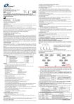
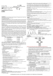
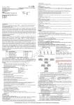
To generate a standard curve on the real-time system, all four dilution standards should be used and
defined as standard with specification of the corresponding concentrations.
Attention:
A. Mix thoroughly before next transfer.
B. The positive control contains high concentration of the target DNA. Therefore, be careful during the
dilution in order to avoid contamination.
9.4 PCR Protocol
The Master Mix volume for each reaction should be pipetted as follows:
M
yB
io
So
ur
1. Intended Use
The Anaplasma phagocytophilum real time PCR kit is used for the detection of Anaplasma
phagocytophilum in whole blood (non-anticoagulant or ticks samples by using real time PCR systems.
2. Principle of Real-Time PCR
The principle of the real-time detection is based on the fluorogenic 5’nuclease assay. During the PCR
reaction, the DNA polymerase cleaves the probe at the 5’ end and separates the reporter dye from the
quencher dye only when the probe hybridizes to the target DNA. This cleavage results in the
fluorescent signal generated by the cleaved reporter dye, which is monitored real-time by the PCR
detection system. The PCR cycle at which an increase in the fluorescence signal is detected initially
(Ct) is proportional to the amount of the specific PCR product. Monitoring the fluorescence intensities
during Real Time allows the detection of the accumulating product without having to re-open the
reaction tube after the amplification.
3. Product Description
Anaplasma phagocytophilum (formerly Ehrlichia phagocytophila), a tick-transmitted pathogen that
infects several animal species, including humans (involved as accidental "dead-end" hosts), is the
causative agent of human granulocytic anaplasmosis (HGA). It is a pathogen of veterinary importance
responsible for tickborne fever of ruminants and for granulocytic anaplasmosis of horses and dogs .
Anaplasma phagocytophilum real time PCR Kit contains a pairs of primers and a probe for the
detection of the HGA through polymerase chain reaction (PCR) in the real-time PCR system. The
master contains reagents and enzymes for the specific amplification of the HGA DNA. Fluorescence
is emitted and measured by the real time systems´ optical unit during the PCR. The detection of
amplified HGA DNA fragment is performed in fluorimeter channel 530nm with the fluorescent
quencher BHQ1. DNA extraction buffer is available in the kit. In addition, the kit contains a system to
identify possible PCR inhibition by measuring the 560nm fluorescence of the internal control (IC). An
external positive control defined as 1×10⁷copies/ml is supplied which allow the determination of the
gene load. For further information, please refer to section 9.3 Quantitation.
4. Kit Contents
Ref.
Type of Reagent
Presentation 25rxns
1
DNA Extraction Buffer
2 vials, 1.5ml
2
HGA Reaction Mix
1 vial, 450μl
3
PCR Enzyme Mix
1 vial, 12μl
4
Molecular Grade Water
1 vial, 400μl
5
Internal control (IC)
1 vial, 30μl
7
6
HGA Positive Control (1×10 copies/ml)
1 vial, 30μl
Analysis sensitivity: 1×10³ copies/ml ;
LOQ:2×10³~1×10⁸ copies/ml
Note: Analysis sensitivity depends on the sample volume, elution volume, nucleic acid extraction
methods and other factors .If you use the DNA extraction buffer in the kit, the analysis sensitivity is
the same as it declares. However, when the sample volume is dozens or even hundreds of times greater
than elution volume by some concentrating method, it can be much higher.
5. Storage
• All reagents should be stored at -20°C. Storage at +4°C is not recommended.
• All reagents can be used until the expiration date indicated on the kit label.
• Repeated thawing and freezing (> 3x) should be avoided, as this may reduce the sensitivity of the
assay.
• Cool all reagents during the working steps.
• Reaction Mix should be stored in the dark.
6. Additionally Required Materials and Devices
• Biological cabinet
• Real time PCR system
• Desktop microcentrifuge for “eppendorf” type tubes (RCF max. 16,000 x g)
• Vortex mixer
• Real time PCR reaction tubes/plates
• Cryo-container
• Pipets (0.5 μl – 1000 μl)
• Sterile filter tips for micro pipets
• Sterile microtubes
• Disposable gloves, powderless
• Biohazard waste container
• Refrigerator and freezer
• Tube racks
om
MBS598302 - Instrument I, II
.c
(For Research Use Only In USA & China)
Anaplasma phagocytophilum Real Time PCR Kit User Manual
ce
Revision No.: ZJ0002
Issue Date: Jul 1st, 2015
• Specimens can be extracted immediately or frozen at -20°C to -80°C.
• Transportation of clinical specimens must comply with local regulations for the transport of
etiologic agents
9. Procedure
9.1 DNA-Extraction
DNA extraction buffer is contained in the kit, please thaw the buffer thoroughly and spin down briefly
in the centrifuge before use. You may use your own extraction systems or commercial kits.
1) Pipet 100μl sample to a 0.5ml tube, add 100μl DNA extraction buffer, close the tube and vortex for
10 seconds. Spin down briefly in a table centrifuge.
2) Incubation the tube for 10 minutes at 100°C.
3) Centrifuge the tube at 13000rpm for 10 minutes. The supernatant contains the DNA extracted and is
used for PCR template.
9.2 Internal Control
It is necessary to add internal control (IC) in the reaction mix. Internal Control (IC) allows the user to
determine and control the possibility of PCR inhibition.
Add the internal control (IC) 1μl/rxn and the result will be shown in the 560nm.
9.3 Quantitation
The kit can be used for quantitative or qualitative real-time PCR. A positive control (1×10⁷copies/ml)
is supplied in the kit.
For performance of quantitative real-time PCR, Standard dilutions must prepare first as follows.
Molecular Grade Water is used for dilution.
Dilution is not needed for performance of qualitative real-time PCR.
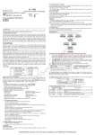
Take positive control (1×10⁷copies/ml) as the starting high standard in the first tube. Respectively
pipette 36ul of Molecular Grade Water into next three tubes. Do three dilutions as the following
figures:
7.
Warnings and Precaution
Carefully read this instruction before starting the procedure.
• For in vitro diagnostic use only.
• This assay needs to be carried out by skilled personnel.
• Clinical samples should be regarded as potentially infectious materials and
should be prepared in a laminar flow hood.
• This assay needs to be run according to Good Laboratory Practice.
• Do not use the kit after its expiration date.
• Avoid repeated thawing and freezing of the reagents, this may reduce the sensitivity of the test.
• Once the reagents have been thawed, vortex and centrifuge briefly the tubes before use.
• Prepare quickly the Reaction mix on ice or in the cooling block.
• Set up two separate working areas: 1) Isolation of the RNA/ DNA and 2) Amplification/
detection of amplification products.
• Pipets, vials and other working materials should not circulate among working units.
• Use always sterile pipette tips with filters.
• Wear separate coats and gloves in each area.
• Do not pipette by mouth. Do not eat, drink, smoke in laboratory.
• Avoid aerosols
8. Sample Collection, Storage and transport
• Collect samples in sterile tubes;
※PCR system without 560nm channel may be treated with 1μl Molecular Grade Water instead of 1μl IC.
1)
The volumes of Reaction Mix and Enzyme Mix per reaction multiply with the number of
samples, which includes the number of controls, standards, and sample prepared. Molecular
Grade Water is used as the negative control. For reasons of unprecise pipetting, always add an
extra virtual sample. Mix completely then spin down briefly in a centrifuge.
2)
Pipet 18μl Master Mix with micropipets of sterile filter tips to each of the real time PCR
reaction plate/tubes. Separately add 2μl DNA sample, positive and negative controls to different
reaction plate/tubes. Immediately close the plate/tubes to avoid contamination.
3)
Spin down briefly in order to collect the Master Mix in the bottom of the reaction tubes.
4)
Perform the following protocol in the instrument:
37°C for 2min
1cycle
Selection of fluorescence channels
94°C for 2min
1cycle
530nm
Target Nucleic Acid
93°C for 5sec, 60°C for 30sec
560nm
IC
40cycles
( Fluorescence measured at 60°C)
10. Threshold setting: Choose Arithmetic as back ground and none as Noise Band method, then
adjust the Noise band just above the maximum level of molecular grade water, and adjust the threshold
just under the minimum of the positive control.
11.Calibration for quantitative detection: Input each concentration of standard controls at the end of
run, and a standard curve will be automatically formed.
12.Quality control: Negative control, positive control, internal control and QS curve must be
performed correctly, otherwise the sample results is invalid.
Channel
Crossing point value
Control
530nm
560nm
Molecular Grade Water
Blank
25~35
Positive Control(qualitative assay)
≤35
——
QS(quantitative detection)
Correlation coefficient of QS curve≤-0.98
13. Data Analysis and Interpretation
The following results are possible:
Crossing point value
Result Analysis
530nm
560nm
1#
Blank
25~35
Below the detection limit or negative
2#
≤38
Positive; and the software displays the quantitative value
——
3#
25~35
Re-test; If it is still 38~40, report as 1#
38~40
4#
Blank
Blank
PCR Inhibition; No diagnosis can be concluded.
For further questions or problems, please contact our technical support
FOR RESEARCH USE ONLY. NOT FOR USE IN DIAGNOSTIC OR THERAPEUTIC PROCEDURES.
• Avoid aerosols
8. Sample Collection, Storage and transport
• Collect samples in sterile tubes;
• Specimens can be extracted immediately or frozen at -20°C to -80°C.
• Transportation of clinical specimens must comply with local regulations for the transport of
etiologic agents
9. Procedure
9.1 DNA-Extraction
DNA extraction buffer is contained in the kit, please thaw the buffer thoroughly and spin down briefly
in the centrifuge before use. You may use your own extraction systems or commercial kits.
1) Pipet 100μl sample to a 0.5ml tube, add 100μl DNA extraction buffer, close the tube and vortex for
10 seconds. Spin down briefly in a table centrifuge.
2) Incubation the tube for 10 minutes at 100°C.
3) Centrifuge the tube at 13000rpm for 10 minutes. The supernatant contains the DNA extracted and is
used for PCR template.
9.2 Internal Control
It is necessary to add internal control (IC) in the reaction mix. Internal Control (IC) allows the user to
determine and control the possibility of PCR inhibition.
Add the internal control (IC) 1μl/rxn and the result will be shown in the HEX/VIC/JOE.
9.3 Quantitation
The kit can be used for quantitative or qualitative real-time PCR. A positive control (1×10⁷copies/ml)
is supplied in the kit.
For performance of quantitative real-time PCR, Standard dilutions must prepare first as follows.
Molecular Grade Water is used for dilution.
Dilution is not needed for performance of qualitative real-time PCR.
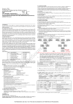
Take positive control (1×10⁷copies/ml) as the starting high standard in the first tube. Respectively
pipette 36ul of Molecular Grade Water into next three tubes. Do three dilutions as the following
figures:
Revision No.: ZJ0002
Issue Date: Jul 1st, 2015
(For Research Use Only In USA & China)
Anaplasma phagocytophilum Real Time PCR Kit User Manual
MBS598302 - Instrument III, IV
For use with ABI Prism®7000/7300/7500/7900/Step One Plus; iCycler iQ™4/iQ™5;
Smart Cycler II;Bio-Rad CFX 96;Rotor Gene™6000; Mx3000P/3005P;MJ-Option2/Chromo4;
LightCycler®480 Instrument
ce
.c
om
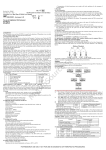
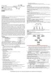
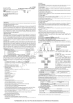
To generate a standard curve on the real-time system, all four dilution standards should be used and
defined as standard with specification of the corresponding concentrations.
Attention:
A. Mix thoroughly before next transfer.
B. The positive control contains high concentration of the target DNA. Therefore, be careful during the
dilution in order to avoid contamination.
9.4 PCR Protocol
The Master Mix volume for each reaction should be pipetted as follows:
M
yB
io
So
ur
1. Intended Use
The Anaplasma phagocytophilum real time PCR kit is used for the detection of Anaplasma
phagocytophilum in whole blood (non-anticoagulant or ticks samples by using real time PCR systems.
2. Principle of Real-Time PCR
The principle of the real-time detection is based on the fluorogenic 5’nuclease assay. During the PCR
reaction, the DNA polymerase cleaves the probe at the 5’ end and separates the reporter dye from the
quencher dye only when the probe hybridizes to the target DNA. This cleavage results in the
fluorescent signal generated by the cleaved reporter dye, which is monitored real-time by the PCR
detection system. The PCR cycle at which an increase in the fluorescence signal is detected initially
(Ct) is proportional to the amount of the specific PCR product. Monitoring the fluorescence intensities
during Real Time allows the detection of the accumulating product without having to re-open the
reaction tube after the amplification.
3. Product Description
Anaplasma phagocytophilum (formerly Ehrlichia phagocytophila), a tick-transmitted pathogen that
infects several animal species, including humans (involved as accidental "dead-end" hosts), is the
causative agent of human granulocytic anaplasmosis (HGA). It is a pathogen of veterinary importance
responsible for tickborne fever of ruminants and for granulocytic anaplasmosis of horses and dogs .
Anaplasma phagocytophilum real time PCR Kit contains a pairs of primers and a probe for the
detection of the HGA through polymerase chain reaction (PCR) in the real-time PCR system. The
master contains reagents and enzymes for the specific amplification of the HGA DNA. Fluorescence
is emitted and measured by the real time systems´ optical unit during the PCR. The detection of
amplified HGA DNA fragment is performed in fluorimeter channel FAM with the fluorescent
quencher BHQ1. DNA extraction buffer is available in the kit. In addition, the kit contains a system to
identify possible PCR inhibition by measuring the HEX/VIC/JOE fluorescence of the internal control
(IC). An external positive control defined as 1×10⁷copies/ml is supplied which allow the determination
of the gene load. For further information, please refer to section 9.3 Quantitation.
4. Kit Contents
Ref.
Type of Reagent
Presentation 25rxns
1
DNA Extraction Buffer
2 vials, 1.5ml
2
HGA Reaction Mix
1 vial, 950μl
3
PCR Enzyme Mix
1 vial, 12μl
4
Molecular Grade Water
1 vial, 400μl
5
Internal control (IC)
1 vial, 30μl
7
6
HGA Positive Control (1×10 copies/ml)
1 vial, 30μl
3
3
Analysis sensitivity: 1×10 copies/ml ;
LOQ:2×10 ~1×10⁸ copies/ml
Note: Analysis sensitivity depends on the sample volume, elution volume, nucleic acid extraction
methods and other factors .If you use the DNA extraction buffer in the kit, the analysis sensitivity is
the same as it declares. However, when the sample volume is dozens or even hundreds of times greater
than elution volume by some concentrating method, it can be much higher.
5. Storage
• All reagents should be stored at -20°C. Storage at +4°C is not recommended.
• All reagents can be used until the expiration date indicated on the kit label.
• Repeated thawing and freezing (> 3x) should be avoided, as this may reduce the sensitivity of the
assay.
• Cool all reagents during the working steps.
• Reaction Mix should be stored in the dark.
6. Additionally Required Materials and Devices
• Biological cabinet
• Real time PCR system
• Desktop microcentrifuge for “eppendorf” type tubes (RCF max. 16,000 x g)
• Vortex mixer
• Real time PCR reaction tubes/plates
• Cryo-container
• Pipets (0.5 μl – 1000 μl)
• Sterile filter tips for micro pipets
• Sterile microtubes
• Disposable gloves, powderless
• Biohazard waste container
• Refrigerator and freezer
• Tube racks
7.
Warnings and Precaution
Carefully read this instruction before starting the procedure.
• For in vitro diagnostic use only.
• This assay needs to be carried out by skilled personnel.
• Clinical samples should be regarded as potentially infectious materials and
should be prepared in a laminar flow hood.
• This assay needs to be run according to Good Laboratory Practice.
• Do not use the kit after its expiration date.
• Avoid repeated thawing and freezing of the reagents, this may reduce the sensitivity of the test.
• Once the reagents have been thawed, vortex and centrifuge briefly the tubes before use.
• Prepare quickly the Reaction mix on ice or in the cooling block.
• Set up two separate working areas: 1) Isolation of the RNA/ DNA and 2) Amplification/
detection of amplification products.
• Pipets, vials and other working materials should not circulate among working units.
• Use always sterile pipette tips with filters.
• Wear separate coats and gloves in each area.
• Do not pipette by mouth. Do not eat, drink, smoke in laboratory.
OR
※ PCR system without HEX/VIC/JOE channel may be treated with 1μl Molecular Grade Water instead of 1μl IC.
1) The volumes of Reaction Mix and Enzyme Mix per reaction multiply with the number of samples,
which includes the number of controls, standards, and sample prepared. Molecular Grade Water is
used as the negative control. For reasons of unprecise pipetting, always add an extra virtual sample.
Mix completely then spin down briefly in a centrifuge.
2) Pipet 36μl (22.5μl for SmartCyclerII) Master Mix with micropipets of sterile filter tips to each of
the Real time PCR reaction plate/tubes. Separately add 4μl(2.5μl for SmartCyclerII) DNA sample,
positive and negative controls to different reaction plate/tubes. Immediately close the plate/tubes to
avoid contamination.
3) Spin down briefly in order to collect the Master Mix in the bottom of the reaction tubes.
4) Perform the following protocol in the instrument:
37°C for 2min
1cycle
Selection of fluorescence channels
94°C for 2min
1cycle
FAM
Target Nucleic Acid
93°C for 15sec, 60°C for 1min
HEX/VIC/JOE
IC
40cycles
( Fluorescence measured at 60°C)
5)
If you use ABI Prism® system, please choose “none” as passive reference and quencher.
10. Threshold setting: just above the maximum level of molecular grade water.
11.Calibration for quantitative detection: Input each concentration of standard controls at the end of
run, and a standard curve will be automatically formed.
12.Quality control: Negative control, positive control, internal control and QS curve must be
performed correctly, otherwise the sample results is invalid.
Channel
Ct value
Control
FAM
HEX/VIC/JOE
Molecular Grade Water
UNDET
25~35
Positive Control(qualitative assay)
≤35
——
QS(quantitative detection)
Correlation coefficient of QS curve≤-0.98
13. Data Analysis and Interpretation :The following results are possible:
Ct value
Result Analysis
FAM
HEX/VIC/JOE
1#
UNDET
25~35
Below the detection limit or negative
2#
Positive; and the software displays the quantitative value
≤38
——
3#
25~35
Re-test; If it is still 38~40, report as 1#
38~40
4#
UNDET
UNDET
PCR Inhibition; No diagnosis can be concluded.
For further questions or problems,please contact our technical support
FOR RESEARCH USE ONLY. NOT FOR USE IN DIAGNOSTIC OR THERAPEUTIC PROCEDURES.