1
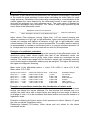
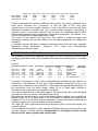
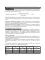
EBV Enzyme Linked Immunosorbent Assay for the cut-off determination of IgA, IgG or IgM Antibodies to Epstein-Barr Virus (EBV) in human serum or plasma. Microwell ELISA USER MANUAL Microwell Method - 96 wells (12 x 8-well antigen coated strips individual breakaway IgA IgG IgM Ref. 01CEBV01 Ref. 01CEBV03 Ref. 01CEBV05 For in vitro Diagnostic Use GENERAL INFORMATION Wavelength Measurement Filter: 450 nm Optional Reference Filter: 600 - 650 nm Incubation Time 70 minutes at 37°/R.T. (30/30/15) Enzyme Conjugate HRP (Horseradish Peroxidase), ready to use Substrate Solution TMB (3,3´,5,5´-Tetramethyl-benzidine), concentrate Sample Serum or Plasma Stability of Samples undiluted: 5 days at 2-8°C; up to 6 months at - 20 °C Shelf life and Stability of Kit Components Kit: 12 months from production date. Kit Components: see expiration date on the label 2 KIT COMPONENTS Microwell plate Negative Control Cut-off Control 12x8 wells coated with EBV antigens in a resealable foil pouch. 1 vial, 1.5 ml, IgA, IgG or IgM, human serum, ready to use Substrate Solution B 1 vial, 3 ml, IgA, IgG or IgM, human serum, ready to use 1 vial, 1.5 ml, IgA, IgG or IgM, human serum, ready to use 1 vial, 12 ml. Anti human IgA, IgG or IgM HRP Conjugate, ready-to-use 1 vial, 0.750 ml. TMB (Tetramethylbenzidine) in DMSO (Dimethylsulfoxide) 1 bottle, 15 ml, contains urea peroxide (corrosive). Sample Diluent 2 bottles, 20 ml each. Additive for Sample Diluent 2 vials, 2ml each Wash Buffer 1 bottle, 20 ml. Contains NaCl and Tween 20. (25x) Concentrate 1 bottle, 15 ml. 1 % v/v sulphuric acid, corrosive. Ready to use Positive Control Enzyme Conjugate Substrate Solution A Stop Solution MATERIALS REQUIRED BUT NOT PROVIDED Deionized or distilled water Graduated cylinders and beakers Wash trays Macropipettes capable of delivering 5 µl to 1000 µl. Multichannel Micropipette Stepper Incubator 37°C+/- 2°C (dry incubator, make sure to correctly seal the wells with the adhesive foil to prevent evaporation which may lead to erroneous results. Microplate reader capable of reading absorbance values at 450 nm. If dual wavelength microplate reader is available, the reference filter should be set at 600-690 nm. Automatic microplate washer capable of dispensing 300 µl. Microplate reader and microplate washers are available from Abiyotek Company. 3 SUMMARY AND EXPLANATION Indirect enzyme immunoassay for the detection of antibodies to Epstein Barr virus (EBV) in human serum and plasma. The enzyme immunoassay is intended for testing individual specimens, not pooled specimens. In vitro diagnosticum, only to be used for in vitro diagnostic purposes by correspondingly educated laboratory personnel. The test can be processed manually or automatically. The barcode identification of each single reagent ensures their correct identification and an accurate automatic processing of the test. DIAGNOSTIC RELEVANCE, DISEASE AND RECOMMENDED LITERATURE Besides detection of the infectious agent, the detection of antibodies to EBV significantly contributes to the serological diagnosis of these infections. The detection of EBV-specific IgG, IgM and IgA antibodies to its major immunodominant antigens has become an important and useful tool for the monitoring and follow-up of patients showing EBV associated diseases several years after primary infection. In the acute phase of infectious mononucleosis, IgM and also IgA and then IgG, IgA or IgM antibodies to EBV early antigens (EA), to viral capsid antigen (VCA) and IgG, IgA or IgM to nuclear antigens (EBNA) appear in sequence. Therefore the presence of IgM/IgA antibodies to VCA or IgM/IgA/IgG antibodies to EA with low or absent EBNA antibodies is indicative of current or recent infection. IgM VCA antibodies are almost always detectable at the early stage of infection and lasts 4 to 8 weeks. The presence of EBNA antibodies are indicative for past infection and appear after 2 to 3 months after infection. Epstein Barr Virus (EBV) infects the majority of people worldwide (> 90 %) and persists as a latent, lifelong infection of long-lived memory B lymphocytes with virus production. EBV infection may demonstrate a wide spectrum of clinical symptoms. The majority of primary EBV infections are transmitted via saliva, occur during childhood, and are subclinical. In young adults, EBV infection may be clinically manifested as Infectious Mononucleosis (IM) with typical symptoms of sore throat, fever, and lymphadenopathy. In addition to IM, EBV causes chronic active EBV infection, B-cell lymphoproliferative disease (a complication of the immunosuppression of transplantation), X-linked lymphoproliferative (XLP) syndrome, a subset of Hodgkin disease, opportunistic nonHodgkin lymphoma, Burkitt lymphoma, unusual hepatitides, nasopharyngeal and other carcinomas, haemophagocytic syndromes, and other diseases/syndromes, including a relative risk for EBV-positive Hodgkin lymphoma in young adults (estimated frequency 1 Hodgkin lymphoma/1000 infectious mononucleosis infections) approximately 4 years after serologically verified infectious mononucleosis. Serological studies indicate that prior to the clinical onset of NPC a high antibody concentration of specific IgA to viral capsid antigens and early antigens is frequently observed. The specific IgA levels increase with progression of the tumor disease and the antibody levels decline responding to therapy. In patients with confirmed clinical remission elevation of specific IgA serological concentration is highly significant for prediction of relapse. The diagnostic significance of IgM and IgA antibodies to EB Nuclear Antigens still has to be established. Infection: The majority of primary EBV infections are transmitted via saliva, occur during childhood, and are subclinical. Infectious agent: Epstein-Barr Virus (EBV, human herpesvirus 4), a member of the Herpesviridae family/genus Lymphocryptovirus of double-stranded DNA viruses. TEST PRINCIPLES Human IgG, IgA or IgM antibodies against EBV virus, if present in the specimen, bind to immobilized EBV virus antigens on the surface of the wells of the microtiterplate, the human antibodies bound are then detected by specific antihuman-IgG, IgA or IgM antibodies labelled to horse radish peroxidase and subsequently revealed by the substrate/chromogen colour reaction. After stopping the colour reaction the initially blue colour turns yellow and the intensity of this yellow colour is measured photometrically (extinction, absorbance, optical density (O.D.)).The intensity of the colour reaction is proportional to the corresponding antibody content CONSTITUTION OF THE REAGENTS Nr. 1 Nr. 2 Nr. 3 Nr. 4 Nr. 5 Nr. 6 Nr. 7 Nr. 8 Nr. 9 Nr. 10 Nr. 11 Microtiterplate wells, coated with EBV virus antigens. Negative Control: contains human IgG, IgA und IgM antibodies in concentrations giving reactivities in the negative range of the test system. Reactivity range/ Index range: Index < 0.60. Buffer: 0.05 M PBSTween 20, pH 7.2. Preservatives: < 0.10 % sodium azide und max. 0.03% Thiomerosal. Colour concentrate: E-104. E-132 max. 0.2 % v/v. Differential Control: contains human IgG, IgA und IgM antibodies in concentrations giving reactivities in the borderline range of the test system. Reactivity range/ Absorbance range (OD-range): 0.080 - 1.000, Index = 1. Buffer: 0.05 M PBS-Tween 20, pH 7.2. Preservatives: < 0.10 % sodium azide und max. 0.03% thiomerosal. Colour concentrate: E-110 max. 0.1 % v/v. Positive Control: contains human IgG, IgA und IgM antibodies in concentrations giving reactivities in the positive range of the test system. Reactivity range/ Index range: Index > 1.40. Puffer: 0.05 M PBSTween 20, pH 7.2. Preservatives: < 0.10 % sodium azide and max 0.03% thiomerosal. Colour concentrate: E-123 max. 0.1% v/v. Diluent buffer: 0.05 M PBS-Tween 20, pH 7.2. Preservatives: max. 0.05% thiomerosal. Colour: Bromphenolblue, max. 20 mg/Liter. Additive for diluent buffer: contains newborn calf serum (NCS), Preservatives: < 0.10 sodium azide and max. 0.05% thiomerosal. Anti-human-IgG, IgA or IgM Peroxidase conjugate: Sheep antibodies against human IgG, IgA or IgMantibodies conjugated with horse radish peroxidase . Contains bovine serum ingredients max. 1.5 %. Preservatives: thymol: max. 0.1 g/Liter, Bronidox L max. 0.4% v/v. Colour: Bromphenolblue. Substrate buffer with substrate, contains urea-peroxide in succinate-borate-buffer. Urea-peroxide is irritant and must be handled cautiously. Chromogen-concentrate 21x conc.: TMB (tetra-methyl-benzidine) in Dimethylsulfoxide (DMSO ). Dimethylsulfoxide and TMB are corrosive respectively minor poisonous and must be handled cautiously. Washing solution concentrate 25x conc, contains 200gr/Liter sodium chloride, 1.25 % Tween 20, max 0.01 % thiomerosal. Stopping solution: sulfuric acid (max. 1% v/v, < 0.2 M). Sulfuric acid is corrosive and must be handled cautiously. REAGENTS DESCRIPTION Closed components of the test kit Opened Microplate strips STORAGE 2...8 °C 2...8 °C Opened components No. 2, 3, 4, 5, 6, 7 2...8 °C Opened substrate buffer (No.8) 2...8 °C STABILITY COMMENTS until expiry date 6 weeks keep storage bag tightly closed avoiding high humidity 12 weeks avoid Temperature stress and contamination 12 weeks avoid direct exposure to light Opened TMB/Chromogen solution 21x conc (No.9) 2…8 °C 12 weeks Specimen diluent (No.5 + No.6), ready to use 2…8 °C 12 weeks Substrate A/B solution (No.8 + No.9), 2…8 °C max. 24 h Washing solution, ready to use 2…8 °C 12 weeks 20…25 °C max. 2 weeks 2…8 °C until expiry date Stop solution avoid direct exposure to light prepare only the necessary volume and avoid contamination prepare only the necessary volume ready to use and avoid direct exposure to light use only a clear solution use only a clear solution SAFETY MEASURES: Warnings, Precautions, Disposal 1 GLP-RULES should always be followed (GLP: Good Laboratory Practice). 2 The testkit is only to be used for in vitro diagnostic purposes and by professional staff only. 3 The use of protective laboratory clothes, protective hand gloves and also protective glasses during the actual manual procedure is recommended,. Do not pipet by mouth. 4 All tested samples should be regarded as potentially infectious and should be handled accordingly. The controls have been derived from donations which have been tested for anti-HIV 1+2, anti-HCV and HBsAg on a single donor basis and have been found non reactive. Nevertheless they should also be handled as potentially infectious. Do not use heat inactivated test specimens. 5 Material of bovine origin used as ingredients in reagents, originate from countries known to be BSE-free at the time of purchase. 6 The controls and the additive for the diluent buffer contain < 0.1 % sodium azide and 0.05 % thiomerosal as preservatives. The dilution buffer contains 0.05 % thiomerosal as preservative. 7 Precautions to be considered using on vitro diagnostic devices containing sodium azide as preservative: Sodium azide is poisonous, swallowing and contact with skin, eyes and mucous tissue is to be avoided. Sodium azide generates explosive azides with heavy metals like copper or lead. Disposing sodium azide-containing waste solutions, always rinse with enough water. 8 Precautions to be considered using in vitro diagnostic devices containing thiomerosal as preservative: Thiomerosal is poisonous, swallowing and contact with skin, eyes and mucous tissue should be avoided. Although thiomerosal is also used in some vaccines as preservative in comparable concentrations, in vitro reagents containing thiomerosal should be handled cautiously. 9 Substrate B with substrate (No. 8), Substrate A concentrate (No. 9) and stopping solution (No. 11) contain irritant and corrosive substances and should be handled cautiously. If contact with skin, eyes or mucous tissue occurs immediately rinse with enough water and consult a physician. 10 All waste solutions should be collected in adequate vessels containing disinfectants capable of inactivating human pathogenic viruses. Follow the corresponding manufacturer’s instructions for use. 11 Disposal: follow the locally ruling safety and disposal laws and regulations for disposal. LIMITATIONS AND CAUSES OF ERROR It is to be considered that under certain specific laboratory working conditions adjustment of alternative incubation periods may be necessary. If reagents are used too cool before reaching room temperature (20…25 °C), a weaker colour development will occur at the end of the test run. On the other hand, if room temperature is high (appr. 30 °C or higher), a stronger colour development will occur at the end of the test run. Under these circumstances the validation criteria of the test run may not be achieved. Periodically check functionality of pipettes and instruments used. The reagents of the testkit are not to be used after its expiry date. Do not use heat inactivated specimens. Avoid testing contaminated samples, strong hemolytic, icteric or lipemic samples, since erroneous results may be obtained. To ensure the performance of the testkit storage conditions and stability of the opened and diluted reagents must be strictly respected as depicted under storage and stability. Reagent No. 1, antigen coated wells, No. 2, negative control, No. 3, differential control, No. 4, positive control and No. 7, conjugate solution are lot specific and are not allowed to be used together with corresponding reagents from another lot. Reagent No. 5, Sample Diluent, No. 6, additive for Sample Diluent, No. 8 Substrate B with substrate, No. 9, chromogen concentrate, No. 10, washing solution and No. 11 stopping solution are not lot specific and may be used, if necessary and respecting the corresponding expiry date, in a test run together with corresponding lot specific reagents (Nr. 1, Nr. 2, Nr. 3, Nr. 4) from another lot. Avoid cross contaminations during manipulations. Never use the same vessel for the ready to use conjugate dilution and the ready to use substrate/chromogen solution. Since TMB turns blue coloured upon oxidation, any contact of the reagents No. 8, No. 9 and No. 11 with heavy metals should be avoided. Also protect TMB solutions from direct light exposure SAMPLE COLLECTION AND HANDLING Plasma or serum collected by venipuncture should be tested within 2 days if stored at …8 °C after reception. Prolonged storage should be done at –20 °C or lower. Avoid repeated thawing and freezing. Samples showing particles should be centrifuged prior to be processed, to avoid possible erroneous results. Contamination should be avoided, since contaminated samples may also lead to erroneous results. Handle all samples as potentially infectious. PREPARATION OF REAGENTS AND TESTSAMPLES All necessary reagents must reach room temperature (20…25 °C) prior to be used. If reagents are used too cool before reaching room temperature (20…25 °C), a weaker colour development will occur at the end of the test run. On the other hand, if room temperature is high (appr. 30 °C or higher), a stronger colour development will occur at the end of the test run. Under these circumstances the validation criteria of the test run may not be achieved, and corrective measures may be necessary (prolonging or shortening the incubation period) Sample diluent (No. 5 + No. 6): 2 ml additive (No. 6) are added to 20 ml diluent buffer (No. 5). Bring only the necessary volume of the ready to use specimen diluent to room temperature. Washing solution, ready to use: The concentrated washing solution (No. 10) is diluted 1 in 25 with deionised water (20 ml concentrate + 480 ml deionised water), use only clear solutions. Microplate strips and wells: Take the necessary amount of strips or wells from the bag after they have reached room temperature. Place the required strips or wells firmly in the frame, make sure they are evenly arrayed in the frame. If required fill the empty plate positions with empty wells or strips (not antigen coated) according to the pipetting or washing device used to avoid overflow of fluid during pipetting or washing steps of the test run. Not required strips or wells (antigen coated) must be transferred into the storage bag, well sealed, avoiding humidity and reset for storage at 2…8 °C. Specimens: Specimens are tested at a 1/21 dilution. Although testing of other body fluids than serum or plasma is possible, specific adjustment of the conditions is needed. Conjugate: always prepare only the amount of conjugate needed plus =< 0.1 ml. Chromogen-Substrate solution.: always prepare only the necessary amount of chromogen-substrate solution plus =<0.1 ml. As an example: to 1 ml substrate buffer 0.050 ml TMB-Chromogen concentrate is added (1/21 dilution). TEST PROCEDURE The test should only be performed by properly trained professional laboratory staff. All reagents must have reached room temperature (22...25 °C) prior to start performing the test run. Incubation periods: Specimens and controls 30 min. at 37 °C, Conjugate 30 min. at 37°C and TMB-Chromogen / Substrate solution 10 to 20 min. at room temperature (22…25 °C). Adaptation of incubation periods to specific internal laboratory needs is possible, according to GLP they should be validated. TEST PROCEDURE (summary) PREPARATION OF REAGENTS / PREPARATION OF TESTPROTOCOL SPECIMEN DILUTION: 1/21 dilution Performing multiple testing of specimens a corresponding volume is prepared in a separate tube or plate: 0.100 ml OF THE SPECIMEN DILUTION ARE PIPETTED Pipetting specimens directly in the plate dispense first 0.200 ml of specimen diluent and then add 0.010 ml specimen 0.200 ml OF SPECIMEN DILUENT + 0.010 ml SPECIMEN 0.100 ml OF THE CONTROLS ARE PIPETTED INCUBATION OF SPECIMENDILUTIONS AND CONTROLS 30 min. at 37 °C WASH 4x PIPETTING OF THE CONJUGATE (0.100 ml) INCUBATION WITH CONJUGATE 30 min. at 37°C WASH 4x PIPETTING OF THE TMB-CHROMOGEN-SUBSTRATE SOLUTION (0.100 ml) INCUBATION WITH TMB-CHROMOGEN-SUBSTRATE SOLUTION (10 …20 min at room temperature, ca. 20…25 °C) PIPETTING OF THE STOPPING SOLUTION (0.100 ml) PHOTOMETRIC MEASUREMENT at 450nm (Ref:630 nm) TESTVALIDATION / EVALUATION 1. Preparation: 2. Test protocol: TESTPROCEDURE (manual) Before starting with the test make sure that each single reagent has reached room temperature ca. 20…25 °C. Check each single reagent to be use for its identity, verify the sequence of dilution and pipetting. Processing more than one strip it is recommended to identify each single strip (eg.: 1,2,3 ...etc.). Prepare a test protocol according to the specimen identification numbers for the dilution and pipetting sequences of the specimens and the controls to be tested. One well is assigned to the negative control, two wells to the differential control and one well to the positive control. If necessary more controls may be scheduled. 3. SPECIMEN PREPARATION, DILUTION, PIPETTING OF SPECIMENS AND CONTROLS, INCUBATION: Specimens, liquid serum or plasma, are prepared to be tested at a 1/21 dilution. Performing multiple testing of specimens a corresponding dilution volume is prepared in a separate tube or plate, then 0.100 ml of the diluted specimen is pipetted into the corresponding well according to the pipetting protocol (it is recommended to pipette in replicates at the beginning to establish the own pipetting accuracy and reproducibility). Are the specimens directly diluted into the wells then 0.200 ml specimen diluent are dispensed first into each well and then 0.010 ml of each specimen are added into the corresponding wells, mix well (it is recommended to pipette in replicates at the beginning to establish the own pipetting accuracy and reproducibility). In addition 0.100 ml of each control are pipetted into their corresponding wells. Pipetting longer series of specimens it is recommended to pipette the controls after reaching half of the series to compensate for pipetting delays. The corresponding controls may also be pipetted at the beginning and at the end of a longer series, for evaluation the mean of the corresponding results is used. After finishing pipetting shake gently the plate, cover it with the adhesive foil and incubate for 30 min. +/- 2 min. at 37 °C +/- 1°C. 4. Preparation: Before finishing the first incubation (specimen incubation) period prepare the necessary volume of conjugate solution. Make sure that the solution has reached room temperature (20...25 °C). For 8 wells 0.8 + 0.1 = 0.9 ml conjugate are needed. 5. Wash step 1: Remove the specimen dilutions and controls form the wells, add 0.300 ml washing solution to each well, wait 1 min. Repeat the sequence removal of the washing solution, adding washing solution and waiting 1 min. 3 times, totally 4 washing cycles. After last removal of washing solution, make sure the wells have been completely voided, eventually tap the plate upside down on absorbent paper. 6. Conjugate: 0.100 ml of the anti-human IgG, IgA or IgM-PO- conjugate solution are pipetted in all wells. After finishing pipetting, cover it with the adhesive foil and incubate for 30 min. +/- 2 min. at 37 °C +/- 1°C. 7. PREPARATION: Before finishing the second incubation (conjugate incubation) prepare the corresponding volume of TMB-Chromogen/substrate solution and keep it in the dark until use ( e.g.: for 8 wells, to 1ml substrate buffer (No.8) add 0.050 ml TMB-Chromogen solution 21x conc.(No.9)). Make sure the solution has reached room temperature (20...25 °C). 8. Wash step 2: Remove the conjugate solution form the wells, add 0.300 ml washing solution to each well, wait 1 min. Repeat the sequence removal of the washing solution, adding washing solution and waiting 1 min. 3 times, totally 4 washing cycles. After last removal of washing solution, make sure the wells have been completely voided, eventually tap the plate upside down on absorbent paper. Wipe carefully the bottom of the strips from outside with absorbent paper, to remove all possible liquid residues that could interfere with photometric reading. 9. TMB-Chromogen/Substrate Incubation: 0.100 ml of the TMB-Chromogen/ Substrate solution are pipetted in all wells. After finishing pipetting, incubate the plate for 15 +- 5 min. at room temperature (20..25°C) avoiding exposure to light (dark chamber, dark box, a closed drawer). Reactive specimens develop a blue colour. 10. Reaction stop: After finishing the TMB-Chromogen/substrate incubation add 0.100 ml Stopping solution to all wells. Reactive specimens turn from blue to yellow. 11. Photometric reading: Photometric reading should be done within 10 to 20 minutes after stopping the colour reaction with a microtiterplate photometer at 450 nm (if possible with the reference wavelength set at 630 nm) Blanking is done against air. 12. Validation: According to the validation criteria (see under Validation). 13. Evaluation: According to the evaluation criteria (see under Evaluation). TESTPROCESSING WITH AUTOMATIC DEVICES Test processing with automatic devices may be carried out according to the assay definition programs of the automatic device in use (e.g.: BEP 2000, EtiMax3000, Evolis, Quickstep among others). The assay definition program allows the bar code identification of the reagents and of the specimens and their sequential process assignment for the entire process of the test. After defining the jobs to be done a list of the corresponding reagents needed is generated .including the necessary reagent volumes and their corresponding containers (Specimen diluent, controls, conjugate, TMB-Chromogen/Substrate solution, stop solution and wash solution). For each single reagent needed the minimal calculated quantities have to be present in the corresponding amounts and in the corresponding bar coded vials to be processed. The barcoded reagent vials prepared are placed in the corresponding reagents’ rack for processing. The racks containing the barcoded specimens and the racks containing the barcoded reagents can then be introduced in the processing area. During introduction of the racks barcode reading is affected and the position of each barcoded reagent and specimen registered. After verifying the necessary amounts of reagents the plate to be processed is requested (it is possible to align a variety of different tests, the only requirement is that the different assays to be processed must have all the same single incubation periods for each incubation). Before inserting the plate in the plate holder make sure that besides the wells needed to be processed in their corresponding positions, empty positions are filled with empty wells to prevent overflow of washing solution in the washing chamber. After inserting the plate it is brought to the pipetting area and the assay/assays job/jobs is/are started. The job is processed in the following way: 1. Dispensing specimen diluent (0.200 ml) in the wells assigned for the specimens. 2. Dispensing specimens in the wells assigned for specimens (0.010 ml in 0.200 ml, 1:21 dilution). 3. Dispensing the controls in the wells assigned for the controls (0.100 ml). 4. Incubation of the specimens and controls: 30 min. +-1 min bei 37 +- 1°C. 5. Washing 6. Dispensing the conjugate (0.100 ml). 7. Incubation with conjugate: 30 min. +- 1 min bei 37 +- 1°C. 8. Washing 9. Dispensing the TMB-Chromogen / Substrate solution (1. cycle 0.025 + 2. cycle immediately there after 0.050 ml). 10. Incubation with TMB-Chromogen / Substrate solution: 15 min. +- 1 min at room temperature (20...25 °C). 11. Stop of the reaction by adding stopping solution (0.100 ml). 12. Photometric reading in integrated photometer. 13. Results may be printed out or further transferred online. The corresponding protocols include validated and evaluated results. VALIDATION OF THE TEST, CORRECTIVE MEASURES, GENERAL CONSIDERATIONS Validation: Results obtained in absorbance units (extinction units, O.D. units) for the controls are used if the values of the differential control are higher than 0.080 and lower than 1.000 (optimally between 0.200 and 0.600) and the deviation of the values obtained for the differential control falls within +- 20 % of the mean value. Additionally the corresponding index value of the negative control must be < 0.6 and the corresponding index value of the positive control must be > 1.4. These criteria apply to all our systems. absorption at 450nm of the corresponding control Index value of the controls = -------------------------------------------------------------------------------Mean absorption at 450nm of the differential control Example of a validation: mean value of the absorption of the differential control 1. value: 0.280, 2. value: 0.320 Mean value: 0.300 controls Absorption (O.D. value) of the negative control Absorption (O.D. value) of the differential control Absorption (O.D. value) of the differential control Absorption (O.D. value) of the positive control O.D.- value 450 nm (Nr. 2)......0.100 (Nr. 3) .....0.280 (Nr. 3) .....0.320 (Nr. 4)......0.600 Index 0.100 / 0.300 = 0.333 0.280 / 0.300 = 0.933 0.320 / 0.300 = 1.067 0.600 / 0.300 = 2.000 If the values obtained are within the range of the validation criteria, then the test run is valid, and evaluation can be performed. If the validation criteria are not met, then the test is not valid and must be repeated. Corrective measures: Before repeating the test, the following possible corrective measures should be considered: 1) Example 1: obtaining too high an absorbance value e.g. 1.6 for the differential control, a correction factor of 0.5 can be applied to all values and the test may be revalidated. This revalidation of the test run only applies if the criteria for the Indexvalue of the positive control (>1.4) and for the negative control (<0.6) also apply. Alternatively if some sample values are above absorbance 3.0 (OVER), a dilution by factor 2 (dispense additionally 0.2 ml stopping solution to each of the stopped wells mix well and then withdraw 0.2 ml from each of them, 1 in 2 dilution) can be performed on all samples to bring ‘OVER’ values in the measuring range of the photometer (measuring range of the photometer should be from 0 to 3). 2) Example 2: obtaining too low an absorbance value for the differential control (not under 0.060) a factor of 2 may be applied and the test run revalidated. This revalidation of the test run only applies if the criteria for the Index-value of the positive control (>1.4) and for the negative control (<0.6) also apply. Alternatively performing the next test run the reaction time can be extended from e.g. 10 min to 20 min or to 30 min. General considerations on peroxidase reactivity: 1) That the peroxidase reaction in our systems is initially practically linear with time and starts levelling off slowly after about 10 to 20 min. 2) Therefore after a reaction time of 20 min the absorbance value of a particular sample will be approximately 2x the value after 10 min. reaction time. 3) This means practically that stopping the reaction of a given sample after 10 min. giving an absorbance value of 0.8, stopping after 20 min. will result in an absorbance value of approximately 1.6. 4) Due to the fact that the course of the reaction is practically linear during the first 10 to 20 min. for all reactivities, low and high, the proportions of the different reactivities to each other remain the same. 5) Incubations at room temperature lead to higher absorbance values at 30…35°C than at 20…25°C during the same time period (approximately 2x higher). According to our test procedure the reaction time for the Chromogen/Substrate incubation is set at 15 min. +- 5 min. for the manual procedure, this means between 10 and 20 min. reaction time. 5) It is to be considered that the reactivity of the conjugate gradually decreases with time; therefore reactivities are set relatively high at the beginning to assure that the validation criteria apply over the entire stability period claimed. 6) Due to these facts it is possible to introduce a corrective factor in case that the absorbance values obtained for the differential control surpasses the upper limit value or remains under the lower limit value, not fulfilling the validation criteria, as specified under corrective measures. Should this possible corrective measures not lead to acceptable results, then the test run has to be repeated. EVALUATION Evaluation of test results can be performed if the validation criteria apply. Evaluation of the results for each specimen is done after calculating the Index value for each single specimen. Calculation of the index value corresponds to a normalization of the results against the value obtained for the differential control in each single test run and may be assigned as a ‘test reference value’. The Index value is obtained by dividing the absorption value (extinction, O.D. value) of each single specimen by the mean value of the differential control. Index = Absorption at 450 nm of a specimen -------------------------------------------------------------------------------Mean Absorption at 450 nm of the differential control (Index of a specimen) Index values (Test reference values) higher than 1.00 are scored reactive and indicate a presence of IgG, IgA or IgM antibodies, Index values lower than 0.90 are scored non reactive and indicate an absence of IgG, IgA or IgM antibodies. Index values between 0.90 and 1.00 are scored questionable. For weakly reactive results it is recommended to consider a confirmatory test or to request a second specimen 10 to 14 days later to be tested in the same test run with the first specimen. Example of a qualitative evaluation Qualitative evaluation is done according to the reactivity of the differential control. All specimens giving Index values higher than that of the differential control are considered as reactive and all giving lower Index values are considered as non reactive. The entire Index range may be divided in ranges with increasing reactivity and to these ranges a diagnostic meaning may be assigned. The higher the reactivity the higher the diagnostic meaning. Mean value of the differential control: 1. value: O.D. 0.280, 2. value: O.D. 0.320, Mean value: O.D. 0.300 Specimen O.D. 450 nm Spec. No. 1........0.080 Spec. No. 2........0.280 Spec. No. 3........0.350 Spec. No. 4........0.500 Spec. No. 5........0.700 Spec. No. 6........1.000 Index/ Test reference value 0.080 / 0.300 = 0.266 0.280 / 0.300 = 0.933 0.350 / 0.300 = 1.167 0.500 / 0.300 = 1.667 0.700 / 0.300 = 2.333 1.000 / 0.300 = 3.333 Index range < 0.900 0.900-1.000 1.000-1.500 1.500-2.000 2.000-3.000 3.000-5.000 Evaluation Ranges non reactive border line weakly reactive reactive highly reactive very highly reactive Example of a quantitative evaluation after introduction of relative units For clinical reports quantitative results in relative units are usually requested to better assess and assign the results obtained. For this purpose the simplest way is to multiply the Index value with a simple factor and assign the new range of values a new range of units. It is to be considered that these relative units are also based on a logarithmic scale. Example: multiplying the Index values of the specimens in above table by 10 gives the new unit values (logarithmic scale): Relatonship between O.D.values, Index values and unit values for the above mentioned results O.D.values: Index values: Units values: Spec. No.1 0.080 0.266 2.66 Spec. No.2 0.280 0.933 9.33 Spec. No.3 0.350 1.167 11.67 Spec. No.4 0.500 1.667 16.67 Spec. No.5 0.700 2.33 23.33 Spec. No.6 1.000 3.333 33.33 Further mathematical evaluation methods of the results, like using a standard curve (with serum dilutions) as a reference, or with the help of the <one point quantification> are also possible. However it has to be kept in mind that all these additional evaluation methods use one common basic operation: calculating a reference value of the basic reactivity with at least one standard before further mathematical transformation (logarithmic, exponential, polynomial, 4 PL Model, etc.) is done to obtain the corresponding relative units. The scales of the relative units found are also divided in reactivity ranges with increasing reactivity that can be related to an increasing probability of a diagnostic indication In principle however all these evaluation methods operate with the same originally measured values (absorption, extinction, O.D. value) and corresponding differentiating reactivity ranges. INTERPRETATION OF RESULTS The probability to assign a diagnostic significance to a given reactivity increases with increasing absorption value, or increasing Index value or increasing value of relative units. EXAMPLE: Specimen O.D. 450 nm Spec. Spec. Spec. Spec. Spec. Spec. No. 1….0.080 No. 2….0.280 No. 3….0.350 No. 4….0.500 No. 5….0.700 No. 6….1.000 Index 0.266 0.933 1.167 1.667 2.333 3.333 Index Range < 0.900 0.900-1.000 1.000-1.500 1.500-2.000 2.000-3.000 >3.000 Relative Units Relative Units (e.g.: Index x 10) Range 2.66 <9 9.33 9 –10 11.67 10 –15 16.67 15 –20 23.33 20 –30 33.33 >30 Evaluation Diagnostic Ranges Significance non reactive --border line --/+ weakly reactive -/++ reactive +++ highly reactive ++++ very highly reactive + + + + + In general the presence of IgG, IgA or IgM antibodies indicates a past infection or vaccination. The detection of IgG, IgA or IgM antibodies during the course of an infection may indicate a current infection, if the results of a parallel determination of two specimens from the same patient, taken 10 to 14 days apart, indicate a seroconversion (conversion from negative to positive). It is to be considered that in the early stage of a seroconversion the results obtained may still fall under the values of the differential control. Borderline and weakly reactive results should be retested together with an additional sample drawn 10 to 14 days apart. If no differences in reactivity are detected no evidence for a current infection may be assigned, if clear increments in reactivity are detected, support for a current infection may be indicated. Very high IgG, IgA or IgM reactivities may indicate the peak of the acute phase of a current infection. The simultaneous detection of IgM- and IgA-antibodies during a seroconversion very strongly support a current infection. Interpretation of serological results should always only be done together with clinical data. EXPECTED RESULTS, REPRODUCIBILITY, PERFORMANCE CHARACTERISTICS REPRODUCIBILITY: Reproducibility of the results of the controls and test specimens in our test systems is calculated according to the mean of the index value (MW), the standard deviation (SA) and the variation coefficient (VK). Standard deviation (SA) Coefficient of variation (VK) = ---------------------------------------Mean (MW) x 100 Repeated determinations of the same samples (minimum n=4) in our test systems allow to define the following ranges for the coefficient of variation of a given index value: Intraassay coefficient of variation of a given index value of a sample should be less than +- 10 %, and not greater than +- 20 %, should this occur, so it is mandatory to review the test conditions and working techniques. Interassay coefficient of variation of a given index value of a sample should be less than +- 10 %, and not greater than +- 25 %, should this occur, so it is mandatory to review the test conditions and working techniques. PERFORMANCE CHARACTERISTICS: Generally the prevalence of IgG, IgA or IgM antibodies in a population depends on the incidence of infection in the various subpopulations at different age groups., incidence is also dependent on the mobility of these subpopulations, on the availability of adequate vaccines, on the geographical location and last but not least on the locally given socio-economic conditions. Reference population The expected values for IgG, IgA und IgM antibody reactivity as adjusted in our systems correspond to the expected distribution of negative and positive samples in a Swiss blood donor population. Depending on the infectious agent the proportion of positive samples for IgG, IgA und IgM may vary between 0…10%, 10…30%, 30…50%, 50…70% and 70…90% or more. The proportion of IgG, IgA und IgM positive samples for EBV virus is adjusted between 70 % and 80 % in our blood donor population. Fig.1 shows the distribution of ELISA results in a blood donor population. Reactivity IgG IgM IgA IgMA % n/N % n/N % n/N % n/N EBV VCA 79.17 57/72 12.5 9/72 0.00 0/72 n.d. EBV EBNA 1 70.88 51/72 12.5 9/72 0.00 0/72 n.d. EBV EAd 75.00 54/72 12.5 9/72 0.00 0/72 n.d. EBV EAr 75.00 54/72 12.5 9/72 0.00 0/72 n.d. Legend: n/N: number of positive samples/total number of tested samples, n.d.: not done. Specificity: To determine specificity, reactivity inhibition tests with inactivated, homologous infected cell suspensions and also with non infected cell suspensions, of positive reacting samples are performed. The reactivity of specific positive samples in the ELISA test is blocked after pre-incubation with homologous infected cell suspensions, but not after pre-incubation with non infected cell suspensions. If the reactivity is also blocked after pre-incubation with the non infected cell suspension a non specific reactivity is indicated. Sensitivity: To determine sensitivity selected samples (when ever possible from patient populations, seroconversion paired samples, vaccination studies, epidemic studies, different endemic regions, etc.) are tested to review and optimize the test settings. Relative specificity and sensitivity: When comparing different ELISA test systems one should always bear in mind that obtained results very much depend on the composition of the tested sample population and also on the characteristics of the antigen preparation used, therefore these results are only indicative for the population of samples selected for this comparative testing. REFERENCES Mackie PL, The Classification of viruses infecting the respiratory tract. Paediatr Respir Rev 2003;4:457-61. Babcock GJ, Decker LL, Volk M., Thorley-Lawson DA. EBV persistence in memory B cells in vivo. Immunity 1998;9:395-404. Yao QY, Rickinson AB, Epstein MA. A reexamination of the Epstein-Barr virus carrier state in healthy seropositive individuals. Int J Cancer 1985;35:35-42. Epstein, M.A., B.B. Achong, and Y.M. Barr. 1964. Virus particles in Cultured Lymphoblasts from Burkitt’s Lymphoma. In: Lancet 1:702-703. Epstein, M.A., Y.M. Barr, and B.G. Achong. 1965. Studies with Burkitt’s Lymphoma. In: Wistar Inst. Sympos. Monogr. 4:69-82. Edwards JM, Woodroof M. EB virus-specific IgA in serum of patients with infectious mononucleosis and of healthy people of different ages Journal of Clinical Pathology 1979;32:1036-1040 Sumava CV. Epstein-Barr virus and infectious mononucleosis: a review. J Clin Immunoassay 1989;12:168-74 Mazeron MC value of anti-Epstein-Barr antibody detection in the diagnosis and management of undifferentiated carcinoma of the nasopharynx. Bull Cancer Radiother 1996; 83 (1):3-7. Shimakage M; Dezawa T; Chatani M. Proper use of serum antibody titers against Epstein-Barr virus in nasopharyngeal carcinoma: IgA / virus capsid antigen for diagnosis and EBV-related nuclear antigen – 2 for follow-up Acta Otolaryngol 2000 Jan.; 120(1): 100-4. Dardari R; Khyatti M; et al. Antibodies to the Epstein-Barr virus transactivator protein as a valuable biomarker in young patients with nasopharyngeal carcinoma Int J Cancer 2000 Apr 1; 86(1): 71-5. ELISA Enzyme Linked Immunosorbent Assay Ankara Biyoteknoloji San. ve Tic. Ltd. Şti Ankara Üniversitesi Teknoloji Geliştirme Böl. Teknokent B. evler Mah. 319 sok. D Blok No: 7 06800 Gölbaşı/Ankara/Turkey Phone: +90312 485 16 88 (pbx) Fax: +90312 484 76 62 e-mail: [email protected]