1
User Manual
TM
FavorPrep
Tissue Total RNA Mini Kit
-- For isolation RNA from animal cells, animal tissues, bacteria,
yeast, paraffin fixed sample, fungi and for RNA clean-up
Cat. No.: FATRK 000
FATRK 001
FATRK 001-1
FATRK 001-2
For Research Use Only
Kit Contents:
Cat. No:
FATRK 000-Mini
(4 preps_sample)
FARB Buffer
Wash Buffer 1
a
Wash Buffer 2 (concentrate)
RNase-free Water
Filter Column
FARB Mini Column
Collection Tube
Elution Tube
Micropestle
User Manual
3 ml
3 ml
1.5 ml
0.5 ml
4 pcs
4 pcs
8 pcs
4 pcs
4 pcs
1
FATRK 001
(50 preps)
25 ml
30 ml
15 ml
6 ml
50 pcs
50 pcs
100 pcs
50 pcs
50 pcs
1
FATRK 001-1
(100 preps)
FATRK 001-2
(300 preps)
45 ml
60 ml
35 ml
6 ml
100 pcs
100 pcs
200 pcs
100 pcs
100 pcs
1
130 ml
170 ml
50 ml x 2
8 ml x 2
300 pcs
300 pcs
600 pcs
300 pcs
300 pcs
1
a
6 ml
60 ml
140 ml
200 ml
Specification:
Principle: mini spin column (silica matrix)
Operation time: 30 ~ 60 minutes
Binding capacity: up to 100 µg total RNA/ column
Column applicability: centrifugation and vaccum
Minimum elution volume: 40 µl
Please Read Important Notes Before Starting Following Steps.
Additional requirment: ß-Mercaptoethanol
70% RNase-free ethanol
6
1. Collect 1 ~ 5 ×10 cells by centrifuge at 300 x g for 5 min at 4 °C. Remove all the supernatant.
-- Note! Do not overload, too much sample will make cell lysis incompletely and lead to lower RNA yield
and purity.
2. Add 350 µl of FARB Buffer and 3.5 µl of ß-Mercaptoethanol to the cell pellet. Vortex vigorously for 1 min to
resuspend the cells completely.
-- Note: If the clump is still visible after vortex, pipet sample mixture up and down to break down the clump.
3. Place a Filter Column to a Collection Tube and transfer the sample mixture to the Filter Column. Centrifuge
at full speed (~ 18,000 x g) for 2 min.
4. Transfer the clarified supernatant from the Collection Tube to a new microcentrifuge tube (not provided),
and measure the volume of the supernatant.
-- Note: Avoid to pipet any debris and pellet when transfering the supernatant.
5. Add 1 volume of 70 % RNase-free ethanol and mix well by vortexing.
6. Place a FARB Mini Column to a Collection Tube and transfer the ethanol added sample mixture (including
any precipitate) to the FARB Mini Column. Centrifuge at full speed for 1 min, discard the flow-through and
return the FARB Mini Column back to the Collection Tube.
7. Optional step: DNase I digestion To eliminate genomic DNA contamination, follow the steps from 7a.
Otherwise, proceed to step 8 directly.
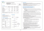
Brief procedure:
Tissue Sample
Homogenization
Sample amount and yield
Sample
Animal cells 6
(up to 5 x 10 )
Yield
(µg)
NIH/3T3
HeLa
COS-7
LMH
10
15
30
12
Animal Tissue
(Mouse/rat)
(up to 30 mg)
Embryo
Heart
Brain
Kidney
Liver
Spleen
Lung
Thymus
Bacteria
E. coli
B. subtilis
Cultured cells
Concentration &
Resuspension
7a. Add 250 µl of Wash Buffer 1 to the FARB Mini Column, centrifuge at full speed for 1 min.
Discard the flow-through and return the FARB Mini Column back to the Collection Tube.
7b. Add 60 µl of RNase-free DNase 1 solution (0.5U/ul, not provided) to the membrane center of
the FARB Mini Column.
Place the column on the benchtop for 15 min.
7c. Add 250 µl of Wash Buffer 1 to the FARB Mini Column, centrifuge at ful speed for 1 min.
Discard the flow-through and return the FARB Mini Column back to the Collection Tube.
7d. After DNase 1 treatment, proceed to step 9.
Lysis (FARB Buffer)
Recommended amount
of sample used
6
1 x 10 cells
centrifuge
Filtration
8. Add 500 µl of Wash Buffer 1 to the FARB Mini Column, centrifugeat at full speed for 1 min.
Discard the flow-through and return the FARB Mini Column back to the Collection Tube.
9. Add 750 µl of Wash Buffer 2 to the FARB Mini Column, centrifuge at full speed for 1 min.
Discard the flow-through and return the FARB Mini Column back to the Collection Tube.
-- Note: Make sure that ethanol has been added into Wash Buffer 2 when first use.
10. Repeat step 9 for one more washing.
11. Centrifuge the FARB Mini Column at full speed for an additional 3 min to dry the FARB Mini Column.
-- Important Step! This step will avoid the residual liquid to inhibit subsequent enzymatic reaction.
12. Place the FARB Mini Column to a Elution Tube (provided, 1.5 ml microcentrifuge tube).
13. Add 40 ~ 100 µl of RNase-free ddH2O to the membrane center of the FARB Mini Column.
Stand the FARB Mini Column for 1 min.
-- Important Step! For effective elution, make sure that RNase-free ddH2O is dispensed on the membrane
center and is absorbed completely.
-- Important : Do not elute the RNA using RNase-free water less than suggested volume (< 40 µl). It will
lower the RNA yield.
14. Centrifuge the FARB Mini Column at full speed for 1 min to elute RNA.
15. Store RNA at -70C.
Add 70% ethanol
centrifuge
10 mg
25
10
10
30
50
35
15
45
centrifuge
9
1 x 10 cells
60
40
centrifuge
Yeast
S. cerevisiae 1 x 10 7 cells
7
(up to 5 x 10 )
25
1. Make sure everything is RNase-free when handling RNA.
2. Buffers provided in this system contain irritants. Wear gloves and lab coat when handling these buffers.
3. Caution: ß-mercaptoethanol (ß-Me) is hazardous to human health. perform the procedures involving ß-Me in
a chemical fume hood.
4. Add required volume of RNase-free ethanol (96~100%) to Wash Buffer 2 when first use.
5. All centrifuge steps are done at full speed (~18,000 x g) in a microcentrifuge.
6. Prepare RNase-free DNase 1 reaction buffer (1M NaCl, 10 mM MnCl2, 20 mM Tris-HCl, pH 7.0 at 25ºC)
and make the final concentration of DNase I to 0.5 U/μl.
Protocol: Isolation of Total RNA from Animal Cells
Preparation of Wash Buffer by adding ethanol (96 ~ 100%)
Ethanol volume for Wash Buffer 2
Important Notes:
RNA Binding
(FARB Mini Column)
optional step:
DNase I digestion
Washing
(Wash Buffer 1)
(Wash Buffer 2 x 2)
RNA Elution
(RNase-free water)
1
v 0515
2
Protocol: Isolation of Total RNA from Yeast
Protocol: Isolation of Total RNA from Animal Tissues
Please Read Important Notes Before Starting Following Steps.
Please Read Important Notes Before Starting Following Steps.
Additional equipment: liquid nitrogen & mortar
a rotor-stator homogenizer or a 20-G needle syringe
ß-Mercaptoethanol
70% RNase-free ethanol
A-1 Weight up to 30 mg of tissue sample. Grind the sample in liquid nitrogen to a fine powder with a mortar
and transfer the powder to a new microcentrifuge tube (not provided).
-- Note! Avoid thawing the sample during weighing and grinding.
A-2. Add 350 µl of FARB Buffer and 3.5 µl of ß-Mercaptoethanol. Homogenize the sample by using a rotor-stator
homogenizer or by passing the sample lysate through a 20-G needle syringe 10 times. Incubate at room
temperature for 5 min.
-- Important step: In order to release more RNA from the harder samples, it is recommended to homogenize
the sample by using suitable homogenize equipment, for example, with a rotot-stator
homogenizer.
A-3. Follow the Animal Cells Protocol starting from step 3.
(Alternative)
B-1. Place up to 30 mg of tissue sample to a microcentrifuge tube. Add 350 µl of FARB Buffer and
3.5 µl of ß-Mercaptoethanol and use provided micropestle to grind the tissue sample thoroughly.
B-2. Homogenize the sample by passing lysate through a 20-G needle syringe 10 ~ 20 times. Incubate at room
temperature for 5 min.
-- For the tissue samples having low cell amount and hard to disrupt, it is recommended to proceed A1-A3
step above.
B-3. Follow Animal Cells Protocol starting from step 3.
Additional requirment: ß-Mercaptoethanol
70% RNase-free ethanol
Enzymatic disruption: Lyticase or zymolase
Sorbitol buffer (1 M sorbitol; 100 mM EDTA; 0.1% ß-ME )
30 °C water bath or heating block
Mechanical disruption: 2 ml screw centrifuge tube
Acid-washed glass beads, 500 ~ 700 µm
7
1. Collect up to 5 x 10 of yeast culture by centrifuge at 5,000 x g for 10 min at 4 °C. Remove all the supernatant.
2A. Enzymtic disruption:
2A-1: Resuspend the cell pellet in 600 µl sorbitol buffer (1 M sorbitol; 100 mM EDTA; 0.1% ß-ME) (not provided).
Add 200 U zymolase or lyticase and incubate at 30 °C for 30 min.
--Note! Prepare sorbitol buffer just before use.
2A-2. Centrifuge at 300 x g for 5 min to pellte the spheroplasts. Remove all the supernatant.
2A-3. Add 350 µl of FARB Buffer and 3.5 µl of ß-Mercaptoethanol to the pellet. Vortex vigorously to disrupt
the spheroplasts for 1 min. Incbuate sample mixture at room temperature for 5 min.
2B. Mechanical disruption:
2B-1. Add 350 µl of FARB Buffer and 3.5 µl of ß-Mercaptoethanol to the pellet and vortex vigorously to
resuspend the cells completely.
2B-2. Transfer the sample mixture to a 2 ml screw centrifuge tube and add 250 mg of acid-washed glass
beads (500 ~ 700 nm) and vortex vigorously for 15 min to disrupt the cells.
3. Follow Animal Cells Protocol starting from step 5.
Protocol: Isolation of Total RNA from Paraffin-embedded tissue
Protocol: Isolation of Total RNA from Bacteria
Please Read Important Notes Before Starting Following Steps.
Additional equipment: xylene & ethanol (96~100%)
liquid nitrogen & mortar
a rotor-stator homogenizer or a 20-G needle syringe
ß-Mercaptoethanol
70% RNase-free ethanol
Please Read Important Notes Before Starting Following Steps.
Additional requirment: ß-Mercaptoethanol
70% RNase-free ethanol
30 °C water bath or heating block
2 ml screw centrifuge tube
Lysozyme reaction solution: (10mg/ ml lysozyme; 20mM Tris-HCl, pH 8.0; 2mM EDTA;
1.2% Trition)
Acid-washed glass beads, 500 ~ 700 µm
9
1. Transfer up to 1x10 cells well-grown bacterial culture to a 2 ml screw centrifuge tube.
8
-- Note! Make sure the amount of total RNA harvested from sample do not excess the column’s binding
capacity (100 µg) when estimate the sample size. Too much sample will make cell lysis
incompletely and lead to lower RNA yield and purity. If RNA amount is hard to determin on
some species, using ≤ 5 x 10 cells as the starting sample size.
2. Descend the bacterial cells by centrifuge at full speed (~18,000 x g) for 2 min at 4 °C. Remove all the
supernatant.
3. Add 100 µl of lysozyme reaction solution. Pipet up and down to resuspend the cell pellet and incubate
at 37°C for 10 min.
4. Add 350 µl of FARB Buffer and 3.5 µl of ß-Mercaptoethanol.
5. Add 250 mg of acid-washed glass beads (500 ~ 700 nm) and vortex vigorously for 5 min to disrupt the cells.
6. Centrifuge at full speed (~18,000 x g) for 2 min to spin down insoluble material. Transfer the supernatant
to a microcentrifge tube (not provided) and measure the volume of the clear lysate.
-- Note! Avoid pipetting any debris and pellet in the Collection Tube.
7. Follow Animal Cells Protocol starting from step 5.
1. Transfer up to 15 mg paraffin-embedded tissue sample to a microcentrifuge tube (not provided).
-- Remove the extra paraffin to minimize the size of the sample slice.
2. Add 0.5 ml xylene, mix well and incubate at room temperature for 10 min.
3. Centrifuge at full speed for 3 min. Remove the supernatant by pipetting.
4. Add 0.25 ml xylene, mix well and incubate at room temperature for 3 min.
5. Centrifuge at full speed for 3 min. Remove the supernatant by pipetting.
6. Repeat step 4 and step 5
7. Add 0.3 ml ethanol (96- 100 %) to the deparaffined tissue, mix gently by vortexing. Incubate at room temperature
for 3 min.
8. Centrifuge at full speed for 3 min. Remove the supernatant by pipetting.
9. Repeat step 7 and step 8.
10. Follow Animal tissue Protocol starting from step 1 for sample disruption then follow Animal Cells protocol starting
from step 3.
Protocol: RNA clean up
Please Read Important Notes Before Starting Following Steps.
Additional equipment: xylene & ethanol (96~100%)
1. Trandfer 100 µl of RNA sample to a microcentrifuge tube (not provided).
-- If the RNA sample is less than 100 µl, add RNase-free water to make the sample volume to 100 µl.
2. Add 300 µl of FARB Buffer and 300 µl of RNase-free ethanol ( 96~100 %) and mix well by vortexing.
3. Place a FARB Mini Column to a Collection Tube and transfer the ethanol added sample mixture to the FARB Mini
Column. Centrifuge at full speed for 1 min and discard the flow-through and return the FARB Mini Column back to
the Collection Tube.
4. Follow Animal Cells Protocol starting from step 8.
3
4