1
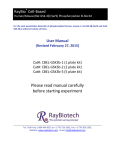
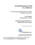
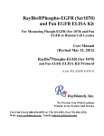
RayBio® Cell‐Based Human EGFR (Tyr1068) Phosphorylation ELISA Kit For the semi‐quantitative detection of phosphorylated human EGFR (Tyr1068) and total EGFR in adherent whole cell lines. User Manual (Revised July 16, 2015) Cat#: CBEL‐EGFR1068‐1 (1 plate kit) Cat#: CBEL‐EGFR1068‐2 (2 plate kit) Cat#: CBEL‐EGFR1068‐5 (5 plate kit) Please read manual carefully before starting experiment Tel: (Toll Free) 1‐888‐494‐8555 or +1‐770‐729‐2992; Fax: +1‐770‐206‐2393; Website: www.raybiotech.com Email: [email protected] Cell‐Based Human EGFR (Tyr1068) Phosphorylation ELISA Kit TABLE OF CONTENTS I. II. III. IV. V. VI. VII. VIII. IX. X. Introduction…………………………………………............................................... How It Works……………………………………………………………………………………. Reagents and Storage.…………....................…………...................... ... Additional Reagents Required…………...................................... Reagent Preparation …………........................................................... Assay Procedure ……………………………………………………………………......... Assay Procedure Summary ………………………………………............... Quality Control Data ………………………………………………..................... References …………………………………………………………………......................... Troubleshooting Guide ……........................................................... 1
RayBio® Cell‐Based EGFR (Tyr1068) ELISA Kit Protocol 2
3
4
4
5
6
9
10
12
13
I. INTRODUCTION Protein phosphorylation is instrumental in the regulation of protein activity within a cell. It plays important roles in the living cells including proliferation, differentiation and metabolism. A large number of protein kinases and phosphatases have been extensively investigated, and have been shown to be involved in signal transduction pathways. The RayBio® Cell‐Based Human EGFR (Tyr1068) Phosphorylation ELISA kit is a very rapid, convenient and sensitive assay kit that can monitor the activation or function of important biological pathways in cells. It can be used for measuring the relative amount of EGFR (Tyr1068) phosphorylation and screening the effects of various treatments, inhibitors (such as siRNA or chemicals), or activators in cultured human cell lines. By determining EGFR protein phosphorylation in your experimental model system, you can verify pathway activation in your cell lines without spending excess time and effort in preparing cell lysate and performing an analysis of Western Blot. In the Cell‐Based EGFR (Tyr1068) ELISA kit, cells are seeded into a 96 well tissue culture plate. The cells are fixed after various treatments, inhibitors or activators. After blocking, Anti‐Phospho‐EGFR (Tyr1068) or Anti‐EGFR (primary antibody) is pipetted into the wells and incubated. The wells are washed, and HRP‐conjugated anti‐rabbit IgG (secondary antibody) is added to the wells. The wells are washed again, a TMB substrate solution is added to the wells and color develops in proportion to the amount of protein. The Stop Solution changes the color from blue to yellow, and the intensity of the color is measured at 450 nm. See Figure 1 below for an illustration. 2
RayBio® Cell‐Based EGFR (Tyr1068) ELISA Kit Protocol II. HOW IT WORKS 1. Add cells
2. Treatment with stimulators
or inhibitors
3. Fixing and blocking
5. HRP-conjugated secondary
antibody
6. Develop with substrate
4. Anti-phospho-protein antibody
or anti-pan-protein antibody
+TMB
Fig.1. Cell-Based protein phosphorylation procedure
3
RayBio® Cell‐Based EGFR (Tyr1068) ELISA Kit Protocol Color
III. REAGENTS AND STORAGE Store entire kit at ≤ ‐20 °C immediately upon arrival. Kit must be used within ITEM COMPONENT 1 PLATE KIT 2 PLATE KIT A B C D E F Uncoated 96‐Well Microplate 20X Wash Buffer A Concentrate 20X Wash Buffer B Concentrate Fixing Solution 30X Quenching Buffer Concentrate 5X Blocking Buffer Concentrate 1000X Rabbit Anti‐phospho (Tyr1068) EGFR Concentrate 1000X Rabbit Anti‐EGFR Concentrate 2000X HRP Conjugated Anti‐Rabbit IgG Concentrate TMB Substrate Stop Solution** 1 plate 2 plates G H I‐1 J K 1 vial (30 ml) 1 vial (30 ml) 1 vial (30 ml) 1 vial (2 ml) 1 vial (20 ml) 1 vial (6 µl) 2 vials (6 µl/ea) 1 vial (6 µl ) 2 vials (6 µl/ea) 1 vial (10 µl ) 2 vials (10 µl/ea) 1 vial (12 ml) 2 vials (12 ml/ea) 1 vial (14 ml) STORAGE AFTER INITIAL THAW*
Room Temperature 2‐8 °C 2‐8 °C (1 month) ‐20 °C 2‐8 °C the 6 month expiration date. Avoid repeated freeze‐thaw cycles. *For up to 3 months (unless otherwise stated) or until expiration date. **Contains 0.2 M Sulfuric Acid IV. ADDITIONAL MATERIALS REQUIRED 1.
2.
3.
4.
5.
6.
7.
8.
9.
A model cell line, protein tyrosine kinase inhibitors, growth factors or cytokines Microplate reader capable of measuring absorbance at 450 nm 37 oC incubator Precision pipettes to deliver 2 μl to 1 ml volumes Adjustable 1‐25 ml pipettes for reagent preparation 100 ml and 1 liter graduated cylinders Absorbent paper Distilled or deionized water Orbital shaker or oscillating rocker 4
RayBio® Cell‐Based EGFR (Tyr1068) ELISA Kit Protocol V. REAGENT PREPARATION NOTE: NOTE: Thaw all reagents to room temperature immediately before use. If wash buffers contain visible crystals, warm to room temperature and mix gently until dissolved. Briefly centrifuge (~1,000g) ITEMS G, H, and I before opening to ensure maximum recovery. ITEM COMPONENT A Uncoated 96‐Well Microplate B 20X Wash Buffer A Concentrate C 20X Wash Buffer B Concentrate D Fixing Solution 30X Quenching Buffer Concentrate E EXAMPLE
No Preparation
N/A Dilute each 20‐fold with distilled or deionized water 25 ml of concentrate + 475 ml of water = 500 ml of 1X working solution No Preparation
Dilute 30‐fold with 1X Wash Buffer A
Dilute 5‐fold with distilled or deionized water
N/A 1 ml of concentrate + 29 ml of wash buffer = 30 ml of 1X working solution
20 ml of concentrate + 80 ml of water = 100 ml of 1X working solution
Dilute each 1000‐fold with 1X Blocking Buffer 6 µl of concentrate + 5594 µl of 1X Blocking Buffer = 6 ml of 1X working solution 5X Blocking Buffer Concentrate G 1000X Rabbit Anti‐phospho (Tyr1068) EGFR Concentrate H 1000X Rabbit Anti‐EGFR Concentrate I‐1 2000X HRP Conjugated Anti‐Rabbit IgG Concentrate Dilute 2000‐fold with 1X Blocking Buffer 10 µl of concentrate + 19990 µl of 1X Blocking Buffer = 20 ml of 1X working solution J K TMB Substrate Stop Solution No Preparation N/A PRIMARY ANTIBODY F SECONDARY ANTIBODY PREPARATION 5
RayBio® Cell‐Based EGFR (Tyr1068) ELISA Kit Protocol VI. ASSAY PROCEDURE: NOTE: ALL incubations and wash steps must be performed under gentle rocking or rotation (~1‐2 cycles/sec). 1. Design your experiment. For example, see Figure 2 below. EGF (ng/ml) 0 20 100 0 20 100 0 20 100 0 20 100 0 min 5 min 10 min 20min Anti‐Phospho‐ Anti‐EGFR Inhibitor + Inhibitor + Anti‐EGFR EGFR Anti‐Phospho‐ (Tyr1068) EGFR (Tyr1068) Fig. 2. Example of plate layout for RayBio® cell‐based assay OPTIONAL: If seeding HUVECs, HMEC‐1 or other loosely attached cells, coat the Uncoated 96‐Well Microplate (ITEM A) by adding 100 μl poly‐L‐Lysine (Recommended Sigma Aldrich, Cat#: P4832) into each well and then follow manufacturer’s instructions. A pre‐coated CellBIND® microplate or other poly‐lysine treated tissue culture plate may be used in place of Item A. 6
RayBio® Cell‐Based EGFR (Tyr1068) ELISA Kit Protocol 2. Seed 100 μl of 20,000 cells into each well of the Uncoated 96‐Well Microplate (ITEM A) provided and incubate overnight at 37oC with 5% CO2. NOTE: The optimal cell number used will vary on the cell line and the relative amount of protein phosphorylation. More or less cells may be used but this must be determined empirically. NOTE: The cells can be starved ~4‐24 hours (depending on cell line) prior to treatment with inhibitors or activators. 3. Apply various treatments, inhibitors (such as siRNA or chemicals) or activators according to manufacturer’s instructions and incubate for the desired time points. NOTE: It is recommended to dissolve inhibitors or activators into serum‐free cell culture medium before treating the cells (unless otherwise stated in the manufacturer’s instructions.) 4. Discard the cell culture medium by flipping the microplate upside down and gently tapping the bottom of the microplate over a sink. 5. Wash by pipetting 200 μl of the prepared 1X Wash Buffer A (see Section V. Reagent Preparation) into each well. Discard the wash buffer (same as step 4) and wash 2 more times for a total of 3 washes using fresh wash buffer each time. After the final wash, gently blot the microplate onto a paper towel to remove any excess/remaining buffer. NOTE: To avoid cell loss, do not pipette directly onto the cells. Instead, gently dispense the liquid down the wall of cell culture wells. Also avoid the use of vacuum suction or too forcefully tapping the microplate when discarding any solution. 6. Add 100 μl of Fixing Solution (ITEM D) into each well and incubate for 20 minutes at room temperature. NOTE: The fixing solution is used to permeabilize the cells. 7. Repeat wash step 5. 7
RayBio® Cell‐Based EGFR (Tyr1068) ELISA Kit Protocol 8. Add 200 μl of the prepared 1X Quenching Buffer (ITEM E) into each well and incubate 20 minutes at room temperature. NOTE: The quenching buffer is used to minimize the background response. 9. Wash 4 times with 1X Wash Buffer A. 10. Add 200 μl of the prepared 1X Blocking Buffer (see Section V. Reagent Preparation) into each well and incubate for 1 hour at 37oC. 11. Wash 3 times with the prepared 1X Wash Buffer B (ITEM C). NOTE: If needed, the microplate may be stored at ‐80 oC for several days after this wash. 12. Add 50 μl of the prepared 1X primary antibody (ITEM G or H) into each corresponding well and incubate for 2 hours at room temperature. 13. Repeat step 11. 14. Add 50 μl of 1X HRP Conjugated secondary antibody (ITEM I) into each well and incubate for 1 hour at room temperature. 15. Wash 3 times with 1X Wash Buffer B. 16. Add 100 μl of the TMB Substrate (ITEM J) into each well and incubate for 30 minutes at room temperature in the dark. 17. Add 50 μl of the Stop Solution (ITEM K) into each well. Read at 450 nm immediately. 8
RayBio® Cell‐Based EGFR (Tyr1068) ELISA Kit Protocol VII. ASSAY PROCEDURE SUMMARY 1. Seed 20,000 cells into each well and incubate overnight. 2. Apply various treatment, inhibitors or activators according to manufacturer’s instructions. 3. Add 100 μl of Fixing Solution into each well and incubate for 20 minutes at room temperature. 4. Add 200 μl of prepared 1X Quenching Buffer and incubate for 20 minutes at room temperature. 5. Add 200 μl of prepared 1X Blocking Buffer and incubate for 1 hour at 37oC. 6. Add 50 μl of prepared 1X primary antibody to each well and incubate for 2 hours at room temperature. 7. Add 50 μl of prepared 1X HRP Conjugated secondary antibody and incubate for 1 hour at room temperature. 8. Add 100 μl TMB Substrate and incubate 30 minutes at room temperature. 9
RayBio® Cell‐Based EGFR (Tyr1068) ELISA Kit Protocol 9. Add 50 μl Stop Solution to each well. Read at 450 nm immediately. VIII. QUALITY CONTROL DATA Representative results of Cell‐Based EGFR (Tyr1068) are shown below: 1. Seeded 20,000 A431 cells into appropriate wells of the microplate. Cells were incubated at 37oC in 5% CO2 overnight. 2. Added 50 μl of different concentrations of stimulators (rhEGF concentration for A431 cells: 0, 20 or 100 ng/ml in serum free DMEM) to appropriate wells (shown below). Then incubated for 10 min at 37oC. 3. Discarded the solution and wash 3 times with 1X Wash Buffer A (200 μl each) immediately. Then tapped the plate upside down to remove all of excess wash buffer. The protocol was then followed as stated. 10
RayBio® Cell‐Based EGFR (Tyr1068) ELISA Kit Protocol Anti-Phospho EGFR (Tyr1068)
Anti-EGFR
0.8
OD=450 nm
0.6
0.4
0.2
0.0
EGF concentrations
0
20
100
(ng/ml)
Fig. 3. A431 cells were stimulated by different
concentrations of EGF for 10 min at 37oC
hEGF 0 10 0 10 (Min) Anti‐EGFR Anti‐phospho‐EGFR (Tyr1068) Fig. 4. Western blot analysis of extracts from 100 ng/ml hEGF treated A431 cells. Phospho‐EGFR (Tyr1068) and EGFR antibodies were used in both detection assays. 11
RayBio® Cell‐Based EGFR (Tyr1068) ELISA Kit Protocol IX. REFERENCES: 1.
2.
3.
4.
5.
Hubbard, S.R. et al. (1994) Nature 372, 746–754. Hackel, P.O. et al. (1999) Curr. Opin. Cell Biol. 11, 184–189. Levkowitz, G. et al. (1999) Mol. Cell 4, 1029–1040. Zwick , E. et al. (1999) Trends Pharmacol. Sci. 20, 408–412. Biscardi, J.S. et al. (1999) J. Biol. Chem. 274, 8335–8343. 12
RayBio® Cell‐Based EGFR (Tyr1068) ELISA Kit Protocol X: TROUBLESHOOTING GUIDE Problem 1. Low signal Cause 1. Improper storage of the ELISA kit 2. Improper dilution 3. Cells drop off from the wells 2. High background 1. Inadequate washing 3. Large CV 2. Too much cells Solution 1. Store the kit according to manual instructions. Keep substrate solution in dark. 2. Ensure correct preparation of antibody and reagents. 3. Some of treatments may make cells drop off the wells. Reduce inhibitor or activator concentration. 1. Be sure to remove all of washing solution and follow the recommendation for washing. 2. Reduce the cell number. 1. Inaccurate pipetting 2. Remaining wash buffer in the well 3. Cells drop off from the wells 1. Check pipette. 2. Remove all of wash buffer. 3. Please don’t directly face the cells with tips when adding reagents or wash buffer. 13
RayBio® Cell‐Based EGFR (Tyr1068) ELISA Kit Protocol NOTES: 14
RayBio® Cell‐Based EGFR (Tyr1068) ELISA Kit Protocol NOTES: 15
RayBio® Cell‐Based EGFR (Tyr1068) ELISA Kit Protocol NOTES: 16
RayBio® Cell‐Based EGFR (Tyr1068) ELISA Kit Protocol This product is for research use only. ©2004 RayBiotech, Inc. 17
RayBio® Cell‐Based EGFR (Tyr1068) ELISA Kit Protocol