Download Forensic Biology: Screening Standard Operating Procedures
Transcript
Forensic Biology: Screening
Standard Operating Procedures
Forensic Analysis Division
Standard Operating Procedures: Biology
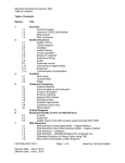
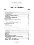
Table of Contents
Table of Contents
1. Overview
2. Facilities
2.1 Work Areas
2.2 Contamination
2.3 Safety
3. Equipment Quality Control and Maintenance
3.1 Maintenance and Calibration
3.2 Equipment
4. Quality Assurance
4.1 Chemical and Reagent Labels
4.2 Quality Control Checks
4.3 Logbook
4.4 Proficiency Testing
5. Evidence Evaluation and Handling
5.1 Case Acceptance and Evaluation
5.2 Evidence Handling
6. Blood Detection
6.1 Phenolphthalein Test Kit
6.2 Luminol
6.3 ABAcard® HemaTrace® Test Devices
6.4 References
7. Semen Detection
7.1 Alternate Light Source
7.2 Acid Phosphatase Spot Test
7.3 Body Fluid Extraction
7.4 Microscopic Spermatozoa Examination
7.5 SERATEC® PSA (p30) Semiquant Test
7.6 Guidelines for Sexual Assault Cases When Contact or Digital Penetration is Alleged
7.7 References
8. Trace Observation/Collection
9. Contact DNA Collection
10. Portioning for DNA Analysis
11. Case Records
11.1 Examination Documentation
11.2 Reports
11.3 Examination Counts
12. Abbreviations
13. Chelex Extraction of DNA
14. Plexor HY Quantification
FAD-BIO-SOP-TOC.1
Revision Date: May 2, 2014
Effective Date: June 1, 2014
Page 2 of 2
Issued by Technical Leader
Forensic Biology: Screening
Overview
Forensic Analysis Division
Standard Operating Procedures: Biology
1
Overview
Overview
The Biology Standard Operating Procedure (SOP) manual specifies procedures for routine
examinations and analyses of biological evidence for human identification. It is approved for
use in the Biology section of the laboratory. Within the scope of that purpose, it is intended to
ensure effective and efficient use of the laboratory facilities for the benefit of all user agencies.
It incorporates the quality assurance elements necessary to ensure the reliability and uniformity
of analyses and reported conclusions.
Each approved revision of this manual will be version-controlled and archived for retrieval by
date of authority. Only the approved revision in effect at the time of analysis governs the
analysis. The official version of this controlled document shall be published on the laboratory’s
intranet webpage and can be viewed from network computers. Any deviation from accepted
protocol requires approval as outlined in the Methods section of the quality manual, and must be
documented in the case file.
This SOP is only one part of the policies and procedures that govern all work performed by the
Biology section. The Biology section will follow the requirements detailed herein, in addition to
the following:
• Parent Agency General Orders/Policy
•
Laboratory Quality Manual
•
Laboratory Safety Manual
•
Laboratory Biology Training Manual
FAD-BIO-SOP-1.1
Revision Date: May 2, 2014
Effective Date: June 1, 2014
Page 2 of 2
Issued by Technical Leader
Forensic Biology: Screening
Facilities
Forensic Analysis Division
Standard Operating Procedures: Biology
2
Facilities
2
Facilities
2
Facilities
The Biology section will be designed to provide adequate space and setup to perform casework
analysis. General facility requirements are described in the Quality Manual.
2.1
Work Areas
The Biology section will have space for evidence examination. The tasks performed in this area
can include: blood and semen fluid identification, swab collections, cutting of stains, portioning
of swabs and cuttings, microscopy and trace evidence collection.
2.2
Contamination
With the facility provided, setup and procedures will be designed to minimize the potential for
DNA contamination. Traffic in and through areas in which testing occurs will be minimized.
Prevention and Decontamination
10% bleach, or DNA Away where bleach may be harmful to instruments or equipment, will be
used to decontaminate utensils and workbenches. Other commercial decontaminants may be
used only if they have been demonstrated to destroy or inactivate DNA.
Wear disposable gloves during all testing and reagent preparation. Change gloves frequently
and whenever gloves may have become contaminated. Discard gloves when leaving a work
area. Use sterile, disposable pipette tips and microcentrifuge tubes. Change pipette tips
between samples. Centrifuge all liquid to the bottom of closed microcentrifuge tubes before
opening.
Clean work surfaces in the evidence examination areas thoroughly with a lab-approved
decontaminant prior to and at the end of each evidence examination session. Use disposable
bench paper whenever possible and change each time it comes in contact with evidence. Use a
clean cutting surface such as weighing paper or piece of paper for evidence. Protect supplies of
this paper from dust and other particulates or aerosols. Clean instruments (e.g., scalpels,
scissors, forceps) with decontaminant between evidence samples. To prevent contamination of
other standards or evidence, handle each piece of evidence one at a time. Liquid samples,
such as blood standards, and wet exhibits shall be allowed to air dry in a method that it will not
come into contact with other pieces of evidence.
Wear gloves when cleaning glassware and plastics. In general, clean glassware/plastics with
an appropriate soap, e.g., Liquinox or Alconox, and water. Rinse with deionized or distilled
water and allow to air-dry inverted. Glassware should be autoclaved. Plastic ware should be
placed under UV light for ~15 minutes. Store glassware and plastic ware after being autoclaved
or being cleaned by UV light.
FAD-BIO-SOP-2.1
Revision Date: May 2, 2014
Effective Date: June 1, 2014
Page 2 of 3
Issued by Technical Leader
Standard Operating Procedures: Biology
2
Facilities
2.3
Safety
There are biological and chemical hazards in the laboratory. Each lab employee is responsible
for familiarity with the Laboratory Safety Manual. Any incident or condition that occurs in or
under the control of the laboratory and that threatens the immediate or future health of any
individual must be immediately brought to the attention of the section supervisor and a
laboratory safety officer. Corrective action related to safety incidents will be defined by
laboratory management.
FAD-BIO-SOP-2.1
Revision Date: May 2, 2014
Effective Date: June 1, 2014
Page 3 of 3
Issued by Technical Leader
Forensic Biology: Screening
Equipment Quality Control and Maintenance
Forensic Analysis Division
Standard Operating Procedures: Biology
3
Equipment Quality Control and Maintenance
3
Equipment Quality Control and Maintenance
3.1
Maintenance and Calibration
In order to provide and maintain the quality of the work provided in the Biology section, it is necessary to
ensure laboratory equipment is in good working order. Routine quality control and maintenance accomplishes
this. The calibration intervals listed below are generally considered to be the minimum appropriate in each
case, provided that the equipment is of good quality and of proven stability and the laboratory has both the
equipment capability and expertise to perform adequate internal checks. More frequent checks are
acceptable. If there is any question concerning the reliability of an instrument or piece of equipment, a
maintenance check should be performed immediately.
Full maintenance, calibration records, and repair activities shall be maintained and recorded in an equipment
calibration and maintenance log. This log will include at a minimum: the date, activity, laboratory personnel
performing or overseeing the activity, non-crime laboratory technician(s) performing or overseeing the activity,
and a record of quality control checks performed to verify operation prior to returning a piece of equipment to
casework use. The section supervisor and QA Manager are responsible for ensuring all systems are checked
and maintained as required. Whenever practical, equipment that requires calibration will be labeled with the
date when last calibrated and the date or expiration criteria when recalibration is due.
Critical equipment is any piece of equipment that must be maintained in a proper working order to ensure the
reliability of results produced. Some critical equipment and associated maintenance requirements can be
found in the Quality Manual. Additional equipment and maintenance requirements are included here.
Equipment Operation Manuals
Equipment operation manuals will be readily available to each examiner approved to use the equipment.
3.2
Equipment
Ultrasonic Cleaner
Ultrasonic cleaners use ultrasonic energy in the form of sound waves to create a mechanical “scrubbing” action
to loosen debris from all surfaces that the solution touches.
This equipment’s condition is routinely maintained for the Biology section’s procedures. The water should be
clear and clean with no evidence of bacterial/fungal growth or rust. The following procedure should be
performed as needed: Wash the inside of the ultrasonic cleaner with detergent and rinse well with water. Fill
bath with the appropriate quantity of diH 2 O water.
Mini-centrifuges
Mini-centrifuges are bench top, unrefrigerated centrifuges that have been designed for quick spin-downs
from tube walls and caps of tubes. These mini-centrifuges are equipped with a circular, one sized rotor. The
maximum speed is 6000 rpm. The relative centrifugal force can be determined as outlined in the
manufacturer’s instructions, if required.
FAD-BIO-SOP-3.1
Revision Date: May 30, 2014
Effective Date: June 1, 2014
Page 2 of 8
Issued by Technical Leader
Standard Operating Procedures: Biology
3
Equipment Quality Control and Maintenance
Please consult the appropriate equipment manual for specific instructions on maintenance and operation of the
mini-centrifuge. These centrifuges will primarily be used for removing liquid from the cap of a microcentrifuge
tube. They will not be used for pelleting cellular material.
Thermometers
All thermometers must be either calibrated or their accuracy must be verified to ensure the temperature is
being accurately measured.
NIST-traceable thermometers must be purchased or re-certified at least once every two years. A NISTtraceable thermometer certified for two years and used for conducting performance checks on equipment shall
require the annual performance check. A NIST-traceable thermometer certified for two years that is not used
for conducting performance checks does not require the annual performance checks and may be used until the
certification expires. A NIST-traceable thermometer to be used beyond its certification date shall be recertified
or be subject to the annual performance check requirements.
The minimum requirements of a performance check of a thermometer used for performing performance checks
may be accomplished: (1) through certification by an outside vendor; or (2) in-house by the comparison of one
or more temperature readings at various time intervals against another NIST-traceable thermometer.
Thermometers that are not NIST-traceable must be verified annually by comparison against a NIST-traceable
thermometer. Any deviation from the NIST-traceable thermometer will be noted and all other readings
adjusted by the same amount, and in the same direction for each thermometer. If the deviation is greater than
3°C, the thermometer should be serviced or replaced.
Thermometers should be handled carefully to avoid breakage. Thermometers may be wiped clean to facilitate
easy and accurate readings. If any discontinuities are detected, repair or replace the thermometer.
Autoclaves
Autoclaves are used to sterilize solutions, glassware, and instruments by subjecting them to high pressure and
high heat simultaneously. Autoclaves also may be used to sterilize biohazard trash prior to discarding. Please
consult the instructions on the outside of the unit for specific information on operation of the autoclave.
Whenever an item is autoclaved, a small piece of autoclave tape must be adhered to the item, to verify correct
functioning of the autoclave. Use proper sterilizer-loading procedures when placing materials in sterilizer
chamber. Those include: all solid containers/ instruments must be placed so that water or air will not be
trapped in them and sufficient space must be left between items to allow steam circulation. The temperature
gauge should read 1200C at the start and during the appropriate cycle.
If the autoclave is not functioning properly, a qualified service technician may repair the instrument.
Alternate Light Source (ALS) Machines
ALS machines are used to presumptively identify semen in forensic casework. The fan air filter, optical filters,
and lenses should be cleaned on a regular basis (approximately every 6 months), where applicable. Please
consult the appropriate equipment manuals for more specific instructions on maintenance and operation of the
ALS machines.
Swab Drying Box E025
FAD-BIO-SOP-3.1
Revision Date: May 30, 2014
Effective Date: June 1, 2014
Page 3 of 8
Issued by Technical Leader
Standard Operating Procedures: Biology
3
Equipment Quality Control and Maintenance
Swab drying boxes are used to dry swabs created in the lab during forensic casework. Before and after each
use, the box should be wiped inside and out with a 10% bleach solution or other lab-approved decontaminant
and dried with a soft cloth. Do not autoclave, immerse, or abrade the unit.
Refrigerators and Freezers
Please refer the Laboratory Quality Manual for refrigerator and freezer maintenance. Reagents and evidence
will be stored separately, either in separate cooling units or in separate space within those units.
Pipettes
Volumetric pipettes are used to accurately and precisely measure the volume of a solution to be delivered. The
accuracy of the measurement is dependent upon the correct technique of the user as well as the proper
maintenance and performance of the pipette. The pipette should be able to provide accuracy, precision,
repeatability and reproducibility over the full range of the pipette’s capabilities. The Biology section is supplied
with pipettes that cover the volume range from 0.1 - 1000 µl. Calibration will be performed at least annually on
these instruments. When a pipette is determined to be performing improperly, it will be returned to the
manufacturer, or another qualified repair technician, so that the problem may be identified and corrected.
Quality control data should include the type of pipette, volume range, model, and serial number.
Procedure
1. Set the volume of the pipette using the knob in the pipetting push-button.
2. Firmly attach the appropriate tip (per manufacturer’s recommendations) to the pipette.
3. Press the push-button to the first positive stop.
4. Holding the pipette vertically, immerse the tip into the sample liquid per manufacturer’s
recommended depth.
5. Release the push-button slowly and smoothly to aspirate the sample.
6. Pausing for a moment, withdraw the tip from the liquid.
7. Depending on the sample, it may be necessary to pre-rinse the tip before aspirating the sample.
8. To dispense the sample, place the tip into the transfer vessel and press the push-button to the
first stop.
9. Pausing for a moment, press the push-button to the second stop to ‘blowout’ or expel any
remaining liquid.
10. Slowly, release the push-button
11. Eject the tip by pressing the ejector button.
Performance Verification/Maintenance
FAD-BIO-SOP-3.1
Revision Date: May 30, 2014
Effective Date: June 1, 2014
Page 4 of 8
Issued by Technical Leader
Standard Operating Procedures: Biology
3
Equipment Quality Control and Maintenance
1. Each pipette should be externally calibrated and certified by an approved calibration vendor
annually.
2. Internal pipette verifications should be performed about 6 months after the external calibration.
3. Pipette performance verification should be documented.
4. To check for accuracy, room temperature deionized water should be pipetted into a weighing
vessel on an analytical balance.
5. The pipette should be checked using 3-points over the full range with 5 replicates at each
volume.
6. Each pipette should fall within the verification tolerance ranges according to Addendum 3A
before it may be used for casework.
7. If a pipette fails a performance verification check, it should be recalibrated. Calibrations are not
performed in-house.
8. Follow manufacturer’s instructions for troubleshooting maintenance if needed.
9. If an analyst has reason that a pipette is not working properly they must:
a. Perform a pipette verification and if the pipette is not in proper working order:
b. Clearly mark the pipette “OUT OF SERVICE”.
c. Inform the section manager. No laboratory case work will be performed using the pipette
until the problem is corrected.
d. Repair or send out the pipette for repairs
e. Verify that the pipette is working correctly and falls with-in the proper tolerances
following routine repair, maintenance or calibration.
f.
Maintain the appropriate documentation and update the log book.
Water Filtration System
Water is used in reagents that are prepared in the laboratory. Therefore, it is necessary to ensure only high
quality, reagent grade water is being utilized. Generally, 15MΩ-cm or greater deionized water is sufficient for
the reagents utilized in our laboratory.
Water quality should be checked on a weekly basis. The following procedure will be utilized:
1. Turn on the deionized water.
2. Allow water to run for 1-2 minutes.
FAD-BIO-SOP-3.1
Revision Date: May 30, 2014
Effective Date: June 1, 2014
Page 5 of 8
Issued by Technical Leader
Standard Operating Procedures: Biology
3
Equipment Quality Control and Maintenance
3. Record both the MΩ-cm reading and the temperature reading displayed on the resistivity monitor in the
Water Quality logbook.
4. If reading is below 15MΩ-cm, call for service. A qualified service technician may repair the system. Label
the system as being “out of service” until the appropriate service is performed and the instrument is
functioning properly.
Balances
Please refer to the Laboratory Quality Manual for the balance calibration schedule.
FAD-BIO-SOP-3.1
Revision Date: May 30, 2014
Effective Date: June 1, 2014
Page 6 of 8
Issued by Technical Leader
Standard Operating Procedures: Biology
3
Equipment Quality Control and Maintenance
Addendum 3A
Acceptable Check (uL)
Average needs to be
between these values
Volumetric
Pipette Range
Percent
0.5-10
5
2.5
2.375
2.625
3
7.5
7.275
7.725
3
10
9.7
13
5
2.5
2.375
2.375
3
7.5
7.275
7.725
3
10
9.7
10.3
3
12.5
12.125
12.875
3
37.5
36.375
38.625
3
50
48.5
51.5
3
5
4.85
5.15
3
15
14.55
15.45
1-10
5-50
2-20
10-100
20-100
20-200
40-200
50-200
100-1000
Check
Volume (uL)
3
20
19.4
20.6
3
25
24.25
25.75
3
75
72.75
77.25
3
100
97
103
3
25
24.25
25.75
3
75
72.75
77.25
3
100
97
103
3
50
48.5
51.5
3
150
145.5
154.5
3
200
194.0
206
3
50
48.5
52
3
150
145.5
154.5
3
200
194.0
206
3
50
48.5
51.5
3
150
145.5
154.5
3
200
194
206
3
250
242.5
257.5
3
750
727.5
772.5
3
1000
970
1030
FAD-BIO-SOP-3.1
Revision Date: May 30, 2014
Effective Date: June 1, 2014
Page 7 of 8
Issued by Technical Leader
Standard Operating Procedures: Biology
3
Equipment Quality Control and Maintenance
200-1000
3
250
242.5
257.5
3
750
727.5
772.5
3
1000
970
1030
FAD-BIO-SOP-3.1
Revision Date: May 30, 2014
Effective Date: June 1, 2014
Page 8 of 8
Issued by Technical Leader
Forensic Biology: Screening
Quality Assurance
Forensic Analysis Division
Standard Operating Procedures: Biology
4
Quality Assurance
4
Quality Assurance
In order to provide and maintain the quality of the work provided in the Biology section, it is
necessary to identify certain reagents as critical. Critical reagents are those that require testing
prior to use on evidentiary samples in order to prevent unnecessary loss of sample. All reagent
preparations and quality control testing must be documented on the appropriate worksheet or
log.
If any critical reagent does not pass all quality control checks, it may not be utilized in casework.
All inconsistencies will be documented and reported to the Section Supervisor. Problems that
cannot be resolved must be reported to the manufacturer.
Reagents and supplies that have passed their expiration dates may not be used on casework
samples. Outdated reagents may be used for training purposes only, but must be clearly
marked as such.
4.1 Chemical and Reagent Labels
Purchased chemicals and reagents will be marked on the container with the date received
and/or date opened. In general, the manufacturer’s labeling will be followed to determine
expiration dates of purchased chemicals and reagents. If no manufacturer expiration exists for
a purchased reagent, it will be considered expired 5 years from the date received and should be
marked on the container.
Prepared reagent labels will include the reagent name, initials of preparer and expiration date.
In general, most solutions prepared in the Biology section shall expire 1 year from the date of
preparation. However, the expiration date of the overall reagent will be no later than the
expiration date of the individual reagent with the nearest expiration date (unless otherwise
specified). Additional information may be documented in a reagent log. The lot number
contains the date prepared and the initials of the preparer, e.g. mmddyyinitials.
4.2 Quality Control Checks
The following shall be tested to confirm that they are working properly with a positive and
negative control each day used:
• Acid Phosphatase Spot Test
•
Alternate Light Source
•
Luminol mixture
•
Phenolphthalein test kit
The following shall be quality control checked to confirm they are working properly with each
new lot received:
• ABAcard® HemaTrace® kits
•
Phosphate Buffered Saline (1X)
•
SERI Christmas Tree Stain
FAD-BIO-SOP-4.2
Revision Date: August 26, 2014
Effective Date: September 1, 2014
Page 2 of 12
Issued by Technical Leader
Standard Operating Procedures: Biology
4
Quality Assurance
•
SERATEC® PSA Semiquant test devices
4.2.1 Acid Phosphatase (AP) Spot Test
Scope
AP Spot Test solution undergoes a color change (Brentamine reaction) in the presence of acid
phosphatase, which is found in highest concentration in semen. Instructions for use and
interpretation are in the AP Spot Test procedure.
Safety
This reagent presents the following hazards: may cause cancer; irritant to eyes, respiratory
system and skin; and corrosive (can cause burns). Wear gloves, lab coat, and mask during
preparation and use. Broken skin should be covered.
Equipment and Supplies
• AP Spot Test premixed powder – purchased
• Darkened or foil wrapped container for storage
• Scale
• Sterile diH 2 O
• Timer
• Known semen sample
• Known non-semen sample
Preparation
1. Measure 10mL of sterile diH 2 O in an appropriate container.
2. Add 0.26g of the purchased AP Spot Test premixed powder.
3. Mix until AP Spot Test premixed powder is completely dissolved in water.
Note: Volume may be smaller or larger than 10mL, but the amount of AP Spot Test
premixed powder added should be adjusted accordingly.
Prior to use each day, the prepared AP solution must be subjected to the appropriate in-house
quality control test as outlined below:
1. A known semen sample will be used as a positive control. This in-house-prepared
sample is typically presented on filter paper.
2. An untreated piece of filter paper or swab moistened with sterile diH 2 O will be used as a
negative control. Ideally, the negative control substrate mimics your sample substrate
as closely as possible.
3. Apply ~ 1 drop of prepared AP solution to both controls.
4. Observe samples for 60 seconds for a color change.
5. The known semen sample must give a strong purple color change indicating a positive
result.
FAD-BIO-SOP-4.2
Page 3 of 12
Issued by Technical Leader
Revision Date: August 26, 2014
Effective Date: September 1, 2014
Standard Operating Procedures: Biology
4
Quality Assurance
6. The untreated filter paper or sterile swab must have no color change indicating a
negative result.
The results of each control test must be recorded on every applicable serology worksheet.
Storage, Labeling, and Expiration
Store prepared solution in a darkened or foil wrapped container. Label with reagent name, lot
number, and expiration date. Reconstituted reagent is stable and sensitive for one day’s use –
make solution fresh daily. Store purchased powder frozen. Purchased powder may be used on
casework samples up to the expiration date stated on the bottle.
4.2.2 Alternate Light Source
Scope
An Alternate Light Source is specially designed for detection of forensic stains, fibers, and
fingerprints. Instructions for use and interpretation are in the Alternate Light Source procedure.
Safety
This instrument presents the following hazards: exposing the skin to the beam of light (directly
from the unit) can cause burns and other skin damage. It is essential that proper eye protection
be provided. Avoid looking directly at the light source, reflected, or refracted light. Remove all
unnecessary reflective surfaces from exam room. Wear goggles, gloves, lab coat, and other
proper laboratory attire.
Mini-Crimescope MCS-400 ALS: If the bulb fails (suddenly stops emitting light), it may leak
vapors for a few minutes. To be safe, immediately leave the room and let the unit run (fans) for
5-10 minutes until vapors are vented.
Equipment and Supplies
• ALS set at 400 or 455 nm
• Orange and/or yellow glasses
• Known semen sample
• Known non-semen sample
Prior to use each day, the ALS must be subjected to the appropriate in-house quality control test
as outlined below:
1. Turn on power switch and ensure the fans are working.
2. Wear the proper glasses.
3. Turn on lamp switch.
4. Darken the examination room.
5. Direct the ALS at the samples.
6. The known semen sample must fluoresce indicating a positive result.
7. The known non-semen sample must not fluoresce indicating a negative result.
FAD-BIO-SOP-4.2
Page 4 of 12
Issued by Technical Leader
Revision Date: August 26, 2014
Effective Date: September 1, 2014
Standard Operating Procedures: Biology
4
Quality Assurance
When ready to shut down the ALS, turn off lamp switch and wait for the unit to cool down (~5
minutes). Then turn off the power switch.
The results of each control test must be recorded on every applicable serology worksheet.
Storage and Expiration
Store equipment in a clean, dry atmosphere. The bulb should be replaced after 3-3.5 years of
non-regular usage.
4.2.3 Luminol
Scope
Catalytic tests for blood are based on the peroxidase-like activity exhibited by the heme group of
hemoglobin. In the case of luminol, the catalytic oxidation of the substrate compound produces
light. Instruction for use and interpretation are in the Luminol procedure.
Safety
This reagent presents the following hazards: luminol is an irritant. Sodium perborate and
sodium carbonate are toxic and irritating. Avoid breathing dust; do not get in eyes, on skin, or on
clothing. Avoid breathing sprayed solution. Wash hands after handling. Gloves must be worn
during testing. Broken skin should be covered.
Equipment and Supplies
• Luminol powder: 3-aminophthalhydrazide
• Sodium carbonate
• Sodium perborate
• Sterile diH 2 O
• Two darkened or foil wrapped spray bottles
• Scale
• Known blood sample
• Known non-blood sample
Preparation (Reagent A)
1. Measure 250 mL of diH 2 O.
2. Add 2.5 g of sodium carbonate.
3. Mix until all of the powder is dissolved.
4. Add 0.5 g of luminol powder.
5. Mix until all of the powder is dissolved.
FAD-BIO-SOP-4.2
Revision Date: August 26, 2014
Effective Date: September 1, 2014
Page 5 of 12
Issued by Technical Leader
Standard Operating Procedures: Biology
4
Quality Assurance
Preparation (Reagent B)
1. Measure 250 mL of diH 2 O.
2. Add 6.5 g of sodium perborate.
3. Mix until all of the powder is dissolved.
Mix equal volumes of Reagents A and B to create a luminol mixture. The mixture is unstable
and must be prepared fresh immediately before use; it is only stable for about one hour.
Prior to use, the luminol mixture must be subjected to the appropriate in-house quality control
test as outlined below:
1. Darken the examination room as completely as possible.
2. Spray luminol mixture onto a known blood sample (prepared in-house).
3. Spray luminol mixture onto a known non-blood sample (cotton swab or filter paper).
4. Observe samples for immediate luminescence.
5. The known blood sample must give an immediate luminescence indicating a positive
result.
6. The cotton swab or filter paper must not produce luminescence indicating a negative
result.
The results of each control test must be recorded on every applicable serology worksheet.
Storage, Labeling, and Expiration
The luminol powder, sodium carbonate and sodium perborate should be stored in a cool, dry,
well-ventilated area away from incompatible substances. The expirations for reagents A and B
(stored separately) are 8 weeks from date solutions were prepared. If stored, the labels must
include, at a minimum, "Luminol, Reagent A" or "Luminol, Reagent B" as appropriate, initials,
the date prepared, and the expiration date. Prior to each use, check the reagents for
precipitate. If a precipitate has formed in either reagent, ensure that the precipitate is back in
solution before use. Luminol mixture should be labeled with the appropriate lot number and
expiration date. The mixture will expire one hour after use.
4.2.4 Phenolphthalein Test Kit
Scope
PHT Test Kit Solutions B and C are used for presumptive blood identification by oxidizing
phenolphthalin (colorless in a basic solution such as the test reagent) to phenolphthalein (pink)
in the presence of heme and hydrogen peroxide. Instructions for use and interpretation are in
the phenolphthalein test kit procedure.
Safety
This reagent presents the following hazards:
FAD-BIO-SOP-4.2
Revision Date: August 26, 2014
Effective Date: September 1, 2014
Page 6 of 12
Issued by Technical Leader
Standard Operating Procedures: Biology
4
Quality Assurance
Solution B: Flammable. Handle with care. Harmful or fatal if swallowed, inhaled, or absorbed
through skin. Avoid contact with eyes, skin, and clothing. Light sensitive! Avoid light, heat,
sparks, and flame. Keep container tightly closed.
Solution C: Handle with care. May be harmful if swallowed, inhaled, or absorbed through skin.
Avoid contact with eyes, skin, and clothing. Avoid heat, sparks, and flame. Keep container
tightly closed.
Wear gloves, lab coat, and mask during preparation and use. Broken skin should be covered.
Equipment and Supplies
• Solution B
• Solution C
• Sterile diH 2 O
• Sterile cotton swab(s)
• Known blood sample
• Known non-blood sample
Prior to use each day, PHT Solutions B and C must be subjected to the appropriate in-house
quality control test as outlined below:
1. Rub a cotton swab moistened with sterile diH 2 O on a known bloodstain sample. This
swab will be used as a positive control.
2. An untreated cotton swab moistened with sterile diH 2 O will be used as a negative
control.
3. Apply ~1 drop of Solution B to each swab.
4. Apply ~1 drop of Solution C to each swab.
5. Observe the swabs for a color change.
6. The cotton swab with the known bloodstain sample must give an immediate pink color
change indicating a positive result.
7. The untreated cotton swab must have no color change indicating a negative result.
The results of each control test must be recorded on every applicable serology worksheet.
Storage, Labeling, and Expiration
PHT test kits can be stored at room temperature. Each analyst will have an aliquot of Solutions
B and C that will be placed in amber bottles stored at room temperature or refrigerated. Each
bottle will be labeled with appropriate reagent name, lot numbers, and expiration dates. The kits
may be used on casework samples up to the expiration date stated on the bottles.
FAD-BIO-SOP-4.2
Revision Date: August 26, 2014
Effective Date: September 1, 2014
Page 7 of 12
Issued by Technical Leader
Standard Operating Procedures: Biology
4
Quality Assurance
4.2.5 ABAcard® HemaTrace® Test Devices
Scope
ABAcard® HemaTrace® test kits are used to determine the presence of human blood in
suspected blood stains. Instructions for use and interpretation are in the ABAcard®
HemaTrace® procedure.
Safety
Wear gloves, lab coat, mask, and other proper laboratory attire. Broken skin should be covered.
Equipment and Supplies
• ABAcard® HemaTrace® test device(s)
• ABAcard® HemaTrace® extraction buffer
• Pipette and pipette tips
• Known blood sample
• Timer
Each new lot of ABAcard® HemaTrace® test kits must be subjected to the appropriate in-house
quality control test as outlined below:
1. Make a 1:106 dilution from a known human blood sample in extraction buffer provided in
kit.
2. Add 150μl of dilution to sample well “S” of one test device to be used as a positive
control.
3. Add 150 µl of extraction buffer from kit to sample well of second test device to be used
as a negative control.
4. Read results up to 10 minutes from application of sample.
5. The 1:106 dilution of known human blood must give a positive result. The extraction
buffer provided in the kit must give a negative result.
Storage, Labeling, and Expiration
The test devices must be stored at room temperature (below 82ºF) and must remain in the
sealed pouch until use. Label each test device with the name of the appropriate sample used.
The test kits may be used on casework samples up to the expiration date stated on the box.
4.2.6 Phosphate Buffered Saline (1X)
Scope
PBS is used as an extraction agent for serological stains. Instructions for use and interpretation
are in the Phosphate Buffered Saline procedure.
FAD-BIO-SOP-4.2
Revision Date: August 26, 2014
Effective Date: September 1, 2014
Page 8 of 12
Issued by Technical Leader
Standard Operating Procedures: Biology
4
Quality Assurance
Safety
This reagent presents the following hazards: causes severe eye, respiratory tract and skin
irritation. Avoid contact with skin and clothing. Wear gloves, lab coat, and other proper
laboratory attire. Broken skin should be covered.
Equipment and Supplies
• OmniPur 10X PBS Premixed Powder
• diH 2 O
• Graduated cylinder
• Two sterile containers for storage
Preparation (10X)
1. Measure 1 liter of diH 2 O.
2. Add 100g of 10X PBS Premixed Powder.
3. Mix until all of the powder is dissolved.
4. Autoclave solution.
Preparation (1X)
1. Measure 100mL of 10X PBS solution.
2. Measure 900 mL of sterile diH 2 O.
3. Mix 10X PBS and sterile diH 2 O.
Each new lot of 1X PBS must be subjected to an appropriate in-house quality control test with
the lot of SERATEC® PSA Semiquant test currently in use. See SERATEC® PSA Semiquant
test quality control procedure for further detail.
Storage, Labeling, and Expiration
1X and 10X PBS solutions can be stored at room temperature. Label each bottle with
appropriate concentration, lot number and expiration date. Expiration for both PBS
concentrations is one year from the date 10X PBS solution was prepared. Both concentrations
of PBS may be used on casework samples up to this expiration date.
4.2.7 SERI Christmas Tree Stain
Scope
SERI Christmas Tree Stain is a Kernechtrot-Picroindigocarmine differential biological stain that
assists in scanning slides for the presence of spermatozoa. The test consists of a two stain
method which dyes sperm heads red, tails green and epithelium green or blue with red nuclei.
Instructions for use and interpretation are in the SERI Christmas Tree Stain procedure.
FAD-BIO-SOP-4.2
Revision Date: August 26, 2014
Effective Date: September 1, 2014
Page 9 of 12
Issued by Technical Leader
Standard Operating Procedures: Biology
4
Quality Assurance
Safety
This reagent presents the following hazards: May cause irritation upon contact. Toxic. Do not
swallow. Do not inhale. Avoid contact with skin and eyes. Wear gloves and proper laboratory
attire. Broken skin should be covered.
Equipment and Supplies
• Microscope slide
• Known semen sample
• Pipette and pipette tips
• Heat block
• Christmas Tree Stain - Solution A and Solution B
• Timer
• Tap water
• 95% ethanol
• Compound microscope
Each new lot of SERI Christmas Tree Stain must be subjected to the appropriate in-house
quality control test as outlined below:
1. Pipette 10 µl of a neat in-house sample onto a glass slide. Fix smear by heating the
slide on the heat block until dry.
2. Stain the slide.
a. Cover the sample area on the slide with Solution A for 10 minutes.
b. Wash with tap water by gentle flooding.
c. Cover the sample area with Solution B for 15 seconds.
d. Wash with tap water by gentle flooding.
e. Flood the slide with 95% ethanol and allow the slide to dry for at least 5 minutes
on the heat block.
3. Examine the slide for the presence of spermatozoa at 1000X magnification. Sperm
heads must be stained red, and any observed tails must be stained green indicating a
positive result.
Storage and Expiration
The stain must be stored at room temperature. The stain may be used on casework samples
up to the expiration date stated on the bottles.
FAD-BIO-SOP-4.2
Revision Date: August 26, 2014
Effective Date: September 1, 2014
Page 10 of 12
Issued by Technical Leader
Standard Operating Procedures: Biology
4
Quality Assurance
4.2.8 SERATEC® PSA Semiquant Test Devices
Scope
SERATEC® PSA Semiquant test devices are used to indicate the possible presence of p30 in
suspected semen stains. Instructions for use and interpretation are in SERATEC® PSA
Semiquant test procedure.
Safety
Wear gloves, lab coat, mask, and other proper laboratory attire. Broken skin should be covered.
Equipment and Supplies
• SERATEC® PSA Semiquant test device(s)
• 1X PBS
• Pipette and pipette tips
• Known semen sample
• Timer
Each new lot of SERATEC® PSA Semiquant Test devices must be subjected to the appropriate
in-house quality control test as outlined below:
1. Make a 1:105 dilution from a known semen sample in 1X PBS.
2. Add 120 µl of dilution to sample well of one test device to be used as a positive control.
3. Add 120 µl of 1X PBS to sample well of second test device to be used as a negative
control.
4. Read results up to 10 minutes from application of sample.
5. The 1:105 dilution of known semen must give a positive result. The 1X PBS must give a
negative result.
6. Mark the stock solution bottle of 1X PBS with all lot numbers of SERATEC® PSA
Semiquant test devices quality control checked with that lot of 1X PBS.
Storage, Labeling, and Expiration
The test devices must be stored at room temperature and must remain in the sealed pouch until
use. Label each test device with the name of the appropriate sample used. The test devices
may be used on casework samples up to the expiration date stated on the sealed pouch.
4.3
Logbook
Reagent preparation logs should include the lot number, the reagent name, the initials of
preparer, and the expiration date. Quality control logs for ABAcard® HemaTrace® kits,
Phosphate Buffered Saline (1X), SERI Christmas Tree Stain, and SERATEC® PSA Semiquant
tests will also contain the test date, signature of the analyst performing the quality control, a
second reader signature, if applicable, and date and any supporting documentation necessary
to demonstrate the reagent met all of the standards.
FAD-BIO-SOP-4.2
Revision Date: August 26, 2014
Effective Date: September 1, 2014
Page 11 of 12
Issued by Technical Leader
Standard Operating Procedures: Biology
4
Quality Assurance
4.4
Proficiency Testing
Proficiency testing and review will follow the requirements of the quality manual. In addition, the
Quality Manager will maintain a copy of the analysis documentation for each proficiency test.
Proficiency tests will be analyzed and interpreted according to standard operating procedures
including technical review. Proficiency test participants will be notified of their final test results.
Analysts will enter into a proficiency test program within 6 months of being deemed competent
on any portion of casework analysis. Proficiency testing should include each technology to the
full extent to which analysts and technicians participate in casework.
Proficiency work is to follow as closely as possible that of normal casework. If symbols are
used in the reporting of data to the proficiency testing agency, they must be defined in the
results submission form. It is also advisable to include comments in the comments section of
the proficiency results form to explain certain results. For example, if a result is reported as
inconclusive, the reason should be presented in the comments section.
During the case file reviews, the proficiency results form (including the screening data and
comments sections), along with the case file, shall be reviewed by both the technical and
administrative reviewers to ensure proper transcription of results by the author of the results
from.
If performing quantification for proficiency samples, the analyst should create his/her own DNA
standards.
Only one proficiency results form will be completed for proficiency tests to ensure that only the
most complete form is submitted to the proficiency testing provider in order to be included in the
provider’s published external summary report. The screening analyst shall complete the
proficiency test results form if the test does not proceed to DNA analysis; the DNA analyst shall
complete the proficiency test results form if the test does proceed to DNA analysis.
FAD-BIO-SOP-4.2
Revision Date: August 26, 2014
Effective Date: September 1, 2014
Page 12 of 12
Issued by Technical Leader
Forensic Biology: Screening
Evidence Evaluation and Handling
Forensic Analysis Division
Standard Operating Procedures: Biology
5
Evidence Evaluation and Handling
5
Evidence Evaluation and Handling
The Biology section provides body fluid identification (semen and blood) and performs the
collection and preservation of trace evidence and possible contact DNA evidence.
5.1
Case Acceptance and Evaluation
Refer to the Quality Manual for procedures regarding the submission of evidence into the Crime
Laboratory. Before a case is worked, the case and the requested examinations will be
evaluated. The examiner should be aware of the requested examinations, and when possible,
the reason(s) for the requested analyses and the quality and quantity of the evidence.
In order to expedite casework, with the exception of sexual assault kits, it is recommended that
5-10 items of evidence should initially be screened for cases containing large volumes of
evidence. Emphasis should be placed on those items believed to be of the most significant
evidentiary value after consultation with the submitting party. Of the items screened, it is
recommended that a maximum of 5 items of evidence continue on to DNA analysis initially.
Additional items may be analyzed at a later time. Individual case needs may dictate that the
initial quantity of examined items is greater than the recommended range presented above.
When submitted in conjunction with a sexual assault kit, clothing evidence does not typically
have to be examined if the kit components yield sufficiently positive samples. (“Sufficiently
positive” is fluid and relevant to the case information.) Clothing should be examined if the
corresponding sexual assault evidence kit does not reveal any positive probative evidence, if
there are multiple suspects, or there is a recent consensual sexual partner. Good judgment
using the case information and/or the client’s specific requests should always supersede this
guideline.
5.2
Evidence Handling
It is not possible to anticipate every situation that may arise or to prescribe a specific course of
action for every case; therefore, the examiner must exercise good judgment based on
experience and common sense. In some cases, the manual offers guidelines for analysis that
must be tempered with the experience of the examiner. However, any portion of a procedure
not explicitly qualified as a guideline may not be modified for use in casework without prior
written approval as outlined in the Quality Manual.
On a sexual assault case, for example, the analyst will have a choice of using conventional
serology for the detection of semen or quantitative PCR for the detection of male DNA. The
evidence type may warrant one method over another. It may be more informative to detect
actual spermatozoa on a non-intimate garment that may have been collected from a common
area than to detect male DNA; alternatively, the detection of male DNA via quantitative PCR on
an intimate swab may be more probative to a particular case.
When ample sample permits, the analyst should consider retesting when “inconclusive” results
are obtained.
Please also refer to the Quality Manual for the Handling of Evidence (5.8).
FAD-BIO-SOP-5.1
Revision Date: May 2, 2014
Effective Date: June 1, 2014
Page 2 of 4
Issued by Technical Leader
Standard Operating Procedures: Biology
5
Evidence Evaluation and Handling
Universal Precautions
Body fluids and extracts may contain infective agents. All analysts should routinely use
appropriate barrier precautions to prevent skin and mucous membrane exposure when in
contact with any body fluids. Gloves should be worn and changed after contact with each piece
of evidence. Masks, lab coats, and protective eyewear should be worn during procedures that
anticipate contact with body fluids to prevent exposure of mucous membranes of the mouth,
nose, and eyes. All analysts should take precautions to prevent injuries caused by scalpels or
other sharp instruments or devices during procedures, when cleaning, or during disposal. After
sharp instruments are used, they should be placed in a puncture-resistant container for
disposal.
Evidence Requiring Latent Print Examination
Submitting parties may request that items be analyzed for latent prints as well as biological
material. In general biological evidence examinations precede any latent print examination.
Appropriate steps should be taken, including consultation with the submitting party and/or a
latent prints examiner, to ensure the appropriate course of action for the evidence presented. In
some cases, it may be best if one type of examination is chosen over another. If an analyst finds
a distinct print during examination, appropriate steps should be taken to preserve the print and
inform the officer.
Storage of Evidence
Biological evidence must be properly stored to preserve biochemicals assayed in body fluid
identifications and DNA typing for current and future analyses. Storage conditions for all types of
evidence present must be considered so that the evidence is not compromised. In addition to
the storage requirements detailed in the Quality Manual, the following procedures will be
followed:
Sexual assault kits may be stored in a cooler, freezer, or dry area at room temperature once
received in the laboratory. After analyses, sexual assault kits may be stored at room
temperature. Sexual assault kits that contain blood or urine specimens are ideally stored in a
refrigerator.
When applicable, wet evidence should be dried upon receipt. Refrigerate or freeze liquid whole
blood specimens until a sample is dried on an appropriate substrate; be careful when frozen
blood has thawed, as it may expand and cause vial breakage. Portions of blood standards
submitted in liquid form should be dried on stain cards within 30 days of receipt by the assigned
analyst. Once the sample is in dried form, the liquid blood tube will be placed back in its original
packaging and returned to the property room.
Cases containing small, dry items may be stored at room temperature or frozen depending on
available space. Larger items such as clothing, bedding, weapons, and other physical evidence
containing bloodstains should be stored in a dry area at room temperature until examination.
FAD-BIO-SOP-5.1
Revision Date: May 2, 2014
Effective Date: June 1, 2014
Page 3 of 4
Issued by Technical Leader
Standard Operating Procedures: Biology
5
Evidence Evaluation and Handling
Consumption of Evidence
The evidence’s quality and quantity will be preserved as much as possible without sacrificing
the quality of the analyses. Good judgment must be exercised to determine the smallest
amount of sample that should be consumed for analysis that will still provide accurate results.
Whenever possible, at least half of the evidence sample will be preserved for possible reanalysis (for both additional screening and additional DNA analysis). When this is not possible,
the appropriate personnel (submitting officer, prosecuting attorney, and/or defense attorney) will
be consulted and written permission will be obtained before the evidence is consumed. All
evidentiary hairs are treated individually and cannot proceed to DNA analysis without
consumption permission.
Chain of Custody
Refer to the Laboratory Quality Manual for chain of custody policies and procedures.
Reference Samples
Whenever possible, reference samples should be collected and retained from suspects,
complainants, witnesses, consensual sex partners, and/or other individuals for the purposes of
elimination. Oral swabs are the preferred reference sample for the Laboratory, but blood is
certainly a viable source of reference material. Anecdotally, Sexual Assault Nurse Examiner
(SANE) protocol dictates that evidentiary oral swabs are not only collected well before
reference/known saliva swabs, but also after oral rinsing to help prevent possible foreign DNA
from contaminating a reference/known saliva sample.
Potential for Contact DNA
Should contact DNA potentially be probative in a case, it is permissible for the examiner to
bypass certain procedures that could possibly remove contact DNA that may be present. For
example, an examiner may choose to skip the presumptive Acid Phosphatase press-out test
and simply portion for the confirmatory microscopy examination in an effort to preserve potential
contact DNA, if both penile and digital penetrations are alleged. Examiners may also choose to
skip serological analysis entirely in an effort to maximize the available DNA. Case records must
clearly document when and why a routine procedure such as the acid phosphatase or
phenolphthalein press-out test is not performed.
FAD-BIO-SOP-5.1
Revision Date: May 2, 2014
Effective Date: June 1, 2014
Page 4 of 4
Issued by Technical Leader
Forensic Biology: Screening
Blood Detection
Forensic Analysis Division
Standard Operating Procedures: Biology
6
Blood Detection
6
Blood Detection
Please refer to SOP #4 (Reagent Quality Control) for quality control testing, storage,
labeling, and expiration guidelines, of the Phenolphthalein Test Kit, Luminol and
ABAcard® HemaTrace® Test Devices.
Many samples submitted to the Crime Laboratory are tested for the presence of blood.
Generally, a sample will be subjected to a presumptive phenolphthalein test. If positive, the
sample will be subjected to a confirmatory test for the presence of human blood. Testing is
typically discontinued on phenolphthalein negative samples.
6.1 Phenolphthalein Test Kit
Scope
Catalytic tests for blood are based on the peroxidase-like activity exhibited by the heme group of
hemoglobin. The test is exceedingly sensitive to minute traces of hemoglobin and its
derivatives but will produce a false positive reaction in the presence of any of a number of
oxidizing substances. Should a color reaction take place, the result only indicates the possible
presence of blood; the test is therefore a presumptive test.
Safety
This reagent presents the following hazards:
Solution B: Flammable. Handle with care. Harmful or fatal if swallowed, inhaled, or absorbed
through skin. Avoid contact with eyes, skin, and clothing. Light sensitive! Avoid light, heat,
sparks, and flame. Keep container tightly closed.
Solution C: Handle with care. May be harmful if swallowed, inhaled, or absorbed through skin.
Avoid contact with eyes, skin, and clothing. Avoid heat, sparks, and flame. Keep container
tightly closed.
Wear gloves, lab coat, and mask during preparation and use. Broken skin should be covered.
Equipment, Materials, and Reagents
• Sterile diH 2 O
• Sterile cotton swab(s) or filter paper
• Phenolphthalein Forensic Test reagents* - Solutions B and C
* Solution A is also included in the Phenolphthalein Forensic Test. However previous testing
showed that using sterile diH 2 O in place of Solution A did not affect the outcome of this test.
Therefore, sterile diH 2 O will be used in place of Solution A.
Procedure
1. Moisten a sterile cotton swab or piece of filter paper with sterile diH 2 O.
2. Rub or press down on the suspected bloodstain with a sterile cotton swab or filter paper
to allow for any possible blood to transfer.
3. Apply approximately 1 drop of Solution B to the swab or filter paper.
FAD-BIO-SOP-6.1
Revision Date: May 2, 2014
Effective Date: June 1, 2014
Page 2 of 6
Issued by Technical Leader
Standard Operating Procedures: Biology
6
Blood Detection
4. Observe briefly to identify color change. A green color is often observed when blood is
present; any other color change should be documented.
5. Add approximately 1 drop of Solution C to the swab or filter paper.
6. Observe the swab or filter paper for an immediate pink color change indicating a positive
reaction.
7. After interpretation, mark the items/areas tested with the results.
8. Discard the swab or filter paper by placing it in the biohazard trash.
Interpretation
Appearance of an immediate pink color change after the application of Solution C is a
presumptive positive result for blood. No color change indicates a presumptive negative result
for blood. A negative result indicates that blood is absent or below the detection threshold.
Substrate controls may be included in body fluid identification tests as appropriate.
6.2 Luminol
Scope
Catalytic tests for blood are based on the peroxidase-like activity exhibited by the heme group of
hemoglobin. In the case of luminol, the catalytic oxidation of the substrate compound produces
light. Because the reagents are applied as a spray over a large area, the test is primarily used
in the visualization of bloodstain patterns. As with similar presumptive blood tests, a number of
oxidizing substances will produce a false positive reaction. Should a positive reaction take
place, the result only suggests the presence of blood; the test is therefore a presumptive test.
Safety
This reagent presents the following hazards: luminol is an irritant. Sodium perborate and
sodium carbonate are toxic and irritating. Avoid breathing dust; do not get in eyes, on skin, or on
clothing. Avoid breathing sprayed solution. Wash hands after handling. Gloves must be worn
during testing. Broken skin should be covered.
Equipment and Supplies
• Luminol powder: 3-aminophthalhydrazide
• Sodium carbonate
• Sodium perborate
• Sterile diH 2 O
• Two darkened or foil wrapped spray bottles
• Scale
Preparation (Reagent A)
1. Measure 250 mL of sterile diH 2 O.
2. Add 2.5 g of sodium carbonate.
3. Mix until all of the powder is dissolved.
FAD-BIO-SOP-6.1
Revision Date: May 2, 2014
Effective Date: June 1, 2014
Page 3 of 6
Issued by Technical Leader
Standard Operating Procedures: Biology
6
Blood Detection
4. Add 0.5 g of luminol powder.
5. Mix until all of the powder is dissolved.
Preparation (Reagent B)
1. Measure 250 mL of sterile diH 2 O.
2. Add 6.5 g of sodium perborate.
3. Mix until all of the powder is dissolved.
Procedure
1. Mix equal volumes of Reagents A and B to create a luminol mixture. The mixture is
unstable and must be prepared fresh immediately before use; it is only stable for about
one hour.
2. Darken the examination room as completely as possible.
3. Spray luminol mixture onto evidence and observe for luminescence (spraying will
deposit a light white film on surfaces).
4. Document or photograph luminescent areas.
5. Re-spray as necessary. The ability to further test the stain decreases with increased or
repeated spraying. Excessive spraying will cause stains to run, therefore, excessive
spraying of non-porous surfaces is not suggested.
6. Dispose of excess solution in the regular sink, flushing with water.
Interpretation
Faint to strong luminescence shows oxidation of the luminol reagent and represents a positive
presumptive result for blood. A negative result (a lack of luminescence) indicates that blood is
absent or below the detection threshold.
Substrate controls may be included in body fluid identification tests as appropriate.
6.3 ABAcard® HemaTrace® Test Devices
Scope
The ABAcard® HemaTrace® test is a qualitative detection method specifically designed for
forensic identification of human blood. Heme is a part of the hemoglobin molecule that is
characteristic of red blood cells found in blood. The ABAcard® HemaTrace® has been shown
to detect as little as 0.05 µg/ml of hemoglobin in 10 minutes.
The sample is added to a sample well where any detectable human hemoglobin (hHb) present
in the sample will bind with mobile monoclonal antihuman Hb antibody which has an attached
pink dye particle. The resultant mobile antigen-antibody complex migrates through an absorbent
strip to an area where an immobile polyclonal antihuman Hb antibody is bound. The mobile
antigen-antibody complex binds to the immobile antibody creating an antibody-antigen-antibody
sandwich. Conjugated pink dye particles become visible in the area of immobilized antibody (in
the test area: “T”) when the hHb concentration in the sample exceeds 0.05 µg/ml. The resultant
FAD-BIO-SOP-6.1
Page 4 of 6
Issued by Technical Leader
Revision Date: May 2, 2014
Effective Date: June 1, 2014
Standard Operating Procedures: Biology
6
Blood Detection
pink band indicates a positive result. As an internal positive control, hHb antibody-dye
conjugates cannot bind to the antibody in the test area “T”, but are captured by an immobilized
anti immunoglobulin antibody present in the control area “C” forming a complex. The captured
pink dye particles will thus form a band in the control area “C”, indicating that the test has
worked properly and that appropriate procedures have been followed.
Hemoglobin from the Family Mustelidae (ferrets) and higher primates also gives a positive
result. The examiner must evaluate the likelihood of blood from either of these sources being
present when this test is used as, forensically, the practical implications of this cross reactivity is
minimal.
The ABAcard® HemaTrace® test may be skipped if, in the analyst’s opinion, consumption for
human origin testing will reduce the potential success of subsequent DNA analysis. Samples
that are of limited size may be documented as Quantity Not Sufficient For Further Analysis
(“QNS”). Only a presumptive screening result will be reported but DNA analysis may be
performed.
Equipment, Materials, and Reagents
•
•
•
•
ABAcard® HemaTrace® Test device – one per sample
ABAcard® HemaTrace® extraction buffer
Pipettes and pipette tips
Timer
Procedure
1. Allow the sample(s) to warm to room temperature if they have been refrigerated.
2. Extract body fluid by placing suspected bloodstain sample into the extraction buffer
provided with the kit and allow to sit for 5-30 minutes. Aged samples may be less
soluble, and should therefore be soaked closer to 30 minutes. The extract(s) may be
placed at room temperature in an ultrasonic cleaner for 5-15 minutes. The analyst must
exercise good judgment regarding the amount of sample to be portioned for each
ABAcard® HemaTrace® test device, providing sufficient sample for subsequent DNA
analysis. Stain intensity and size are a few characteristics that should be considered.
Generally, up to approximately ¼ of a swab or up to approximately (1 cm)2 of a stain
may be consumed for this analysis.
3. For each sample, unwrap and label an ABAcard® HemaTrace® test device.
4. Add 150 µl of supernatant to the sample well “S” of the device.
5. Results may be read up to 10 minutes from application of the supernatant. Positive
results can be seen as early as 2 minutes.
6. Record the ABAcard® HemaTrace® test results on the Serology Results worksheet.
7. Have a second qualified analyst examine the ABAcard® HemaTrace® test device and
verify the documented test result(s) by initialing and dating the appropriate Serology
Results worksheet.
Interpretation
FAD-BIO-SOP-6.1
Revision Date: May 2, 2014
Effective Date: June 1, 2014
Page 5 of 6
Issued by Technical Leader
Standard Operating Procedures: Biology
6
Blood Detection
There are two lines that appear on the test device:
1. Control “C” line: the presence of this line confirms the integrity of the test components
and the test procedure. It is expected for all tests and must be present. Absence of the
“C” line is an invalid result and the test should be repeated if adequate sample remains.
2. Test “T” line: A line indicates the presence of human hemoglobin and should be
documented as a positive result. The absence of a line or a line that appears after 10
minutes is a negative result. A negative result indicates that blood is absent or below the
detection threshold.
Substrate controls may be included in body fluid identification tests as appropriate.
Note: the presence of the “High Dose Hook Effect” may give false negative results due to the
presence of a high concentration of human hemoglobin in the sample. When huge amounts of
human Hb bind to the antibody to form an antigen-antibody complex but also free Hb migrates
towards the test area “T”, the antibody in the test area “T” is blocked by this free Hb. As a
result, the mobile antigen-antibody complex with the pink color cannot bind to the antibody and
the pink line cannot form in the test area “T”, even though a lot of Hb is actually present. In
such cases where human blood is strongly suspected, the extract may be retested using a 1:10
or 1:100 fold dilution.
6.4 References
1. Abacus Diagnostics, Inc. ABAcard® HemaTrace® For the Forensic Identification
of Human Blood Technical Information Sheet.
2. Gaensslen, Robert E. 1983. Sourcebook in Forensic Serology, Immunology, and
Biochemistry. U.S. Department of Justice, National Institute of Justice. Sections
10.3.2 and 10.3.3.
3. Lee, Henry C. 1982. Identification and grouping of bloodstains. In: Forensic
Science Handbook, Volume 1. Richard Saferstein, ed. Prentice-Hall, Inc.,
Englewood Cliffs, New Jersey. Chapter 7, p. 273.
4. Lytle, L. T. 1978. Chemiluminescence in the visualization of forensic bloodstains.
Journal of Forensic Sciences 23(3): 550-562.
5. Saferstein, R. Forensic Science Handbook: Identification and Grouping of
Bloodstains, (Prentice-Hall, Inc., 1982), p. 274.
6. Stoilovic, M. 1991. Detection of semen and blood stains using Polilight as a light
source. Forensic Science International 51: 289-296.
FAD-BIO-SOP-6.1
Revision Date: May 2, 2014
Effective Date: June 1, 2014
Page 6 of 6
Issued by Technical Leader
Forensic Biology: Screening
Semen Detection
Forensic Analysis Division
Standard Operating Procedures: Biology
7
Semen Detection
7
Semen Detection
Please refer to SOP #4 (Reagent Quality Control) for quality control testing, storage,
labeling, and expiration guidelines, of the Acid Phosphatase Spot Test, Alternate Light
Source, SERI Christmas Tree Stain, Phosphate Buffered Saline (1X), and SERATEC® PSA
Semiquant test devices.
Many samples submitted to the Crime Laboratory are tested for the presence of semen.
Generally, a sample will be subjected to a presumptive acid phosphatase test, followed by a
confirmatory microscopic examination for the presence of spermatozoa. If the sample is not
microscopically positive, a third test, the test for the presence of prostate-specific antigen, may
be warranted and executed.
7.1 Alternate Light Source
Scope
The Alternate Light Source (ALS) lamps within this lab will be used for detection of forensic
semen stains. These lamps provide intense light of specific wavelengths through a hand-held
wand.
Some dried semen stains on cloth are detectable visually because their color, off-white or
yellow, is different from that of the material on which the semen has been deposited. However,
on some substrates, semen stains are not readily visible. Under intense blue light (~450 nm),
semen stains typically fluoresce. Blood absorbs light and will not fluoresce; fibers and other
bodily fluids can typically fluoresce. Should fluorescence take place, the result only suggests the
presence of semen; the test is therefore a presumptive test.
Equipment, Materials, and Reagents
• Alternate light source (ALS) set at 450-455 nm.
• Orange and/or yellow glasses
Procedure
1. Turn on power switch and ensure the fans are working.
2. Wear the proper glasses given and darken the examination room.
3. Turn on lamp switch.
4. Direct the ALS at the evidence samples.
5. Document areas of apparent fluorescence on the evidence.
When ready to shut down the machine, turn off lamp switch and wait for the unit to cool down
(~5 minutes). Then turn off the power switch.
Interpretation
FAD-BIO-SOP-7.1
Revision Date: May 2, 2014
Effective Date: June 1, 2014
Page 2 of 10
Issued by Technical Leader
Standard Operating Procedures: Biology
7
Semen Detection
Should a stain fluoresce, the result only indicates the possible presence of semen.
Substrate controls may be included in body fluid identification tests as appropriate.
7.2 Acid Phosphatase Spot Test
Scope
Acid phosphatase (AP) is found in relatively large quantities in semen and its presence is
indicative of the possible presence of semen in a stain. In the following procedure, acid
phosphatase is detected by a color-change reaction. Acid phosphatase liberates the phosphate
from α-naphthyl phosphate, and the released naphthol combines with tetrazotized o-dianisidine
to form a purple azo dye.
The AP test is semi-quantitative. A stronger reaction is more likely to indicate semen.
However, because acid phosphatase occurs in other body fluids, most notably vaginal
secretions, this is only a presumptive test. The presence of semen in the sample can
subsequently be confirmed by the presence of spermatozoa.
Equipment, Materials, and Reagents
• Sterile diH 2 O
• Sterile cotton swab(s) or filter paper
• AP Spot Test reagent
• Timer
• Non-porous surface
• Clips (optional)
AP Procedure for Swabs
Note: Begin at step 1 when it is necessary to transfer the suspected semen sample from an item
of evidence to a cotton swab or swabs for ease of testing. Otherwise, begin at step 3 following
the procedure below:
1. Moisten cotton swab(s) with sterile diH 2 O.
2. Swab the suspected semen stain from the evidence.
3. Moisten filter paper with sterile diH 2 O.
4. Press the swabs firmly onto the filter paper.
5. Apply one or two drops of the AP Spot Test reagent to the filter paper.
6. Observe the treated filter paper for up to 60 seconds for a color change.
7. After interpretation, document the test results onto the serology worksheet(s).
8. Discard used filter paper by placing it in the biohazard trash.
AP Mapping Procedure
This procedure can be used when larger items of evidence are presented.
FAD-BIO-SOP-7.1
Revision Date: May 2, 2014
Effective Date: June 1, 2014
Page 3 of 10
Issued by Technical Leader
Standard Operating Procedures: Biology
7
Semen Detection
1. Spread the clothing item, bedding, towel, or other item flat onto a non-porous surface
(use clips if necessary).
2. Moisten filter paper with sterile diH 2 O.
3. Lay the moist filter paper over the item or area of the item to be tested. Press the paper
firmly against the item for approximately 30 seconds to 5 minutes to allow for any
possible acid phosphatase to transfer to the paper.
4. Mark the position of the paper on the item.
5. Remove the filter paper.
6. Spray or apply drop(s) of AP test reagent to the filter paper.
7. Observe the treated filter paper for up to 60 seconds for a color change.
8. After interpretation, mark the tested areas with results.
9. Discard used filter paper by placing it in the biohazard trash.
Interpretation
Should a color reaction take place within 60 seconds, the result only indicates the possible
presence of semen; the test is therefore a presumptive test.
Grade the reaction according to the time it takes for the color to appear as follows:
• 0-15 seconds:
4+
• 16-30 seconds:
3+
• 31-45 seconds:
2+
• 46-60 seconds:
1+
• No change, or color change after more than 60 seconds: negative
A negative result indicates acid phosphatase is absent or below the detection threshold.
Substrate controls may be included in body fluid identification tests as appropriate.
7.3 Body Fluid Extraction
Scope
Possible body fluid stains are removed from the substrate and dissolved in PBS for testing. The
extract is used to collect cellular debris from a stain for spermatozoa examination and can also
be used for p30 testing.
Microscopic examination for spermatozoa or p30 testing may be skipped if, in the analyst’s
opinion, consumption for these tests will reduce the potential success of subsequent DNA
analysis. Samples that are of limited size may be documented as Quantity Not Sufficient for
Further Analysis (“QNS”). Only a presumptive screening result will be reported but DNA
analysis may be performed.
Equipment, Materials, and Reagents
• Microcentrifuge tube(s)
FAD-BIO-SOP-7.1
Revision Date: May 2, 2014
Effective Date: June 1, 2014
Page 4 of 10
Issued by Technical Leader
Standard Operating Procedures: Biology
7
Semen Detection
•
•
•
•
•
•
Forceps (optional)
Scalpel and/or scissors
Mini-centrifuge
1X PBS
Pipette and pipette tips
Ultrasonic cleaner
Procedure
1. Add sample to a microcentrifuge tube. The analyst must exercise good judgment
regarding the amount of sample to be portioned for stain extraction, providing sufficient
sample for DNA analysis. Stain intensity and size are a few characteristics that should
be considered. Generally, up to approximately ¼ of a swab or up to approximately (1
cm)2 of a stain may be consumed for this procedure.
2. Add approximately 200 µl of 1X PBS to the sample. More PBS may be required to
ensure the sample is fully submerged.
3. The extract(s) may be placed at room temperature in an ultrasonic cleaner for 5-15
minutes. Alternatively, the extracts may be soaked for 30 minutes - 24 hours in
refrigerated storage.
4. Centrifuge the extract(s) briefly.
5. The extract, containing any cellular debris, will be used for microscopic spermatozoa
examination and may also be used for p30 testing.
6. Store the extract refrigerated, when necessary, for up to 24 hours. Beyond 24 hours the
extract should be stored frozen.
Substrate controls may be included in body fluid identification tests as appropriate.
7.4 Microscopic Spermatozoa Examination
Scope
Spermatozoa (sperm) detected on an evidence sample confirm the presence of semen; the test
is therefore a confirmatory test. This procedure uses the SERI Christmas Tree Stain which is a
Kernechtrot-Picroindigocarmine differential biological stain that assists in scanning slides for the
presence of spermatozoa. The test consists of a two stain method which dyes the tip of sperm
heads pink, the bottom of sperm heads red, the tails green or blue and any epithelium green
with red nuclei.
Human sperm are flagellated with a total length of about 50 µm. The sperm cell head generally
is oval, flattened at the anterior end, with dimensions about 4.6 µm x 2.6 µm x 1.5µm.
FAD-BIO-SOP-7.1
Revision Date: May 2, 2014
Effective Date: June 1, 2014
Page 5 of 10
Issued by Technical Leader
Standard Operating Procedures: Biology
7
Semen Detection
Identifying characteristics for spermatozoa:
Size (head is approximately 4.6 µm x 2.6 µm x 1.5µm)
Morphology (oval to round or teardrop-shaped structure)
Acrosomal cap at tip of sperm (unstained or lightly stained)
Staining (dark pink to red head, with unstained or lightly stained acrosomal cap; tails
green or blue, if present)
5. Presence of a flagella or tail (may not be present)
1.
2.
3.
4.
EQUIPMENT, MATERIALS, AND REAGENTS
• Microscope slide
• Body fluid extract
• Pipette and pipette tips
• Heat block
• Christmas Tree Stain - Solution A and Solution B
• Timer
• Tap water
• 95% ethanol
• Compound microscope
• Immersion oil
Procedure
If slide has already been prepared, proceed to step #4.
1. Pipette 10 µl of the supernatant from the body fluid extract onto a glass slide. Fix smear
by heating the slide on the heat block for approximately 5 minutes.
2. Stain the slide.
a. Cover the sample area on the slide with Solution A for 10 minutes.
b. Wash with tap water by gentle flooding.
c. Cover the sample area with Solution B for 15 seconds.
d. Wash with tap water by gentle flooding.
FAD-BIO-SOP-7.1
Revision Date: May 2, 2014
Effective Date: June 1, 2014
Page 6 of 10
Issued by Technical Leader
Standard Operating Procedures: Biology
7
Semen Detection
e. Flood the slide with 95% ethanol and allow the slide to dry for at least 5 minutes
on the heat block.
3. Examine the slide for the presence of spermatozoa at 1000X magnification.
4. Document the approximate number of sperm seen on the slide. Once the count
exceeds 25 spermatozoa observed, the documented count can be “25+”, “>25”, or
“TNTC”. The presence of only one spermatozoon must be verified by a second qualified
analyst who must initial and date the examination documentation.
5. If too much cellular material and/or debris is potentially obscuring any spermatozoa on
the slide for the analyst to make a determination about the presence or absence of
sperm, the following procedure may be performed:
a. Centrifuge the sample to pellet any cells. Remove and save supernatant for p30
analysis.
b. Wash the cell pellet 1x with 500µl of sterile diH 2 O or 1X PBS, pipetting the pellet
up and down or vortexing.
c. Centrifuge again to pellet any cells.
d. Re-suspend in 50 to 300µl of sterile diH 2 O or 1X PBS, again by pipetting up and
down or vortexing.
a. Use the new suspension to make another slide, repeating steps 2-5.
Examination documentation must reflect the creation of multiple slides.
INTERPRETATION
Sperm heads stain red and the tails stain green by this procedure. A minimum of one sperm
head should be identified to confirm the presence of semen. A search of the entire slide is
required before negative results for spermatozoa can be reported. If the slide contains too much
cellular material or debris for the analyst to determine the presence or absence of sperm, the
analyst may then report the result as “inconclusive”. Alternatively, if sperm-like objects are
observed, but one or more of the identifying characteristics are lacking, the slide may be
reported as “inconclusive”.
It is recommended that “inconclusive” samples proceed to DNA analysis, along with positive
samples.
7.5 SERATEC® PSA (p30) Semiquant Test
Scope
The cells that line the ducts of the prostate make a glycoprotein known as p30 or prostatespecific antigen (PSA). The protein is secreted into seminal fluid to a concentration of
approximately 0.2 - 3.0 mg/ml, and its detection may indicate the presence of semen. PSA may
be found at very low concentrations in vaginal fluid (0.4-1.25 ng/ml) and is therefore a
presumptive test.
FAD-BIO-SOP-7.1
Revision Date: May 2, 2014
Effective Date: June 1, 2014
Page 7 of 10
Issued by Technical Leader
Standard Operating Procedures: Biology
7
Semen Detection
The SERATEC® PSA Semiquant test is a detection method specifically designed for forensic
identification of p30. This test will be used qualitatively only in this laboratory. The test is
capable of detecting p30 in a concentration range of at least 2ng/ml - 100µg/ml PSA.
SERATEC® PSA Semiquant test contains two monoclonal murine anti-PSA antibodies as active
compounds. One of these antibodies is immobilized at the test region on the membrane. The
upstream control region and the region of the internal standard (between control and test
region) contain immobilized polyclonal goat anti-mouse antibodies. The amount of antibody at
the internal standard is adjusted to a color intensity of the line, which is equal to the color
intensity of the test line at a PSA concentration of 4 ng/ml. A glass fiber pad downstream of the
membrane is used for sample loading and transmission to a second fiber pad with the dried and
gold-labeled second monoclonal murine anti-PSA antibody. PSA at the sample will bind to the
remobilized gold-labeled antibody and form a PSA-gold-labeled-anti-PSA-antibody complex.
Through the capillary effect of the membrane, the reaction mixture including the complex
mixture is carried upwards with the fluid. In any case, the colored gold-labeled anti-PSAantibody will bind to the anti-mouse-antibody at the control region and the region of the internal
standard, thus developing two red lines (one at the control region and one at the region of the
internal standard). These two lines are independent of the existence of PSA in the sample and
indicate only the correct execution of the test.
If the sample contains PSA, the PSA-gold-labeled- anti-PSA-antibody complex will bind to the
immobilized monoclonal antibody of the test result region that recognizes another epitope on the
PSA molecule (sandwich complex). The binding is indicated by the formation of an additional
line. Thus a PSA positive sample will show three colored lines in the result window.
Equipment, Materials, and Reagents
1. SERATEC® PSA Semiquant test device – one per sample
2. Body fluid extract
3. Pipettes and pipette tips
4. Timer
Procedure
1. Allow previously extracted sample(s) to warm to room temperature if they have been
refrigerated.
2. For each sample, unwrap and label a SERATEC® PSA Semiquant test device.
3. Add 120 µl of the supernatant to the sample well of the device.
4. Results may be read up to 10 minutes from application of the supernatant.
5. Record the SERATEC® PSA Semiquant test results on the Serology Results worksheet.
6. Have a second qualified analyst examine the SERATEC® PSA Semiquant test device
and verify the documented test result(s) by initialing and dating the appropriate Serology
Results worksheet.
Interpretation
There are three lines that appear in the test device “result window”:
FAD-BIO-SOP-7.1
Page 8 of 10
Issued by Technical Leader
Revision Date: May 2, 2014
Effective Date: June 1, 2014
Standard Operating Procedures: Biology
7
Semen Detection
1. Control “C” line: the presence of this line confirms the integrity of the test components
and the test procedure. It is expected for all tests and must be present. Absence of the
“C” line is an invalid result and the test should be repeated if adequate sample remains.
2. Internal Standard line: The intensity of this line correlates with a p30 concentration of
approximately 4 ng/ml. It is expected for all tests and must be present. The absence of
the internal standard line is also an invalid result and the test should be repeated if
adequate sample remains.
3. Test “T” line: A line indicates the presence of semen and should be documented as a
positive result. The absence of a line or a line that appears after 10 minutes is a negative
result or p30 concentration is below the detection limit.
Samples that produce a negative result but show strong positive acid phosphatase activity must
be diluted to an appropriate dilution and be re-tested to ensure the High Dose Hook Effect is
not occurring (see SOP #6.3 ABAcard® HemaTrace® Test Devices).
7.6 Guidelines for Sexual Assault Cases When Contact or Digital Penetration is Alleged
It is not possible to anticipate every situation that may arise or to prescribe a specific course of
action for every case; therefore, the examiner must exercise good judgment based on
experience and common sense. The following are intended only as guidelines for cases in
which digital penetration or other contact may have occurred. Specific case information may
suggest a course of action other than what is prescribed below.
Competent (ex: can clearly recall incident):
External swabs with clear notes on why it was collected (ex: “he kissed my breast”):
• Bathed: NTC, add bathed statement in report
• Not bathed: retain
External swabs without clear notes on why it was collected (outer envelope only states
“Breast swabs”):
• Press-out; do not retain negatives
Intimate swabs:
Penile and other penetration:
• Within 24 hours, snippet test; retain regardless of semen results
• Beyond 24 hours, press-out; do not retain negatives
Non-penile penetration:
• Within 24 hours, retain
• Beyond 24 hours, NTC
Incompetent (ex: mentally challenged/infants/children):
• Snippet test, retain regardless of semen results
Unconscious (ex: doesn’t know what happened, drunk/drugged):
• External swabs:
o Bathed: NTC, add bathed statement in report
o Not bathed: retain
• Intimate swabs:
o Press-out; do not retain negatives
FAD-BIO-SOP-7.1
Page 9 of 10
Issued by Technical Leader
Revision Date: May 2, 2014
Effective Date: June 1, 2014
Standard Operating Procedures: Biology
7
Semen Detection
o
Unless specifically states a digital/contact situation:
o Within 24 hours, snippet test; retain regardless of semen results
o Beyond 24 hours, press-out; do not retain negatives
Any information indicating that semen may be present should always be considered and
samples processed accordingly, regardless of above guidelines (ex: “he ejaculated on my
neck”, shorts, etc.).
7.7 References
1. http://www.micropticsl.com/eng/products/sperm_analysis_sca_morphology.html.
Accessed July 21, 2012.
2. http://www.historyforkids.org/scienceforkids/biology/cells/transportation2.htm.
Accessed July 21, 2012.
3. Edwin L. Jones, Jr. (2005). The Identification of Semen and Other Body Fluids.
In R. Saferstein (Ed.), Forensic Science Handbook, Volume II. (pp. 329-399). Mt.
Laurel, New Jersey: Pearson/Prentice Hall.
4. Baechtel, S. F. 1988. The identification and individualization of semen stains. In:
Forensic Science Handbook, Volume 2. Richard Saferstein, ed. Prentice-hall,
Inc., Englewood Cliffs, New Jersey. Chapter 7, p. 349.
5. Gaensslen, Robert E. 1983. Sourcebook in Forensic Serology, Immunology, and
Biochemistry. U.S. Department of Justice, National Institute of Justice. Sections
10.3.2 and 10.3.3
6. Kind, Stuart S. 1957. The use of acid phosphatase in searching for seminal
stains. Journal of Criminal Law, Criminology, and Police Science. 47(5):597-600
7. Omniprint™ 1000A-110 Operating Instructions
8. SERATEC® PSA Semiquant Test. In-vitro diagnostic test for professional
forensic use for the detection of seminal fluid by the semi-quantitative
determination of PSA (Prostate-specific antigen). Product insert, revised June,
2011.
9. Smith, Charles. 1979. An acid phosphatase reagent for use in searching large
areas for seminal stains. Unpublished
10. Stoilovic, M. 1991. Detection of semen and blood stains using Polilight as a light
source. Forensic Science International 51: 289-296.
11. Stone, I. C. 1972. Staining of spermatozoa with Kernechtrot and
picroindigocarmine for microscopical identification. Document CIL No. 2,
Southwestern Inst. Forensic Sci., Criminal Investigation Laboratory (USA).
FAD-BIO-SOP-7.1
Revision Date: May 2, 2014
Effective Date: June 1, 2014
Page 10 of 10
Issued by Technical Leader
Forensic Biology: Screening
Trace Observation/Collection
Forensic Analysis Division
Standard Operating Procedures: Biology
8
Trace Observation/Collection
8
Trace Observation/Collection
8.1 Scope
This procedure may be used when necessary for the preservation of trace evidence prior to
forensic biology analysis. Examples of typical trace evidence in criminal cases include
fingerprints, glove prints, hairs, cosmetics, plant fibers, mineral fibers, synthetic fibers, glass,
paint chips, soils, botanical materials, gunshot residue, explosives residue, and volatile
hydrocarbons (arson evidence). However, the reporting of trace may be limited to apparent
hairs and fibers.
The Locard Exchange Principle presumes that when a person comes into contact with an object
or person, a cross-transfer of material, sometimes trace evidence, occurs. While this laboratory
does not currently examine trace evidence, it does make every effort to collect and preserve it
for potential future analysis. To do so, trace evidence is generally collected prior to the
commencement of biological analysis.
Dedicated trace collection room doors should remain closed at all times. The rooms will be
vacuumed and the floors damp mopped at least once per week when in use. Lab coats will be
provided in the trace rooms for analysis. Lab coats used in other areas of the laboratory will not
be taken into the trace rooms due to possible contamination. For any method used, tables and
tools will be cleaned with bleach solution (or other lab-approved cleaning solution) before each
use to remove any extraneous debris. At a minimum, tools will be cleaned and new bench
paper will be used for each item when multiple items are received packaged together.
Generally, for any method used, a clean sheet of butcher paper will be used on the table for
each evidence package opened.
Evidence from victim(s) and suspect(s) will be examined in separate rooms to prevent cross
contamination. The examiner will change lab coats between the examination of items from
victim(s) and suspect(s). If separate rooms are not available, items from victim(s) and
suspect(s) can be processed in the same room after a period of 7 days.
8.2 Procedure
Equipment, Materials, and Reagents
• Butcher paper
• Tweezers
• Plastic zipper bags
• Clear packing tape or trace evidence lifters
There are several options available for ensuring that trace evidence is properly
collected/stored/preserved. These are as follows:
1. Visual examination: locate and remove trace evidence from the evidence item with
tweezers. Recovered trace will be placed in a suitable labeled container (zipper bags or
on tape) to prevent loss or contamination of the sample.
FAD-BIO-SOP-8.1
Revision Date: May 2, 2014
Effective Date: June 1, 2014
Page 2 of 3
Issued by Technical Leader
Standard Operating Procedures: Biology
8
Trace Observation/Collection
2. Tape Lifting: clear tape is pressed over the area potentially bearing trace evidence. This
application is repeated until all areas of interest are covered. New tape should be used
once the tape in current use loses its stickiness. Tape(s) will be placed on the inside
surface of a clean cut open zipper bag or a sheet protector. The bag/protector will be
labeled as to its contents.
3. Retaining in butcher paper: Trace evidence may also be preserved by retaining the
evidence item in butcher paper. In some instances, however, it may be most
appropriate to repackage the item using the original packaging. For example, a swab
with an embedded hair or fiber may be repackaged in the original swab carton. The
method of packaging selected should prevent damage, deterioration, and/or loss of the
trace evidence. The evidence item may only be re-opened in a clean trace environment
to prevent contamination and/or loss.
Place all collected trace evidence in the original evidence container with the item from which it
was collected.
The observation or collection of trace evidence must be documented on the appropriate
examination documentation. Examiners should not make any attempt to identify on their
worksheets what was collected (i.e. hair, fiber, etc) unless they also possess current training in
the disciplines of Trace Evidence. However, it is permissible for sample descriptions to include
adjectives such as “apparent” or “-like” when describing trace evidence (e.g., hair-like or
apparent fibers).
8.3 Reference
1. Saferstein, R. (2007). Criminalistics: An Introduction to Forensic Science, Ninth Edition.
Upper Saddle River, New Jersey: Pearson/Prentice Hall.
FAD-BIO-SOP-8.1
Revision Date: May 2, 2014
Effective Date: June 1, 2014
Page 3 of 3
Issued by Technical Leader
Forensic Biology: Screening
Contact DNA Collection
Forensic Analysis Division
Standard Operating Procedures: Biology
9
Contact DNA Collection
9
Contact DNA Collection
9.1 Scope
This procedure may be used, when necessary, for the collection of contact DNA evidence prior
to forensic biology analysis. This laboratory does not currently “screen” for the presence of
contact DNA through a method such as microscopic examination for the presence of epithelial
cells. Contact DNA is generally very limited in nature and consumption during screening may
further reduce the potential success during DNA analysis. For this reason, when an item is
suspected of containing contact DNA, efforts to collect possible contact DNA will be made.
These samples may then be submitted for DNA analysis.
9.2 Equipment, Materials, and Reagents
• Sterile cotton swabs
• Sterile reagent grade water
• Swab drying box (optional)
9.3 Procedure
1. Moisten swab(s) with sterile diH 2 O.
2. Swab the suspected area(s) on the evidence with the tip(s) of the cotton swab(s).
Efforts to concentrate the possible contact DNA on the tip of the swab should be made.
This will hopefully serve to increase chances of DNA success not only by concentrating
the possible DNA but also in the absence of visible staining, later portioning will more
likely capture DNA if the correct area of the swab is selected for portioning. As the area
to be swabbed increases in size, so too may the number of swabs needed to adequately
collect the evidence.
3. The wet swab may be followed by a dry swab on non-absorbent surfaces.
4. Avoid any suspected semen or blood stains. These sources of biological material
generally contain more DNA and will potentially “mask” any lower level contact DNA.
These stains may be processed separately.
5. Allow swab(s) to completely dry in a swab drying box for 30 minutes. Alternatively, the
swab(s) may be air dried overnight in a secure and clean environment.
Sometimes, such as in the case of fingernail scrapings and clippings, suspected bloodstains
cannot always be avoided. In such instances, the analyst will look for any visible red/brown
staining and document accordingly prior to swabbing the item. It may be appropriate to forgo
blood or semen detection in order to maximize potential contact DNA for DNA anlaysis.
FAD-BIO-SOP-9.1
Revision Date: May 2, 2014
Effective Date: June 1, 2014
Page 2 of 2
Issued by Technical Leader
Forensic Biology: Screening
Portioning for DNA Analysis
Forensic Analysis Division
Standard Operating Procedures: Biology
10
Portioning for DNA Analysis
10
Portioning for DNA Analysis
10.1 Scope
After serological analysis, any evidence that will be retained in the laboratory can be portioned
for potential DNA analysis. Please refer to SOP #5 (Evidence Evaluation and Handling) for
guidelines on general evidence handling, and the consumption of evidence in particular. These
should be treated as guidelines only; good judgment and case specifics may warrant a deviation
from what is prescribed below.
10.2 Equipment, Materials, and Reagents
• Scalpel or scissors
• Microcentrifuge tube(s)
• Weighing paper or butcher paper
10.3 Procedure
NOTE: Make every effort to preserve at least half of the sample for re-analysis.
unavoidable, written permission must be obtained.
1. Set the evidence onto paper.
If this is
2. Portion the evidence according to the chart below:
Swabs
Results
Portions
P30 (+) only, no spermatozoa
~ ½ of total swab(s)
<10 spermatozoa
~ ½ of total swab(s)
≥ 10 spermatozoa
Remainder of swab cut for p30
or micro
HemaTrace (+)
~ ½ remainder of swab cut for
HemaTrace
QNS
~ ½ of total swab(s)
Contact
~ ½ of total swab(s)
Bloodstain card
~ 0.5 cm 2 from center of stain
Known saliva swabs
~ ½ of 1 swab
FAD-BIO-SOP-10.1
Page 2 of 3
Revision Date: May 2, 2014
Effective Date: June 1, 2014
Issued by Technical Leader
Standard Operating Procedures: Biology
10
Portioning for DNA Analysis
Clothing
Results
Portions
<10 spermatozoa or p30 (+) only
~ 1 cm 2 cutting
≥ 10 spermatozoa
~ 0.5 cm 2 cutting
HemaTrace (+)
~ 0.5 – 1 cm 2 cutting
QNS
~ ½ of the stain
3. Place portion into a microcentrifuge tube.
4. Label microcentrifuge tube with case number, item number, and initials.
FAD-BIO-SOP-10.1
Revision Date: May 2, 2014
Effective Date: June 1, 2014
Page 3 of 3
Issued by Technical Leader
Forensic Biology: Screening
Case Records
Forensic Analysis Division
Standard Operating Procedures: Biology
11
Case Records
11
Case Records
11.1
Examination Documentation
The notes and other documentation must support the conclusions of the examiner. The
laboratory report must communicate both the analytical results and the conclusions of the
examiner, conveying the essence of what he or she would say if asked for an expert opinion in
court. Decisions may be made by police officers, attorneys and the courts based on the report
alone without examiner clarification, so the report should be able to stand alone. Some case
record requirements can be found in the Quality Manual, any additional case record
requirements are included here.
Refer to the quality manual for chain-of-custody policies and procedures, and documentation of
chain-of-custody, as well as documentation required in all Laboratory case records.
Documentation must be in such a form that another qualified examiner or supervisor, in the
absence of the primary examiner, would be able to evaluate what was done and interpret the
data. The reviewer of the case should be able to determine from the notes that sufficient
testing, relevant testing, and correct methods of testing were used. To this end, all
documentation of procedures, standards and controls used, observations made, results of tests
performed, charts, graphs, photographs, sketches, etc. that are used to support the examiner’s
conclusions must be preserved as a record. Observations, data, and calculations shall be
recorded at the time they are made; the date of an in-house photograph shall be included on the
photograph or associated examination documentation. Examination records shall be of a
permanent nature. If a written examination record is created (or if original observations are
made) on non-traditional media (for example: sticky notes, paper towels, gloves, etc.), then
either the original media will be retained or an electronic scan/picture of the original will be
retained in the case record. Once an electronic scan/picture is created, the original hardcopy
may be destroyed. Examination documentation should reflect the name and/or initials of the
individual who performed the work.
Appropriately completed SOP worksheets should be used during the analyses. Examination
documents should have notes that help in the identification of the item of evidence. A written
description may suffice for some items, whereas others may need a drawing, sketch, or
photograph.
If an item is submitted for immediate analysis, such as a mobile phone that will be swabbed for
possible contact DNA and immediately returned to the submitting party, it is not necessary for
the evidence to be sealed and it may not be possible to establish a proper seal and/or to be
marked with a unique identifier. However, case record documentation must include identifying
information that will permit a later identification of the item handled. For example, in the
absence of analyst markings on the actual item of evidence, a photograph of the mobile phone’s
serial number, along with a photo of the mobile phone, will enable later identification of the
actual item handled.
Items collected at autopsy do not always include the name of the complainant, as it may be
unknown at the time of the autopsy. If morgue evidence is received without the name of the
complainant, the evidence should be described using the Medico-Legal number (ML#) and/or
“unknown”. Alternatively, if written notice is provided by the investigator or other case agent, the
complainant name provided may be used in the evidence descriptions, in conjunction with the
FAD-BIO-SOP-11.2
Revision Date: July 19, 2014
Effective Date: July 19, 2014
Page 2 of 4
Issued by Technical Leader
Standard Operating Procedures: Biology
11
Case Records
ML#. This written notice must be maintained in the case file and may be referred to in the
report.
Each examination worksheet(s) should include the following: initials, case number, controls,
dates of analyses, item names/numbers/quantity, results, and disposition. Items that are not
tested or opened should be described to the extent to which they can be. A written description
may suffice for some items, whereas others may need a drawing or photograph. Descriptions of
evidence should be clear and sufficient to permit the later identification of an item. They may
include any of the following: tears, cuts, missing buttons, possible bullet holes, location, and
length of possible stab holes, soiling condition, degree of degradation, type of material,
significant foreign material, etc. Significant stains must be drawn or documented in a manner
which clearly demonstrates the location, relationship to other stains, reactions to screening
tests, etc. Abbreviations may be used; see SOP #12 for a listing of approved abbreviations.
If needed, the location of the substrate blank control and its relationship to the body fluid stain in
question must be clearly documented. The type of material or fabric the stain is on may be
significant.
11.2
Reports
Report the results of any tests performed. If presumptive tests are positive but confirmatory
tests are not performed, this fact should be reported. For some items, it may be useful to report
what area of the item was tested. Also include trace examination, whether collected or
observed. Instructions should be included in the report as to requesting trace analysis if needed.
Note if any items listed were not analyzed.
It may be appropriate to include in the report a request for the submission of known samples
from any of the following: suspects, complainants, witnesses, consensual sex partners, and/or
other individuals for the purposes of elimination.
Although interpretation of individual tests are discussed in the appropriate protocols, analysis
results must be considered together to ensure that conclusions take into account all reasonable
possibilities. During analysis, the examiner must continuously monitor results for problems and
inconsistencies that may ultimately affect final conclusions. Therefore, careful review of notes
and results by the examiner should be undertaken before conclusions are drawn. A supervisor
or qualified examiner may assist in the developing of the appropriate conclusions. Slightly
different situations may lead to different conclusions. When in doubt, consult with other
qualified examiners and/or the supervisor.
Reports will include the disposition of all items of evidence, whether they are retained within the
laboratory or returned to the submitting agency.
11.3 Examination Counts
Information related to the number of examinations performed during each analysis can be
documented on the “Serology Examination Count Sheet” in LIMS. This information can be used
by laboratory management to accurately capture metrics such as laboratory capacity, volume of
specific requests, and sectional productivity.
FAD-BIO-SOP-11.2
Revision Date: July 19, 2014
Effective Date: July 19, 2014
Page 3 of 4
Issued by Technical Leader
Standard Operating Procedures: Biology
11
Case Records
The following is a summary of how totals for this form should be calculated:
Category
Number of Items
ALS Exams
AP
Sperm searches
p30
PHT
HemaTrace
Trace collection/observation
Bloodstain cards
Swabbings/cuttings
Portions for DNA
Total examinations
How to Calculate
Number of items listed on the report
Number of items viewed with ALS (excluding controls)
Number of swabs/stains tested (excluding controls)
Number of slides viewed
Number of cards tested
Number of swabs/stains tested (excluding controls)
Number of cards tested
Number of items with trace collected or trace observed
Number of bloodstain cards made
Number of cuttings or swabs collected
Number of portions created
Total of examinations – LIMS will calculate
FAD-BIO-SOP-11.2
Revision Date: July 19, 2014
Effective Date: July 19, 2014
Page 4 of 4
Issued by Technical Leader
Forensic Biology: Screening
Abbreviations
Forensic Analysis Division
Standard Operating Procedures: Biology
12
Abbreviations
12
Abbreviations
In addition to standard scientific abbreviations, as well as those found in the dictionary,
the following abbreviations are defined here and acceptable for use on case work
documentation. Non-standard scientific abbreviations, those not found in the dictionary,
and abbreviations not contained on this list must be defined on each examination
document on which it is used.
Ø
~
ALS
AP
APP
none
approximately
alternate light source
acid phosphatase
apparent
FNSC
FTC
HPS
HRS
HT
fingernail scrapings/clippings
found to contain
hospital patient sticker
hours
HemaTrace®
PR
PR#
PREP
PT
Q
AR
administrative review
INC
inconclusive
QNS
BC
barcode
INC #
R/B
BSC
bloodstain card
IPC
REP
representative
BUC
c
buccal
containing
IT
Incident Number
Internal positive or PCR
control
item
HPD Property Room
payroll number
preparation
patient
questioned sample
quantity not sufficient
(for further analysis)
reddish/brown
REC’D
K
known standard sample
RMS
CAPT
COMM
COMPL
captain
communication
complainant
KBS
KSS
LG
RPR
S/SEC
SA
CONT’D
continued
LIMS
S-CELLS
sperm cells
CS
CSP
CTRL
D
DOB
diH 2 O
DISP
ECD
Controlled Substances
Consensual sex partner
control
depleted
date of birth
deionized water
disposition
excessive cellular debris
LT
MED
MICRO
MFG
N/A
NEG
NSO
NTC
known buccal swabs
Known saliva swabs
large
Laboratory Information
Management System
lieutenant
medium
sperm search by microscopy
manufacturer
not applicable
negative
no stains observed
no testing conducted
received
Records Management
System
return to property room
second
sexual assault
SCRN
SF
SGT
SM
ST
STC
SUPPL
SUSP
E-CELLS
epithelial cells
NTO
no trace observed
SWIFS
EF
EMS
ENV
EtOH
EVID
EXP
F
FA
FCN
FNS
epithelial fraction
Evidence Management System
envelope
ethanol
evidence
expiration
frozen
Firearms
Forensic case number
fingernail scrapings
NSTO
NVB
OFTC
OLO
PBS
PG/PP
PHT
PKGD/PKG’D
POS/+
POSS
no significant trace observed
no visible bloodstain
opened, found to contain
online offense system
phosphate buffered saline
page/pages
phenolphthalein
packaged
positive
possible
TEMP
TNTC
TOX
TR
UNK
VAG
W/
WIF
YOA
Screening
sperm fraction
sergeant
small
stain
said to contain
supplement
suspect
Southwestern Institute
of Forensic Sciences
temperature
too numerous to count
Toxicology
technical review
unknown
vaginal
with
walk-in freezer
years of age
FAD-BIO-SOP-12.1
Revision Date: May 2, 2014
Effective Date: June 1, 2014
Page 2 of 2
Issued by Technical Leader
Forensic Biology: Screening
Chelex Extraction of DNA
Forensic Analysis Division
Standard Operating Procedures: Biology
13
Chelex Extraction of DNA
13
Chelex Extraction of DNA
This procedure will allow for the extraction of DNA from samples containing semen. The Chelex
extraction is an efficient and quick method to obtain the DNA from the samples. The addition of
DTT will allow for the digestion of samples that contain semen. Samples may or may not
contain semen.
Safety
Body fluids, tissues, and extracts may contain infective agents. Use universal precautions
during evidence handling. Follow instructions for reagent preparation. Gloves must be worn
during testing. Clothing may protect unbroken skin; broken skin should be covered.
Standards, Control, and Calibration
One reagent blank must be processed for each extraction batch as a negative control.
Please refer to SOP #5 (Evidence Evaluation and Handling) for guidelines on general evidence
handling, and the consumption of evidence in particular.
Equipment, Materials, and Reagents
• scissors, scalpel, tweezers
• 5% Chelex
• Proteinase K (10 mg/ml)
• ~1 M DTT
• tubes – 1.5 ml, microcentrifuge
• microcentrifuge with rotor for 2 ml tubes at room temperature
• water bath or dry bath ~56.0 °C
• water bath or dry bath ~100.0 °C
• vortex
• stir plate
Procedure
Portioning
A portion of all swabs provided will be taken for Chelex extraction.
1.
2.
3.
4.
Set the evidence onto paper.
Portion ~1/8 of all swabs for a particular piece of evidence.
Place all portions into a microcentrifuge tube.
Label microcentrifuge tube with case number, item number, and initials.
Chelex Extraction
1. Make sure Chelex beads are evenly distributed in solution.
a. If necessary, re-cap and mix stock by inversion periodically during processing.
b. Alternatively, chelex solution may be made in a sterile bottle and a reagent stir
bar used to keep the beads distributed.
2. Add approximately 200 uL of 5% Chelex solution.
FAD-BIO-SOP-13.1
Page 2 of 3
Issued by Technical Leader
Revision Date: May 2, 2014
Effective Date: June 1, 2014
Standard Operating Procedures: Biology
13
Chelex Extraction of DNA
a. Ensure the substrate is completely saturated. Additional Chelex solution may be
needed to completely submerge the swab.
b. If additional Chelex is needed, the same volume must be added to reagent blank
3. Add 10 uL of Proteinase K and 15.6 uL of ~1M DTT
4. Vortex sample for ~10 seconds.
5. Incubate at ~56.0 °C for 30-45 minute.
6. Vortex
7. Incubate at ~100 °C for 8 minutes. Take care to ensure that microcentrifuge tube lids
remain closed during the ~100 °C incubation.
8. Briefly vortex, then centrifuge the tube to pull beads to the bottom of the tube.
a. Chelex beads and substrates should be pulled to the bottom of the tube. If a
supernatant layer is not visible then centrifuge again. A higher centrifugation
setting may be necessary. A pipette tip may be used to force the substrate down
prior to centrifugation.
b. Alternatively, if substrate will not stay in the bottom of the tube, use a large bore
pipette to remove at least 75 ul of supernatant to a new tube. Centrifuge the new
tube to pellet any beads. Continue with step 9.
9. Carefully transfer ~50 uL of supernatant into a new 1.5ml microcentrifuge tube. Be sure
to not transfer any Chelex beads.
a. If any of the beads are transferred, pipette the supernatant back into the Chelex
solution. Repeat steps 7 – 9. Use a new final tube if supernatant is added back
to the Chelex solution.
10. Store samples refrigerated (up to 2 weeks) or frozen (>2 weeks) until ready to perform
Plexor HY analysis.
FAD-BIO-SOP-13.1
Revision Date: May 2, 2014
Effective Date: June 1, 2014
Page 3 of 3
Issued by Technical Leader
Forensic Biology: Screening
Plexor HY Quantification
Forensic Analysis Division
Standard Operating Procedures: Biology
14
Plexor HY Quantification
14
Plexor HY Quantification
The Promega Plexor HY kit contains all necessary reagents for the simultaneous realtime PCR amplification, detection, and quantification of human autosomal and human
male DNA in a sample. The results can be used to determine the presence of male
DNA in samples with overwhelming female DNA.
The DNA quantification assay combines three separate assays:
• A multicopy human autosomal DNA assay
• A multicopy human male DNA assay
• An internal PCR control assay
The kit works with the Applied Biosystems 7500 Real-Time PCR System and SDS
Software v1.2.3 for data collection. Data analysis is accomplished by exporting the data
to the Plexor Analysis Software v1.5.6.7.
Safety
Body fluids, tissues, and extracts may contain infective agents. Use universal
precautions during evidence handling. Follow instructions for reagent preparation.
Gloves should be worn during testing. Clothing may protect unbroken skin; broken skin
should be covered.
Kit Contents and Storage
The Plexor HY kit comes in two sizes, 200 and 800 reactions with a 20 µL reaction
volume. Store the entire kit at -15°C to -25°C upon receipt. Store the male genomic
DNA standard at 2°C to 8°C after first thaw, the remaining kit should continue to be
stored at -15°C to -25°C between uses.
Plexor® HY System 200 reactions
Product Size Cat. # DC1001
Includes:
• 2 × 1ml
Plexor® HY 2X Master Mix
• 200µl
Plexor® HY 20X Primer/IPC Mix
• 150µl
Plexor® HY Male Genomic DNA Standard, 50ng/µl
• 2 × 1.25ml Water, Amplification Grade
Plexor® HY System 800 reactions
Product Size Cat. # DC1000
Includes:
• 8 × 1ml
Plexor® HY 2X Master Mix
• 4× 200µl
Plexor® HY 20X Primer/IPC Mix
• 3 × 150µl Plexor® HY Male Genomic DNA Standard, 50ng/µl
• 5 × 1.25ml Water, Amplification Grade
FAD-BIO-SOP-14.1
Revision Date: May 2, 2014
Effective Date: June 1, 2014
Page 1 of 12
Issued by Technical Leader
Standard Operating Procedures: Biology
14
Plexor HY Quantification
NOTE: Keep Primer/IPC Mix and Master Mix protected from direct exposure to light.
Excessive exposure to light may affect the fluorescent dyes.
Creating a Plexor HY worksheet
1. Open the "Plexor HY Worksheet" template (an Excel spreadsheet).
2. Type sample names into the sample sheet. Ensure each standard is present and
in duplicate.
3. Print the file for documentation in the case file(s).
4. Save the excel file with initials and date of run (i.e. AB092012)
5. Close excel.
Preparing the DNA Quantification Standards
Make a standard curve dilution series consisting of:
Standard
ng/µL
Male Genomic DNA Standard
50
B
10
C
2
D
0.4
E
0.08
F
0.016
G
0.0032
To create this series of standards, perform the following tasks:
FAD-BIO-SOP-14.1
Revision Date: May 2, 2014
Effective Date: June 1, 2014
Page 2 of 12
Issued by Technical Leader
Standard Operating Procedures: Biology
14
Plexor HY Quantification
1. Label 6 microcentrifuge tubes B, C, D, E, F, and G.
2. Dispense 40 µL of TE into the six microcentrifuge tubes.
3. Vortex the Male Genomic DNA Standard for 3 to 5 seconds
a. Allow the Male standard to warm to room temperature.
b. If this is the first use of the standard, thaw the DNA.
4. Prepare Standards B through G:
a. Pipette 10 µL of the Male Genomic DNA Standard to the tube for Standard
B.
b. Mix the dilution thoroughly
c. Using a new pipette tip, add 10 µL of the prepared standard to the tube for
the next standard (e.g. 10 µL of Standard B into the tube for Standard C;
the resulting mixture will contain 10 µL of the previous standard and 40 µL
of TE)
d. Mix the dilution thoroughly
e. Repeat steps 4c and 4d until the dilution series is complete
NOTE: Any unused standard can be labeled with the analyst's initials and date of
creation (e.g. ABC010101) and stored at 2°C to 8°C for up to two weeks.
Reaction and Plate Setup Procedure
1. Allow the Plexor HY kit components to thaw to room temperature. Mix the 2X
Master Mix and 20X Primer/IPC Mix by brief vortexing.
2. Make a set of positive standards as described in the Previous section (entitled
"Preparing the DNA Quantification Standards").
a. If standards exist and are less than two weeks old, you may use them
instead of making new standards.
3. Prepare a Master Mix consisting of the following reagents and volumes:
a. The “Plexor HY Worksheet” will include the necessary volumes for the
master mix
b. The worksheet includes a correction factor for pipetting loss
Component
Volume Per
Reaction (µL)
Plexor HY 2X Master Mix
11
Plexor HY 20X Primer/IPC Mix
1.1
Water, Amplification Grade
7.7
4. Vortex the Master Mix for 3 to 5 seconds, then briefly centrifuge the tube.
5. Dispense 18 µL of the Master Mix into each reaction well.
a. Make sure the plate is correctly oriented, A1 will be to the top left
FAD-BIO-SOP-14.1
Revision Date: May 2, 2014
Effective Date: June 1, 2014
Page 3 of 12
Issued by Technical Leader
Standard Operating Procedures: Biology
14
Plexor HY Quantification
6. Add 2 µL of each sample, standard, or control (TE for negative control) into the
applicable wells.
7. Seal the reaction plate with the adhesive cover.
8. Centrifuge the plate at ~3000 rpm for about 20 seconds in a plate centrifuge with
plate holders to remove any bubbles.
9. Load the plate into the plate holder of the 7500. Ensure that the plate is correctly
aligned in the holder.
Push the tray door to open it.
Insert the plate into the plate holder.
Load standard 96-well plates with the notched A12 position at the top-right of the tray.
FAD-BIO-SOP-14.1
Revision Date: May 2, 2014
Effective Date: June 1, 2014
Page 4 of 12
Issued by Technical Leader
Standard Operating Procedures: Biology
14
Plexor HY Quantification
Close the tray door.
Starting the 7500 Real-Time PCR System
Overview
Starting the 7500 Real-Time PCR System involves:
1. Starting the Computer, if not already on
2. Powering on the Instrument
3. Starting the 7500 SDS Software
Starting the Computer
1. Open the laptop computer by pushing in the front center button, holding it,
and lifting the lid.
2. Press the power button on the computer.
3. Enter the username associated with the computer, if applicable.
4. If required, type the corresponding password in the password field.
NOTE: Wait for the computer to finish starting up before powering on the 7500
instrument.
FAD-BIO-SOP-14.1
Revision Date: May 2, 2014
Effective Date: June 1, 2014
Page 5 of 12
Issued by Technical Leader
Standard Operating Procedures: Biology
14
Plexor HY Quantification
Powering on the Instrument
1. Press the power button on the lower right front of the 7500 instrument.
2. The indicator lights on the lower left of the front panel cycle through a power on
sequence.
3. When the green power indicator is lit (not flashing), communication is established
between the computer and the instrument.
NOTE: If the green power-on indicator is flashing or the red error indicator is lit, see the
Applied Biosystems 7300/7500/7500 Fast Real-Time PCR System Installation and
Maintenance Guide (PN 4347828)
Starting the 7500 SDS Software
1. Start the 7500 System Software. Double click on the software icon.
2. The software starts and displays the word "Disconnected" in the status bar on the
bottom-right corner. The status changes to "Connected" only after the New
Document Wizard is completed, the software is initialized, and the software is
connected to the 7500 instrument. If the connection is successful, the software
displays “Connected” in the status bar on the bottom-right corner.
Creating a Plate Document and Starting the Run
1. Click File > New
2. Click Browse below the Template box.
FAD-BIO-SOP-14.1
Revision Date: May 2, 2014
Effective Date: June 1, 2014
Page 6 of 12
Issued by Technical Leader
Standard Operating Procedures: Biology
14
Plexor HY Quantification
3. Navigate to the Desktop and select the “Plexor HY” template
4. Click Open (or double-click the template file)
NOTE: The following box will appear any time you open, close, save, or analyze
the file. Always click yes to navigate pass the box.
5.
6.
7.
8.
Click File > Save As
Name the run with initials and date (i.e. AB092012)
Save the file to the Desktop
Click on the Instruments tab and click Start to start the run.
9. Ensure that the Estimated Time Remaining shows a time before leaving the
room.
FAD-BIO-SOP-14.1
Revision Date: May 2, 2014
Effective Date: June 1, 2014
Page 7 of 12
Issued by Technical Leader
Standard Operating Procedures: Biology
14
Plexor HY Quantification
Analyzing run and Exporting files with 7500 SDS Software
1. When the run is complete the instrument will display a successful run completion
message.
2. Click okay
3. Omitting unused wells
a. Select the unused wells according to your worksheet on the Results tab
b. With the well selected, hit Ctrl + M
c. The omitted wells will now show an “X” through them
4. On the menu bar, select Analysis > Analyze.
a. Alternatively the green arrow in the menu can be clicked
5. Exporting Files
a. Select File > Export > Delta Rn
b. Save the file with the run name and the suffix “-delta” (i.e. AB092012delta)
i. Save the file to the Desktop with the run file
c. Select File > Export > Dissociation > Raw & Derivative data
d. Save the file with the run name and the suffix “-diss” (i.e. AB092012-diss)
i. Save the file to the Desktop with the run file
e. Do NOT open the export files. This will corrupt them for later use.
6. Close the 7500 run file and 7500 SDS software
a. Click Yes to Save if prompted to do so.
7. Move the three saved files to the appropriate month and year folder of
\\travfs01\CrimeLab\SHARED\BiologyInstruments\7500PCR\Plexor
HY
8. Remove and discard the plate from the 7500
9. Turn off the 7500
Plexor HY Analysis software
Note: The following instructions are suitable on software that has been appropriately
initialized
Software set up and file import
1. Start the Plexor Analysis Software
2. Select File > Import New Run
3. Fill in the Assay Name with the same name as the 7500 run file (i.e. AB092012)
4. Make sure that the instrument is set to Applied Biosystems 7500 SDS v1.4 &
prior
5. Click Next
6. Fill in the Operator Name, initials are acceptable
a. The remaining fields can be left blank
FAD-BIO-SOP-14.1
Revision Date: May 2, 2014
Effective Date: June 1, 2014
Page 8 of 12
Issued by Technical Leader
Standard Operating Procedures: Biology
14
Plexor HY Quantification
7. Click Next
8. Click the upper Browse button to add the Amplification file
9. Navigate to the previously saved “Delta Rn” file (i.e. AB092012-delta)
10. Select file and click open
11. Click the lower Browse button to add the Melt file
12. Navigate to the previously saved “Raw & Derivative data” file (i.e. AB092012diss)
13. Select file and click open
14. Under the Advanced Options
a. Click Edit under Analysis Template
b. Select Defaults > Reset to Default
c. Click OK
15. Click Finish
16. A window will appear with the run data
Data Analysis and Report Generation
1. Make sure the FL-Autosomal Tab is selected
2. With all samples selected, go to Edit > Add Standard Curve
a. Alternatively you can hit the “d” key. Caps lock cannot be on.
3. Select the CO560-Y Tab
4. With all samples selected, go to Edit > Add Standard Curve
a. Alternatively you can hit the “d” key. Caps lock cannot be on.
5. Select the Sample Names Tab
a. Open the Excel Plexor HY worksheet saved previously
b. Select and Copy the wells that were used for the run
c. Return to the Plexor Analysis software
6. Select Edit > Paste Sample Names from Template
a. Alternatively you can hit the “Ctrl + t” key
7. Click Accept Changes in the bottom right of the window
FAD-BIO-SOP-14.1
Page 9 of 12
Issued by Technical Leader
Revision Date: May 2, 2014
Effective Date: June 1, 2014
Standard Operating Procedures: Biology
14
Plexor HY Quantification
8. Click on the Standard Curves Tab
a. Evaluate the standard curves as follows
i. Click on the “FL-Autosomal” tab
1. The Autosomal curve, the slope must be between -3.1 to 3.7 and R2 ≥ 0.990
ii. Click on the “CO560-Y” tab
1. The Y curve, the slope must be between -3.0 to -3.6 and R2
≥ 0.990
NOTE: A limited number of outlier points may be omitted from the curve to bring these
values within acceptable range. If these values are not within range, the procedure must
be re-run. Removal of more than 3 data points must be approved by the Technical
Leader. Points can be removed by changing the sample type under the “PCR Curves”
tab. Refer to Plexor HY manual for more detail.
9. Select Forensics > Set Normalization and IPC parameters
a. Do not change any settings
10. Click Ok on the window that pops up
a. Give the software a minute and the window will change to the Forensic
report tab under the Reports tab
11. Check that the first location is A1.
a. Click on Location so the samples will be sorted by location
12. Select File > Save Analysis File
13. Title the file with initials and run date (i.e. AB092412)
14. Save the file to the appropriate month and year folder of
\\travfs01\CrimeLab\SHARED\BiologyInstruments\7500PCR\Plexor
HY
Data interpretation
1. Check that the IPC Status column is “OK” for all samples.
NOTE: A “Check IPC” value indicates the possible presence of inhibitors. For
any samples that say “Check IPC”, select that sample and examine the PCR
curves under the PCR Curves tab. The absence of an IPC value under the
CR610-IPC tab and a quantification value for a sample indicates either complete
inhibition or a failure of the amplification reaction. The sample may be rerun.
Alternatively, a dilution may be made and run again with Plexor HY. While the
dilution would contain fewer inhibitors, it would also contain less DNA and may
not be a true negative.
2. Check that the Curve Status column is “OK” for all samples.
a. Depending on sample type (standard, non-template control, or sample)
three other options may be displayed depending on sample type
i. Check STD – displayed for samples designated as standard
controls
ii. Check NTC – displayed for samples designated as non-template
controls
FAD-BIO-SOP-14.1
Page 10 of 12
Issued by Technical Leader
Revision Date: May 2, 2014
Effective Date: June 1, 2014
Standard Operating Procedures: Biology
14
Plexor HY Quantification
iii. Check Melts – displayed for sample designated as unknowns
NOTE: “Check” messages will only be displayed on samples that have
concentration values. The message indicates that the melt curve needs to be
assessed for that sample.
b. Examine the melt curves for samples that are NOT “OK”.
i. Melt curves need to be assessed for both Autosomal and Y results.
c. Melt curves can be examined under the “PCR Curves” tab. The melt
curve is the bottom graph
NOTE: Melt curves are a measure of the PCR products produced during
amplification. If a sample has a low level DNA concentration, but no melt curve,
then the detected fluorescence is most likely from non-specific fluorescence and
not from amplifiable DNA.
d. When examining the melt curves in the “PCR Curves” tab three different
calls are available in the “Tm?” column on the right side
i. Yes – the melt curve is within the designated range
ii. No – the melt curve is not within the designated range
iii. No Call – the melt curve is within the designated range, but there is
insufficient amplification product to cross the threshold
e. Documentation of why melt curves were not “OK” will need to be made on
the report print out.
3. Review raw data for all samples and controls. Examine amplification curves for
unusual morphology. Samples with unusual curves may be re-run or reported as
inconclusive.
NOTE: It is recommended that the nearest standards above and below the
unknown sample be used for comparison of curve morphology.
4. Return to the “Forensic” tab under the “Reports” tab
5. Check that all negative controls (including reagent blanks) have less than 5 x 10⁻³
(0.005) ng/µl of detectable DNA present. If negative control values exceed 0.005
ng/ul, then consult with the Technical Leader on available options.
6. Close the Plexor run file by File > Close Current Analysis.
a. Alternatively you can click the red X in the upper right run file window.
This is just above the printer icon.
7. Save results if asked to do so.
8. Reopen the file by File > Open Analysis File
FAD-BIO-SOP-14.1
Revision Date: May 2, 2014
Effective Date: June 1, 2014
Page 11 of 12
Issued by Technical Leader
Standard Operating Procedures: Biology
14
Plexor HY Quantification
a. Alternatively you can click the file icon in the menu bar
9. Navigate to the saved file and select it. Click Open
10. Return to the “Forensic” tab under the “Reports” tab if not already there
11. Make sure all samples are selected. All samples will be highlighted blue.
a. All samples can be selected by clicking one sample and hitting Ctrl + A
12. Select the print icon
on the right
13. Under Page Setup, change the orientation to Portrait
14. Click Print
15. Close the Plexor run file and software.
a. Save any changes if prompted to do so
16. Add any comments to the report that are necessary. Examples include:
a. If any points were dropped from the standard curve.
b. Results of melt curve investigations
17. Place a copy of the report containing the results in the case file.
Reporting and Sample Retention
1. Samples that have less than 5 x 10⁻³ (0.005) ng/µl of detectable male DNA
present shall be reported as inconclusive.
2. Inconclusive samples may proceed to DNA analysis depending on the availability of
additional male DNA positive samples within a particular case.
FAD-BIO-SOP-14.1
Revision Date: May 2, 2014
Effective Date: June 1, 2014
Page 12 of 12
Issued by Technical Leader


