1
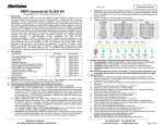
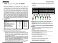
BioVision RBP4 (human) Competitive ELISA Kit I. II. (Catalog #K4911-100; 100 assays; Store kit at 4°C) Description: Retinol binding protein (RBP) 4 is the only specific transport protein for vitamin A in the circulation whose function is to deliver vitamin to target tissues. In obesity and type 2 diabetes, the expression of Glut4 is significantly impaired in adipocytes. Glucose transport via Glut4 is the rate-limiting step for glucose use by muscle and adipose tissue. Adipocyte-specific deletion of Gluts leads to notable elevation of mouse RBP4 causing systemic insulin resistance, and that reduction of RBP4 improves insulin resistance. This identified a novel role of RBP4 in regulating insulin action and RBP4 is recorded as an adipocyte-derived hormone. The RBP4 (human) ELISA Kit is to be used for the in vitro quantitative determination of human RBP4 in serum, urine and cell culture supernatant. This assay is a competitive ELISA which utilizies a 96-well microtiter plate which was pre-coated with a human RBP4. A purified polyclonal which recognizing native human RBP4 reacts with a series of predetermined recombinant human RBP4 standard proteins or the test samples under competition in the human RBP4-coated plate. Their relative reactivity is plotted with that of the standard proteins. This ELISA is specific for the measurement of natural and recombinant human RBP4. It does not cross-react with mouse RBP4, rat RBP4, human adiponectin, rat adiponectin, human resistin, human vaspin, human clusterin, human leptin, human IL-23, human IL-33, human GPX3, human Nampt, human ANG1, human ANG2, human ANGPTL3, human ANGPTL4, human ANGPTL6, human FABP4, human RELM-β, rat RELM-α, mouse Nampt, human PAI-1. The assay range is 0.001 – 5 µg RBP4/ml and a detection limit of 1 ng/ml (based on adding two standard deviations to the mean value of the (50) zero standards). Kit Contents: Component Pre-coated Microtiter Plate Wash Buffer (10X) Diluent (5X) Detection Antibody Detector 100X (Hrp conjugated anti-IgG) Human RBP4 Standard (lyophilized, 5 g) Human RBP4 QC Sample (lyophilized) Substrate Solution I (TMB) Substrate Solution II (Peroxidase) Stop Solution Plate Sealers For research use only rev. 2/14 100 Assays 1 ea (12 x 8 well strips) 50 ml 50 ml 12 ml 150 µl 1 vial 1 vial 6 ml 6 ml 12 ml 3 each III. Part Number K4911-100-1 K4911-100-2 K4911-100-3 K4911-100-4 K4911-100-5 K4911-100-6 K4911-100-7 K4911-100-8 K4911-100-9 K4911-100-10 K4911-100-11 Storage Conditions: Reagents must be stored at 2 - 8°C when not in use. Bring reagents to room temperature before use. Do not expose reagents to temperatures greater than 25°C. IV. Assay Procedure (Read the ENTIRE Protocol Before Proceeding) 1. Test Samples/Standards/QC Sample: (We recommend these be run in duplicate) a) Serum: Use a serum separator tube. Let samples clot at room temperature for 30 min before centrifugation for 20 min at 1000 x g. Assay freshly prepared serum or store serum in aliquots at -20°C for future use. Avoid repeated freeze/thaw cycles. Serum should be diluted in Diluent 1X. Samples containing visible precipitates must be clarified before use. As a starting point, 1/100 dilution is suggested. If samples fall outside the assay range a lower or higher dilution may be required. b) Urine: Aseptically collect the urine, voided directly into a sterile container. Assay immediately or aliquot and store at -20°C. Avoid repeated freeze/thaw cycles. Urine should be diluted in Diluent 1X. Samples containing visible precipitates must be clarified before use. Note: Serum, Plasma, Urine or Cell Culture Supernatant have to be diluted in Diluent 1X. Samples containing visible precipitates must be clarified before use. c) QC Sample: Reconstitute human RBP4 QC Sample with 1 ml of dH2O. Mix the QC Sample to ensure complete reconstitution. Allow to sit for a minimum of 15 min. The QC Sample is ready to use-do not dilute it (refer to the C of A for current QC Sample concentration). BioVision Incorporated 155 S. Milpitas Boulevard, Milpitas, CA 95035 USA d) e) f) Standards: Reconstitute human RBP4 Standard with 1 ml of dH2O to produce a stock solution (24 ng/ml). Mix the Stock solution to ensure complete reconstitution. Allow to sit for a minimum of 15 min. The reconstituted standard should be aliquoted and stored at -20°C. Prepare 1X Diluent: Dilute 5X Diluent 1:4 with dH2O. Prepare Standard Curve using 2-fold serial dilutions with 1X Diluent: 5 µg/ml To obtain 5 µg/ml 2.5 µg/ml 1 µg/ml 0.5 µg/ml 0.25 µg/ml 0.1 µg/ml 0.01 µg/ml 0.001 µg/ml Add --300 µl of RBP4 (5 µg/ml ) 200 µl of RBP4 (2.5 µg/ml ) 300 µl of RBP4 (1 µg/ml ) 300 µl of RBP4 (0.5 µg/ml ) 200 µl of RBP4 (0.25 µg/ml ) 50 µl of RBP4 (0.1 µg/ml ) 50 µl of RBP4 (0.01 µg/ml ) 300 µl 300 µl 200 µl 2.5 µg/ml 1 µg/ml 0.5 µg/ml 300 µl 0.25 µg/ml 200 µl 0.1 µg/ml Into --300 µl of 1X Diluent 300 µl of 1X Diluent 300 µl of 1X Diluent 300 µl of 1X Diluent 300 µl of 1X Diluent 450 µl of 1X Diluent 450 µl of 1X Diluent 50 µl 0.01 µg/ml 50 µl 0.001 µg/ml 2. Reagent Preparation: (Prepare just the appropriate amounts for the assay) a) 1X Wash Buffer: Dilute 10X Wash Buffer 1: 9 with dH2O to obtain 1X Wash Buffer. b) 1X Diluent: Dilute 5X Wash Buffer 1: 4 with dH2O to obtain 1X Diluent. c) 1X Detector: Dilute 100X Detector 1: 99 with 1X Diluent to obtain 1X Detector. d) Substrate Solution: Equal volumes of Substrate Solutions I and II must be mixed together. Note: The diluted Detector must be used within 1 hr of preparation; Substrates I and II should be mixed within 15 min of use (Protect Substrate Solution from light) 3. Assay Protocol: a) Determine the number of 8-well strips needed for assay and insert them into the frame for current use. The extra strips should be resealed in the foil pouch and can be stored at 4°C for up to 1 month. b) Add 50 µl of the Standards, Samples and QC Sample into the appropriate wells in duplicate. c) Cover plate with plate sealer and incubate for 1 hr at 37°C. d) Aspirate and wash x 3 with 300 µl of 1X Wash Buffer. e) Warm Detection Antibody to room temperature. Add 100 µl to each well and tap gently on the side of the plate to mix. f) Cover plate with plate sealer and incubate for 1 hr at 37°C. g) Aspirate and wash x 3 with 300 µl of 1X Wash Buffer. h) Add 100 µl of the 1X Detector to each well. i) Cover plate with plate sealer and incubate for 1 hr at 37°C. j) Remove plate from 37°C, aspirate and wash x 5 with 300 µl of 1X Wash Buffer. k) After last wash, tap inverted plate on a stack of paper towels. Complete removal of liquid is essential for good performance. l) Add 100 µl of the mixed Substrate Solution to each well. m) Allow the color to develop at room temperature in the dark for 20 min. n) Stop the reaction by adding 100 µl of Stop Solution to each well. o) Tap the plate gently to ensure thorough mixing. The substrate reaction yields a blue solution that turns yellow when Stop Solution is added. Caution: Stop Solution is a Corrosive Solution p) Measure the OD at 450 nm in an ELISA plate reader within 30 min. Tel: 408-493-1800 | Fax: 408-493-1801 www.biovision.com | [email protected] Page 1 of 2 BioVision 3. Calculations: a) Average the duplicate readings for each Standard, QC Sample and Test Sample. b) Generate a Standard Curve by plotting the average absorbance on the vertical (Y) axis vs. the corresponding concentration (µg /ml) on the horizontal (X) axis. (See Typical Data below) c) Calculate the Test Sample RBP4 concentrations by interpolation of the Standard Curve regression curve as shown below in the form of a 4-parameter logistic equation. d) If the Test Samples were diluted, multiply the interpolated values by the dilution factor to calculate the corrected human RBP4 concentrations. For research use only rev. 2/14 Expected values: RBP4 levels in plasma & serum range from 10 to > 70 mg/ml (healthy donors) Technical Hints and Limitations: • It is recommended that all standards, QC sample and samples be run in duplicate. • Do not combine leftover reagents with those reserved for additional wells. • Reagents from the kit with a volume less than 100 µl should be centrifuged. • Residual wash liquid should be drained from the wells after last wash by tapping the plate on absorbent paper. • Crystals could appear in the 10X solution due to high salt concentration in the stock solutions. Crystals are readily dissolved at room temperature or at 37°C before dilution of the buffer solutions. • Once reagents have been added to the 8-well strips, DO NOT let the strips DRY at any time during the assay. • Keep Substrate Solution protected from light. • The Stop Solution consists of phosphoric acid. Although diluted, the Stop Solution should be handled with gloves, eye protection and protective clothing. Troubleshooting: PROBLEM VI. Performance Characteristics: 1. Intra-assay Precision: (5) samples of known concentration were assayed in replicates (4) times to test precision within an assay. Samples Mean (µg/ml) SD CV (%) n 1 16.42 0.43 2.64 4 2 19.14 0.70 3.63 4 3 22.29 1.01 4.51 4 4 25.31 1.34 5.31 4 5 45.76 4.22 9.22 4 2. Inter-assay Precision: (5) samples of known concentration were assayed in (4) separate assays to test precision between assays. Samples Mean (µg/ml) SD CV (%) n 1 15.39 0.54 3.48 4 2 21.48 1.54 7.15 4 3 24.98 1.62 6.50 4 4 26.89 2.31 8.58 4 5 31.88 3.28 10.27 4 3. Linearity: Different human samples containing RBP4 were diluted several fold (1/00 to 1/800); recoveries ranged from 81 % to 110 %. Expected Observed % of Samples Sample Dilution (µg/ml) (µg/ml) Expected 1 : 100 14.59 14.59 100 1 : 200 7.29 7.95 109 1 1 : 400 3.65 4.01 110 1 : 800 1.82 1.95 107 1 : 100 23.09 23.09 100 1 : 200 11.55 9.81 85 2 1 : 400 5.77 4.71 82 1 : 800 2.89 2.33 81 1 : 100 31.03 31.03 100 1 : 200 15.52 13.55 87 3 1 : 400 7.76 6.69 86 1 : 800 3.88 3.26 84 BioVision Incorporated 155 S. Milpitas Boulevard, Milpitas, CA 95035 USA POSSIBLE CAUSES SOLUTIONS Check that all reagents have been added in the correct order. Use an automated plate washer Washes too stringent if possible. Incubation times should be Incubation times inadequate followed as indicated in the manual. Plate reader settings not Verify the wavelength and filter optimal setting in the plate reader. Use recommended incubation Incorrect assay temperature temperature. Bring substrates to room temperature before use. Concentration of detector too Use recommended dilution high factor. Ensure all wells are filling wash Inadequate washing buffer and are aspirated completely. Completely aspirate wells Wells not completely aspirated between steps. Be sure that reagents are Reagents poorly mixed thoroughly mixed. Be sure that reagents were Omission of reagents prepared correctly and added in the correct order. Check pipetting technique and Dilution error double-check calculations. Omission of key reagent No signal or weak signal High background Poor standard curve Unexpected results FOR RESEARCH USE ONLY! Not to be used on humans. Tel: 408-493-1800 | Fax: 408-493-1801 www.biovision.com | [email protected] Page 2 of 2