1
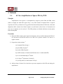
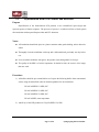
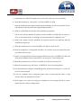
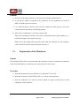
SPOLIGOTYPING SPOLIGOTYPING USER'S MANUAL A PCR-Based Method to Simultaneously Detect and Type Mycobacterium tuberculosis Complex Bacteria FOR RESEARCH USE ONLY This kit is not intended for food, drug, household, agricultural or cosmetic use. Use of this product must be supervised by a technically qualified individual. Ocimum Biosolutions does not warrant or guarantee that its products are merchantable or satisfactory for any particular purpose, nor free from any claim of foreign or domestic patent infringement by a third party, and there are no warranties, express or implied, to such effect. Ocimum Biosolutions will not be liable for any incidental, consequential or contingent damages involving their use. Page 2 of 22 Spoligotyping Kit User Manual Table of Contents 1. 1.1. 1.2. GENERAL DESCRIPTION ............................................................................................................ 4 PRINCIPLE OF THE SPOLIGOTYPING METHOD ......................................................................................... 4 PRACTICAL USE ..................................................................................................................................... 7 2. 2.1. 2.2. KITS ................................................................................................................................................... 8 AVAILABLE SPOLIGOTYPING PRODUCTS ................................................................................................ 8 KIT CONTENTS (IM9701) ...................................................................................................................... 8 3. METHODS ........................................................................................................................................ 9 3.1. IN VITRO AMPLIFICATION OF SPACER DNA BY PCR ............................................................................. 9 Principle ......................................................................................................................................................... 9 Procedure ....................................................................................................................................................... 9 Remarks ........................................................................................................................................................ 10 3.2. HYBRIDIZATION WITH PCR PRODUCT AND DETECTION ....................................................................... 11 Purpose ........................................................................................................................................................ 11 Notes: ........................................................................................................................................................... 11 Procedure: ................................................................................................................................................... 11 Remarks ........................................................................................................................................................ 13 3.3. REGENERATION OF THE MEMBRANE .................................................................................................... 13 Purpose ........................................................................................................................................................ 13 Procedure ..................................................................................................................................................... 13 4. ADDITIONAL REAGENTS .......................................................................................................... 14 5. SUPPLIES REQUIRED FOR RLB............................................................................................... 15 6. TROUBLESHOOTING ................................................................................................................. 16 7. FAQ (FREQUENTLY ASKED QUESTIONS) ............................................................................ 17 8. REFERENCES ................................................................................................................................ 18 9. TEMPLATE DNA ISOLATION METHODS .............................................................................. 20 General Remarks .......................................................................................................................................... 20 A. Preparation of Chromosomal DNA ......................................................................................................... 20 B. Preparation of Lysates from Colonies ..................................................................................................... 21 C. Extraction of Total DNA from Clinical Samples...................................................................................... 21 D. Isolation of Genomic DNA from Paraffin-Embedded Tissues ................................................................. 22 Remarks ........................................................................................................................................................ 22 Page 3 of 22 Spoligotyping Kit User Manual 1. General Description 1.1. Principle of the Spoligotyping Method The typing method described in this protocol is based on DNA polymorphism present at one particular chromosomal locus, the "Direct Repeat" (DR) region, which is uniquely present in Mycobacterium tuberculosis complex bacteria. This locus was first described by Hermans et al.5 who sequenced this region in Mycobacterium bovis BCG, the strain used worldwide to vaccinate against tuberculosis. The DR region in M. bovis BCG consists of directly repeated sequences of 36 base pairs, which are interspersed by non-repetitive DNA spacers, each 35 to 41 base pairs in length. The number of copies of the DR sequence in M. bovis BCG was determined to be 49. In other M. tuberculosis complex strains the number of DR elements was found to vary significantly. The vast majority of the Mycobacterium tuberculosis strains contain one or more IS6110 elements in the DR region (Fig.1). Figure 1: Structure of the DR locus in the genome of M. tuberculosis H37Rv and M. bovis BCG P3. The green rectangles depict the 36 bp Direct Repeat (DR). In contrast to the DRs, the spacers are usually present only once in the DR region, but occasionally some are found twice, either separated by one or by several DR's and other spacers. One DR and its neighboring non-repetitive spacer is termed “Direct Variant Repeat” (DVR). When the DR regions of several strains were compared, it was observed that the order of the spacers is about the same in all strains, but deletions and/or insertions of spacers and DR's occur (Fig. 2)2. The mechanism, by which spacers and copies of DR are generated, is unknown. With the method described here, the presence or absence in the DR region of 43 spacers of known sequence can be detected by hybridization of PCR-amplified spacer DNA to a set of immobilized oligonucleotides, Page 4 of 22 Spoligotyping Kit User Manual representing each of the unique spacer DNA sequences. This method will be referred to as spoligotyping (from spacer oligotyping)7. Figure 2: Schematic presentation of the polymorphism in DR regions of different M. tuberculosis complex strains. Blocks of DVR are missing in one strain when compared to another. The spacer order remains about the same. By spoligotyping one can detect the presence or absence of spacers of known sequence. The first step in the method is to amplify the DR region of a given strain by PCR. The primers used are based on the sequence of the DR, and allow the amplification of the spacer(s) between the DR targets (Fig. 3). The obtained PCR products differ in length because of two reasons. First, the product contains several spacers and the DR's in between if the primers anneal to DR's not next to each other. Second, the product itself can act as a primer, and become elongated with one or more DVRs. Therefore, the PCR product provides no reliable information about spacer order or total length of the DR region. A biotin labeled reverse primer is used, so that all the reverse strands synthesized are biotin labeled. Figure 3: Principle of the in vitro amplification of DNA within the DR region of M. tuberculosis complex bacteria. The use of the 2 primers, a and b, for in vitro amplification, will lead to the amplification of any spacer or a stretch of neighbouring spacers and DRs. Page 5 of 22 Spoligotyping Kit User Manual Oligonucleotides derived from the known spacers in the DR cluster are covalently linked to an activated membrane in parallel lines. PCR products are hybridized perpendicular to the oligo lines. After hybridization the membrane is incubated in streptavidin peroxidase, which binds to the biotin label on the PCR products. Detection of hybridization signals is optimized by the enhanced chemiluminescence (ECL) detection system (of course any biotin-detection method can be used when optimized). The peroxidase present on the streptavidine catalyzes a reaction resulting in the emission of light which can be detected by autoradiography of the membrane. We refer to this type of blot as reversed line blot (Fig. 4). Figure 4 Overview of the spoligotyping method. Page 6 of 22 Spoligotyping Kit User Manual An example of a result of the spoligotyping method used to analyze a variety of clinical isolates is shown in Fig. 5. Figure 5: A typical spoligotyping result of M. tuberculosis H37Rv, M. bovis BCG P3 and 38 different clinical isolates. A membrane with 43 spacer oligonucleotides was used (vertical lines). The spacer oligonucleotides were derived from the spacers of M. bovis BCG P3, M. tuberculosis H37Rv. 1.2. Practical Use Spoligotyping may offer an alternative for typing Southern blotting when rapid results are required. The method is in particular useful to simultaneously detect and type M. tuberculosis complex bacteria in clinical samples (suspected nosocomial infections, outbreaks in prisons, etc.). The level of differentiation by spoligotyping is less compared to IS6110 fingerprinting for strains having five or more IS6110 copies, but higher for strains with less than five copies. Thus spoligotyping is a preferred method to type M. bovis strains, which usually contain only one or two IS6110 copies. Note that M. bovis can be recognized by the absence of reactivity with spacers 39-43 (Fig. 5). Page 7 of 22 Spoligotyping Kit User Manual 2. Kits 2.1. Available Spoligotyping Products Catalog Number Description IM9701 Spoligotyping kit with membrane, primers and controls IM9702 Spoligotyping kit without controls and primers MN45 Mini-blotter for spoligotyping PC200 Foam cushions (100 per pack) PC20 Foam cushions (10 per pack) Accessories for Mini-blotter 2.2. Kit Contents (IM9701) This kit for spoligotyping consists of four vials, one membrane and one manual. 1) Two vials containing the positive controls: a) Positive control 1 (M. tuberculosis strain H37Rv) b) Positive control 2 (M. bovis BCG P3) Both positive controls are 10ng/µL. For PCR amplification, 2µL of each positive control should be added to the PCR reaction mixture (see section 3.1). The volume in each vial is 15µL and should therefore be sufficient for approximately 7 PCR reactions. 2) Two vials containing the primers for PCR amplification: a) Primer DRa (biotinylated) b) Primer DRb The primers are shipped lyophilized. To prepare both primers for PCR amplification (see section 3.1), both primers should be dissolved in 1.72 mL MQ water. For storage see also the remarks b) and c) from section 3.1. The volume in each vial should be sufficient for approximately 430 PCR reactions. 3) One Spoligo-membrane The membrane is shipped in 20 mM EDTA. Please avoid dehydration at any time, because this will rapidly decrease the quality of the membrane (see section 3.3, item 3). Store the membrane at + 4°C for optimal storage condition. Page 8 of 22 Spoligotyping Kit User Manual 3. Methods 3.1. In Vitro Amplification of Spacer DNA by PCR Principle Amplification of the spacers is accomplished by using the primers DRa and DRb, which enable to amplify the whole DR region. Only a very small amount of template DNA is required. Typically the PCR is performed on 10 ng purified chromosomal Mycobacterial DNA but, with minor adaptations, DNA extracts from clinical samples or lysed bacteria (from freezer or Löwenstein) can also serve as template (see also section 7). The PCR products are labeled with biotin, because primer DRa is biotinylated. Procedure 1) Dilute the DNA samples to the required concentration. Always include chromosomal DNA of M. tuberculosis strain H37Rv and M. bovis BCG P3 as positive controls. Use water as a negative control. 3) Prepare the reaction mixture: a) x μL template DNA (20 ng) 4 μL primer DRa (20 pmol) b) 4 μL primer DRb (20 pmol) c) 4 μL dNTP-mixture (2.5 mM each dNTP, final conc. 0.2 mM each dNTP) 5 μL 10x concentrated Super Tth buffer 0.1 μL Super Tth polymerase (5 units/μL) 33-x μL MQ water (to a final volume of 50 μL) 4) Add one drop of mineral oil to the tubes to prevent evaporation of the PCR-mix during the amplification. Page 9 of 22 Spoligotyping Kit User Manual 5) Place the tubes in a PCR-apparatus for amplification, and perform the following temperature cycling: d,e,f) 3 min 96°C 1 min 96°C 1 min 55°C 20x 30 sec 72°C 5 min 72°C Remarks a) Preparation of the mixture has to take place in a laboratory free of mycobacterial PCR products containing the DR sequences. b) Primer DRa is biotinylated and should be stored at +4°C. Repeated freeze-thawing of the biotinylated primer results in weaker Spoligo patterns. c) Primer DRb should be stored in small aliquots at -20°C. d) For amplification of the DR-cluster from extracts of clinical samples, the number of cycles can be increased to 40. e) For amplification of the DR-cluster from heat-killed cells, the number of cycles can be increased to 30 (see also section 7B, Preparation of lysates from colonies). f) PCR products can be used immediately, but can also be stored at -20ºC to be used later. Page 10 of 22 Spoligotyping Kit User Manual 3.2. Hybridization with PCR Product and Detection Purpose Hybridization of the biotin-labeled PCR products to the immobilized spacer-oligos that represent spacers of known sequence. The presence of spacers is visualized on film as black squares after incubation with streptavidin-peroxidase and ECL-detection. Notes: All incubations should take place in a plastic container under gentle shaking, unless otherwise stated. Thoroughly clean the miniblotter with soap and a dedicated brush, preferably one day before use. Never touch the membrane with gloves, the powder causes background. Use forceps. The quality of the SDS is of critical importance. It should be fresh, do not store it for longer than one week. Procedure: 1) All buffers should be pre-warmed before use. Prepare the following buffers from concentrated stocks, using de-mineralized water for dilution (quantities for one membrane): 250 mL 2xSSPE/0.1% SDS, 60ºC 250 mL 2xSSPE/0.5% SDS, 60ºC 250 mL 2xSSPE/0.5% SDS, 42ºC 250 mL 2xSSPE, room temperature 2) Add 20 μL of the PCR products to 150 μL2xSSPE/0.1% SDS. Page 11 of 22 Spoligotyping Kit User Manual 3) Heat-denature the diluted PCR product for 10 min at 99°C and cool on ice immediately. 4) Wash the membrane for 5 min at 60°C in 250 mL2xSSPE/0.1% SDS. 5) Place the membrane and a support cushion into the miniblotter in such a way that the slots are perpendicular to the line pattern of the applied oligonucleotides. a) 6) Remove residual fluid from the slots of the miniblotter by aspiration. 7) Fill the slots with the diluted PCR product (avoid air bubbles!) and hybridize for 60 min at 60ºC on a horizontal surface (no shaking!). Avoid contamination of neighboring slots. b) 8) Remove the samples from the miniblotter by aspiration and take the membrane from the miniblotter using forceps. 9) Wash the membrane twice in 250 mL2xSSPE/0.5% SDS for 10 min at 60°C . 10) Place the membrane in a rolling bottle and allow it to cool down to prevent inactivation of the peroxidase in the next step. 11) Add 2.5 μL streptavidin-peroxidase conjugate (500U/ml) to 10 mL of 2xSSPE/0.5% SDS, and incubate the membrane in this solution for 45 to 60 min at 42°C in the rolling bottle. 12) Wash the membrane twice in 250 mL of 2xSSPE/0.5% SDS for 10 min at 42°C. 13) Rinse the membrane twice with 250 mL of 2xSSPE for 5 min at room temperature. 14) For chemiluminiscent detection of hybridizing DNA, incubate the membrane for 1 min in 20 mL ECL detection liquid. c) 15) Cover the membrane with a transparent plastic sheet or Saran-wrap and expose a light sensitive film to the membrane for 20 min. d) 16) If the signal is too weak or too strong, the membrane can be used again directly to expose another film for a shorter or longer period. Page 12 of 22 Spoligotyping Kit User Manual Remarks a) Do not reuse the support cushions. You can order more support cushions from us. b) If less than 45 samples are applied to the miniblotter, fill one neighboring slot with 2X SSPE/0.1%SDS to prevent cross-flow. c) Use a dedicated plastic container. Do not use this container for other purposes, since some reagents decrease the intensity of the Spoligo patterns. d) If the result is unsatisfactory, you can try to improve this. Black spots (background) possibly occur due to contamination during filter handling (e. g. touched with fingers). Start again from step 8. Blank areas in the spoligo patterns possibly indicate that the membrane was not completely soaked with ECL detection liquid. Start again from step 13. 3.3. Regeneration of the Membrane Purpose The hybridized PCR product is dissociated from the membrane in order to regenerate the membrane for the next hybridization. A membrane can be regenerated for at least 5 times. Procedure 1) Wash the membrane twice by incubation in 1% SDS at 80°C for 30 min. 2) Wash the membrane in 20 mM EDTA pH 8, for 15 min at room temperature. 3) Store the membrane at 4°C until use (sealed in plastic or wrapped in Saran-wrap, to avoid dehydration of the membrane). Page 13 of 22 Spoligotyping Kit User Manual 4. Additional Reagents Not Supplied along with standard items; need to be ordered separately 20xSSPE Store at room temperature for no longer than 6 months. 2xSSPE Dilute 20xSSPE ten times with de-mineralised water. 10% SDS 10 g SDS / 100 mL de-mineralised water. 2xSSPE/0.1%SDS Add 100 mL 20xSSPE and 10 mL 10% SDS to 890 mL de-mineralised water. 2xSSPE/0.5%SDS Add 100 mL 20xSSPE and 50 mL 10% SDS to 850 mL de-mineralised water. Page 14 of 22 Spoligotyping Kit User Manual 5. Supplies required for RLB 1. 2. 3. 4. 5. 6. 7. 8. Maxi 14 Hybridization Oven with Shaking Platform Miniblotter MN45 (Cat. No. MN45) and Miniblotter accessories X-ray films preferably Hyperfilm ECL (18 x 24 cm), but others work as well Incubation box fitting the membrane - Common heat resistant household Tupperware. Exposure cassette (24 cm X 30 cm) 20 x SSPE, 4 ltr. SDS specially pure (500 g) Plate Cushions for Miniblotter (Cat No. PC2 (10 per pack); PC200 (100 per pack)) For all the above, please contact us at: Ocimum Biosolutions 6th Floor, Reliance Classic Building Road No 1, Banjara Hills, Hyderabad, India Tel.: +91(40) 6698 6700 Fax: +91(40) 6662 7205 E-mail: [email protected] Website: www.ocimumbio.com Super Taq DNA polymerase (5000 U) Streptavidin-POD-conjugate ECL detection liquid Page 15 of 22 Spoligotyping Kit User Manual 6. Troubleshooting 1) No hybridization signal detected: Analyze 5 μL of the PCR product on a 2% agarose gel. A ladder pattern should be visible. If a ladder pattern is visible, check the labeling of the PCR product by spotting it onto a membrane, followed by incubation with streptavidin peroxidase. 2) High background (stripes): Clean the miniblotter thoroughly using a dedicated brush, and soak the apparatus, preferably overnight, in a soap solution. Contact us in case you want help in procuring this. 3) High background (spots): strip the membrane again, and test it with PCR products of the control strains. If stripping does not lead to a lower background, the membrane should not be used anymore. Page 16 of 22 Spoligotyping Kit User Manual 7. FAQ (Frequently Asked Questions) Q. When running the PCR reactions on a gel, I do not see any product. Is that normal? A. Sometimes no DNA is observed looking at a gel. Because spoligotyping is more sensitive than gel electrophoresis, you may find a good signal on the membrane. Q. What is the orientation of the membrane? A. Each membrane is marked by date of production. This date is written in the upper right corner when then membrane was labeled from left to right. When hybridizing you PCR products, this date should therefore be vertically. When you overlay your film, mark your film or bend the corner at which this date is written. Q. We already have SDS in our laboratory. Can we use this without any problem? A. Some users obtain good results with their own SDS. Some companies however produce SDS that gives some problems in obtaining clear blots. The kit is evaluated with the SDS used in our laboratories and we therefore recommend using this particular SDS. Use a 10% stock solution (RT) with a expiration date of around a month. Q. Is the kit only available with the biotinylated Dra-primer? A. Since Ocimum Biosolutions is a producer of biomolecules as DNA, peptides and PNA, we can have any other haptens coupled to the Dra primer as well (eg. DIG, DNP,etc.). However, these haptens have not been evaluated with the spoligotyping technique. If you prefer using other haptens, please inquire about the possibilities. Q. When incubating the PCR products at 60°C, is it possible that you get evaporation or crosscontamination of the different PCR products? A. We have never experienced these features. Holding the blotter horizontally is the most important thing in hybridizing your PCR products. Page 17 of 22 Spoligotyping Kit User Manual 8. References 1) Aranaz, A., E. Liebana, A. Mateos, L. Dominquez, D. Vidal, M. Domingo, O. Gonzalez, E. F. Rodriguez-Ferri, A. E. Bunschoten, J. D. A. van Embden, and D. Cousins. 1996. Spoligotyping of Mycobacterium bovis strains from cattle and other animals: a tool for epidemiology of tuberculosis. J. Clin. Microbiol. 34:2734-2740. 2) Groenen, P. M. A., A. E. Bunschoten, D. van Soolingen, and J. D. A. van Embden. 1993. Nature of DNA polymorphism in the direct repeat cluster of Mycobacterium tuberculosis; Application for strain differentiation by a novel method. Mol. Microbiol.10(5):1057-1065. 3) Goguet de la Salmoniere, H., M. Li, G. Torrea, A. Bunschoten, J. van Embden, and B. Gicquel. 1996. Spoligotyping: evaluation of this method by studying the transmission of Mycobacterium tuberculosis. Combination with inter-IS6110 PCR for typing M. tuberculosis complex strains. Submitted 4) Goyal, M., N. A. Saunders, J. D. A. van Embden, D. B. Young, and R. J. Shaw. 1997. Differentiation of Mycobacterium tuberculosis isolates by Spoligotyping and IS6110 restriction fragment length polymorphism. J. Clin. Microbiol. 35:647-651. 5) Hermans, P. W. M., D. van Soolingen, E. M. Bik, P. E. W. de Haas, J. W. Dale, and J. D. A. van Embden. 1991. The insertion element IS987 from M. bovis BCG is located in a hot spot integration region for insertion elements in M. tuberculosis complex strains. Infect. Immun. 59:2695-2705. 6) Heyderman, R. S., M. Goyal, P. Roberts, S. Ushewokunze, S. Zizhou, B. G. Marshall, R. Makombe, J. D. A. van Embden, P. R. Mason, and R. J. Shaw. 1997. The epidemiology of sputum-positive pulmonary tuberculosis in Harare, Zimbabwe - analysis by spoligotyping. Submitted. Page 18 of 22 Spoligotyping Kit User Manual 7) Kamerbeek, J., L. M. Schouls, A. Kolk, M. van Agterveld, D. van Soolingen, S. Kuijper, A. E. Bunschoten, H. Molhuizen, R. Shaw, M. Goyal, and J. D. A. van Embden. 1997. Simultaneous detection and strain differentiation of Mycobacterium tuberculosis for diagnosis and epidemiology. J. Clin. Microbiol. 35:907-914. 8) Kaufhold, A., A. Podbielski, G. Baumgarten, M. Blokpoel, J. Top, and L. Schouls. 1994. Rapid typing of group A streptococci by the use of DNA amplification and nonradioactive allele specific oligonucleotide probes. FEMS Microbiol. Lett. 119:19-26. 9) Mangiapan, G., M. Vokurka, L. Schouls, J. Candranel, D. Lecossier, J. van Embden, and A. Hance. 1996. Sequence capture - PCR improves the detection of mycobacterial DNA in clinical specimens. J. Clin. Microb. 34:1209-1215. 10) Samper, S., C. Martin, A. Pinedo, A. Rivero, J. Blázquez, F. Baquero, D. van Soolingen, and J. van Embden. 1997. Detection of international transmission of multidrug-resistant tuberculosis by using DNA fingerprint databases. Submitted. 11) Van Soolingen, D., L. Qian, P. E. W. de Haas, J. T. Douglas, H. Traore, F. Portaels, H. Z. Qing, D. Enkhasaikan, P. Nymadawa, and J. D. A. van Embden. 1995. Predominance of a single genotype of Mycobacterium tuberculosis in countries of East Asia. J. Clin. Microb. 33:3234-3238. Page 19 of 22 Spoligotyping Kit User Manual 9. Template DNA Isolation Methods General Remarks For handling clinical samples that are to be used in a PCR, it is recommended to work in a room specially equipped for this purpose (over-pressure, laminar-flow hood). Always use negative controls. A. Preparation of Chromosomal DNA Typically, isolation and purification of chromosomal Mycobacterium DNA is done using the CTAB method: 1) Transfer at least one loop full of cells into an Eppendorf tube containing 400 μL of 1xTE. 2) Heat 20 min at 80°C to kill the cells, and cool to room temperature. 3) Add 50 μL10 mg/mL lysozyme, vortex and incubate at least 1 hr at 37°C. 4) Add 75 μL10 % SDS/proteinase K solution (5 μL proteinase K, 10 mg/mL and 70 μL 10% SDS), vortex shortly and incubate 10 min at 65°C. 5) Add 100 μL 5M NaCl. 6) Add 100 μL CTAB/NaCl solution (4.1 g NaCl and 10 g CTAB [N-cetyl-N,N,N,trimethylammoniumbromide] in 100 mL distilled water), which is pre-warmed at 65°C. Vortex until the liquid content becomes white ("milky"). Incubate 10 min at 65°C. 7) Add 750 μL of chloroform/isoamyl alcohol (24:1), vortex 10 sec, and centrifuge at room temperature for 5 min, 12,000 g. 8) Transfer the aqueous supernatant to a fresh microcentrifuge tube. 9) Add 450 μL isopropanol. 10) Incubate 10 minutes on ice. 11) Centrifuge 15 minutes at room temperature. Page 20 of 22 Spoligotyping Kit User Manual 12) Discard the supernatant and wash the pellet with 1 mL of 70% ethanol and centrifuge (approximately 5 min at room temperature). 13) Discard the supernatant and dry the pellet. 14) Redissolve the pellet in 20 μL of 1xTE buffer. The DNA can be stored at 4°C until further use. B. Preparation of Lysates from Colonies 1) Resuspend 2 loops of cells in 250 μL 1xTE in an Eppendorf tube. 2) Kill the cells by incubation at 80°C for 1 hour. 3) Centrifuge the tube at 13000 rpm for 2 min, discard the supernatant and resuspend the pellet in 500 μL of 150 mM NaCl. Repeat this step twice. 4) Discard the supernatant and resuspend the pellet in 25 μLof distilled water or 1x TE. C. Extraction of Total DNA from Clinical Samples 1) Bring the sample (max 1 mL or 1 cm3 ) aseptically in a 10 mL tube with 2-3 mL of digestion buffer (500 mM Tris HCl, pH 9.0, 20 mM EDTA, 10 mM NaCl, 1%SDS) and incubate overnight at 60°C. 2) Vortex the sample for 20 sec, add 0.5 mL phenol to 0.9 mL sample and vortex for 20 seconds. 3) Centrifuge for 5 minutes at max speed. 4) Transfer the aqueous phase to a fresh tube, containing 0.5 mL phenol, vortex for 20 seconds and centrifuge for 5 minutes at maximum speed. 5) Transfer the aqueous phase (approx. 350 μL) to a fresh tube containing 35 μL3 M NaAc and 800 μL absolute ethanol, mix and incubate for 20 minutes at -20°C. 6) Centrifuge for 30 minutes at room temperature, max speed. 7) Discard the supernatant and wash the pellet with 500 μL 70% ethanol, centrifuge for 5 minutes at max speed. 8) Discard the supernatant, dry the pellet, resuspend the DNA in 50-200 μL 1x TE and store at 20°C until further use. Page 21 of 22 Spoligotyping Kit User Manual D. Isolation of Genomic DNA from Paraffin-Embedded Tissues Sample preparation of DNA from the paraffin-embedded tissues is successful, but remains time consuming. The technique involves two major steps, deparaffinization and protein digestion, each of which involves several centrifugations and washes and requires multiple tube transfers. We use a method which is a one step procedure without protein digestion: 1) Add 150 µL of a 5% Chelex suspension to a 14 µm paraffine-embedded tissue section. a, b) 2) Vortex thoroughly. The section should be completely covered with the Chelex suspension. 3) Heat the mixture 30 minutes at 100°C. The paraffin then appears floating on the surface of the solution. 4) Centrifuge 10 minutes at 13000 x g. 5) Transfer the solution beneath the paraffin, containing the extracted DNA, to a clean microcentrifuge tube. c) 6) The PCR is run with two different dilutions of the extracted DNA: 10 µL undiluted DNA and 10 µL DNA of an 1:4 dilution, in 50 µL PCR-mix. Remarks a) To prepare the sections use another scalpel or knife for each sample, the microtome and the knife should be disinfected with 1N HCl for two minutes after each sample to prevent contamination. Cut a negative control between every sample. b) The aim of Chelex®100 treatment is to remove metal ions. Chelex® 100 is stable for at least 2 years when stored sealed in the original container at 22°C. If left in the hydrogen form for more than a few hours, the Chelex has a tendency to lose chelating capacity. c) Avoid transferring the Chelex. Chelex will bind Mg2+ in the PCR-mix. Page 22 of 22 Spoligotyping Kit User Manual