1
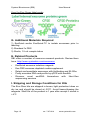
Exo-GLOW™ Exosome Labeling Kits Cat #s EXOR100A-1, . EXOG200A-1, EXOC300A-1 User Manual System Biosciences (SBI) 265 North Whisman Rd. Mountain View, CA 94043 Tel: 888.266.5066 (Toll Free in US) 650.968.2200 Fax: 650.968.2277 E-mail: [email protected] Web: www.systembio.com Store Kits at -20ºC upon receipt A limited-use label license covers this product. By use of this product, you accept the terms and conditions outlined in the Licensing and Warranty Statement contained in this user manual. Exo-Glow™ Exosome Labeling Kits Cat. #s EXOR100A-1, EXOG200A-1, EXOC300A-1 Contents I. Introduction ..............................................................................1 A. Exosome Overview .................................................................1 B. The Exo-Glow kits for labeling exosomes...............................2 C. Exo-Glow Protocols ................................................................3 D. Sample Exo-Glow IP bead data .............................................6 E. Exo-Glow exosomes added to target cells .............................8 F. How the exosome labels work ................................................9 G. Additional Materials Required ..............................................10 H. Related Products ..................................................................10 I. Shipping and Storage Conditions for Kits ..............................10 II. References ............................................................................11 III. Technical Support ..............................................................12 VII. Licensing and Warranty information ..................................13 I. Introduction A. Exosome Overview Exosomes are 60 - 180 nm membrane vesicles secreted by most cell types in vivo and in vitro. These microvesicles are produced by the inward budding of multivesicular bodies (MVBs) and are released from the cell into the microenvironment following the fusion of MVBs with the plasma membrane. Exosomes are extracellular, nanoshuttle organelles that facilitate communication between cells and organs. Exosomes are found in blood, urine, 888-266-5066 (Toll Free) 650-968-2200 (outside US) Page 1 System Biosciences (SBI) User Manual amniotic fluid, breast milk, malignant ascites fluids and contain distinct subsets of RNAs and proteins depending upon the cell type from which they are secreted, making them useful for biomarker discovery. SBI has engineered tools and nextgeneration sequencing services to accelerate the study of exosomes, exosome protein and RNA biomarkers. B. The Exo-Glow kits for labeling exosomes The Exo-Glow kits allow researchers to fluorescently-label isolated exosomes to track cellular interaction and uptake. You will first enrich for all exosomes using ExoQuick (serum, plasma, ascites samples) or ExoQuick-TC (cell media, urine, spinal fluid) or immunopurify specific exosome subpopulations using SBI’s ExoFlow IP kits. The isolated exosomes are then resuspended and incubated with either the 10x Exo-Red or 10x Exo-Green labeling stain. The Exo-Red stain is based on an Acridine Orange (AO) chemistry. AO is membrane permeable and fluorescently-labels single-stranded RNAs inside of exosomes. These Red exoRNAs can then be monitored for delivery into target cells via the exosomes using fluorescent microscopy. The Exo-Green stain is based on Carboxyfluorescein succinimidyl diacetate ester (CFSE) chemistry. CFSE is also membrane permeable. When CFSE enters exosomes it is hydrolyzed by exosome esterases known to be within exosomes that remove the diacetate residues (Savina A., et al., 2002). This activates the CFSE to fluoresce green and is then coupled to the amino ends of proteins. This approach allows researchers to track exoProtein transfer into target cells using fluorescent microscopy. Fluorescently label exosome internal RNA Red or internal exosome proteins Green for tracking. Page 2 ver. 7-071614 www.systembio.com Exo-Glow™ Exosome Labeling Kits Cat. #s EXOR100A-1, EXOG200A-1, EXOC300A-1 C. Exo-Glow Protocols Materials provided: Exo-Red (10x in DMSO) : 1 ml per kit Exo-Green (10x in water) : 1 ml per kit ExoQuick-TC : 2 ml per kit Description Cat# Size Exo-Red exosome RNA fluorescent label EXOR100A-1 20 rxns Exo-Green exosome protein fluorescent label EXOG200A-1 20 rxns Exo-Red + Exo-Green exosome cargo fluorescent label Combo Pack (10 rxns of each label) EXOC300A-1 20 rxns Exosome sample preparation Isolate exosomes from serum or media (isolated by ExoQuick, ExoQuick-TC or ultracentrifugation methods), resuspend the exosome pellet in 500 ul of 1x PBS. The recommended exosome protein input should be approximately 100 - 500 ug per labeling reaction. Each Exo-Glow reaction (rxn) equates to labeling 1.5 x 10^8 exosomes. Protocol for labeling exosomes: 1. Add 50 ul 10x Exo-Red or Exo-Green to 500 L volume of resuspended exosome suspension in 1x PBS in a 1.5 ml eppendorf tube. 2. Mix it well by flicking/inversion. Do not vortex 3. Incubate the exosome solution in 37°C for 10 minutes (rotation not necessary). 4. To stop labeling reaction, add 100 ul of the ExoQuick-TC reagent to the labeled exosome sample suspension and mix by inverting 6 times. 5. Place the labeled exosome sample on ice (or at 4°C) for 30 minutes. 888-266-5066 (Toll Free) 650-968-2200 (outside US) Page 3 System Biosciences (SBI) User Manual 6. Centrifuge the sample for 3 minutes at 14,000 rpm in a microfuge. 7. Remove the supernatant with excess label and resuspend the labeled exosome pellet in 500 ul 1x PBS. 8. The labeled exosomes are ready to be monitored for tracking. Adding Exo-Glow exosomes to cells: 1. Add at least 100 ul of labeled exosomes to approximately 1x10^5 cells per well in a 6-well culture plate (as an example). You can scale this ratio up or down depending upon your experimental requirements. 2. Incubate for 2 to 24 hours, visualize with fluorescent microscopy using the following excitation/emission guidelines below. Exosome Label Excitation Emission Filter setting Exo-Red 460 nm 650 nm (red) Typical RFP filter set Exo-Green 494 nm 521 nm (green) Typical GFP filter set NOTE: ExoQuick and ExoQuick-TC for exosome isolation purposes are not provided in the Exo-Glow kits and can be purchased separately. The following ExoQuick products are recommended for exosome concentration prior to Exo-Flow purification. Page 4 ver. 7-071614 www.systembio.com Exo-Glow™ Exosome Labeling Kits Cat. #s EXOR100A-1, EXOG200A-1, EXOC300A-1 Description Size Catalog # ExoQuick Serum exosome precipitation solution (5 ml) 75 reactions EXOQ5A-1 ExoQuick Plasma prep and Exosome precipitation kit (5 ml ExoQuick plus Thrombin) 75 reactions EXOQ5TM-1 Thrombin Plasma prep for Exosome precipitation 100 reactions TMEXO-1 ExoQuick Serum exosome precipitation solution (20 ml) 300 reactions EXOQ20A-1 ExoQuick-TC for Tissue Culture Media and Urine (10 ml) 10 reactions EXOTC10A-1 ExoQuick-TC for Tissue Culture Media and Urine (50 ml) 50 reactions EXOTC50A-1 Exosome isolation protocols using ExoQuick reagents Combine your biofluid sample containing exosomes with ExoQuick or ExoQuick-TC using the guidelines shown in the Table below. Mix the ExoQuick precipitation reagent with the biofluid sample by inversion and place at 4°C for 30 minutes to overnight, then recover the exosomes in a pellet with a low speed spin. Please refer to the ExoQuick or ExoQuick-TC User manuals for more details. Recommended amounts of exosomes provided in Table. Biofluid Sample volume ExoQuickTC volume Resuspend exosome pellet Volume to use in Exo-Flow Urine 10 ml 2 ml 500 L PBS 100 L/rxn Spinal fluid 10 ml 2 ml 500 L PBS 100 L/rxn Culture media 10 ml 2 ml 500 L PBS 100 L/rxn 888-266-5066 (Toll Free) 650-968-2200 (outside US) Page 5 System Biosciences (SBI) User Manual Biofluid Sample volume ExoQuick Serum 250 L 63 L Plasma 250 L 63 L Ascites fluid 500 L 120 L Resuspend exosome pellet 500 L PBS 500 L PBS 250 L PBS Volume to use in ExoFlow 100 L/rxn 100 L/rxn 100 L/rxn Amount of exosomes to use The number of exosomes in a given biofluid will vary depending upon the sample itself. There are abundant levels of exosome in serum, less in cell culture medium and urine. Use the guidelines in the Tables above as a starting point. You can typically add about 100-500 g protein of isolated exosomes per Exo-Glow reaction. D. Sample Exo-Glow IP bead data Exosomes were first isolated from the tissue culture medium from HEK293 cells grown in Exo-FBS exosome-depleted media supplement with standard DMEM. The cells were grown to 8090% confluency in a 10 cm cell culture dish. The secreted exosomes were isolated as stated in the protocol for ExoQuick-TC above. The exosome pellet was resuspended in 1 ml 1x PBS and contained an exosome protein content of 1 ug/ul. About 500 ul of this exosome suspension was labeled with 50 ul of either 10x ExoRed or Exo-Green for 10 minutes at 37°C. The exosomes were reisolated using the addition of 100 ul ExoQuick-TC and precipitation at 5°C for 30 minutes on ice or 4°C. The labeled exosome pellets were resuspended in 500 ul 1x PBS. CD63-coupled magnetic beads from SBI’s Exo-Flow IP kit (cat# EXOFLOW32A-CD63). Briefly, 50 ul of CD63 magnetic beads were incubated with 100 ul of the labeled exosomes overnight at 4°C on a rotator. The following day, the bead/labeled exosomes were placed on a magnetic plate for 5 minutes and then washed with 100 ul 1x wash buffer once. Then, 100 ul 1x PBS was added to the IP well and placed on the magnetic rack for 2 minutes to position the beads at Page 6 ver. 7-071614 www.systembio.com Exo-Glow™ Exosome Labeling Kits Cat. #s EXOR100A-1, EXOG200A-1, EXOC300A-1 the bottom of the wells. The beads with captured, labeled exosomes were imaged using fluorescent microscopy. 888-266-5066 (Toll Free) 650-968-2200 (outside US) Page 7 System Biosciences (SBI) User Manual E. Exo-Glow exosomes added to target cells The same labeled exosomes from (E. above) were added to target HEK 293 cells in culture. The cells were plated at about 1x10^5 cells in a standard 6 well culture plate. Approximately 100 ul of the labeled exosome suspension was added per 6 well with cells and allowed to interact/uptake for the time indicated in the following data figures. Page 8 ver. 7-071614 www.systembio.com Exo-Glow™ Exosome Labeling Kits Cat. #s EXOR100A-1, EXOG200A-1, EXOC300A-1 F. How the exosome labels work The Exo-Red exosome label is based on Acridine Orange chemistry and is a nucleic acid selective fluorescent cationic dye. It is cell-permeable, and interacts with DNA by intercalation and RNA by electrostatic attractions. When bound to DNA, Exo-Red is very similar spectrally to fluorescein, with an excitation maximum at 502 nm and an emission maximum at 525 nm (green). When Exo-Red associates with RNA, the excitation maximum shifts to 460 nm (blue) and the emission maximum shifts to 650 nm (red). This allows for the labeling of internal exosome RNAs and the ability to monitor RNA delivery to cells via exosomes. The Exo-Green stain is based on Carboxyfluorescein succinimidyl diacetate ester (CFSE) chemistry. CFSE is also membrane permeable. When CFSE enters exosomes it is hydrolyzed by esterases known to be within exosomes that remove the diacetate residues. This activates the CFSE to fluoresce green and is then coupled to the amino ends of proteins. This approach allows researchers to track exosome Protein transfer into target cells using fluorescent microscopy. 888-266-5066 (Toll Free) 650-968-2200 (outside US) Page 9 System Biosciences (SBI) User Manual How the Exo-Green label works G. Additional Materials Required 1) ExoQuick and/or ExoQuick-TC to isolate exosomes prior to labeling. 2) Standard 1x PBS 3) Sterile, 1.5 mL sample tubes H. Related Products SBI offers a number of exosome research products. Review them here: http://www.systembio.com/exosomes • • • • • ExoQuick exosome isolation reagents Exo-FBS exosome-depleted media supplement Detect and quantitate exosomes with Antibodies and ELISAs Purify exosome RNA and profile by qPCR with SeraMir Discover novel exoRNA biomarkers with Next-Gen sequencing services I. Shipping and Storage Conditions for Kits The Exo-Glow kits are shipped in brown, light protection tubes on dry ice and should be stored at -20°C. Avoid freeze-thawing the reagents. Shelf life of the product is 1 year after receipt if stored in +4°C. Page 10 ver. 7-071614 www.systembio.com Exo-Glow™ Exosome Labeling Kits Cat. #s EXOR100A-1, EXOG200A-1, EXOC300A-1 II. References Savina A, Vidal M, Colombo MI. The exosome pathway in K562 cells is regulated by Rab11. J Cell Sci. 2002 Jun 15;115(Pt 12):2505-15. Huang X, Yuan T, Tschannen M, Sun Z, Jacob H, Du M, Liang M, Dittmar RL, Liu Y, Liang M, Kohli M, Thibodeau SN, Boardman L, Wang L. Characterization of human plasma-derived exosomal RNAs by deep sequencing. BMC Genomics. 2013 May 10;14:319. Kurbegovic A, Trudel M. Progressive development of polycystic kidney disease in the mouse model expressing Pkd1 extracellular domain. Hum Mol Genet. 2013 Jun 15;22(12):2361-2375. Bhattarai N, McLinden JH, Xiang J, Landay AL, Chivero ET, Stapleton JT. GB Virus C Particles Inhibit T Cell Activation via Envelope E2 Protein-Mediated Inhibition of TCR Signaling. J Immunol. 2013 May 17. [Epub ahead of print]. Narayanan A, Iordanskiy S, Das R, Van Duyne R, Santos S, Jaworski E, Guendel I, Sampey G, Gerhart E, Iglesias-Ussel M, Popratiloff A, Hakami R, Kehn-Hall K, Young M, Subra C, Gilbert C, Bailey C, Romerio F, Kashanchi F. Exosomes derived from HIV-1 infected cells contain TAR RNA. J Biol Chem. 2013 May 9. [Epub ahead of print]. Carlos Salomon, Luis Sobrevia, Keith Ashman, Sebastian E. Illanes, Murray D. Mitchell and Gregory E. Rice. The Role of Placental Exosomes in Gestational Diabetes Mellitus, Gestational Diabetes-Causes, Diagnosis and Treatment. Dr. Luis Sobrevia (Ed.), ISBN: 978-953-51-1077-4, InTech, DOI: 10.5772/55298. Skrzypek K, Tertil M, Golda S, Ciesla M, Weglarczyk K, Collet G, Guichard A, Kozakowska M, Boczkowski J, Was H, Gil T, Kuzdzal J, Muchova L, Vitek L, Loboda A, Jozkowicz A, Kieda C, Dulak J. Interplay between heme oxygenase-1 and miR-378 affects non- 888-266-5066 (Toll Free) 650-968-2200 (outside US) Page 11 System Biosciences (SBI) User Manual small cell lung carcinoma growth, vascularization and metastasis. Antioxid Redox Signal. 2013 Apr 25. [Epub ahead of print]. Haney MJ, Zhao Y, Harrison EB, Mahajan V, Ahmed S, et al. Specific Transfection of Inflamed Brain by Macrophages: A New Therapeutic Strategy for Neurodegenerative Diseases. PLoS ONE 8(4): e61852. doi:10.1371/journal.pone.0061852. Hu G, Gong AY, Roth AL, Huang BQ, Ward HD, Zhu G, Larusso NF, Hanson ND, Chen XM. Release of Luminal Exosomes Contributes to TLR4-Mediated Epithelial Antimicrobial Defense. PLoS Pathog. 2013 Apr;9(4):e1003261. Katakowski M, Buller B, Zheng X, Lu Y, Rogers T, Osobamiro O, Shu W, Jiang F, Chopp M. Exosomes from marrow stromal cells expressing miR-146b inhibit glioma growth. Cancer Lett. 2013 Feb 15. pii: S0304-3835(13)00131-6. doi: 10.1016/j.canlet.2013.02.019. Choi DS, Kim DK, Kim YK, Gho YS. Proteomics, transcriptomics, and lipidomics of exosomes and ectosomes. Proteomics. 2013 Feb 11. doi: 10.1002/pmic.201200329. [Epub ahead of print]. Munch EM, Harris RA, Mohammad M, Benham AL, Pejerrey SM, Showalter L, Hu M, Shope CD, Maningat PD, Gunaratne PH, Haymond M, Aagaard K. Transcriptome Profiling of microRNA by Next-Gen Deep Sequencing Reveals Known and Novel miRNA Species in the Lipid Fraction of Human Breast Milk. PLoS One. 2013;8(2):e50564. III. Technical Support For more information about SBI products and to download manuals in PDF format, please visit our web site: http://www.systembio.com For additional information or technical assistance, please call or email us at: Page 12 ver. 7-071614 www.systembio.com Exo-Glow™ Exosome Labeling Kits Cat. #s EXOR100A-1, EXOG200A-1, EXOC300A-1 System Biosciences (SBI) 265 North Whisman Rd. Mountain View, CA 94043 Phone: (650) 968-2200 (888) 266-5066 (Toll Free) Fax: (650) 968-2277 E-mails: General Information: [email protected] Technical Support: [email protected] Ordering Information: [email protected] VII. Licensing and Warranty information Limited Use License Use of the Exo-Glow™ Kits (i.e., the “Product”) is subject to the following terms and conditions. If the terms and conditions are not acceptable, return all components of the Product to System Biosciences (SBI) within 7 calendar days. Purchase and use of any part of the Product constitutes acceptance of the above terms. The purchaser of the Product is granted a limited license to use the Product under the following terms and conditions: The Product shall be used by the purchaser for internal research purposes only. The Product is expressly not designed, intended, or warranted for use in humans or for therapeutic or diagnostic use. The Product may not be resold, modified for resale, or used to manufacture commercial products without prior written consent of SBI. This Product should be used in accordance with the NIH guidelines developed for recombinant DNA and genetic research. 888-266-5066 (Toll Free) 650-968-2200 (outside US) Page 13 System Biosciences (SBI) User Manual Purchase of the product does not grant any rights or license for use other than those explicitly listed in this Licensing and Warranty Statement. Use of the Product for any use other than described expressly herein may be covered by patents or subject to rights other than those mentioned. SBI disclaims any and all responsibility for injury or damage which may be caused by the failure of the buyer or any other person to use the Product in accordance with the terms and conditions outlined herein. Limited Warranty SBI warrants that the Product meets the specifications described in this manual. If it is proven to the satisfaction of SBI that the Product fails to meet these specifications, SBI will replace the Product or provide the purchaser with a credit. This limited warranty shall not extend to anyone other than the original purchaser of the Product. Notice of nonconforming products must be made to SBI within 30 days of receipt of the Product. SBI’s liability is expressly limited to replacement of Product or a credit limited to the actual purchase price. SBI’s liability does not extend to any damages arising from use or improper use of the Product, or losses associated with the use of additional materials or reagents. This limited warranty is the sole and exclusive warranty. SBI does not provide any other warranties of any kind, expressed or implied, including the merchantability or fitness of the Product for a particular purpose. SBI is committed to providing our customers with high-quality products. If you should have any questions or concerns about any SBI products, please contact us at (888) 266-5066. © 2014 System Biosciences (SBI), All Rights Reserved. Page 14 ver. 7-071614 www.systembio.com