Download Testing Instructions - American College of Radiology
Transcript
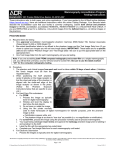
Stereotactic Breast Biopsy Accreditation Program 1891 Preston White Drive, Reston, VA 20191-4397 Testing Instructions Your cover memorandum describes the type of testing that your facility is currently undergoing. If you are going through INITIAL accreditation, accreditation RENEWAL or REINSTATEMENT, you must submit both clinical and phantom images. If you are REPEATING a test on which you received a deficiency, you must only submit images from the deficient test. PHANTOM IMAGE AND DOSE A. Required Items for testing 1. One approved accreditation phantom: the ACR Mammography Accreditation Phantom (Gammex (RMI) 156, Nuclear Associates (Fluke) 18-220, or CIRS 015) or the Mini Digital Stereotactic Phantom (Gammex (RMI) 156D or Nuclear Associates (Fluke) 18250) 2. One blue Landauer dosimeter card for each mammography unit to be tested. Each unit is assigned a specific dosimeter with a unique serial number. Please keep the card away from radiation. Each card contains: a. One Test Dosimeter b. One Control Dosimeter 3. One padded envelope addressed to Landauer and marked “FILM-DO NOT X-RAY.” (Within each envelope you will find the blue dosimeter card.) 4. Bar-coded identification labels to be affixed to the phantom image and the Test Image Data form. IMPORTANT: These labels are for a specific unit and are marked “Phantom Image” and “Test Image Data.” Be sure to put the appropriate label on the appropriate item. 5. One Test Image Data form for each stereotactic breast biopsy unit you are testing. B. Procedure IMPORTANT: This is a 2-step procedure. First, make 1 or more phantom images without a dosimeter; then make 1 separate image of the phantom with the dosimeter. See the specific instructions for your phantom below. 1. Phantom images (without dosimeter) a. Prepare your system for a stereotactic procedure Mount the stereotactic localization device with its collimator and remove the grid (if necessary). Select the exposure mode that you typically use for obtaining scout images (either automatic exposure control [AEC] or manual). If AEC is used, be sure the AEC detector is under the center of the phantom. Select the kVp, mAs (for manual modes), focal spot, target, and filter used for stereotactic localization of a 4.2 cm thick compressed breast of 50% adipose and 50% glandular tissue. b. Instructions for the Mini Digital Stereotactic Phantom Orient the phantom as shown in Figure 1. Take 1 image using your typical technique for a scout image (AEC or manual mode). Record all technique factors on the Test Image Data form. Chest Wall Side Figure 1 Imaging the Mini Digital Stereotactic Phantom This document is copyright protected by the American College of Radiology. Any attempt to reproduce, copy, modify, alter or otherwise change or use this document without the express written permission of the American College of Radiology is prohibited. Page 1 of 7 S:\AccredMaster\SBBAP\Umbrella Testing Materials\SBBAP Testing Instructions.docx Revised: 9/9/14 c. Instructions for the ACR Mammography Accreditation Phantom If you can remove the biopsy collimator and compression plate or have a large enough field-of-view to image the entire insert on 1 image, you may submit 1 image, as long as the entire insert is visible. If you have a large enough field-of-view to image the entire insert on 1 image but the needle holder appears on the image, you may submit 2 images: one with the needle holder as far to the left as possible, the other with the needle holder in the far right position. If you cannot remove the biopsy collimator, the system is restricted to the biopsy aperture, or the system is a dedicated stereotactic breast biopsy unit with small field-of-view digital detector, you must submit 4 images as shown in Figure 2 in order to image all of the test objects. Take 4 overlapping sections of the phantom. Use your typical technique for a scout image (AEC or manual mode). Record all technique factors on the Test Image Data form. Chest Wall Side Figure 2 Imaging the ACR Mammography Accreditation Phantom This document is copyright protected by the American College of Radiology. Any attempt to reproduce, copy, modify, alter or otherwise change or use this document without the express written permission of the American College of Radiology is prohibited. Page 2 of 7 S:\AccredMaster\SBBAP\Umbrella Testing Materials\SBBAP Testing Instructions.docx Revised: 9/9/14 2. Phantom Image (with dosimeter) a. Place the phantom and dosimeter as indicated in Figure 3a for the ACR Mammography Accreditation Phantom or Figure 3b for the Mini Digital Stereotactic Phantom. (You may wish to make preliminary exposures of the phantom without the dosimeter to determine correct positioning of the phantom.) Expose the phantom and dosimeter. For units with AEC, use a manual technique as close as possible to the resultant AEC technique obtained in step B.1. (Do not use an AEC technique; the additional attenuation of the beam by the dosimeter will result in an excessive dose measurement.) For manual systems, use the same technique used in step B.1. for the phantom. Record all technique factors on the Test Image Data form. WARNING: Each dosimeter may be exposed on the phantom once and once only; repeat exposures will yield excessive dose values. The single test image must include both the phantom and dosimeter properly positioned. If a mistake is made, another dosimeter is available for a small fee. Call the ACR for more information. 3. Hardcopy a. Print the phantom images on film or high quality photographic paper (as long as the paper image is of equal quality to a transparency). CDs are not accepted. b. One phantom image must show the entire dosimeter. c. For digital: Print the image(s) as close to “true size” as possible (i.e., without magnification or minification). Do not print the phantom images on the same film that contains the clinical images. Only print and submit images on 10 x 12-in. films (or smaller). If you can only print on larger size film, trim the film so that it is no larger than 10 x 12 in. (but be sure the required area of the phantom image is on the film). Films that are too large will be returned to the facility. d. For screen-film, process the image(s) using the processor normally used for your stereotactic clinical images. e. Place 1 phantom barcode label on each phantom image. (i.e., if you made 5 phantom images with the ACR Accreditation Phantom, use phantom labels 1-5; if you made 2 phantom images with the Mini Digital Stereotactic Phantom, only use phantom labels 1 and 2.) This document is copyright protected by the American College of Radiology. Any attempt to reproduce, copy, modify, alter or otherwise change or use this document without the express written permission of the American College of Radiology is prohibited. Page 3 of 7 S:\AccredMaster\SBBAP\Umbrella Testing Materials\SBBAP Testing Instructions.docx Revised: 9/9/14 CLINICAL IMAGES A. (PLEASE NOTE THAT ANY IMAGE MISSING THE REQUIRED INFORMATION WILL FAIL) Required items for testing 1. Bar-coded identification labels to be affixed to the clinical images and the Test Image Data form. IMPORTANT: These labels are for a specific unit and are marked with the procedure image under evaluation. Use the appropriate label on each item. 2. One Test Image Data form (clinical image section) for each stereotactic unit you are testing. The enclosed labels show when your testing materials are due to the ACR. Failure to meet this due date will jeopardize completion of your accreditation. If your facility is renewing its accreditation, we cannot guarantee completion before your ACR certificate expires. B. Procedure 1. General a. Submit 1 BI-RADS® Category 4 or 5 calcification biopsy case that provides an example of your facility’s best work. Select the images based on the criteria described in the Clinical Image section of the Program Requirements. The biopsied calcifications must be visible on all submitted images. The biopsy may have been performed with a gun-needle biopsy probe, a vacuum suction biopsy probe or another FDA-approved core biopsy device. (Lateral arm units are acceptable only if the lateral arm device is the only option for biopsy and the needle can be seen in relation to the calcifications in 2 views.) Although valuable for some calcifications, “target-on-scout” images are not acceptable for accreditation because reviewers cannot assess the needle position on 2 views. Select a case no older than 6 months from the date on your application. b. Clinical images may be submitted on film or high quality photographic paper (as long as the paper image is of equal quality to a transparency). CDs are not accepted. Only print and submit images on 10 x 12-in. films (or smaller). If you can only print on larger size film, trim the film so that it is no larger than 10 x 12 in. (but be sure the breast image is all on the film). Films that are too large will be returned to the facility. c. You may submit original mammograms or copies (as long as the copies clearly demonstrate the target calcifications). Mammograms must have been taken sometime in the 60 days immediately prior to the date of the biopsy. Mammogram copies must be good quality and clearly labeled with the patient ID and procedure date. Digital mammograms must be printed “true size” (i.e., without magnification or minification) or with a scale. The calcifications to be biopsied must be clearly circled on each projection. IMPORTANT: If the target calcifications are not circled on all mammogram projections, or more than one group is circled, the case will not be evaluated because the reviewers cannot be certain which calcifications are the targets for the biopsy. d. e. f. g. h. i. All images (stereotactic breast biopsy and mammogram) submitted must be from the same patient. Do not submit images that are obtained on models or volunteers. Do not include the radiology or pathology reports with the clinical cases. These will not be sent to the ACR reviewers. Clearly label each image with: PLEASE NOTE THAT ANY IMAGE MISSING THE REQUIRED INFORMATION WILL FAIL The patient’s first and last names (required) Identification number and/or date of birth (required) Examination date (required) Facility name (required) Facility location (city, state and zip) Designation of left or right breast (required) Annotation of mammographic view (e.g., CC, MLO/ML/LM) Technologist’s identification number or initials Label each image with the enclosed barcode labels. (See the Labeling Guide on the last page.) During accreditation review, ACR radiologist reviewers will assess the following case attributes: Visualization of the calcifications in the pre-biopsy mammogram Appropriateness of the pre-fire stereo pair (as applicable) Appropriateness of the pre-biopsy (post-fire) stereo pair (as applicable) Appropriateness of the specimen radiograph Exam identification (The ACR will keep all patient information confidential.) REMEMBER: ACR reviewers assume that the case you submitted for accreditation is an example of your best work. Do not send images you believe are less than that. Consequently, your supervising physician must review the case before submission. This document is copyright protected by the American College of Radiology. Any attempt to reproduce, copy, modify, alter or otherwise change or use this document without the express written permission of the American College of Radiology is prohibited. Page 4 of 7 S:\AccredMaster\SBBAP\Umbrella Testing Materials\SBBAP Testing Instructions.docx Revised: 9/9/14 2. 3. Gun-needle biopsy probe - required images: a. A 2-view mammogram (CC and MLO/ML/LM) performed prior to the stereotactic procedure that includes the entire breast. Circle the calcifications to be biopsied on each image. Print the images “true size” (i.e., without magnification or minification) or with a scale. Label the images “Calc Mammo 1” and “Calc Mammo 2” using the ACR barcode labels. b. A specimen radiograph demonstrating calcium. Label the image “Specimen Radiograph” using the ACR barcode label. c. A Pre-Fire stereo pair demonstrating needle positioning; the calcifications must be visible on both stereotactic views. Label the images “Calc Pre Fire Str Pair” using the ACR barcode labels. Vacuum-suction biopsy probe or other FDA-approved core biopsy device (e.g., intact tissue devices) - required images: a. A 2-view mammogram (CC and MLO/ML/LM) performed prior to the stereotactic procedure that includes the entire breast. Circle the calcifications to be biopsied on each image. Print the images “true size” (i.e., without magnification or minification) or with a scale. Label the images “Calc Mammo 1” and “Calc Mammo 2” using the ACR barcode labels. b. A specimen radiograph demonstrating calcium. Label the image “Specimen Radiograph” using the ACR barcode labels. c. One of the following (as applicable to your practice): i. A Pre-Biopsy (post fire) stereo pair demonstrating needle positioning for tissue acquisition (preferred); the calcifications must be visible on both stereotactic views. Label the images “Calc Pre Biopsy Str Pair” using the ACR barcode labels. OR (if pre-biopsy (post-fire) stereo pair is not possible or the needle on the pre-biopsy (post-fire) images obscures calcifications) ii. A Pre-Fire stereo pair demonstrating needle positioning for tissue acquisition; the calcifications must be visible on both stereotactic views. Label the images “Calc Pre Fire Str Pair” using the ACR barcode labels. IMPORTANT MESSAGE ABOUT LABELS: Labels help us keep track of your films. If you would like to submit additional mammography views that demonstrate the calcifications (e.g., magnification views), use the blank image labels included in this package. If you wish to submit other stereo pairs to demonstrate the positioning of the biopsy device (even if the calcifications are not visible on them) you may do so, but use one of the blank image labels. (If you label these additional images with one of the ACR’s preprinted stereo pair with barcode labels, you may fail accreditation because the reviewer will score the wrong image.) Do not make copies of the existing barcode labels. Use 1 label per image and fill in all the blanks on each label. Retain a copy of the completed label form with the blanks filled in for your records. MAILING INSTRUCTIONS A. Mail the exposed dosimeter and control dosimeters in the pre-addressed, bubble envelope to: Landauer, Inc. ATTN: Mammography Analysis Laboratory 2 Science Road Glenwood, IL 60425-1586 B. Return the QA Questionnaire, Test Image Data form, phantom images, and clinical images to the following address by a traceable method: Stereotactic Breast Biopsy Accreditation Program American College of Radiology 1891 Preston White Drive Reston, VA 20191-4397 The images submitted for review will be returned once the accreditation evaluation is complete. However, you should maintain copies of all images as well as a record of the patient names whose clinical images were sent for accreditation purposes until you receive official notification your accreditation is approved. This document is copyright protected by the American College of Radiology. Any attempt to reproduce, copy, modify, alter or otherwise change or use this document without the express written permission of the American College of Radiology is prohibited. Page 5 of 7 S:\AccredMaster\SBBAP\Umbrella Testing Materials\SBBAP Testing Instructions.docx Revised: 9/9/14 Stereotactic Breast Biopsy Accreditation Program 1891 Preston White Drive, Reston, VA 20191-4397 Labeling Guide Submit 1 calcification case with any probe. Attach the bar-coded labels to the designated images as described below. The calcifications must be visible on all submitted images. Be sure that the labels do not to cover any pertinent clinical or identification information on either the mammogram or the stereo images. Stereotactic Breast Biopsy Accreditation Clinical Images PLEASE NOTE THAT ANY IMAGE MISSING THE REQUIRED INFORMATION WILL FAIL Gun-Needle Biopsy Probe IMAGES BARCODE LABELS 2-view mammogram with the calcifications clearly circled Calc Mammo 1 Calc Mammo 2 Specimen radiograph Calc Specimen Pre-fire stereo pair demonstrating needle position Calc Pre Fire Str Pair Post-fire stereo pair demonstrating needle position NA Vacuum-Suction Biopsy Probe or Other FDA-Approved Core Biopsy Device IMAGES Required for all submissions BARCODE LABELS 2-view mammogram with the calcifications clearly circled Calc Mammo 1 Calc Mammo 2 Specimen radiograph Calc Specimen Pre-biopsy (post-fire) stereo pair demonstrating needle position (preferred) Choose one stereo pair Calc Pre Biopsy Str Pair OR Pre-fire stereo pair demonstrating needle position Calc Pre Fire Str Pair This document is copyright protected by the American College of Radiology. Any attempt to reproduce, copy, modify, alter or otherwise change or use this document without the express written permission of the American College of Radiology is prohibited. Page 6 of 7 S:\AccredMaster\SBBAP\Umbrella Testing Materials\SBBAP Testing Instructions.docx Revised: 9/9/14 Gun-Needle Biopsy Probe: Pre-fire stereo pair 1 Film STERETOACTIC BREAST BIOPSY LABELING SAMPLES PLEASE NOTE THAT ANY IMAGE MISSING THE REQUIRED INFORMATION WILL FAIL IMPORTANT: Be sure that the ACR bar-coded labels are placed below the requested image without covering any pertinent clinical or identification information on either the mammograms or the stereotactic images. If stereo pairs are printed on 2 separate films, complete and affix an Additional Image Label nd to the 2 film. 2 Film Vacuum-Suction Biopsy Probe or Other FDA-Approved Core Biopsy Device: Pre-biopsy (post-fire) stereo pair 1 Film Vacuum-Suction Biopsy Probe or Other FDA-Approved Core Biopsy Device: Pre-fire stereo pair 1 Film 2 Film 2 Film This document is copyright protected by the American College of Radiology. Any attempt to reproduce, copy, modify, alter or otherwise change or use this document without the express written permission of the American College of Radiology is prohibited. Page 7 of 7 S:\AccredMaster\SBBAP\Umbrella Testing Materials\SBBAP Testing Instructions.docx Revised: 9/9/14


