1
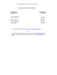
Signosis Innovative Plate Assay Solutions Stat1 Luciferase Reporter HeLa Stable Cell Line Catalog Number SL-0004 (For Research Use Only) Introduction STAT1 exerts a complex array of functions on both tumor cells and the immune system and is usually considered as a tumor suppressor. STAT1 is a central mediator of type II (gamma) IFNs, a family of multifunctional secreted proteins involved in cell growth regulation and antiviral and immune defense. IFNgamma (IFNγ), through JAK1 and JAK2, mainly triggers prolonged STAT1 activation that induces gene expression by binding to gamma-activated sequences (GAS). Signosis has developed Stat1 luciferase reporter stable cell line, which can be used to monitor the activation of Stat1 in response to the stimuli, such as IFNgamma. Principle of the assay The cell line was established by transfection of Stat1 luciferase reporter vector along with hygromycin expression vector followed by hygromycin selection. The hygromycin resistant clones were subsequently screened for oncostatin induced luciferase activity. The clone with the highest fold induction (50 fold) was selected and expanded to produce this stable cell line. Fig.1: Stable cell line diagram Materials provided Handling cells upon arrival One vial of 5 X 10^6 cells, at passage 2, in Freezing Media (store the vial in liquid nitrogen until it is ready to be thawed). Material required but not provided Dulbecco’s Modified Eagle’s Medium (DMEM) Fetal Bovine Serum (FBS) Penicillin (10,000 units/ml) Streptomycin (100ug/ml) Hygromycin B (Roche) Freezing media Luciferase reporter system (Promega E-1500) It is strongly recommended that you propagate the cells by following instructions as soon as possible upon arrival. Genetic instability is a common in all transfected cells, therefore, it is critical to prepare numbers of frozen stocks at early passages. Prepare Complete Growth Media: DMEM (in high glucose + sodium pyruvate + Lglutamine + Phenol Red) + Penicillin (100 units/mL) Streptomycin (100ug/ml) + 10% FBS + Hygromycin (100ug/ml) Signosis, Inc. • 1700 Wyatt Drive Suite 10-12 • Santa Clara, CA 95054 • Tel 408 747 0771 • Fax 408 864 2182 Initial Culture Procedure Important: The first propagation of cells should be for generating stocks for future use. Cells undergo genotypic changes resulting in reduced responsiveness over time in normal cell culture conditions. Therefore, it is critical to prepare an adequate number of frozen stocks at early passages. Quickly thaw cells in a 37 oC water bath with careful agitation. 2. Add 10 ml Complete Growth Media to a sterile 15 ml centrifuge tube then add entire contents of the vial to the media. 3. Spin at 2,000 rpm for 5 minutes. 4. Discard supernatant. 5. Add 1ml of Complete Growth Medium to suspend pellet. 6. Add 10ml of Complete Growth Medium to culture dish and transfer suspended pellet to culture dish containing Complete Growth Medium. 7. Pipette cells up and down to ensure the transfected cells are mixed well in the medium. 8. Place the culture dish with cells in a humidified incubator at 37 oC with 5% CO2. 9. Change media every 2-3 days using Complete Growth Media. 10. When cells reach 90% confluency (usually within 1 week), prepare frozen stocks and continue to propagate the rest of the culture for future assays. 11. Transfer vials from -80oC to liquid nitrogen for long term storage. 1. Assay procedure The following procedure should be followed as a guideline. You will need to optimize the assay conditions based upon your experimental set up. 1. 2. 3. 4. 5. 6. 7. 8. 9. 10. 11. 12. 13. The day before performing the assay, trypsinize the cells and plate each well of a 96-well plate with 5 X 10^4 cells in 100ul. Incubate the plate in a humidified incubator at 37 oC with 5% CO2 overnight. Prepare inducing reagent, such as Oncoststin, at the optimal concentration of 100ng/ml in Complete Growth Medium. Add inducing reagent media mixture to each experimental well and incubate for 8 hours to produce maximal induction. Remove the media by aspiration. Wash wells gently with 1X PBS Add 50ul lysis buffer to each well. Incubate cells in lysis buffer for a few minutes at room temperature. Rock culture dish several times to ensure complete coverage of the cells with lysis buffer. Pipette up and down to ensure complete lysis of cells. Perform one freeze-thaw cycle at -80oC and room temperature. Gently pipet up and down 2-3 times to mix. Transfer 20ul of each lysate to a new 96-well plate for the luciferase assay. Add 100ul of luciferase substrate to each well and gently pipette up and down. Immediately read the plate in a luminometer. Prepare frozen stocks 1. 2. 3. 4. 5. 6. 7. 8. 9. Carefully remove the culture media from cells by aspiration. Rinse cells with PBS, being careful to not dislodge attached cells. Then remove PBS by aspiration. Add 2ml of 0.25% Trypsin/0.53mM Tris-EDTA solution to the culture dish. Let the dish incubate with Trypsin for a few minutes or detached. Confirm detachment by observation under the microscope. Add 10ml of pre-warmed Complete Growth Media and gently pipette up and down to break the clumps. Transfer cells to a 15ml conical centrifuge tube and centrifuge at 2000 rpm for 5 minutes to collect the cells. Aspirate the culture media and resuspend cells at a density of 5 x 10^6 cells/mL in freezing media. Aliquot 1ml cells into cryogenic vials. Place vials in a freezing container and store at -80oC overnight. Data Example Fig 2. Figure: Stat1 Stable cell line Analysis. The cells were seeded on a 96-well plate for overnight with DMEM including 10% FBS. The cells treated with or without 100ng/ml IFNgamma for 8 hours, and then subjected to luciferse assay. Signosis, Inc. • 1700 Wyatt Drive Suite 10-12 • Santa Clara, CA 95054 • Tel 408 747 0771 • Fax 408 864 2182