1
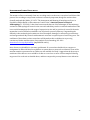
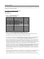
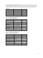
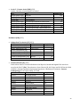
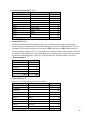
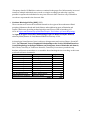
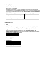
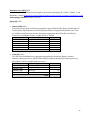
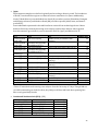
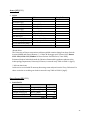
Cranial Nonmetric Trait Database User Guide Nancy S. Ossenberg, PhD. Professor Emeritus, Department of Biomedical and Molecular Sciences Queen’s University Data and Government Information Centre, Queen’s University Library 2013 TABLE OF CONTENTS Introduction .................................................................................................................................................................................... 2 Acknowledgements...................................................................................................................................................................... 3 Record Layout ................................................................................................................................................................................ 4 Traits: Description and Criteria for Scoring ................................................................................................................... 13 1. Sutural Variants ............................................................................................................................................................... 14 2. Traits Related to Nerves, Arteries and Veins ....................................................................................................... 16 3. Variations at the Craniovertebral Border ............................................................................................................. 19 4. Hypostotic traits .............................................................................................................................................................. 22 5. Hyperostotic Traits ........................................................................................................................................................ 23 6. Dental Variants................................................................................................................................................................. 25 Intraobserver Scoring Replicability ................................................................................................................................... 26 Geographic Samples.................................................................................................................................................................. 27 Publications.................................................................................................................................................................................. 38 Bibliography................................................................................................................................................................................. 40 Citation ........................................................................................................................................................................................... 41 Data Processing .......................................................................................................................................................................... 42 1 INTRODUCTION Inspired by the example of the late Dr. W.W. Howells of Harvard University who published his lifetime's worldwide collection of cranial measurements on the internet, I here offer my files of cranial nonmetric data as a freely available resource. The tables were constructed in the program Excel. They are available also in comma-delimited format (comma-separated values, CSV) from which the user can import them into any program of choice. The tables can be used to study the morphological features themselves; i.e. patterns of variation according to age, sex, side and population, as well as intertrait correlation and the effects of artificial cranial deformation. Additionally, I have tried to provide sufficient provenience to facilitate exploration of various ethnogenetic problems. Investigators can pull the tables apart and re-assemble the component samples in any way appropriate to their particular purpose. Additionally, researchers are encouraged to explore methods of biodistance analysis alternative to the Smith’s Mean Measure of divergence (MMD) used in my own ethnogenetic studies. Reflecting my own long-time ethnogenetic research interest, the regions best represented in the tables are the Arctic and Northwestern North America. Since 1991 many of these museum collections have been repatriated under the terms of NAGPRA (Native American Graves Protection and Repatriation Act) and are no longer available for original research. Therefore, I hope that this website may be particularly useful in supplementing existing osteobiological records for the Native peoples of these regions. 2 ACKNOWLEDGEMENTS My fascination with skeletal morphology and variation was fostered by graduate studies 1958-1969 at the University of Toronto's Department of Anatomy under the supervision of Dr. James E. Anderson. Jim, in turn, had been influenced greatly by his mentor in Anatomy, Dr. John Boileau Grant. I dedicate this website to their memory. Queen's University, Kingston, Ontario (1973-1998) and the University of Alberta, Edmonton (19691973) where I taught Anatomy, provided support for my research. Summer 2000 with a travel grant from Central Washington University, Ellensburg, I recorded data for a collection of remains from the Plateau region of North America. During 2001-2002 with a visiting research professorship at Tohoku University, Sendai, Japan, I was able to explore questions of northeast Asian ethnogenesis using nonmetric cranial traits. Grant support by The National Science and Engineering Research Council, the Boreal Institute of the University of Alberta, and The Canada Council are acknowledged in my published articles. The collaboration over many years of Bill Orme, Kingston computing programmer, in making MMD and other calculations has been indispensable for my research. Debra Komar, Queen's Anatomy graduate student, 1994-1996, working from the original paper records, transcribed all my data up to that point to electronic files in the program PARADOX. I am grateful for her care and accuracy in completion of this huge task. My major objective e to put all the data on a website has been realized through endorsement by Queen's University Libraries, with the approval and encouragement of Sharon Murphy, Head, Academic Services. The expertise and work required to transcribe the data files and to design the website was provided by Jeff Moon, Data and Government Information Librarian, and his associate, Alexandra Cooper. I am indebted to them for their meticulous oversight and enthusiastic collaboration on the project. The original paper records also may be consulted by researchers. These are in filing cases stored in Queen's University Archives, under the auspices of Paul Banfield, Queen's University Archivist. Throughout my research career I have studied collections at many museums and other institutions in North America and abroad; I gratefully acknowledge the assistance of their curators and other institutional staff. As for the more than 8000 individuals represented in these files whose remains I have been privileged to study: I hope that their descendants will recognize the gratitude and respect with which I make these data available for posterity. 3 RECORD LAYOUT Record count = 8016 Number of Variables = 84 Columns 2 - 7 contain provenience for the crania. Arranged in nested order from broadest to most specific the columns provide information for each individual according to geographical origin, culture period, and cultural or tribal affiliation. For records marked "not confirmed" there was not enough documentation to definitely identify the tribe or band. GP1 – World Region NA AS EU AF SP Native America and Greenland Northeast Asia Eurasia Africa South Pacific GP2 – Major Region, Country Native America and Greenland AR NW NE SW SA Arctic Northwest, Native America Northeast, Native America Southwest, Native America South America Northeast Asia JA NEA Japan Continental Northeast Asia Eurasia ARM BR BV CZ Armenia Britain Bavaria Czechoslovakia EU FR GE HU Europe France Germany Hungary IC IN IT RU Iceland India Italy Russia Africa AM E N S W African Americans East Africa North Africa (Sudan) Southern Africa West Africa 4 GP3 – Country, Specific Region AL AT AU C CAM CAN CAR CH CHAT CHN EAR GAB GHA HK ILL IND KEN LIB MON MQ N Aleutian Islands Athapaskan territories Australia Central Japan Cameroon Canada Central Arctic Chile Chatham Island North China Eastern Arctic Gabon Ghana Hokkaido Illinois India Kenya Liberia Mongolia Marquesas North Japan Tohoku District: Fukushima, Yamagata, Miyagi prefectures NAL NFL NIG NMV NN NPC NZ ONT PEC PLN PLT PT S SAL SIB SIE SLS SUD TAN TF USA W North Alaska Newfoundland Nigeria Northern Mississippi Valley Northern North Japan Tohoku District: Akita, Iwate, Aomori prefectures North Pacific Coast New Zealand Ontario Pecos Plains Plateau Patagonia Southern Africa South Alaska Siberia Siena St. Lawrence Island, Siberia (Chukotka) Sudan Tanzania Terra del Fuego United States of America West Japan 5 GP4 – General Culture Period, Population or Tribe ABR AI AK Aborigines (Australia) Ainu Alaska, Athapaskans MAO MDV MID Maori Mediaeval Period Middle Period (Kerma) ANC Ancient Period (Kerma) MM Middle Missouri Period ARCH ARIK AS ATQ BK BL BRZ C CA CH CHU CK CLA DK DK-nc DO E EDO MON MOR MRQ MW NM NP OB OKH ONT OR PMJ HD IK IP IRN JOM Archaic Period Arikara Assiniboin Antique Age (Armenia) Birnirk Blackfoot Bronze Age (Armenia) Central Aleut Canada, Athapaskans Cheyenne Chukchi Chinook Classical Period (Kerma) Dakota Sioux Dakota Sioux – not confirmed Dorset Eastern Aleut Edo Period Japanese birth dates approximately 1600-1874 Haida Ipiutak Inupik speakers Iron Age (Armenia) Jomon PR PU PUE ROM SH SL SW TH TL TUN W YAY Mongolians Moriori Marquesans Middle Woodland Period Namu Nez Perce Old Bering Sea Okhotsk Ontario, 19th Century British origin Oregon Athapaskans Post-Meiji Japanese birth dates 1875 and later Prince Rupert Harbour Punuk Pueblo Roman Period (Hungary) Sahaptin Salish Southwest Athapaskans Thule Tlingit Tungus Western Aleut Yayoi KHO Khoisan YKG Yukaghir KO LW MAN Kodiak Islanders Late Woodland Period Manchurians YM YP Yamhill Yupik speakers 6 GP5 – Specific Culture Period, Archaeological Designation, or Sub-tribal Affiliation Site – Specific Site Specific site as stated in the documents of the curating museum. CatalogNo – Museum’s catalog number Catalog number of the skull assigned by the museum Museum Name of the curating institution, date(s) of my survey. AMNH American Museum of Natural History, New York 1975, 2003 CHB Department of Anatomy, Chiba University School of Medicine, Chiba City, Japan 2002 CMC CWU FM IEA IESP KYO KYU LPR MUN NSM O O4 PAN PMH Canadian Museum of Civilization, Gatineau, Quebec 1963, 1965, 1976, 1980, 1991, 1993, 1994 Central Washington State University, Ellensburg, Washington 2000 Field Museum, Chicago, Illinois 1975 Institute for Ethnography, Armenian Academy of Sciences, Yerevan 1994 Institute for Ethnography, Russian Academy of Sciences, St. Petersburg 1981 Kyoto University, Faculty of Science, Laboratory of Physical Anthropology, Japan 1981 Department of Anatomy, Faculty of Medicine, Kyushu University, Fukuoka, Japan 2002 Laboratory for Plastic Reconstruction, Russian Academy of Sciences, Moscow 1981 Memorial University of Newfoundland, St. John's, Newfoundland 1993 National Science Museum, Tokyo 1981, 2002 United States National Museum, Smithsonian Institution, Washington, D.C. 1963, 1964, 1970 United States National Museum, Smithsonian Institution, Washington, D.C. 1995 Panum Institute, Copenhagen 1995 Peabody Museum, Harvard University, Cambridge, Massachusetts 1994 7 QU ROM SAP SEN SFU SIEN STT TKM TKO UAB UAF UCT UG UIN UMA UMN UOR WHO Department of Anatomy, Queen's University, Kingston, Ontario 1975 Royal Ontario Museum, Toronto, Ontario 1958-1960 Department of Anatomy, Sapporo Medical University, Sapporo, Japan 2001 Department of Anatomy and Anthropology, Tohoku University School of Medicine, Sendai, Japan 2001-2002 Department of Anthropology, Simon Fraser University, Burnaby, British Columbia 1989 Department of Anatomy, University of Siena, Japan 1985 St Thomas Church, Belleville, Ontario 1991 University of Tokyo, University Medical Museum, Tokyo 1981 University of Tokyo, University Museum, Tokyo 1981, 2002 Department of Anthropology and Department of Anatomy, University of Alberta, Edmonton, Alberta 1971-1972 Department of Anthropology, University of Alaska, Fairbanks, Alaska 1995 Laboratory of W.S. Laughlin, University of Connecticut, Storrs, Connecticut 1997 Department of Anthropology, University of Geneva 1995 Department of Anthropology, University of Indiana, Bloomington, Indiana 1964 Department of Anthropology, University of Manitoba, Winnipeg, Manitoba 1964 Department of Anthropology, University of Minnesota, Minneapolis, Minnesota 1964 University of Oregon, Museum of Natural History, Eugene, Oregon 1994 W.H. Over Museum, Pierre, South Dakota 1964 8 AgeC – Age cohort 0 1 2 3 4 5 6 8 9 Neonate to five years Six to eight years Nine to eleven years Twelve to fifteen Sixteen to twenty Twenty-one to twenty-nine Thirty and older Child Adult AgeY – Age at death in years Age estimated by a physical anthropologist. Age in years followed by “d” indicates documented age at death for an Anatomy dissecting-room subject, or individual retrieved from a cemetery. Sex 0 2 4 Male Indeterminate Female 9 Deformation Variables Deformation data were originally coded in the variable DeformOriginal. This variable was recoded into two new variables - DeformGrade and DeformLRS. This was done to separate information about deformation itself and the side it appeared on (left, right, symmetrical). The original variable, DeformOriginal, was retained on the dataset. DeformOriginal 0 Undeformed 1 Minimal deformation 2 Slight deformation 3 Moderate deformation 4 Extreme deformation 1L Minimal deformation – left side 2L Slight deformation – left side 3L Moderate deformation – left side 4L Extreme deformation – left side 1R Minimal deformation – right side 2R Slight deformation – right side 3R Moderate deformation – right side 4R Extreme deformation – right side 1S Minimal deformation – symmetrical 2S Slight deformation – symmetrical 3S 4S Moderate deformation – symmetrical Extreme deformation – symmetrical DeformGrade – Grade of deformation 0 Undeformed 1 Minimal deformation 2 Slight deformation 3 Moderate deformation 4 Extreme deformation 5 Deformation – no gradation 999 Presence or absence of deformity was not recorded DeformLRS – Side of most pronounced occipital flattening (left, right, symmetrical) L Left side > right side R Right side > left side S Symmetrical deformation [blank] Symmetry/asymmetry of deformation was not recorded NO-OBS Presence or absence of deformation was not recorded 10 Trait List See TRAITS: DESCRIPTION AND CRITERIA FOR SCORING descriptions of the traits. METO APIC INCA OMBL OMBR ASTL ASTR PNBL PNBR POSL POSR PCTB ODON TRFS PHAR HYPL HYPR PCPL PCPR ICCL ICCR SQSL Metopic suture Apical bone Os inca Occipito-mastoid ossicle, left Occipito-mastoid ossicle, right Asterionic ossicle, left Asterionic ossicle, right Parietal notch bone, left Parietal notch bone, right Posterior condylar canal absent, left Posterior condylar canal absent, right Precondylar tubercle(s) Odonto-occipital articulation Transverse fissure of basiocciput Pharyngeal fossa Hypoglossal canal bridged or double, left Hypoglossal canal bridged or double, right Paracondylar process, left Paracondylar process, right Intermediate condylar canal, left Intermediate condylar canal, right Parietal process of temporal squama, left SQSR Parietal process of temporal squama, right SPSL SPSR MARL Squamoparietal synostosis, left Squamoparietal synostosis, right Marginal foramen of tympanic plate, left MARR Marginal foramen of tympanic plate, right TYML TYMR FSPL Tympanic dehiscence, left Tympanic dehiscence, right Dehiscent wall of foramen spinosum or foramen ovale, left FSPR Dehiscent wall of foramen spinosum or foramen ovale, right LPFL LPFR CIVL Foramen in lateral pterygoid plate, left Foramen in lateral pterygoid plate, right Pterygospinous bridge complete (foramen of civinini), left CIVR PTBL Pterygospinous bridge complete (foramen of civinini), right Pterygobasal spur or bridge, left PTBR Pterygobasal spur or bridge, right CLNL CLNR Clinoid bridging, left Clinoid bridging, right 11 SOFL SOFR FRGL FRGR TRSL TRSR OPTL OPTR ORBL ORBR CONL CONR JAPL JAPR M3UL Supraorbital foramen, left Supraorbital foramen, right Frontal groove(s), left Frontal groove(s), right Trochlear spur, left Trochlear spur, right Accessory optic canal, left Accessory optic canal, right Orbital suture variant, left Orbital suture variant, right Infraorbital suture variant, left Infraorbital suture variant, right Transversozygomatic suture, left Transversozygomatic suture, right Upper third molar congenitally absent, left M3UR Upper third molar congenitally absent,right MENL MENR MHBL Accessory mental foramen, left Accessory mental foramen, right Mylohyoid bridge, left MHBR Mylohyoid bridge, right BUCL Retromolar foramen, left BUCR M3LL M3LR Retromolar foramen, right Lower third molar congenitally absent, left Lower third molar congenitally absent, right TRML Three-rooted mandibular first molar, left TRMR ATAL ATAR ATBL ATBR Three-rooted mandibular first molar, right Atlas bridging, condylar process to posterior arch, left Atlas bridging, condylar process to posterior arch, right Atlas bridging, condylar to transverse process, left Atlas bridging, condylar to transverse process, right 12 TRAITS: DESCRIPTION AND CRITERIA FOR SCORING Descriptions of the traits and bibliographical references are provided in my published articles, 1969 to 2006 (see PUBLICATIONS); also in Hauser, G. and G.F. DeStefano. 1989. Epigenetic Variants of the Human Skull, Schweizerbart, Stuttgart. For some traits, definitions and description provided by other authors are cited (see BIBLIOGRAPHY). The traits are classed in six categories: 1. Sutural Variants 2. Traits Related to Nerves, Arteries and Veins 3. Variations at the Craniovertebral Border 4. Hypostotic Traits 5. Hyperostotic Traits 6. Dental variants These categories are not mutually exclusive; traits in categories 1 – 3 generally have a secondary classification as either 4 hypostotic or 5 hyperostotic, while certain traits classed primarily as hyperostotic also have a relationship to nerves or blood vessels. Some well-known traits have been excluded from the files. These are the tori (maxillary, mandibular, palatal and auditory) likely to be strongly influenced by behaviour or environmental factors. Others traits generally familiar to researchers I have found too ambiguous in expression, too difficult to score, or simply too invariant to be useful in ethnogenetic studies. Scoring of Traits General rule for all traits: trait present = 1 trait absent = 0 presence or absence of trait indeterminate = 9 The exception to this rule is where a trait is scored 2 or 3 indicating degree of expression or subtype of the feature. 13 1. Sutural Variants METO Metopism An interfrontal suture persisting after the normal age (~ 8 years) for closure is scored 1. The common case where only the supranasal portion persists is scored 0. INCA Os inca A persisting mendosal (biasterionic) suture dividing the occipital squama into an upper and lower portion is scored 1. The upper portion may be subdivided into segments (Epigenetic Variants, Fig 15, i-w), presence of one or more of which is scored 1. A lateral trace of the biasterionic suture is scored 0. APIC Os apicis (apical bone) Any ossicle at lambda (Epigenetic Variants, Fig. 15, a-d) is scored as 1. The distinction between this feature and the median segment of an Os inca (Epigenetic Variants, Fig 15, t) depends on size. OMB Occipito-mastoid bone An ossicle of any size in the occipito-mastoid suture and which does not touch asterion is scored 1. AST Asterionic bone An ossicle of any size touching asterion is scored 1. PNB Parietal notch bone An ossicle of any size in the parietal notch is scored 1. SPS Squamo-parietal synostosis Fusion of the squamo-parietal suture in whole or part is scored 1. Partial fusion involving the posterior one-third is the most common expression of this rare anomaly (Ossenberg, 1976, Pl. 2D). ORB Orbital suture variant The inferior orbital fissure is situated between the floor and lateral wall of the orbit. The anterior end of the fissure is usually bounded bythe junction of the maxilla with the zygomatic bone, scored 0. Alternatively, a tongue of the greater wing of sphenoid intrudes to form a spheno-maxillary contact or short suture at this site, a variant scored 1 (Kozintsev, A.G. 1992. “Ethnic Epigenetics: a New Approach”. Homo 43: 213-244). This site can be examined either by looking into the orbit from the frontal aspect, or by examining the sutures in the anterior wall of the infratemporal fossa. My data are based on the latter approach. CON Infraorbital suture variant Confluence of the infraorbital suture with the zygomaxillary suture at or below the orbital margin is scored 1. (Epigenetic Variants, Fig. 11 b,c; Ossenberg 2006, Fig. 3; Kozintsev AG 1992. “Ethnic Epigenetics: a New Approach”. Homo 43: 213-244). In cases where the infraorbital suture appears to be obliterated – a normal age change – its remnants can sometimes be made visible by wetting the area. 14 JAP Os japonicum A complete transversozygomatic suture dividing the bone into an upper portion and a lower (Os japonicum) portion is very rare in most populations (with the exception of the indigenous populations of Japan) and is scored 2. A posterior trace of the suture 2mm or more in length is scored 1. 15 2. Traits Related to Nerves, Arteries and Veins POS Postcondylar canal A postcondylar canal which in life transmits a vein communicating between the sigmoid sinus and the suboccipital plexus is usually present bilaterally. A right or left side in which a canal or foramen of any size pierces the condylar fossa is scored 0; absence of the postcondylar canal on that side is scored 1. SQS Squamous style The parietal process of the temporal squama is essentially a bony manifestation of an arterial variant. The middle temporal artery normally arises from the auriculotemporal, pierces the temporalis fascia and then runs up deep to the temporalis, its course marked by a vertical groove on the side of the cranial vault above the root of the zygomatic arch. The variant is the origin of the middle temporal artery from the middle meningeal in the middle cranial fossa. The anomalous artery then exits the skull via a “squamoparietal canal” formed between the beveled surfaces of the squamoparietal suture. The canal usually, though not in every case, extends upwards for a short distance by means of a flat style-like projection of the squama. Above its point of exit the artery etches a telltale pattern of branching groves on the parietal bone while the normal vertical groove above the zygomatic root is usually (not always) absent (1969, p 114; 1976, Pl 2B; Epigenetic Variants p. 191 and Pl xxviii a). This variant is scored 1. Other variants involve branches of the middle meningeal directly piercing the temporal squama or the parietal bone; however, these are extremely rare and were not recorded. MAR Marginal foramen of the tympanic plate (1969, p 48; 1976, Pl 4B) This bony foramen during life encloses the auriculotemporal nerve branch just before it pierces the cartilage of the external auditory meatus to supply the skin lining the meatus. The lateral margin of the tympanic plate, though rough and porous in all individuals, in some cases shows a distinct groove at the mid-point of its inferior surface; the groove is scored 0. The variant scored 1results from bony spicules growing from the edges of a well-defined groove and meeting to form a foramen. Spurs which do not quite meet are scored 0. In order to see the canal which is more or less sagittally oriented the skull must be tilted obliquely from the basal view (from which tympanic dehiscence is observed). LPF Lateral pterygoid plate foramen (1969 p. 136; 1976 Pl 3 A, B) This is a round or oval foramen 1-2 mm in diameter piercing the lateral pterygoid plate close to its posterior border and roughly at its mid-point or, more superiorly, near the roof of the infratemporal fossa. During life it transmits the mandibular nerve branch and/or vascular structures supplying the medial pterygoid muscle. SOF Supraorbital foramen A branch of the frontal nerve and associated vessels supplying the skin of the forehead and scalp in some crania exit the orbit through a bony foramen or canal piercing the superior orbital margin. In some cases the canal is deep, its external opening as much as 15 mm above the orbital margin; deepest canals tend to occur in the lateral, rather than in the middle or supratrochlear portion of the margin. More commonly the feature is a foramen seemingly formed when spicules of bone growing from the 16 edges of a deep notch meet; these tend to occur in the middle portion of the margin or towards its medial end. Any such canal or foramen which communicates between the roof of the orbit and the external surface of the frontal bone is scored 1. Two or more such features on the same side are scored 2. A deep notch in the orbital margin – even where the spicules of bone almost meet – is scored 0. Openings for diploic veins are scored 0. FRG Frontal grooves (Epigenetic Variants p. 48; Pl VII a, b) Frontal grooves scored 1 are usually single, sometimes multiple, grooves impressed into the lateral portion of the frontal bone by branches of the supraorbital nerve and/ or vessels running upwards from the orbital margin to enter the skin of the forehead and scalp. Frontal grooves often occur in association with a deep supraorbital canal, but they also occur independently of the presence of a canal or foramen. In some cases, vague meandering grooves run transversely on the frontal bone; such cases are scored 0. OPT Accessory optic canal (1969 p. 118; 1976 PL I A, B) This rare anomaly is essentially a bony manifestation of an ophthalmic artery variant. Normally, the ophthalmic artery branches off the internal carotid immediately after the latter pierces the dural roof of the cavernous sinus; it then enters the orbital cavity through the optic canal, lying on the floor of the canal below and lateral to the optic nerve. In the anomalous case the ophthalmic artery arises from the cavernous portion of the internal carotid and enters the orbit through an accessory optic canal or notch in the stout postero-inferior root of the lesser wing of the sphenoid which forms the floor of the optic canal. The complete accessory optic canal is a round canal approximately 2 mm in diameter piercing the floor of the optic canal, scored 2. An incomplete accessory optic canal scored 1 is the case where the floor of the optic canal is either notched or deficient; i.e. abnormally slender. According to the position and orientation of the accessory optic canal (or notch) it appears that the postero-inferior root of the lesser wing during development is obstructed by, and must chondrify around, the anomalous ophthalmic artery. MEN Mental foramen double The mental nerve and associated vessels arise in the mandibular canal and exit the mandible via the mental foramen to supply the skin of the lip and chin region. Usually there is a single mental foramen, scored 0. The case of two or more foramina is scored 1. BUC Retromolar foramen (Ossenberg, 1987) This trait relates to variations in the course and connexions of the buccal nerve, a branch of the anterior division of the mandibular nerve carrying sensory fibres from the cheek and buccal gingiva. The buccal nerve usually passes medial to the tendon of the temporalis at its attachment to the coronoid process and temporal crest. In some individuals sensory nerve branches from the molar roots run upwards to pierce the bone of the retromolar fossa and join the buccal nerve (1987, Fig 2A). In some cases, in its passage backwards from the cheek, the buccal nerve (or part of it) pierces the 17 bone of the retromolar fossa, receives accessory sensory branches from the molar roots and then joins the inferior alveolar nerve close to the opening of the mandibular canal (1987, Fig 2B). Any retromolar foramen as described above is scored 1. Very rarely, the buccal nerve runs through a conspicuous temporal crest canal whose anterior opening is in the upper portion of the retromolar fossa and whose posterior opening is behind the temporal crest (1987, Fig 2C); this rare anomaly is scored 2. 18 3. Variations at the Craniovertebral Border The exoccipital and supraoccipital portions of the occipital bone develop in paraxial mesoderm with four somites serially homologous with those of the atlas and axis vertebrae. The basiocciput, including the anterior portions of the occipital condyles, develops from the parachordal cartilages surrounding the cranial end of the notochord. Certain morphological traits at the craniovertebral border result from variations in the number, or in the normal pattern of fission and coalescence, of these primordial elements (Barnes, Ethne. 1994 Developmental Defects of the Axial Skeleton in Paleopathology. University Press of Colorado; Ossenberg 1969, p. 145-185; Wackenheim, Auguste. 1974. Roentgen Diagnosis of the Craniovertebral Region. New York, Springer-Verlag). PCTB Precondylar tubercles (1969 p. 161 - ; Epigenetic Variants p. 134 -; Fig 22 b, c; Pl XIXg; Pl XX f, h) Precondylar tubercles are bilateral small bony protuberances on the basiocciput near the anterior margin of the foramen magnum. As a manifestation of the occipital vertebra they are thought to result from “cranial shift” at the craniovertebral border; i.e. the cartilaginous primordia of the occipital bone fail to integrate completely, so that the anterior margin of the foramen magnum retains morphological features reminiscent of the anterior arch of the atlas (Barnes 1994). Precondylar tubercles vary; but most cases are small discrete paired tubercles, usually of unequal size, situated close to the midline and separated from each other by a narrow gap. In other cases paired bony ridges extend medially towards each other from the antero-superior portions of the condyles. Very rarely, the condylar articular suface extends medially onto such a precondylar ridge for articulation with an anomalous anterior arch of atlas. Any such bony tubercle or ridge is scored 1. (The Basilar tubercle (1969 p.165) is a median protuberance extending backwards into the foramen magnum and, as it theoretically represents an ossified notochord remnant, it is a trait distinct from the PCT. This feature was scored but not included in my tables). ODON Odonto-occipital articulation (1969 p. 157) This is a rare oval facet 5-6 mm wide on the anterior margin of the foramen magnum for articulation with the tip of the odontoid process, scored 1. It is flat or shallowly concave and, in contrast to the condyles, is barely raised above the plane of the foramen magnum. (This trait is distinct from Condylus tertius (Epigenetic Variants, Fig. Pl XXg) - also a rare anomaly - which articulates with a facet on the anterior arch of the atlas and is located on the basiocciput anterior to the foramen magnum. Condylus tertius was not recorded separately in my studies, though it is possible that a few cases were scored as Precondylar tubercles). TRFS Transverse fissure of basiocciput (1969, p.159 - ; Barnes, 1994 p. 84, Fig. 3.25) This rare anomaly is a transversely oriented slit or dehiscence penetrating the basiocciput on one or both sides. Sometimes the slit is isolated within the occipital bone; more often it extends laterally to the petro-occipital synchondrosis giving the basiocciput a “waisted” apprearance. Any such feature is scored 1. Barnes (1994) places this anomaly among those resulting from cranial border shift. 19 PHAR Pharyngeal fossa (1969, p. 168 -; 1976, Pl 5A; Epigenetic Variants, p. 137; Pl XX b, f) Pharyngeal fossa is a median fovea in the basiocciput anterior to the pharyngeal tubercle. The smallest is a shallow pit about 1 mm in diameter barely indenting the bone and is scored 1. Any pit more conspicuous than the minimal expression is scored 2. The largest tend to be tub-shaped and may be up to 3 mm in both circumference and depth. A related variant, extremely rare and scored 3, is called the Median basilar canal; it tunnels obliquely from its external opening at the site of the Pharyngeal fossa to its internal opening on the clivus of the skull. The fossa and the canal may be related to remnants of the notochord. HYP Hypoglossal canal bridged or double (1969, p. 145- ; Epigenetic Variants, p. 120-) The hypoglossal (anterior condylar) canal gives passage to cranial nerve XII and occasional vascular structures. It is usually single and undivided. The presence of two hypoglossal canals; or a case where the canal is partly occluded by a bony bridge – whether at the internal or the external aperture or anywhere within the canal - is scored 1. Any partial expression of a bridge (spurs) is scored 0. This trait is one of the manifestations of the occipital vertebra theoretically resulting from incomplete coalescence of the occipital somites; i.e. cranial border shift. PCP Paracondylar process (1969, p. 151 - ; 1976 Pl C, D; Epigenetic Variants, p. 128 -, Pl XIX g) Lateral to the posterior portion of the occipital condyle the surface of the jugular process giving attachment to the rectus capitis lateralis muscle is usually flat or gently convex, and fairly smooth in texture. In some crania a Paracondylar process projects from this surface. The feature scored 1 is the minimal expression of the PCP, a very small conical tubercle or eminence. A conspicuously large process; or one of any size with an articular facet for the transverse process of the atlas (or with an epitransverse process, Barnes 1994 p. 89) whether the articular facet on the PCP is discrete from - or continuous with - that of the occipital condyle is scored 3. In subadults the PCP may bear a flattened corrugated facet resembling the epiphyseal surface at the end of a growing limb bone, suggesting a temporary cartilaginous joint with the atlas or with a separate PCP epiphysis; such a feature is scored 3. Any process larger than the minimal expression but not qualifying for a 3 is scored 2. Paracondylar processes (and Precondylar tubercles) are often involved in cases of the rare anomaly Atlanto- occipital assimilation. I described cases of this anomaly in the cranial series I examined but have not included it in the tables. According to Barnes (1994) Atlanto-occipital assimilation represents caudal shift at the cranio-vertebral border. However, Paracondylar process by itself likely represents persistence of the transverse element of the occipital vertebra; i.e. cranial shift at the border. Because of the range of expressions of PCP, some of which involve reciprocal anomalies of the atlas, perhaps it would be best to classify all such expressions including Atlanto-occipital assimilation neither as cranial shift nor as caudal shift, but more generally as hypoplasia at the craniovertebral border. 20 Because of its wide range in expression I have found Paracondylar process to be one of the most difficult features to score and, out of exasperation, many times contemplated scrapping it. Yet, I persisted in recording it not only because of its inherent morphological and clinical significance, but also because with scoring refinements PCP may prove to be a powerful population discriminator. ATA Atlas bridging, type A A bony bridge on the atlas extending from the condylar process to the posterior arch and spanning the groove for the vertebral artery (Epigenetic Variants, Fig. 17h) is scored 1. Incomplete bridges - spurs which do not meet – (Epigenetic Variants, Fig.17 e-g) are scored 0. ATB Atlas bridging, type B A bony bridge on the atlas extending from the condylar process to the transverse process and spanning the groove for the vertebral artery (Epigenetic Variants, Fig.17d) is scored 1. Incomplete bridges – spurs which do not meet (Epigenetic Variants, Fig. 17, a-c) - are scored 0. Both types of bridging can occur on the same vertebra. An atlas bridge of either type occasionally forms a synovial articulation with a Paracondylar process. According to Barnes (1994) atlas bridges are thought to result from caudal shift at the craniovertebral border (but see the note above under PCP). 21 4. Hypostotic traits These features represent arrested morphogenesis; i.e. retention of a fetal or immature stage beyond the age when development is normally completed and adult morphology attained (Ossenberg, 1970). TYM Tympanic dehiscence (1969, p. 34-; Epigenetic Variants, p. 143-) At birth the tympanic membrane is very superficial on the skull, framed by a C-shaped bony ring. Subsequently, growth laterally from the anterior portion of the C-shaped ring forms the floor of the auditory meatus. Up to the age of about five years there is a normal developmental dehiscence in the floor of the meatus which is gradually filled in by tiny finger-like processes of bone derived from its margins which give it a transitory cribriform appearance. The tympanic plate attains its full lateral growth during in adolescence by which time the dehiscence normally has been obliterated. Except for its rough and porous lateral margin, the mature tympanic plate normally is not perforated. The feature scored 1 is an aperture in the middle of the plate ranging in size from a pinhole, to a dehiscence as large as 5 mm. The margins of the larger ones often have a jagged or lacerated appearance. In some cases the dehiscent area is cribriform, in other cases may consist of two or more pinholes. FSP Deficient wall of the foramen spinosum and / or ovale (1969, p. 38; Epigenetic Variants, p. 149-) The roots of the sphenoid greater wing in the region of foramen rotundum and the pterygoid canal are preformed in cartilage; the other portions of the greater wing ossify in membrane. Early in the human fetus neither the foramen spinosum nor foramen ovale are differentiated: the mandibular nerve, middle meningeal artery and associated structures make their exit from, or entry to, the middle cranial fossa through the foramen lacerum medium as in the adult forms of other mammals. Subsequently, bone encroaches on and surrounds the neurovascular structures thereby separating the foramen ovale, foramen spinosum (and occasionally an emissary foramen of Vesalius anterior to foramen ovale) from each other and from the sphenopetrous fissure. Various expressions of arrested morphogenesis in this region are recognized: foramen ovale and spinosum are confluent, either foramen communicates with the sphenopetrous fissure, both foramina open into the fissure, or any combination of these deficiencies. The communications vary from the merest suture-like slit, to large deficiencies in the walls. Foramen spinosum confluent with sphenopetrous fissure is the trait most commonly seen. Any such variant is scored 1. 22 5. Hyperostotic Traits These features are characterized by an excess of ossification over the normal condition; i.e. ossification into structures normally membrane, cartilage, ligament or dura (Ossenberg 1969, 1970). ICC Intermediate (lateral) condylar canal (1969, p. 74- ; 1976, Pl. 6 A, B; Epigenetic Variants, p. 126-) A small vein commonly connects the beginning of the internal jugular and the anterior condylar (hypoglossal) emissary vein with the postcondylar emissary vein or suboccipital plexus. This vein runs backwards in a groove lateral to the base of the occipital condyle. In some crania a bony crest from the lateral lip of the groove grows medially to fuse with the base of the condyle whereby the groove for all of its length - or more commonly for a short portion - is converted to a canal one to two mm in diameter. Any such canal is scored 1. A spur or crest which fails to fuse with the condyle is scored 0. Because of the orientation of the ICC its openings especially in the case of a long canal of small diameter easily escape notice; the skull should therefore be tilted slightly to an oblique position to make the observation. CIV Pterygospinous bridge (Foramen of Civinini) (1969, p. 50 - ; 1976, Pl 3 A, B; Epigenetic Variants, p.156-) The pterygospinous ligament stretches from a point near the middle of the posterior border of the lateral pterygoid plate to, or to some point near, the spine of the sphenoid. The variant scored 1 is complete ossification of this ligament. A case where spurs extend towards each other but do not actually join is scored 0. The Pterygospinous bridge forms a foramen, more or less sagittally oriented, and situated below and medial to the foramen ovale. This anomalous bony foramen varies in size and shape according to the extent of ossification of the structures forming its margins (i.e. the plate, ligament and spine) and may be subdivided into two or more apertures, completely, or partially by means of bony spurs. The trait LPF (Lateral pterygoid plate foramen) often occurs with a Pterygospinous bridge, but can also occur independently. PTB Pterygobasal bridge (1969, p. 53 - ; 1976, Pl 3 C,D; Epigenetic Variants, p. 156 -, Pl XXIV g, h, i) A ligament commonly stretches from the posterior border of the lateral ptyerygoid plate near its root, to a point on the greater wing of sphenoid lateral to the foramen ovale. The ligament likely gives attachment to fibres of the upper head of the lateral pterygoid muscle, and stretches below and protects the masseteric and deep temporal branches of the mandibular nerve. Occasionally, as they course laterally from the foramen ovale on the greater wing of sphenoid these nerves for a short distance lie in an approximately 5 mm wide shallow sulcus. Independent of the presence or not of a sulcus, the ligament may ossifiy completely or partially. Minimal expression scored 1, is a tiny sharp forward- pointing spur on the greater wing lateral to the foramen ovale; also scored 1 is the case where a shallow sulcus is present and deepened slightly by a bony spur or crest seemingly pinched up from its posterior margin. Full expression scored 3 is either complete ossification of the ligament or spurs that almost connect, with only a slit-like gap between them. An expression larger than minimal yet not sufficient to merit a 3 is scored 2. Though situated close to each other on the 23 roof of the infratemporal fossa Pterygospinous bridge, situated medial to the foramen ovale, cannot be confused with Pterygobasal bridge which lies lateral to the foramen; they are distinct and independent traits. CLN Clinoid bridges (1969, p. 57 - ; 1976, PL 5 C, D; Epigenetic Variants, p. 162, Pl XXIV b, c) On each side of the sella turcica the clinoid processes – anterior, posterior and middle (inconstant) – are normally joined by “ligaments” of the dura reinforcing the attachments of the tentorium cerebelli. The anomalous case is where two or three of the clinoid processes are joined by a bony bridge instead of by dura. When the anterior and middle processes are so joined the carotico- clinoid canal is formed, enclosing the internal carotid artery as it bends upwards to pierce the dural roof of the cavernous sinus: this variant, AM, is scored 1. A bridge uniting the anterior and posterior processes, AP, is scored 2. The case where all three processes are joined, AMP, is scored 3. Also included in these categories are cases in which the bridge appears to be interrupted; i.e. slender bony bars with flattened ends approach each other so closely that they appear to form a tiny joint. Any lesser spur expression is scored 0. The region of the sella turcica is observed with a pen-light and dental mirror inserted through the foramen magnum. TRS Trochlear spur (1969, p. 62- ; 1976, PI 1 C; Epigenetic Variants, p. 64, PI IXf) This variant is a small spine on the upper medial wall of the orbit at the site of attachment of the fibrocartilaginous pulley for the tendon of the superior oblique muscle of the eyball. It represents ossification into one of the two ligaments –most commonly the ligament of the posterior-superior horn - attaching the cartilaginous arc of the pulley to the frontal bone. The bony spur varies from barely perceptible to well-developed: any expression is scored 1. MHB Mylohyoid bridge (1969, p.66- ; 1974; 1976, Pl 6 C, D; Epigenetic Variants, p. 234-) Ossification of the sphenomandibular ligament at its insertion on the medial surface of the mandibular ramus converts the mylohyoid groove to a bony canal enclosing the mylohyoid nerve and vessels, a variant scored 1. The mylohyoid canal varies in length from 2 to 25 mm and may be interrupted into two or more segments. Rarely, the mylohyoid canal opens superiorly at the level of the mandibular foramen. In this case its opening is often shielded by an extension backwards of the lingula (the extension also representing ossification into the sphenomandibular ligament). Such high-opening mylohyoid canals, especially if they are long ones, can easily be overlooked. Mylohyoid bridges starting at the level of the mandibular foramen were noted separately on my scoring sheets but included with the other MHB variants scored 1 in the tables. 24 6. Dental Variants Three dental variants were included in my survey. These were chosen because, unlike most morphological features of the crown for which scoring requires unworn teeth and the expertise of a dental anthropologist, these are fairly easy to assess and are observable in most crania. M3U Upper third molar suppressed Congenital absence or suppression of the third molar is scored 1. A juvenile case where the molar crown had still been forming in its crypt at time of death is scored 0; as is any case where the molar is present, or where the empty socket shows it had fallen out post-mortem, or where the alveolus shows evidence that the molar had been lost ante-mortem. A peg-shaped third molar or one greatly reduced in size was noted on my scoring sheets but entered in the tables as 0. M3L Lower third molar suppressed The same protocol as above applies to scoring M3L. TRM Three-rooted mandibular first molar (Turner, C.G. II. 1971. “Three-rooted Mandibular First Permanent Molars and the Question of American Indian Origins”. American Journal of Physical Anthropology 34: 229-242) Normally the first permanent mandibular molar has two roots: this case is scored 0. The variant scored 1 is the presence of an extra, distolingual root. This variant is scored most easily by counting the root sockets in the case where the tooth had been lost. When the molar was still in the jaw, gently rocking the tooth will usually permit it to be elevated sufficiently to observe the roots at the level of their bifurcation. The extra root usually, but not always, angles more towards the lingual surface of the alveolus than does the mesial root. X-rays to assess TRM were not used in my survey. I started to record TRM routinely in 1981. However, I had the opportunity in the 1990’s during visits to the USNM and CMC, to re-examine samples scored previously and update these for Orbital suture variant, Infraorbital suture variant as well as for Three-rooted mandibular first molar. 25 INTRAOBSERVER SCORING REPLICABILITY The measure of how consistently from one recording session to the next a researcher has followed the protocol for recording a trait present or absent is extremely important, though few workers have formally analyzed this (Molto, J.E. 1979. “The Assessment and Meaning of Intraobserver Error in Population Studies Based on Discontinuous Cranial Traits”. American Journal of Physical Anthropology 51: 333-344). It has often been asserted that one of the advantages of discontinuous variants for anthropological research is the ease with which they can be scored and standardized. This is not a valid assumption: the wide range of expression in size and/or position of several of the traits means that it can be difficult to establish a cut-off point for presence/absence. Compounding the difficulty is that archaeological remains are often incomplete, damaged, or affected by preservatives and storage methods. My research with nonmetric traits has occupied a span of some forty years. The usefulness of these data to other researchers will depend on their confidence in my scoring consistency. Results and discussion of my analysis are in Ossenberg, N.S. 2011. Intraobserver Scoring Replicability. [unpublished article]. Inter-observer replicability is even more problematic. If a researcher should wish to compare or amalgamate his data with mine he would have to ensure that our protocols are identical. Some of the museum samples represented in my Tables are still available for study. I suggest that the best way to ameliorate inter-observer error would be for him to record one or more of these samples following my protocol for each trait as detailed above; and then compare his present/absent scores with mine. 26 GEOGRAPHIC SAMPLES The Cranial Nonmetric Trait Database is based on 27 files organized by geographic region. Each of the original files is indicated by a bullet. NATIVE AMERICA AND GREENLAND (NA) (GP1) Arctic (AR) (GP2) Arctic 1 – South Alaska (SAL) (GP3) Alaska Peninsula to Golovin Bay GP4 GP5 Sample Size Port Moller 8 Yupik –speakers (YP) Naknek River 61 Yupik –speakers (YP) Nushagak River 72 Yupik –speakers (YP) Kuskokwim River, middle 66 Yupik –speakers (YP) Kuskokwim River, lower 62 Yupik –speakers (YP) Nelson Island 32 Yupik –speakers (YP) Nunivak Island 107 Yupik –speakers (YP) Hooper’s Bay 29 Yupik –speakers (YP) Yukon River, middle 43 Yupik –speakers (YP) Yukon River, lower 51 Yupik –speakers (YP) Norton Sound 39 Yupik –speakers (YP) Golovin Bay 100 Total 670 Arctic 2 – St. Lawrence Island, Siberia (Chukotka) (SLS) (GP3) The assignment of the USNM St Lawrence Island remains to Old Bering Sea, Punuk or recent Yupik-speakers was based on the 1996 report of the Repatriation Office. According to an article by H.-G. Bandi and R. Blumer (Investigations by Swiss Archaeologists on St. Lawrence Island, Alaska. 2002. In: Archaeology of the Bering Strait Region, edited by D.E. Dumond and R.L. Bland, University of Oregon Anthropological Papers no. 59: 25-59.) most of the remains excavated by Hans-Georg Bandi and colleagues and curated at the University of Geneva belonged to the Punuk cultural group. A few were associated with Old Bering Sea cultural objects. I assigned the individual crania in my files according to this information. Importantly, the extensive radiocarbon dates provided by R. Blumer (Radiochronological Assessment of Neo-Eskimo Occupations on St. Lawrence Island. In: Archaeology of the Bering Strait Region, edited by D.E. Dumond and R.L. Bland, University of Oregon Anthropological Papers no. 59: 61-106.) indicate that Old Bering Sea and Punuk were to a large extent contemporaneous but distinct sociocultural units on St. Lawrence. Of the many burials in the Ekven Cemetery, Chukotka, I recorded data for 77. I placed these all in a single “Old Bering Sea” file. However, according to an article by M.M. Bronshtein and K.A. Dneprovsky (The Northeastern Chukchi Peninsula during the Birnirk and Early Punuk Periods. In: 27 Archaeology of the Bering Strait Region, edited by D.E. Dumond and R.L. Bland, University of Oregon Anthropological Papers no. 59: 153-166.) the representation of cultural groups in this cemetery is much more complicated including elements of Punuk and Birnirk as well as Old Bering Sea. GP4 GP5 Sample Size Old Bering Sea (OB) St. Lawrence Island 17 Punuk (PU) St. Lawrence Island 120 Yupik-speakers (YP) St. Lawrence Island 129 Old Bering Sea (OB) Siberia Chukotka 77 Yupik-speakers (YP) Siberia Chukotka 80 Total 423 Arctic 3 – North Alaska (NAL) (GP3) Seward Peninsula to Point Barrow GP4 GP5 Sample Size 52 Inupik- speakers (IP) Kauwerak 40 Inupik- speakers (IP) Wales 72 Inupik-speakers (IP) Shishmarev Point Hope, Ipiutak 56 Ipiutak (IK) 61 Inupik-speakers (IP) Point Hope, Tigara 50 Inupik-speakers (IP) Point Hope, recent Point Barrow, Birnirk 44 Birnirk (BK) 86 Inupik-speakers (IP) Point Barrow, recent 461 Total Arctic 4 – Central Arctic (CAR) (GP3) Mackenzie Delta to Ellesmere Island GP4 GP5 Mackenzie Inupik- speakers (IP) Caribou Inupik- speakers (IP) Copper Inupik-speakers (IP) Netsilik Ipiutak-speakers (IP) Hudson’s Bay Thule (TH) Sadlermiut Inupik-speakers (IP) Baffin Inupik-speakers (IP) Iglulik Inupik-speakers Sample Size 70 16 12 3 140 104 14 58 417 Total 28 Arctic 5 – Eastern Arctic (EAR) (GP3) Quebec, Labrador, Newfoundland, Greenland GP4 GP5 Sample Size Inupik-speakers (IP) Labrador 120 Inupik-speakers (IP) Quebec 2 Dorset (DO) Newfoundland 6 Inupik-speakers (IP) Greenland West North Greenland Polar 45 Inupik-speakers (IP) Greenland West Central - Upernavik area 58 Inupik-speakers (IP) Greenland West South – Uumannalik area 36 Inupik-speakers (IP) Greenland East South – Ammassalik area 69 Inupik-speakers (IP) Greenland East North – Scoresbysund area 61 397 Total Northwest (NW) (GP2) Athapaskan Territories (AT) (GP3) GP4 GP5 Alaska, Athapaskans (AK) Southeast Alaska, Athapaskans (AK) Yukon River Canada, Athapaskans (CA) Oregon, Athapaskans (OR) Southwest Athapaskans (SW) Apache Southwest Athapaskans (SW) Navajo Sample Size 12 82 19 45 28 26 212 Total Aleutian Islands (AL) (GP3) Radiocarbon dates for certain individuals in the Shiprock, Umnak and Kagamil files have been noted in the Field 7 (Site). These dates are from: Coltrain, J.B., M.G. Hayes and D.H. O’Rourke. 2006. Hrdlička’s Aleutian Population-Replacement Hypothesis: A Radiometric Evaluation. Current Anthropology 47, No.3 (June 2006): 537-548. GP4 GP5 Sample Size Eastern Shumagin Islands 13 Eastern Amaknak Island 36 Eastern Unalaska Island 35 Eastern Shiprock 47 Eastern Umnak Island 120 Central Kagamil Island 130 Central Andreanov Islands 53 Central Rat Islands 53 Western Near Islands 54 541 Total 29 North Pacific Coast (NPC) (GP3) GP4 GP5 Kodiak Lower – Blue Kodiak Middle – Red Kodiak Upper - Black Tlingit Prince Rupert Harbour Haida Haida Central, East Haida Central, West Haida North Haida South Namu Sample Size 46 92 72 50 85 18 43 30 38 31 29 534 Total Plateau (PLT) (GP3) Most Plateau heads had been shaped in infancy by cradleboard and bandages producing the fronto-occipital (sometimes the fronto-lambdoid) type of artificial cranial deformation. For each individual I assessed, deformation was recorded as: DEF (deformed), or UDF (undeformed); or, more specifically as degrees (0, 1, 2, 3, 4); and L (posterior flattening more pronounced on the left; frontal bone flattening greater on the right), R (posterior flattening more pronounced on the right; frontal bone flattening greater on the left), or S (symmetrical). This information is in Field 13 (DeformOriginal). GP5 Sample Size Chinook 36 Nez Perce 63 Sahaptin 57 Salish 63 Yamhill (Kulapaya) 21 Total 240 Plains (PLN) (GP3) Historic 19th century tribes of the northern Plains. GP4 GP5 Sample Size Arikara 71 Assiniboin 33 Blackfoot 15* Cheyenne 29 Dakota 8 Dakota Santee 30 Dakota Santee – not confirmed 2 Dakota Teton 81 Dakota Wiciyela 18 Dakota Wiciyela – not confirmed 9 Total 296 30 *Frequency data for 82 Blackfoot crania are contained in the paper files. Unfortunately, in several instances, multiple individuals were scored on a single recording form whereby it was not possible to separate the individuals for entry into electronic files. Therefore only 15 Blackfoot records are represented in the electronic files. Northern Mississippi Valley (NMV) (GP3) These remains were retrieved from burial mounds in the region of the northeastern Plains’ periphery (Minnesota, North and South Dakota, and neighbouring parts of Manitoba and Ontario). References providing archaeological provenience are cited in Ossenberg, N.S. 1974. In Origins and Relationships of Woodland Peoples: The Evidence of Cranial Morphology. In: Aspects of Upper Great Lakes Anthropology: Papers in Honor of Lloyd A. Wilford, edited by Elden Johnson. St. Paul, Minnesota Historical Society: 15-39. Archaeological classification of sites is subject to disagreement and revision (Myster, Susan M.T. 2001. Ten Thousand Years of Population Relationships at the Prairie-Woodland Interface: Cranial Morphology in the Upper Midwest and Contiguous Areas of Manitoba and Ontario, Ph.D. Thesis, University of Tennessee: Knoxville, Tennessee). Provenience provided in my database will permit researchers to re-assemble the archaeological sites according to their own preferred taxonomic framework. GP4 GP5 Sample Size Late Woodland period Arvilla phase, north 86 Late Woodland period Arvilla phase, south 80 Late Woodland period Blackduck phase, north 64 Late Woodland period Blackduck phase, south 45 Late Woodland period Devil’s Lake phase 36 Late Woodland period Manitoba phase 85 Late Woodland period Melita phase 45 Late Woodland period Mille Lacs phase 59 Late Woodland period phase indeterminate 34 Middle Missouri tradition Big Stone phase 12 Total 546 31 Northeast (NE) (GP2) Northeastern North America Illinois, Ontario, Newfoundland Of the Illinois Hopewell crania 37 showed deformation of the bifronto-occipital type, while 63 were judged by me to be undeformed (Ossenberg, Nancy S. 1970. The Influence of Artificial Cranial Deformation on Discontinuous Morphological Traits. American Journal of Physical Anthropology 33 (3): 357-371.). GP3 GP4 GP5 Sample Size Illinois (ILL) Middle Woodland period Hopewell 100 Ontario (ONT) Late Woodland period Iroquois 72 Ontario (ONT) Middle Woodland period 3 Newfoundland (NFL) Archaic period Maritime 41 Total 216 Southwest (SW) (GP2) Pecos Pecos Pueblo Checking my deformation categories for Pecos against those on Hooton’s original file cards, I noticed that my assessment usually placed a cranium in a higher category; i.e. where Hooton had assigned a +, mine would be ++. Provenience for the crania with respect to location in the site or Glaze (i.e. time-level) was taken from Morgan, M.E. 2010. Pecos Pueblo Revisited: The Biological and Social Context. Papers of the Peabody Museum of Archaeology and Ethnology 85, Harvard University, Cambridge, Massachusetts. GP3 Sample Size Pecos Pueblo (PEC) 168 South America (SA) (GP2) South America GP3 Sample Size 34 Chile (CH) 12 Patagonia (PT) 14 Terra del Fuego (TF) 60 Total 32 NORTHEAST ASIA (AS) (GP1) Analysis based on the Northeast Asian samples is presented in Ossenberg, N.S., Dodo, Y., Maeda, T. and Kawakubo, Y. 2006. Ethnogenesis and Craniofacial Change in Japan from the Perspective of Nonmetric Traits. Anthropological Science 114, no. 2: 99-115. Japan (JA) (GP2) Jomon (JOM) (GP4) Jomon sites are grouped according to geographic region. Hokkaido (HK), North (Tohoku District), Central (Tokai and Kanto Districts), West (Kyushu, Shikoku, Chugoku and Kinki Districts). These are middle to final Jomon period sites with dates ranging from about 3500 BC to 300 BC. EpiJomon sites in Hokkaido are later, roughly 300 BC to 700 AD. GP3 GP5 Sample Size Hokkaido (HK) Epi-Jomon 27 Hokkaido (HK) 34 North (N) 62 Central (C) 61 West (W) 83 Total 267 Ainu (AI) (GP4) The Ainu sites in Hokkaido were aggregated geographically: Northeast (Kushiro, Nemuro, Abashiri and Soya provinces, and Kunashiri Island), Southeast (Hidaka and Tokachi provinces), West (Rumoi, Ishikari and Shiribeshi provinces). GP3 GP5 Sample Size Northeast (NE) 50 Hokkaido (HK) Southeast (SE) 36 Hokkaido (HK) West (W) 31 Hokkaido (HK) 31 Hokkaido (HK) 148 Total 33 Japan Wajin Japanese samples were defined regionally and according to historic period. The boundaries of North, Central and West regions are identical to those stated above for Jomon. Additionally, North (Tohoku District) was subdivided into: North (N) a southern portion (Fukushima, Yamagata and Miyagi prefectures) and Northern North (NN) a northern portion (Akita, Iwate, and Aomori prefectures). Some individuals represented in this table had been retrieved from archaeological sites. Others had been University Anatomy dissecting-room subjects; and for those subjects whose age had been documented, age at death in years is entered in Field 11 (AgeY) and indicated as “d”. GP3 Northern North Japan (NN) Northern North Japan (NN) North Japan (N) North Japan (N) GP4 Edo Period (EDO) Post Meiji Period (PMJ) (birth dates 1875+) Edo Period (EDO) Post Meiji Period (PMJ) (birth dates 1875+) Central (C)* Central (C) Central (C) Medieval Period (MDV) Edo Period (EDO) Central (C) Edo Period (EDO) Central (C) Central (C) West (W) West (W) Edo Period (EDO) PMJ (birth dates 1875+) Yayoi (YAY) Edo Period (EDO) West (W) West (W) Edo Period (EDO) PMJ (birth dates 1875+) GP5 Middle Period ( birth dates 18th Century) Sample Size 42 12 Late Period (birth dates 1800-1874) Early Period (birth dates 17th Century) Middle Period (birth dates 18th Century) Late Period (birth dates 1800-1874) Middle Period (birth dates 18th Century) Late Period (birth dates 1800-1874) Total 78 75 47 69 52 36 29 87 31 45 46 61 710 *These 47 individuals are dissecting-room subjects from the University of Tokyo. Though I did not record documented age at death for these, they likely would have had birth dates spanning Late Edo to early post-Meiji Periods. Continental Northeast Asia (NEA) (GP2) GP3 GP4 North China (CHN) Manchuria (MAN) Mongolia (Mon) Mongolia (MON) Siberia (SIB) Chukchi (CHU) Siberia (SIB) Okhotsk (OKH) Siberia (SIB) Tungus (TUN) Siberia (SIB) Yukaghir (YKG) Sample Size 72 62 54 37 83 27 Total 335 34 EURASIA (EU) (GP1) India (IN) (GP2) These crania were imported from India by the University of Alberta for student use in Anatomy courses. GP3 Sample Size 129 India (IND) Armenia (ARM) (GP2) “Catalog numbers” represent the numbered order in which I scored the Armenian series crania. The Museum catalog numbers were not clear. GP5 Sample Size 74 Bronze Age (BRZ) 42 Iron Age (IRN) 20 Antique Age (ATQ) 136 Total Europe (EU) (GP2) GP2 Sample Size 7 Bavaria (BV) 13 Czechoslovakia (CZ) 4 Europe, unspecified country (EU) 1 France (FR) 7 Germany (GE) 14 Russia (RU) 46 Total Hungary (HU) (GP2) GP4 Sample Size 10 Roman Period (ROM) 58 Medieval Period (MDV) 68 Total Italy (IT) (GP2) These are crania of Anatomy Department dissecting-room subjects curated at the University of Siena. The subject’s documented age at death is entered in Field 11 (AgeY) . GP3 Sample Size 88 Siena (SIE) Iceland (IC) (GP2) Hafrsfjarthor Churchyard. 1200-1563 AD, excavated by Stefansson. GP4 Sample Size 51 Medieval (MDV) 35 Britain (BR) (GP2) Ontario, Canada Burials were archaeologically excavated from the pioneer cemetery, 1821-1873, of St. Thomas Anglican Church in Belleville, Ontario, Canada as a prelude to clearing the land for new construction at the Church. Most of the records had been lost. For most individuals the age at death was estimated by students in the Anthropology Department of McMaster University. The estimated age is entered in my tables in Field 11 (AgeY). For the few individuals with documented age at death this is indicated by a “d” after the age. Under the direction of the late Shelley Rae Saunders of McMaster University extensive analyses were performed on these skeletons prior to their reburial in 1994, and provided the subject for several published reports. GP3 GP4 Sample Size Canada (CAN) Ontario (ONT) 280 36 AFRICA (AF) (GP1) Africa GP2 West Africa (W) West Africa (W) West Africa (W) West Africa (W) West Africa (W) East Africa (E) East Africa (E) Southern Africa (S) North Africa (N)* African Americans (AM)** GP3 Cameroon (CAM) Gabon (GAB) Ghana (GHA) Liberia (LIB) Nigeria (NIG) Kenya (KEN) Tanzania (TAN) Southern Africa (S) Sudan (SUD) United States of America (USA) Total Sample Size 12 2 7 33 2 29 27 47 65 86 64 374 *North Africa The University of Geneva excavations at Kerma yielded remains dating from three Periods: Ancient, Middle and Classic (Bonnet, C., L. Chaix, M. Honegger and C. Simon. 1995. Kerma: 1993-1994, 1994-1995, Soudan. La Revue Genava. Nouvelle Serie, Tome XLIII). Estimated death of individuals made by Christian Simon and his graduate students at the Anthropology Department, University of Geneva, is entered in my Table in Field 11 (AgeY). * *African Americans In this series are included 25 anatomy dissecting-room subjects from the Terry Collection. For these crania the recorded age at death is entered in my Table in Field 11 (AgeY). SOUTH PACIFIC (SP) (GP1) South Pacific GP3 GP4 Australia (AU) Marquesas (MQ) Chatham Island (CH) New Zealand (NZ) Aboriginal (AB) Marquesans (MRQ) Moriori (MOR) Maori (MAO) Total Sample Size 55 78 22 48 203 37 PUBLICATIONS Thesis Ossenberg, N.S. 1969. Discontinuous Morphological Variation in the Human Cranium. Ph.D. Thesis. University of Toronto: Toronto, Ontario. Articles Ossenberg, Nancy S. 1970. The Influence of Artificial Cranial Deformation on Discontinuous Morphological Traits. American Journal of Physical Anthropology 33 (3): 357-371. Ossenberg, N.S. 1974. The Mylohyoid Bridge: An Anomalous Derivative of Meckel's Cartilage. Journal of Dental Research 53 (1): 77-82. Ossenberg, N.S. 1974. Origins and Relationships of Woodland Peoples: The Evidence of Cranial Morphology. In: Aspects of Upper Great Lakes Anthropology: Papers in Honor of Lloyd A. Wilford, edited by Elden Johnson. St. Paul, Minnesota Historical Society: 15-39. Ossenberg, Nancy S. 1976. Within and Between Race Distances in Population Studies Based on Discrete Traits of the Human Skull. American Journal of Physical Anthropology 45 (3): 701-715. Ossenberg, N S. 1977. Congruence of Distance Matrices Based on Cranial Discrete Traits, Cranial Measurements, and Linguistic-Geographic Criteria in Five Alaskan Populations. American Journal of Physical Anthropology 47 (1): 93-8. Ossenberg, N.S. 1981 Mandibular Torus: A Synthesis of New and Previously Reported Data and a Discussion of its Cause. In: Contributions to Physical Anthropology, 1978-1980, edited by Jerome S. Cybulski. Ottawa, Ontario, National Museums of Canada: 1-52 Ossenberg, N S. 1981. An Argument for the Use of Total Side Frequencies of Bilateral Nonmetric Skeletal Traits in Population Distance Analysis: the Regression of Symmetry on Incidence. American Journal of Physical Anthropology 54 (4): 471-9. Ossenberg, Nancy S. 1987. Retromolar Foramen of the Human Mandible. American Journal of Physical Anthropology 73 (1): 119-128. Ossenberg, Nancy S. 1986. Isolate Conservatism and Hybridization in the Population History of Japan: The Evidence of Nonmetric Cranial Traits. In: Prehistoric Hunter-Gatherers in Japan: New Research Methods 27, edited by Takeru Akazawa and C. Melvin Aikens: 199. Ossenberg, Nancy S. 1992. Native People of the American Northwest: Population History from the Perspective of Skull Morphology. In: The Evolution and Dispersal of Modern Humans in Asia, edited by Takeru Akazawa, Kenichi Aoki, and Tasuku Kimura. Japan, Hokusen-sha Publishing Co.: 493-530. 38 Ossenberg, Nancy S. 1994. Origins and Affinities of the Native Peoples of Northwestern North America: The Evidence of Cranial Nonmetric Traits. In: Method and Theory for Investigating the Peopling of the Americas. Corvallis OR: Center for the Study of the First Americans. Department of Anthropology, Oregon State University: 79-115. Ossenberg, N. S. 2005. Ethnogenesis in the Central and Eastern Arctic: A Reconstruction Based on Cranial Nonmetric Traits. In: Contributions to the Study of the Dorest Palaeo-Eskimos, edited by Patricia D. Sutherland. Gatineau, Quebec, Canadian Museum of Civilization: 33-56. Ossenberg N.S., Dodo, Y., Maeda, T. and Kawakubo, Y. 2006. Ethnogenesis and Craniofacial Change in Japan from the Perspective of Nonmetric Traits. Anthropological Science 114, no. 2: 99-115. Ossenberg, Nancy S. 2011. Intraobserver Scoring Replicability. [unpublished article]. 39 BIBLIOGRAPHY Articles Bandi, H.-G. and R. Blumer. 2002. Investigations by Swiss Archaeologists on St. Lawrence Island, Alaska. In: Archaeology of the Bering Strait Region, edited by D.E. Dumond and R.L. Bland, University of Oregon Anthropological Papers no. 59: 25-59. Blumer, R. 2002. Radiochronological Assessment of Neo-Eskimo Occupations on St. Lawrence Island. In: Archaeology of the Bering Strait Region, edited by D.E. Dumond and R.L. Bland, University of Oregon Anthropological Papers no. 59: 61-106. Bronshtein, M.M. and K.A. Dneprovsky. 2002. The Northeastern Chukchi Peninsula during the Birnirk and Early Punuk Periods. In: Archaeology of the Bering Strait Region, edited by D.E. Dumond and R.L. Bland, University of Oregon Anthropological Papers no. 59: 153-166. Coltrain, J.B., M.G. Hayes and D.H. O’Rourke. 2006. Hrdlicka’s Aleutian Population-Replacement Hypothesis: A Radiometric Evaluation. Current Anthropology 47, No.3 (June 2006): 537-548. Kozintsev, A.G. 1992. Ethnic Epigenetics: a New Approach. Homo 43, 1993: 213-244. Molto, J.E. 1979. The Assessment and Meaning of Intraobserver Error in Population Studies Based on Discontinuous Cranial Traits. American Journal of Physical Anthropology 51: 333-344. Turner, C.G. II. 1971. Three-rooted Mandibular First Permanent Molars and the Question of American Indian Origins. American Journal of Physical Anthropology 34: 229-242. Books Barnes, Ethne. 1994. Developmental Defects of the Axial Skeleton in Paleopathology. University Press of Colorado. Bonnet, C., L. Chaix, M. Honegger and C. Simon. 1995. Kerma: 1993-1994, 1994-1995, Soudan. La Revue Genava. Nouvelle Serie, Tome XLIII. Hauser, G. and G.F. DeStefano. 1989. Epigenetic Variants of the Human Skull. Schweizerbart, Stuttgart. Morgan, M.E. 2010. Pecos Pueblo Revisited: The Biological and Social Context. Papers of the Peabody Museum of Archaeology and Ethnology 85. Harvard University, Cambridge, Massachusetts. Myster, Susan M.T. 2001. Ten Thousand Years of Population Relationships at the PrairieWoodland Interface: Cranial Morphology in the Upper Midwest and Contiguous Areas of Manitoba and Ontario, Ph.D. Thesis, University of Tennessee: Knoxville, Tennessee. Wackenheim, Auguste. 1974. Roentgen Diagnosis of the Craniovertebral Region. New York, Springer-Verlag. 40 CITATION The publishing of analysis and results from research using this data is permitted in research communications such as scholarly papers, journals and the like. The authors of these communications are required to cite the author as the source of these data, and to indicate that the results or views expressed are those of the author/authorized user. This survey data should be referenced as follows: Ossenberg, Nancy S. 2013. Cranial Nonmetric Trait Database. Kingston, Ontario, Canada: Nancy S. Ossenberg [producer]. Queen’s University Library, Data and Government Information Centre [distributor]. 41 DATA PROCESSING The original 27 files received from Dr. Ossenberg were ‘stacked’ one on top of the other to create a single Excel spreadsheet. A new column was added at the beginning of the spreadsheet, containing the name of the original file (of the 27) from which that ‘record’ was obtained. Two additional columns of data were added (representing ‘deformity’ and ‘side of deformity’) based on a combined deformity/side of deformity variable in the original data. The original variable was retained on the final dataset. Ambiguities, inconsistencies, and obvious errors in these data and/or coding were verified/clarified/corrected in close consultation with Dr. Ossenberg. The resulting ‘omnibus’ file was imported into SPSS. Variable labels and value labels were added using an SPSS Syntax file (included in the metadata for this data). Any questions about the dataset or its use should be directed to: Data and Government Information Centre, Stauffer Library, Queen’s University Kingston ON K7L 5C4 Phone: (613) 533-6000 ext. 77481 Fax: (613) 533-6401 Email: [email protected] http://library.queensu.ca/webdoc 42


![1 No modo parado, prima [SETUP].](http://vs1.manualzilla.com/store/data/006083259_1-3de954a1ad61f9876a0e4f81f9c2cd22-150x150.png)
![1 No modo parado, prima [SETUP]. - Wiki Karat](http://vs1.manualzilla.com/store/data/006083260_1-661b4f9fb10cd2383726a866e7981720-150x150.png)