1
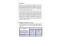
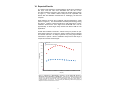
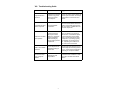
GFP-Certified™ FluoForte™ Calcium Assay Kit for microplates Instruction Manual Cat. No. ENZ-51020-KP010 Starter Pack , for 10 x 96-well plates Cat. No. ENZ-51020-KP100 High-Throughput, for 100 x 96-well plates For research use only. Rev. 1.0 August 2009 Notice to Purchaser The GFP-Certified™ FluoForte™ Calcium Assay Kit is a member of the CELLestial™ product line, reagents and assay kits comprising fluorescent molecular probes that have been extensively benchmarked for live cell analysis applications. CELLestial™ reagents and kits are optimal for use in demanding imaging applications, such as confocal microscopy, flow cytometry and HCS, where consistency and reproducibility are required. This product is manufactured and sold by ENZO LIFE SCIENCES, INC. for research use only by the end-user in the research market and is not intended for diagnostic or therapeutic use. Purchase does not include any right or license to use, develop or otherwise exploit this product commercially. Any commercial use, development or exploitation of this product or development using this product without the express prior written authorization of ENZO LIFE SCIENCES, INC. is strictly prohibited. Limited Warranty These products are offered under a limited warranty. The products are guaranteed to meet appropriate specifications described in the package insert at the time of shipment. Enzo Life Sciences’ sole obligation is to replace the product to the extent of the purchase price. All claims must be made to Enzo Life Sciences, Inc. within five (5) days of receipt of order. Trademarks and Patents Enzo, CELLestial, GFP-Certified and FluoForte are trademarks of Enzo Life Sciences, Inc. FLIPR is a trademark of Molecular Devices. CellLux is a trademark of PerkinElmer. FDSS is a trademark of Hamamatsu Photonics. Several of Enzo’s products and product applications are covered by US and foreign patents and patents pending. Contents I. Introduction ............................................................... 1 II. Reagents Provided and Storage.............................. 1 III. Additional Materials Required ................................. 2 IV. Safety Warnings and Precautions........................... 2 V. Methods and Procedures ......................................... 3 A. CELL PREPARATION.......................................................... 3 B. PREPARATION OF GFP-CERTIFIED™ FLUOFORTE™ DYE LOADING SOLUTION .................................................. 3 C. CALCIUM MOBILIZATION ASSAY ......................................... 4 VI. Expected Results ..................................................... 5 VII. References ................................................................ 6 VIII. Troubleshooting Guide ........................................... 7 I. Introduction The calcium ion is an important second messenger involved in many physiological and signal transduction processes within cells. Rhod-2 has been the most popular red fluorescent Ca2+ indicator for in-cell measurement of agonist-stimulated and antagonist-inhibited calcium signaling in high-throughput screening applications.1-4 However, its relatively weak fluorescence signals have limited its application in some challenging cell lines and with certain membrane receptors. Enzo Life Sciences’ GFP-Certified™ FluoForte™ Calcium Assay Kit provides a homogeneous fluorescence-based assay for detecting intracellular calcium mobilization across a broad spectrum of biological targets. Relative to other commercially available red fluorescent dyes, GFP-Certified™ FluoForte™ dye is the brightest and most sensitive fluorescent calcium indictor. The kit provides a homogeneous mix-and-read, no-wash calcium mobilization assay. The homogenous cell-based assay for calcium offers fewer steps, lower variability and an easier protocol for adherent and non-adherent cell lines. In addition, it requires neither a washing step, nor exogenous addition of a quencher dye, which could adversely effect receptor-ligand interaction kinetics.5 II. Reagents Provided and Storage All reagents are shipped on dry ice. Upon receipt, the kit should be stored at -20°C, protected from light. When stored properly, these reagents are stable for six months from date of receipt. Avoid repeated freezing and thawing. Quantity Reagent ENZ-51020-KP010 (for 10 plates) ENZ-51020-KP100 (for 100 plates) 1 vial 10 vials Reagent B: Dye efflux inhibitor 10 x 1 mL 10 x 10 mL Reagent C: Hanks’ buffer with 20 mM HEPES (HHBS) 1 x 100 mL Not included Reagent A: GFP-Certified™ FluoForte™ dye, lyophilized 1 III. Additional Materials Required A fluorometric imaging plate reader capable of performing quantitative optical screening for cell-based kinetic assays. (Molecular Devices FLIPR, PerkinElmer CellLux, Hamamatsu FDSS system, or similar instrumentation) Calibrated, adjustable precision pipetters, preferably with disposable plastic tips Deionized water Anhydrous DMSO Serum (optional). Growth medium (e.g. Dulbecco’s modified Eagle medium, D-MEM) Assay Plates: 96- or 384-well black-wall, clear bottom plates or 1536well low base black-wall, clear bottom plates, 1536-well lids Compound plates: 96-well or 384-well polypropylene plates, 1536-well polystyrene plates IV. Safety Warnings and Precautions This product is for research use only and is not intended for diagnostic purposes. Some components of this kit may contain hazardous substances. They can be harmful if ingested or absorbed through the skin and may cause irritation to the eyes. The reagents of the kit should be treated as possible mutagens and should be handled with care and disposed of properly. Observe good laboratory practices. Gloves, lab coat, and protective eyewear should always be worn. Never pipet by mouth. Do not eat, drink or smoke in the laboratory areas. All blood components and biological materials should be treated as potentially hazardous and handled as such. They should be disposed of in accordance with established safety procedures. To avoid photobleaching, perform all manipulations in low light environments or protected from light by other means. 2 V. Methods and Procedures Brief Summary of Assay Work Flow Prepare cells. Remove medium. Add GFP-Certified™ FluoForte™ dye loading solution. Incubate plate for 1 hour. Add test agents. Monitor fluorescence at Ex/Em=530/570 nm NOTE: PLEASE READ THE ENTIRE PROCEDURE BEFORE STARTING. Allow all reagents to be used to warm to room temperature before proceeding. Upon thawing of solutions, gently hand-mix or vortex the reagents prior to use to ensure a homogenous solution. A. CELL PREPARATION 1. Adherent Cells. The day before the experiment, plate the cells overnight in growth medium using 4 x 10⁴ to 8 x 10⁴ cells per well at a plating volume of 100 µL per well for 96-well plates, or using 1 x 10⁴ to 2 x 10⁴ cells per well at a plating volume of 25 µL per well for a384-well plates. After overnight incubation, remove the growth medium from the cell plates. Then proceed to section C, page 4. NOTE: It is important to remove the growth medium in order to minimize background fluorescence, and compound interference with serum or culture media. 2. Non-adherent Cells. On the day of the experiment, centrifuge the cells from the culture medium and then resuspend the cell pellets in GFP-Certified™ FluoForte™ dye-loading solution (see section B). Plate the cells using 1.25 x 10⁵ to 2.5 x 10⁵ cells per well at a plating volume of 100 µL per well for 96-well plates, or 3 x 10⁴ to 6 x 10⁴ cells per well at a plating volume of 25 µL per well for 384-well plates. Centrifuge the plates at 800 rpm for 2 minutes, with brake off, prior to starting the experiments. Proceed to section C, page 4. NOTE: Each cell line should be evaluated on an individual basis to determine the optimal cell density for the intracellular calcium mobilization assay. B. PREPARATION OF GFP-CERTIFIED™ FLUOFORTE™ DYE LOADING SOLUTION The following procedure is for preparation of GFP-Certified™ FluoForte™ dye loading solution for use in 1 plate. Before starting, equilibrate Reagent A (GFP-Certified™ FluoForte™ dye), a vial of Reagent B and Reagent C (HBSS) to room temperature. 3 1. Reagent A Stock Solution. Add 100 µL DMSO to the vial containing Reagent A. Mix well. NOTE: 10 µl of the reconstituted Reagent A is enough for 1 plate. The remaining unused, reconstituted Reagent A can be aliquoted and stored at ≤ -20ºC for at least one month if stored properly. The tubes (preferably amber vials) should be capped tightly. Avoid exposure to light and repeated freeze-thaw cycles. 2. 1X Assay Buffer. Mix well 9 mL of Reagent C (HHBS) with the contents of 1 vial of Reagent B. NOTE: The 100 plates kit does not include the HBSS Buffer. Be sure to prepare this reagent prior to starting any procedure. 10 mL of 1X Assay Buffer is sufficient for one plate. Unused buffer may be stored at ≤ -20oC up to 1 month . Avoid exposure to light and repeated freeze-thaw cycles. 3. GFP-Certified™ FluoForte™ Dye Loading Solution. Add 10 µL of Reagent A Stock Solution (from step B-1) to 10 mL of 1X Assay Buffer (from step B-2). Mix well. This working solution is stable for at least 2 hours at room temperature. C. CALCIUM MOBILIZATION ASSAY 1. Obtain prepared cell plates (see section A). 2. Add GFP-Certified™ FluoForte™ Dye Loading Solution to each well (100 µL/well (for 96-well plates) or 25 µL/well for 384-well plates). 3. Incubate the cell plates for 45 minutes in a 37°C cell incubator, and then incubate for another 15 minutes at room temperature. NOTE: The incubation time should be optimized for each cell line. The incubation time should be limited to 1~2 hours. DO NOT wash the cells after dye loading. 4. Prepare the compound plates by dissolving the compound in the buffer of choice. The GFP-Certified™ FluoForte™ Calcium Assay is optimized for an agonist addition at one-fifth of the final volume. 5. Run the calcium flux assay by monitoring the fluorescence at Ex=530 nm/Em=570 nm with a fluorometric imaging plate reader. NOTE: Faster addition speeds can lead to better mixing of compounds and lower signal variance across the plate. Make sure to follow the recommended experimental setup parameters provided by the instrument manufacturer before reading the plate. It is also important to run the signal test before the experiment. Different instruments have their own intensity range. Adjust the signal test intensity to the level of 10% to 15% of the maximum instrument intensity counts. For example, the maximum fluorescence intensity count for FLIPR-384 is 65,000, so the instrument settings should be adjusted to have its signal test intensity around 7,000 to 10,000. 4 VI. Expected Results In a side-by-side comparison of GFP-Certified™ FluoForte™ and Rhod-2 AM dyes, CHO M1 cells were stimulated with 100 nM of ATP. The GFP-Certified™ FluoForte™ dye yields much brighter signal. (shown in Figure 1). This enables calcium assays that are impossible with Rhod-2 AM and facilitates measurements of challenging cell lines and receptors. Dose response curves for ATP in CHO M1 cells gave similar EC50 values (shown in Figure 2). This demonstrates consistent pharmacology among the assays. However, relative fluorescence units (delta RFU) of GFPCertified™ FluoForte™ is much higher than Rhod2-AM and it has an approximately 10 times larger assay window than Rhod-2 AM for HTS applications. Overall GFP-Certified™ FluoForte™ Calcium Assay kit provides an optimized assay method for monitoring G protein-coupled receptors (GPCRs) and calcium channels.6 Its ability to generate very strong signal enables researchers to perform calcium mobilization assays with a wide range of receptor and calcium channel targets. 16000 Rhod‐2™, AM GFP‐Certified™ FluoForte™ Reagent 14000 12000 RFU (Max‐Min) 10000 8000 6000 4000 2000 0 0:00:00 0:00:17 0:00:35 0:00:52 0:01:09 0:01:26 0:01:44 Time (sec) Figure 1: Comparisons of GFP-Certified™ FluoForte™ kit and Rhod-2 AM detection of intracellular calcium mobilization in CHO-M1 cells. CHO cells were seeded overnight in 40,000 cells per 100 µL per well in a 96-well black wall/clear bottom Costar plate. The growth medium was removed, and cells were incubated with 100µl of GFP-Certified™ FluoForte™ assay reagent, or 5 µM Rhod-2 AM for 1 hour at 37°C. ATP (20 µL/well) was added using a Biotek two syringe pump dispenser to achieve concentrations of 100 nM. 5 Figure 2: ATP Dose Response Curves in CHO-M1 cells. CHO cells were seeded overnight at 40,000 cells per 100 µL per well in a 96-well black wall/clear bottom microplate. The cells were incubated with 100 µL of Enzo’s GFP-Certified™ FluoForte™ dye and 5 µM Rhod-2 AM for 1 hour at 37°C. ATP (20 µL/well) was added a Biotek two syringe pump dispenser to achieve the final indicated concentrations. No significant difference in EC50 of ATP between GFP-Certified™ FluoForte™ and Rhod-2 AM was observed. The GFP-Certified™ FluoForte™ dye generated much higher intensity signal and larger assay window. VII. References 1. Martin VV, Beierlein M, Morgan JL, Rothe A, Gee KR. (2004) Novel fluo-4 analogs for fluorescent calcium measurements. Cell Calcium, 36, 509. 2. do Ceu Monteiro M, Sansonetty F, Goncalves MJ, O'Connor JE. (1999) Flow cytometric kinetic assay of calcium mobilization in whole blood platelets using Fluo-3 and CD41. Cytometry, 35, 302. 3. MacGowan GA, Du C, Glonty V, Suhan JP, Koretsky AP, Farkas DL. (2001) Rhod-2 based measurements of intracellular calcium in the perfused mouse heart: cellular andsubcellular localization and response to positive inotropy. J Biomed Opt, 6, 23. 4. Perez-Terzic C, Stehno-Bittel L, Clapham DE. (1997) Nucleoplasmic and cytoplasmic differences in the fluorescence properties of the calcium indicator Fluo-3. Cell Calcium, 21, 275. 5. Du C, MacGowan GA, Farkas DL, Koretsky AP. (2001) Calcium measurements in perfused mouse heart: quantitating fluorescence and absorbance of Rhod-2 by application of photon migration theory. Biophys J, 80, 549. 6. Greimers R, Trebak M, Moutschen M, Jacobs N, Boniver J. (1996) Improved four-color flow cytometry method using fluo-3 and triple immunofluorescence for analysis of intracellular calcium ion ([Ca2+]i) fluxes among mouse lymph node B- and T-lymphocyte subsets. Cytometry, 23, 205. 6 VIII. Troubleshooting Guide Problem Potential Cause Suggestion High baseline fluorescence Contributions to baseline fluorescence by growth medium and organic anion transport Remove the medium before adding the indicator dye to the wells. Use the dye solution within 2 hours at room temperature. Untreated cells have calcium response Inconsistent DMSO concentration Make sure that the buffer used for the negative control wells have the same final concentration of DMSO as those in the test compounds. Response is smaller than expected Agonists and antagonists may stick to the pipette tips. Experimental setup parameters (Ex/Em wavelength) and dye loading time are not optimized. Ensure Ex/Em =530/570nm. Use 0.1% of BSA in all compound buffer diluents. Fast addition speeds are recommended to ensure better mixing of compounds and improved cell response. Dye loading typically takes between 30 minutes and one hour. Optimizing the conditions for each cell line is recommended. Well to well variation observed Incorrect dispenser and experimental setup parameter used. Use instrument manufacturer’s recommended dispenser and setup parameters (i.e., volume, height and speed of dispensing) for compound addition. Fluorescence drop upon compound addition Dislodging the cells during addition Decrease the rate of addition or seed fewer cells in the wells to avoid this problem. 7 www.enzolifesciences.com