1
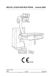
Operating Instructions
Uromat 3000
Initial version: English
11/99
-1 von 44-
0116 7321
Revision 00 © 1999 Hans Pausch Röntgengerätebau Graf-Zeppelin-Str.1 D-91056 Erlangen ALL RIGHTS RESERVED Ru
CONTENTS
Page
Important Note
3
Safety-technical Information
- Regulations
- Product Safety
- Electrical Safety
- Mechanical Safety
- Crush Zones
- Radiation Safety
- Explosion Protection
- Electromagnetic Interference, EMI
- Classification per IEC 601-1-1
- EC Conformance
- Environmental Conditions during Operation
- Disposal
3
4
4
4
5
6
6
6
6
6
6
Design Features
- Design
7
General
- Brief Description
- Areas of Application
8
9
- Space Requirements
- Room Height
- Connection
- Line Power
- Attenuation AL Equivalency Values
10
10
10
10
10
Setup
Operating Controls
- Location
- Unit Movements
- Meaning of Symbols/Function
- Setting the Working Position for Cassette Exposure
- Settings for I.I. Fluoroscopy / Cassette Exposure
- Optional Accessories
11
14
15
23
27
32
Maintenance
- Important Note
- Checks Performed by the User
- Checks Following Customer Service
- Cleaning
- Disinfection
- EEC Guideline 93/42 Regarding Medical Products
38
38
38
39
39
40
Error Codes
- Cause, Troubleshooting
41
Location of Identification Labels
- Labeling
0116 7321
42
- 2 von 44 -
11/99
Revision 00 © 1999 Hans Pausch Röntgengerätebau Graf-Zeppelin-Str. 1 D-91056 Erlangen ALL RIGHTS RESERVED Ru
IMPORTANT NOTE:
Proper use of this product requires that operating personnel have knowledge of the
OPERATING INSTRUCTIONS; these must be carefully studied prior to starting up
the equipment.
This radiographic unit may be operated only by persons who have the required
technical understanding of radiation safety or an adequate knowledge of radiation
safety and who have been instructed in the use of the radiographic unit.
The operator is always responsible for maintaining regulations that apply for operation of the radiographic unit.
SAFETY-TECHNICAL REMARKS:
Regulations
If legally specified rules exit for operation of the radiographic equipment, it is the
obligation of the user to observe them.
In the interest of safety for the patient, for operating personnel and for third parties,
the checks that are intended to maintain the operational safety and functionality of
the product must be performed in intervals of 12 months in accordance with the
maintenance instructions.
We request that you contact your customer service organisation regarding this
performance.
If national regulations or guidelines require that these checks be observed in
shorter intervals, it is absolutely necessary that they be observed .
Modifications and expansions of the product must correspond to legal regulations
as well as to generally accepted rules applicable to the technology.
As a manufacturer of radiological equipment, we can assume responsibility for the
safety-technical features of the unit only if:
we perform maintenance, repair and modification ourselves or performance is by
agents that we have authorised to do this for us, and if components that affect the
safety fail, they are replaced by original replacement parts.
If this work is performed by a subcontractor, we recommend that verification of the
type and extent of the work, and if applicable, information about any changes to
nominal values or of the operating range be requested, along with the date, company name and signature.
Prior to treatment operation, the user must be sure that all safety-relevant devices
are functional and that the product is operational.
11/99
-3 von 44-
0116 7321
Revision 00 © 1999 Hans Pausch Röntgengerätebau Graf-Zeppelin-Str.1 D-91056 Erlangen ALL RIGHTS RESERVED Ru
If the user of the radiographic unit wishes to combine it with other equipment, components or assembles, and this possibility cannot be seen from the technical data,
he must ensure that the safety of the patient as well as of operating personnel is
not adversely affected by the intended combination by contacting us as the manufacturer or by consulting a technical expert.
PRODUCT SAFETY
Electrical Safety
Only trained maintenance personnel may remove the covers and cover panels on
the radiographic unit.
This radiographic unit may be used only in medical rooms that meet the requirements of VDE 0107.
It is designed for a permanent connection to all-pole isolation from line power (ICE
601, Chap. 57.1).
Mechanical Safety
Please make sure that neither the patient nor you can touch moving parts of the
radiographic unit or that articles of clothing can become caught in such parts.
Make sure that all objects are removed from the movement range of the radiographic unit.
Crush Zones
The highlighted locations in the following sketch indicate dangerous locations at
which the patient or the operator can be injured by crushing or sharp hits.
See also the opposite page
Caution:
If the cover panel is blocked during motorised movement of the tube unit support
arm, the unit controller indicates Error E 2.
Corrective measure:
press and then disengage the emergency stop switch.
0116 7321
- 4 von 44 -
11/99
Revision 00 © 1999 Hans Pausch Röntgengerätebau Graf-Zeppelin-Str. 1 D-91056 Erlangen ALL RIGHTS RESERVED Ru
Radiation Safety
The unit has a footswitch with which radiation can be triggered.
Exposure can be initiated by the unit footswitch or from the radiation-protected location of the generator.
Regarding this, the general radiation safety regulations must be observed.
We also recommend:
1.) Keep the tube current as low as possible.
2.) Limit the radiation field as wide as possible.
3.) Maintain the max. possible distance.
4.) Do not forget radiation safety measures for patients.
11/99
-5 von 44-
0116 7321
Revision 00 © 1999 Hans Pausch Röntgengerätebau Graf-Zeppelin-Str.1 D-91056 Erlangen ALL RIGHTS RESERVED Ru
Explosion Protection
This unit is not intended for operation in areas where there is a risk of explosion.
Only such skin cleaning agents whose gas-air mixture is not flammable may be
used.
Electromagnetic Interference (EMI)
The unit meets the EMI specifications of EC Guideline 89/336.
The limit values for electrical noise measurement per EN 55011, Group 1, Class B
and the requirements for imperviousness to noise per EN 50082-1, levels 2 and 5
are maintained.
Classification per IEC 601-1-1
The unit conforms to the type of protection against electrical shock of protection
class 1 and to the level of protection, Type B.
EC Conformance:
This radiological unit meets the basic requirements according to the specifications
of EC Guideline 93/42 of the Council for Medical Products per Article 11, Section 3
and to the procedure listed in Appendix II.
The CE symbol applies only for the product without the X-ray components.
Additional information can be obtained on request from:
Hans Pausch
Röntgengerätebau
Qualitätssicherung
Postfach 28 60
D-91016 Erlangen
Fax: ..49 9131 99 24 22
Environmental Conditions for Operation
Ambient temperature range
Relative humidity in the range
Atmospheric pressure in the range
10° C to 40° C
20% to 80%
700 hPa to 1100 hPa
Disposal:
This product is manufactured in accordance with the latest environmental standards.
Legal disposal regulations may apply. To avoid hazards to the environment and persons, we request that you contact customer service before taking the product permanently out of operation.
0116 7321
- 6 von 44 -
11/99
Revision 00 © 1999 Hans Pausch Röntgengerätebau Graf-Zeppelin-Str. 1 D-91056 Erlangen ALL RIGHTS RESERVED Ru
Design Features
- Design
(The illustration shows the right-handed model, the left-handed model is the mirror image)
A
B
C
D
E
F
G
H
J
K
L
M
N
O
P
R
S
T
U
V
11/99
X-ray tube unit - collimator
Tube unit support arm, moveable
Unit table with four-way tabletop
Unit base
Manual control unit
Leg support
Elbow supports
Flush bowl or rinse bag mount
Table extension
Footrest for table extension
Cassette shaft cover
Micturation seat
Emergency stop switch
Head cushion with mount
Paper roll with holder
Patient handgrips
Footswitch for exposure and fluoroscopy
Multi-function footswitch
Grip handle
Tilt angle indicator/ position memory display / error display
-7 von 44-
0116 7321
Revision 00 © 1999 Hans Pausch Röntgengerätebau Graf-Zeppelin-Str.1 D-91056 Erlangen ALL RIGHTS RESERVED Ru
General Remarks
Brief Description
The modern compact design of the Uromat 3000 requires little space and can be installed
anywhere in the room to conform to practical requirements.
The electronic components of the unit as well as the vertical drive and the tilt drive for the
table unit are located in the unit column, which as the central component, is secured to the
concrete floor using 4 expansion bolts.
The height-adjustable patient table can be tilted from the 20° Trendelenburg position to the
88° vertical position, with an automatic stop in the horizontal position. The horizontal position can be adjusted from 122.5 cm to 68 cm.
The tabletop on the patient table has a motorised floating movement, with an automatic
stop in the middle position. The size of the tabletop is 76 x 120 cm.
The movement range of the tabletop is 24 cm at both the head end and foot end, and ±13
cm transversely.
The tabletop has lateral OP rails to mount accessories.
The tabletop can be lifted up and the elevated side parts, together with the table mat, ensure optimum protection against water as well as problem-free cleaning.
Because of the automatic tabletop compensation (optional during installation), the working
level remains constant in the examiner's eye level, even when the tabletop is tilted, as long
as the vertical limit is not reached.
Very precisely positioned exposures can be made using the built-in cassette Bucky.
The minimum film-skin distance of 60 mm assures the best geometric exposure relationship.
Image intensifiers of up to 40 cm or 16„ from well-known manufacturers can be installed.
The image intensifier is mechanically attached to the X-ray tube unit and can be moved
longitudinally a max. of 30 cm (depending on the I.I.) by the auxiliary motor.
Activities under fluoroscopic control can be made in the image intensifier mode. Depending
on the I.I. model, the minimum achievable table height changes.
The lift, tilt, transverse, and tube system movements are designed for smooth running using
an electronic controller, and for soft stop into a position and soft start out of a position.
The park position for the X-ray tube unit makes working easier and allows unrestricted patient access and unrestricted view of the patient.
Unit movements are initiated by the easy-to-position large-surface footswitch or by easily
viewed switches in the manual control unit.
The manual control unit is stored in the opening provided for it in the patient table.
Optional Accessories
The tabletop extension is inserted into the foot-end openings (with sensing by switches).
The footrest can be hooked onto the rungs provided in the tabletop extension.
The micturation seat is inserted into the foot-end openings (with sensing by switches).
0116 7321
- 8 von 44 -
11/99
Revision 00 © 1999 Hans Pausch Röntgengerätebau Graf-Zeppelin-Str. 1 D-91056 Erlangen ALL RIGHTS RESERVED Ru
The elbow supports can be inserted into the foot-end holder blocks and can be pivoted
into any desired working position.
The stainless steel flush bowl or the rinse bag mount can be hooked into the holder studs
provided.
The leg supports, the arm rests, the patient grips, the shoulder rests, the infusion bottle stand, the paper roll holder as well as the compression belt can be installed on the
later OP rails.
The Uromat 3000 permits:
- problem-free mounting by the patient onto the table
- easy and comfortable positioning of the patient
- optimum adjustment of the unit table to the preferred working height
- attachment of a wide variety of accessories
- excellent conditions for good image quality
- free positioning of the footswitch in the working area
- optimum operating and working convenience
- easy cleaning
The Uromat 3000 is:
- waterproof against drip and sprayed water from above
Field of Application
The Uromat 3000 is a general-purpose urology unit for radiological, gynecological,
as well as urological diagnostics and therapy.
The following applications can be practiced easily:
- Urograms with injection or infusion
- Retrograde pyelography
- Cystosgraphy
- Uretography
- Cystosgraphy of micturation
- Cystoscopy
- Endoscopy
- Percutaneous nephroscopy
- Transurethral resection
- Urethro-renoscopy
Caution: The above-listed applications are sometimes performed while the
patient is anesthetized or in combination with auxiliary equipment
which requires the highest degree of attention by the user.
In such circumstances, the instructions of the auxiliary equipment
must be followed, and appropriate accessories must be used.
The tabletop may be tilted a max. of only 30° while the patient is
anesthetized.
11/99
-9 von 44-
0116 7321
Revision 00 © 1999 Hans Pausch Röntgengerätebau Graf-Zeppelin-Str.1 D-91056 Erlangen ALL RIGHTS RESERVED Ru
Setup
Space Requirement
The unit is designed for stationary operation.
The space required is approx. 370 cm X 195 cm
In addition, a minimum spacing of 20 cm must be maintained between the unit column and the wall.
Room Height
The min. room height for the Uromat 3000 is 260 cm.
Power Line Connection
The power line connection must be made over a 20 mA ground fault interrupter installed on-site. The room installation must conform to VDE 0107.
In all countries outside the Federal Republic of Germany, the legally specified national regulations must take precedence.
The unit is designed for single-phase AC voltage with a permanent installation and
is equipped for permanent installation using an all-pole isolator from the power
source (ICE 601, Chap. 57.1).
It can be connected to the following line voltages without a pretransformer:
Nominal voltage:
1N
Nominal current:
Nominal frequency:
Nominal line power rating:
Heat dissipation:
115/200/208/230/240 V AC
13/7,5/7,2/6,5/6,25 A
50/60 Hz
1500 VA
240 Watt
Water Intake - Waste Water
(with use of the flush bowl or rinse bag)
Water line connection:
Waste water line:
1/2"
1"
AL Equivalency Value
The attenuation equivalency value of the tabletop (patient table) is 1.0 mm.
Measured according to:
DIN EN 60601-1-1-3 at 100 kV and a half value layer of 3.7 mm AL
and FDA 21 CFR § 1020.30 (n) at 100 kV and a half value layer of 2.7 mm AL.
0116 7321
- 10 von 44 -
11/99
Revision 00 © 1999 Hans Pausch Röntgengerätebau Graf-Zeppelin-Str. 1 D-91056 Erlangen ALL RIGHTS RESERVED Ru
Operating Elements
Location / Unit Movements / Meaning of Symbols - Function
Location
(The illustration shows the right-handed version, the left-handed version is the mirror image)
1
2
3
4
5
6
7
8
9
11/99
Handlebar for X-ray tube support arm
Switch to release the support arm
Cassette shaft flap
Cassette Bucky
Emergency stop switch
Multi-function footswitch
Exposure footswitch
Fluoroscopy footswitch
Manual control unit
-11 von 44-
0116 7321
Revision 00 © 1999 Hans Pausch Röntgengerätebau Graf-Zeppelin-Str.1 D-91056 Erlangen ALL RIGHTS RESERVED Ru
0116 7321
6
Multi-function footswitch
11
12
13
14
15
16
17
18
19
20
21
22
Footswitch, tilt table down
Footswitch, tilt table up
Footswitch, store
Footswitch, recall
Footswitch, move tabletop to the left
Footswitch, move tabletop to the right
Footswitch, move tabletop to the head end
Footswitch, move tabletop to the foot end
Footswitch, lower tabletop
Footswitch, raise tabletop
Footswitch, move radiographic system to the head end
Footswitch, move radiographic system to the foot end
- 12 von 44 -
11/99
Revision 00 © 1999 Hans Pausch Röntgengerätebau Graf-Zeppelin-Str. 1 D-91056 Erlangen ALL RIGHTS RESERVED Ru
9 Manual control unit
11/99
-13 von 44-
30
30a
31
32
33
34
34a
35
36
37
38
Collimator manual control
LED, collimator man. control
Open collimator, length
Close collimator, width
Switch on light localizer
Close collimator, both sides
Collimator closed
Close collimator, length
Open iris diaphragm
Open collimator, width
Close iris diaphragm
41
42
43
44
45
45a
LED, zoom step off
LED, zoom step one
LED, zoom step two
Zoom step selector
Invert image button
LED, image inversion
51
52
53
54
55
56
61
61a
62
62a
63
63a
64
64a
65
65a
66
66a
67
67a
68
68a
69
69a
70
LED, memory location 1
LED, memory location 2
LED, memory location 3
Store button
Recall button
Reset button
Raise table
LED, raise table limit
Lower table
LED, lower table limit
Tilt up table
LED, tilt up table limit
Tilt table down
LED, tilt down table limit
Move X-ray system, foot end
LED, end of X-ray sys., foot.
Move X-ray sys. to head end
LED, end of X-ray sys., head.
Move tabletop to head end
LED, end of tabletop, head.
Move tabletop to foot end
LED, end, tabletop, foot.
Move tabletop to left
LED, end, tabletop, left
Move tabletop to right
0116 7321
Revision 00 © 1999 Hans Pausch Röntgengerätebau Graf-Zeppelin-Str.1 D-91056 Erlangen ALL RIGHTS RESERVED Ru
Unit Movements
(The illustration shows the right-handed version, the left-handed version is the mirror image)
A
B
C
D
E
F
G
H
I
K
L
M
0116 7321
X-ray tube unit support arm, working position
X-ray tube unit support arm, park position
X-ray system, head end
X-ray system, foot end
Raise table
Lower table
Tilt up table
Tilt down table
Move tabletop to head end
Move tabletop to foot end
Move tabletop left, toward stand column
Move tabletop right, away from stand column
- 14 von 44 -
11/99
Revision 00 © 1999 Hans Pausch Röntgengerätebau Graf-Zeppelin-Str. 1 D-91056 Erlangen ALL RIGHTS RESERVED Ru
Meaning of Symbols - Function
Raise Table
Press the button 61 on the manual
control unit or on the footswitch 20 and
release when the desired position is
reached.
Movement switches off automatically
when the max. table height is reached
and LED 61a lights up yellow.
If LED 61a lights up red, an error has
occurred in the vertical drive.
Lower Table
Press the button 62 on the manual
control unit or on the footswitch 19 and
release when the desired position is
reached.
Movement switches off automatically at
the min. table height, and the LED 62a
lights up yellow.
If LED 62a lights up red, a malfunction has occurred in the vertical drive.
Tilt Table Up
Press the button 63 on the manual control unit or on the footswitch 12 and
release when the desired position is
reached.
Tilting up movement switches off automatically in the end position and LED
63a lights up yellow.
Tilting down movement switches off
automatically when the horizontal position is reached and LED's 64a and 63a
light up green.
Movement is also switched off if the
table exceeds the required min. space
to the floor.
If LED 63a lights up red, an error has occurred in the tilt drive.
11/99
-15 von 44-
0116 7321
Revision 00 © 1999 Hans Pausch Röntgengerätebau Graf-Zeppelin-Str.1 D-91056 Erlangen ALL RIGHTS RESERVED Ru
Tilt Table Down
Press the button 64 on the manual
control unit or on the footswitch 11 and
release when the desired position is
reached.
Tilting down movement switches off
automatically in the end position and
LED 64a lights up yellow.
Tilting is switched off automatically in
the horizontal position and LED's 64a
and 63a light up green.
If the table exceeds the minimum required floor spacing, movement is also
switched off.
If LED 64a lights up red, an error has
occurred in the tilt drive.
Tube Unit Support Arm, Park / Exposure Position
Press switch 2 on control arm 1 and
move the tube unit - collimator into the
park or exposure position.
Release of fluoroscopy or exposure is
not possible while in the park position.
Move Tabletop to the Right
Press switch 70 on the manual control
unit or footswitch 16 and release it
when in the desired position.
Motorised movement switches off
automatically when in the end position
and LED 70a lights up yellow.
In the middle position, LED’s 70a and
69a both light up green.
To continue movement: release footswitch 16 or switch 70 and press it
again.
If LED 70a lights up red, an error has
occurred in the transverse drive.
0116 7321
- 16 von 44 -
11/99
Revision 00 © 1999 Hans Pausch Röntgengerätebau Graf-Zeppelin-Str. 1 D-91056 Erlangen ALL RIGHTS RESERVED Ru
Move Tabletop to the Left
Press switch 69 on the manual control
unit or footswitch 15 and release it when
in the desired position.
Motorised movement switches off automatically when in the end position and
LED 69a lights up yellow.
In the middle position, LED’s 70a and
69a both light up green.
To continue movement: release footswitch 15 or switch 69 and press it
again.
If LED 69a lights up red, an error has
occurred in the transverse drive.
Move Tabletop to the Head End
Press switch 67 on the manual control
unit or footswitch 17 and release it in the
desired location.
Movement is automatically switched off
when in the end position and LED 67a
lights up yellow.
When in the middle position, LED’s 67a
and 68a both light up green.
To continue movement: release footswitch 17 or switch 67 and press it
again.
If LED 67a lights up red, an error has
occurred in the longitudinal drive.
Move Tabletop to the Foot End
Press switch 68 on the manual control
unit or footswitch 18 and release it when
in the desired position.
Movement is switched off automatically
in the end position and LED 68a lights
up yellow.
When the middle position is reached,
LED’s 67a and 68a both light up
green.
To continue movement: release footswitch 18 or switch 68 and press it
again.
If LED 68a lights up red, an error has
occurred in the longitudinal drive.
11/99
-17 von 44-
0116 7321
Revision 00 © 1999 Hans Pausch Röntgengerätebau Graf-Zeppelin-Str.1 D-91056 Erlangen ALL RIGHTS RESERVED Ru
Move Tube Unit System to the Head End
Press switch 66 on the manual control
unit or footswitch 21 and release it
when in the desired position.
Movement is switched off automatically in the end position and LED 66a
lights up yellow.
If LED 66a or 65a blinks, an error has
occurred in the system drive; if both
are on at the same time, the system is
in the exposure position.
Move Tube Unit System to the Foot End
Press switch 65 on the manual control
unit or footswitch 22 and release it
when in the desired position.
Movement is switched off automatically in the end position and LED 65a
lights up yellow.
If LED 66a or 65a blinks, an error has
occurred in the system drive; if both
are on at the same time, the system is
in the exposure position.
Position Memory
Save the three memory positions that
are available: longitudinal table position, transverse table position, table
height, table tilt angle as well as tube
unit position at the same time.
To save the positions, press switch 54
on the manual control unit or footswitch 13 (the LED blinks yellow) until
the LED lights up green after 2 seconds. When the 4th position is saved,
the 1st position is overwritten.
0116 7321
- 18 von 44 -
11/99
Revision 00 © 1999 Hans Pausch Röntgengerätebau Graf-Zeppelin-Str. 1 D-91056 Erlangen ALL RIGHTS RESERVED Ru
Recall the Position Memory and Move into Position
Press the recall switch 55 on the manual control unit or footswitch 14 until
the memory location recalled is
reached (the corresponding LED
blinks yellow) and after approx. 2 seconds, all movement axes move to their
stored positions. After reaching all end
positions, the corresponding LED
lights up green.
Clear the Position Memory
Press the reset switch 56 on the manual control unit. All memory locations
will be cleared.
Change I.I. Image Size
Press the zoom selection switch 44 on
the manual control unit as many times
as required until the corresponding
LED 41 - 43 lights up green.
11/99
-19 von 44-
0116 7321
Revision 00 © 1999 Hans Pausch Röntgengerätebau Graf-Zeppelin-Str.1 D-91056 Erlangen ALL RIGHTS RESERVED Ru
Activate / Deactivate I.I. Image Inversion
Press the image inversion switch 45
on the manual control unit until LED
44a lights up.
To deactivate, press the image inversion switch 45 again until the green
LED 44a goes off.
Switch on Light Localizer
Press switch 33 on the manual control
unit.
Open Iris Diaphragm
Press switch 36 on the manual control
unit and when the desired iris opening
is reached, release it.
0116 7321
- 20 von 44 -
11/99
Revision 00 © 1999 Hans Pausch Röntgengerätebau Graf-Zeppelin-Str. 1 D-91056 Erlangen ALL RIGHTS RESERVED Ru
Close Iris Diaphragm
Press switch 38 on the manual control
unit and release it when the desired
iris opening is reached.
Open Collimator
Press switch 31 or 37 on the manual
control unit and release it when the
desired collimator opening is reached.
Close Collimator
Press switch 32 or 35 on the manual
control unit and release it when the
desired collimator opening is reached.
If the collimator is closed, LED 34a
lights up green.
11/99
-21 von 44-
0116 7321
Revision 00 © 1999 Hans Pausch Röntgengerätebau Graf-Zeppelin-Str.1 D-91056 Erlangen ALL RIGHTS RESERVED Ru
Close both sides of the Collimator
Press switch 34 on the manual control
unit and release it when the desired
collimator opening is reached.
If the collimator is closed, LED 34a
lights up green.
Switch Off the Automatic Collimator
Press switch 30 on the manual control
unit; LED 30a lights up green.
Insert - Remove the Cassette
Open the cassette shaft cover L and
pull out the cassette tray to the stop.
Insert the cassette between the tension jaws into the desired position and
secure it in place with the latching
lever.
Insert the cassette tray to the stop
and close the cassette shaft cover
again.
Remove the cassette in the reverse
order of above.
0116 7321
- 22 von 44 -
11/99
Revision 00 © 1999 Hans Pausch Röntgengerätebau Graf-Zeppelin-Str. 1 D-91056 Erlangen ALL RIGHTS RESERVED Ru
Setting the Working Position for Cassette Exposure
Caution:
If there is a dangerous situation, press the emergency stop switch. The switch locks in
place and all motorised movements are interrupted.
After eliminating the dangerous situation, disengage the switch by turning the latch
ring to the right.
Lower Table
Press switch 62 on the manual control
unit or footswitch 19 and release it when
in the desired position.
When at the min. table height, the
movement is switched off automatically
and LED 62a lights up yellow.
Park X-ray Tube Unit
Press switch 2 on the control arm 1 and
move the tube unit - collimator into the
park position.
11/99
-23 von 44-
0116 7321
Revision 00 © 1999 Hans Pausch Röntgengerätebau Graf-Zeppelin-Str.1 D-91056 Erlangen ALL RIGHTS RESERVED Ru
Positioning the Patient on the Table
Lay the patient on the tabletop.
Instruct the patient not to grab onto
the tabletop.
Install the accessories required for the
examination or the exposure.
Moving the X-ray Tube Unit into the Exposure Position
Press switch 2 on the control arm 1
and move the tube unit - collimator
into the exposure position.
Attention:
The X-ray collimator unit must be
caught in operation.
Moving the Table into the Working Position
Press the footswitch 20 or switch 61
and release it when the table is in the
desired position.
Press footswitch 11 or 12 or switch 63
or 64 and release it when the table is
at the desired tilt angle.
0116 7321
- 24 von 44 -
11/99
Revision 00 © 1999 Hans Pausch Röntgengerätebau Graf-Zeppelin-Str. 1 D-91056 Erlangen ALL RIGHTS RESERVED Ru
Inserting the Cassette
Open the cassette shaft cover L and
pull the cassette tray out to the stop.
Insert the cassette between the tension jaws in the desired position and
secure it in place with the latching
lever.
Insert the cassette tray all the way to
stop and close the cassette shaft
cover again.
Centering the Exposure Subject
Switch on the light localizer in the collimator with the switch 33 and set the
cassette size with switches 31 and 37
or 32 and 35 per the scale.
Move the exposure subject into the
beam path by moving the tabletop
longitudinally and transversely.
Making an Exposure
Set or check the exposure data at the
generator.
Triggering the X-ray exposure is possible at both the generator and at the
footswitch 7.
Check for readiness to make the exposure.
Instruct the patient:
Take a breath and hold it!
Press the exposure switch and release
it only when the exposure is completed.
11/99
-25 von 44-
0116 7321
Revision 00 © 1999 Hans Pausch Röntgengerätebau Graf-Zeppelin-Str.1 D-91056 Erlangen ALL RIGHTS RESERVED Ru
Note
Do not forget radiation protection measures for the patient
(lead rubber apron, gonad protector, etc.)!
Removing the Cassette
Open the cassette shaft cover L and
pull the cassette tray all the way out to
stop. Release the cassette by turning
the latching lever and remove the
cassette.
Insert the cassette tray all the way to
the stop and close the cassette shaft
cover again.
End of the Examination
If applicable, move the table into the
horizontal position and lower the table.
Press switch 2 on the control arm 1
and move the tube unit - collimator
into the park position.
Remove any accessories that are not
needed or that may be in the way
when removing the patient from the
table.
Have the patient dismount the table.
0116 7321
- 26 von 44 -
11/99
Revision 00 © 1999 Hans Pausch Röntgengerätebau Graf-Zeppelin-Str. 1 D-91056 Erlangen ALL RIGHTS RESERVED Ru
Settings for I.I. Fluoroscopy / Cassette Exposure
Caution:
If there is a dangerous situation, press the emergency stop switch. The switch locks
into position and all motorised movements are interrupted.
After eliminating the dangerous situation, the switch can be released by turning the
release ring to the right.
Lowering the Table
Press switch 62 on the manual control
unit or footswitch 19 and release it when
in the desired position.
Movement is switched off automatically
at the min. table height and LED 62a
lights up yellow.
Parking the X-ray Tube Unit
Press switch 2 on the control arm 1
and move the tube unit - collimator
into the park position.
11/99
-27 von 44-
0116 7321
Revision 00 © 1999 Hans Pausch Röntgengerätebau Graf-Zeppelin-Str.1 D-91056 Erlangen ALL RIGHTS RESERVED Ru
Placing the Patient on the Table
Lay the patient on the table.
Instruct the patient not to grab onto
the tabletop.
Install the accessories for the examination or the exposure.
Moving the X-ray Tube Unit into the Exposure Position
Press switch 2 on the control arm 1
and pull the tube unit - collimator into
the exposure position.
Moving the Table into the Working Position
Press footswitch 20 or switch 61 and
release it when the table is in the desired height.
Press footswitch 11 or 12 or switch 63
or 64 and release it when the table is
at the desired tilt angle.
0116 7321
- 28 von 44 -
11/99
Revision 00 © 1999 Hans Pausch Röntgengerätebau Graf-Zeppelin-Str. 1 D-91056 Erlangen ALL RIGHTS RESERVED Ru
Inserting the Cassette
Open the cassette shaft cover L and
pull the cassette tray out until it
reaches stop. Insert the cassette between the gripper jaws in the desired
position and engage it in place with
the latch lever.
Insert the cassette tray until it reaches
the sop and close the cassette shaft
cover again.
Selecting Image Intensifier Format - Image Size
Press the zoom switch 44 on the manual control unit as often as required
until the corresponding LED 41 - 43
goes on.
Setting I.I. Image Inversion
Press the image inversion switch 45
on the manual control unit until LED
44a goes on.
To deactivate this, press the image
inversion switch 45 again until LED
44a goes off.
11/99
-29 von 44-
0116 7321
Revision 00 © 1999 Hans Pausch Röntgengerätebau Graf-Zeppelin-Str.1 D-91056 Erlangen ALL RIGHTS RESERVED Ru
I.I. Fluoroscopy
Set the fluoroscopy data at the generator.
Press the fluoro footswitch 8.
If needed, move the tabletop longitudinally or transversely.
Making an Exposure
Triggering of radiological exposure is
possible both at the generator and
from the footswitch 7.
Set or check the exposure data at the
generator.
Check exposure preparations.
Instruct the patient:
Take a deep breath and hold it!
Press the exposure switch and release it only when exposure is completed.
Note
Do not forget radiation protection measures for the patient (lead rubber apron, gonad protector, etc.)!
0116 7321
- 30 von 44 -
11/99
Revision 00 © 1999 Hans Pausch Röntgengerätebau Graf-Zeppelin-Str. 1 D-91056 Erlangen ALL RIGHTS RESERVED Ru
Removing the Cassette
Open the cassette shaft cover L and
pull the cassette tray out all the way to
stop. Release the cassette by pressing
the latch lever and remove it.
Insert the cassette tray all the way to
stop and close the cassette shaft
cover again.
End of the Examination
If applicable, move the table into the
horizontal position and lower the table.
Press switch 2 on the control arm 1
and move the tube unit - collimator into
the exposure position.
Remove any accessories that are not
needed or that may be in the way
when removing the patient from the
table.
Have the patient dismount the table.
11/99
-31 von 44-
0116 7321
Revision 00 © 1999 Hans Pausch Röntgengerätebau Graf-Zeppelin-Str.1 D-91056 Erlangen ALL RIGHTS RESERVED Ru
Optional Accessories:
Patient Table Mat
The patient table mat is used as a
patient cushion and is secured in
place with built-in magnets.
Head - Back Cushion
The holder for the head cushion is
secured to the table rail by tightening
the knob A.
Paper Roll Holder
The paper roll holder is secured to the
head-end mount by tightening the
knob B.
0116 7321
- 32 von 44 -
11/99
Revision 00 © 1999 Hans Pausch Röntgengerätebau Graf-Zeppelin-Str. 1 D-91056 Erlangen ALL RIGHTS RESERVED Ru
Patient Handgrips
The patient handgrips are slid into the
table rails and secured in place by
tightening the knob C.
Tabletop Extension
The tabletop extension is inserted into
the two openings on the table frame E
using the two studs D and automatically snaps in place.
When in the latched position, the tabletop extension is sensed by
switches.
To remove it, press the two release
levers F and remove the tabletop extension from the rear.
Attention:
Both pivots must be caught audibly
Footrest
The footrest is hooked into the rungs
G of the tabletop extension in the desired location.
Removing the footrest is done in four
steps, in the sequence 1 - 4.
11/99
-33 von 44-
0116 7321
Revision 00 © 1999 Hans Pausch Röntgengerätebau Graf-Zeppelin-Str.1 D-91056 Erlangen ALL RIGHTS RESERVED Ru
Leg Supports, Standard Version
The leg supports are inserted into
each of the table rails and secured in
place by tightening the knob H.
Coxafix Leg Supports
The leg supports are inserted into
each of the table rails and secured in
place by tightening the knob H.
See also the operating instructions
for the leg supports.
Elbow Supports
The elbow supports are inserted into
the mount provided for it and can be
pivoted into the working position.
0116 7321
- 34 von 44 -
11/99
Revision 00 © 1999 Hans Pausch Röntgengerätebau Graf-Zeppelin-Str. 1 D-91056 Erlangen ALL RIGHTS RESERVED Ru
Shoulder Rests
The shoulder rests are inserted into
the table rails and secured in place by
tightening the knob L.
Flush Bowl
The flush bowl is hooked onto the two
studs D with the hooks of the table
frame.
D
To remove it, disengage the latching
studs and take off the flush bowl.
D
Rinse Bag Mount
The bracket for the rinse bag mount is
inserted completely into the two holes.
Then the rinse bag is placed over the
bracket from behind, the connector
bracket is inserted through the tab of
the rinse bag and hooked into the bag.
11/99
-35 von 44-
0116 7321
Revision 00 © 1999 Hans Pausch Röntgengerätebau Graf-Zeppelin-Str.1 D-91056 Erlangen ALL RIGHTS RESERVED Ru
Micturation Seat
The micturation seat is inserted into
the two openings in the table frame E
with the two studs D and snaps
automatically in place.
When in the latched position, the
micturation seat is sensed by the
switch.
To remove it, press the two release
levers F and remove the micturation
seat towards the back.
Attention:
Both pivots must be caught audibly
Infusion Bottle Stand
The infusion bottle stand is attached
to the table rail and secured in place
with the knob O.
Arm Rest - Infusion Arm Rest
The armrest is attached to the table
rail and secure in place with the knob
P.
0116 7321
- 36 von 44 -
11/99
Revision 00 © 1999 Hans Pausch Röntgengerätebau Graf-Zeppelin-Str. 1 D-91056 Erlangen ALL RIGHTS RESERVED Ru
Compression Band
Installation:
Insert take-up roll B into the wall-side
profile rail in the tabletop. Secure it in
the working position using the knob
C on the opposite side.
Insert tensioner A into the front rail.
Secure the tensioner in the working
position opposite the take-up B with
knob C.
Press the release latch F. Unroll the
band and stretch it across the patient.
Wrap the stretch band once around
the shaft of the take-up roll B. Den
Insert bow D into the slot of shaft G.
Turn knob E and roll up/tension the
compression band.
To release the band:
Press release latch F, unroll the tension band and remove it.
11/99
-37 von 44-
0116 7321
Revision 00 © 1999 Hans Pausch Röntgengerätebau Graf-Zeppelin-Str.1 D-91056 Erlangen ALL RIGHTS RESERVED Ru
MAINTENANCE:
Important Note:
As with every piece of technical equipment, this radiographic unit also requires
regular maintenance and care to increase the operating reliability of the unit.
Checks Performed by the User:
The user must check the radiographic system for deficiencies as described below.
If there are functional deficiencies or other differences from normal operating behavior, switch the unit off immediately and contact customer service.
The unit may be put back into operation only after all deficiencies have been
corrected.
Daily Checks:
Display lamps or LED's, tilt angle indicator/position memory display, manual control
unit, multi-function footswitch, exposure and fluoroscopy footswitch, control bar for
tube support arm, labels and warning labels and good condition of all visible parts.
Weekly Checks:
All cables and their connectors for damage or cable breaks.
Per the Radiation Regulations
The constancy test.
Checks Performed by Customer Service:
Maintenance respectively Repairs may always be carried out by qualified personnel being authorised by us do so.
To obtain problem-free operation of the unit as well as to ensure safety for patients
and operating personnel, technical maintenance should be performed by customer
services in 12-month intervals.
See "Technical Maintenance" in the Installation Instructions.
Caution:
If there is a failure of parts that may affect the safety of the unit, original replacement parts must be used.
We recommend that written verification of the type and extent of the work performed be requested from the person performing the work, and if applicable, including changes to nominal data or of the operating range, and with date, company
name and signature.
0116 7321
- 38 von 44 -
11/99
Revision 00 © 1999 Hans Pausch Röntgengerätebau Graf-Zeppelin-Str. 1 D-91056 Erlangen ALL RIGHTS RESERVED Ru
CLEANING:
Before cleaning the system, switch it off.
Clean the space between the tabletop and the table.
Move the table into the horizontal position. Remove all
accessories from the table
accessory rail.
Grab the tabletop at the foot
end and lift it up (the tabletop
is held in the opened position
by gas springs).
Clean all visible parts. Then
press down on the tabletop
against the pressure of the
gas springs.
Caution: risk of crushing
Plastic surfaces may be cleaned only with soapy water because other solutions
(e.g. with high alcohol content) can dull the surface and cause it to become brittle.
No caustic, solvent or abrasive cleaners or polishes may be used. Water or any
other liquid may not get inside the unit because this can cause short-circuits in the
electrical installation and to avoid corrosion of parts.
Painted parts and aluminum surfaces may only be wiped down with a damp cloth
and wiped dry with a cotton cloth.
Chromed parts may only be wiped down with a dry cotton cloth.
DISINFECTION:
Prior to disinfecting it, switch off the system.
Only those disinfection methods that correspond to applicable regulations and
guidelines as well as to explosion protection measures may be used.
No caustic, solvent or volatile disinfectants may be used.
Spray disinfectants are not recommended because there is a possibility that disinfectant can penetrate into the radiographic unit.
If you use a disinfectant that can form an explosive gaseous mixture, they
must have evaporated before the system is switched on again.
The following disinfectants have been tested and approved:
Tego 103, Kosolin, Misty Multi-Purpose Disinfectant Cleaner, Misty Multi-Purpose
Disinfectant Cleaner II, Misty Disinfectant and Deodorant RTU, Precise Hospital
foam Cleaner Disinfectant.
11/99
-39 von 44-
0116 7321
Revision 00 © 1999 Hans Pausch Röntgengerätebau Graf-Zeppelin-Str.1 D-91056 Erlangen ALL RIGHTS RESERVED Ru
EEC Guideline 93/42 Regarding Medical Products
Article 12
Special Procedure for Systems and Treatment Equipment
(1) Differing from Article 11, this article applies for systems and treatment equipment.
(2) Every natural or legal person who assembles products which bear the CE symbol, with the intention of putting them into use in the form of a system or as treatment equipment corresponding to their specified purpose and within their intended
defined application, must provide a statement of content that
a) in mutual agreement, they have tested the products in accordance with the
manufacturer's instructions and have performed the work steps in accordance with
these instructions;
b) they have packaged the system or treatment equipment and have provided specific user instructions, including detailed manufacturer instructions;
c) The entire procedure was internally monitored and checked in an appropriate
manner.
If the conditions as stated in Paragraph 2 have not been met, as would be the case
when the system or the treatment equipment includes products which do not bear
the CE symbol, or when the selected combination of products no longer corresponds to its original intended purpose, the system or treatment equipment shall be
considered a separate product and, as such, is subject to the detailed specifications of Article 11.
The user is responsible for observance and performance of national differences in
EC countries!
0116 7321
- 40 von 44 -
11/99
Revision 00 © 1999 Hans Pausch Röntgengerätebau Graf-Zeppelin-Str. 1 D-91056 Erlangen ALL RIGHTS RESERVED Ru
Error Codes
If there are malfunctions of the
unit, they are indicated by error
numbers in the display or on the
operational unit by color changes
of the LED's.
The following is a list of error
codes and corrective measures
for them.
Error
number
E 1
Position
1
2
3
Cause
Emergency stop pressed
Error correction
Disengage emergency stop
Increase distance to the floor
E 12
E 13
System can not be moved into the exposure position (risk of I.I. collision)
Tube unit support arm in park position
during fluoroscopy or exposure request
Bucky cover not closed
A valid film format not detected
E 14
E 15
E 16
Attention! Double exposure
System and cassette not centered
Exposure aborted
E 20
E 21
Collision of image intensifier
Collision of X-ray tube unit
Movement possible only in opposite direction
Movement possible only in opposite direction
E 30
E 31
Table accessory no correctly snapped in
Table accessory malfunctioning
Snap the accessory in position
Check the accessory
Fatal system errors
Call service and report the error number
E 10
E 11
E 40
E 50
E 60
E 70
E 80
E 90
F 1 to
F 99
Move the support arm into the exposure position
Close Bucky cover
Insert film
Insert Bucky all the way
Change the film
None
Hold exposure footswitch pressed until exposure is completed
If LED's 61a, 62a, 63a, 64a, 67a, 68a, 69a, 70a on the manual control unit light up
red or if the two LED's, 65a and 66a blink yellow, a fatal error has occurred that
can be corrected only by the service technician.
11/99
-41 von 44-
0116 7321
Revision 00 © 1999 Hans Pausch Röntgengerätebau Graf-Zeppelin-Str.1 D-91056 Erlangen ALL RIGHTS RESERVED Ru
Location of Identification Labels
Labeling:
See also the opposite page
0116 7321
- 42 von 44 -
11/99
Revision 00 © 1999 Hans Pausch Röntgengerätebau Graf-Zeppelin-Str. 1 D-91056 Erlangen ALL RIGHTS RESERVED Ru
Sign board table:
1. Type plate
2. CE- lable
IP X1
3. Pay attention
4. Do not sit down - max. load 30 kg
Max. 30 kg/ 66 lb.
5. Attention: Crash zones
11/99
6. Inherent Filtration
-43 von 44-
0116 7321
Revision 00 © 1999 Hans Pausch Röntgengerätebau Graf-Zeppelin-Str.1 D-91056 Erlangen ALL RIGHTS RESERVED Ru
Notes:
We reserve the right to make changes resulting from technical advances.
0116 7321
- 44 von 44 -
11/99
Revision 00 © 1999 Hans Pausch Röntgengerätebau Graf-Zeppelin-Str. 1 D-91056 Erlangen ALL RIGHTS RESERVED Ru