1
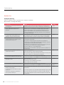
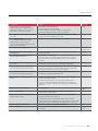
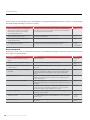
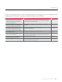
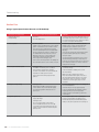
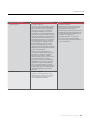
Chapter 21 | Troubleshooting Karen N. Atwood and Dako Technical Support Group Immunohistochemistry is a multi-step process that requires specialized Section One is a compilation of common problems encountered training in the processing of tissue, the selection of appropriate when using immunohistochemical-staining reagents, the underlying reagents and interpretation of the stained tissue sections. In general, causes of staining failure and recommended corrective actions. The IHC staining techniques allow for the visualization of antigens chart is divided into sections describing little or no staining, general by sequential application of a specific antibody to the antigen, a background staining and limited background staining. secondary antibody to the primary antibody, an enzyme complex and a chromogenic substrate. The enzymatic activation of the chromogen results in a visible reaction product at the antigen site. Because of its highly complex nature, the causes of unexpected negative reactions, undesired specific staining or undesired background could be difficult Section Two presents a method of systematically adding one IHC reagent at a time to determine at which stage non-specific or undesired staining may be occurring in a peroxidase, streptavidinbiotin staining system. to isolate. The information contained in this chapter should enable Section Three is a simple chart used to define the type of tissue you to rapidly pinpoint and resolve problems encountered during the specimen, the IHC staining and ancillary reagents already in place staining procedure. in the laboratory, and the staining protocol used by the laboratory personnel. You are encouraged to copy this chart and use it to help troubleshoot any problems you may encounter with your staining systems. Section Four is a guide to reading a manufacturers’ specification sheet for IVD antibodies. This includes general information for use in immunohistochemistry including fixation, recommended visualization systems, recommended titer and diluent, pretreatment, and selection of required controls. IHC Staining Methods, Fifth Edition | 137 Troubleshooting Section One Inadequate Staining Little or no staining of controls or specimen tissue, except for counterstain. May show little or no background staining. Possible Cause Solution See Page Primary antibody or labeled reagent omitted. Reagent used in wrong order. Repeat the procedure using the manufacturer’s staining system specification sheet or standard operating procedure reagent checklist as established by the individual laboratory. 57-60 Excessively diluted or excessively concentrated reagents; inappropriate incubation time and temperature. Determine correct concentration for each reagent. Depending on the degree of staining obtained, if any, a 2- to 5- fold change in concentration may be needed. Incubation temperature and incubation time are inversely proportional and will affect results. To determine optimal incubation protocol, vary either the time or temperature for each reagent in the IHC staining system. Generally, incubation times can be extended if little or no background was detected. 7, 11-12 Primary antibody diluted with inappropriate buffer. Use of PBS or TBS as an antibody diluent. Lack of stabilizing or carrier protein. Detergent in diluent. Check formula and compatibility of antibody diluent. A change of ion content and/or pH of the antibody diluent can cause a diminution in the sensitivity of the antibody. Addition of NaCl should be avoided. This problem is primarily seen with monoclonal antibodies. 57-60 Primary antibody defective; one or several secondary or ancillary reagents defective. Do NOT use product after expiration date stamped on vial. Replace defective or expired antibody; repeat staining protocol, replacing one reagent at a time with fresh, in-date reagents. 7-8 Dissociation of primary antibody during washing or incubation with link antibodies Use of alcohol-based counterstain and/or alcoholbased mounting media with aqueous–based chromogens. Excessive counterstaining may compromise proper interpretation of results. Incorrect preparation of substrate-chromogen mixture. Store products according to each product specification sheet or package insert. If using a neat or concentrated antibody, and directed by the manufacturer to store frozen, it may be aliquoted to avoid repeated freezing and thawing. For freezing, use a -70 °C to -80 °C freezer or a non frost free -20 °C freezer. A frost free freezer will cycle on and off which will cause damage to the antibody¬. Do not freeze ready-to-use or customer diluted products. Follow manufacturer recommendations on product specification sheets, package inserts and reagent labels. A feature of low affinity antibodies: Polyclonal primary antiserum: Attempt staining at low dilutions. Monoclonal primary antibody: Replace with higher affinity antibody of identical specificity. Re-optimize incubation times for washing buffer and link antibody. Repeat staining, using water-based counterstain and mounting media. Use a permanent chromogen, such as DAB/DAB+, that is not affected by organic solvents. Use a counterstain that: Will not excessively stain tissue sections. Can be diluted so as not to obliterate the specific signal. Sodium azide in reagent diluent or buffer baths for immunoperoxidase methodologies. IHC Staining Methods, Fifth Edition 115-121 Reduce incubation time of the counterstain. Repeat substrate-chromogen treatment with correctly prepared reagent. Staining intensity is decreased when excess DAB/DAB+ is present in the working reagent. Specification Sheet Check compatibility of buffer ingredients with enzyme and substrate-chromogen reagents. 57-60 Repeat staining. Commercial phosphate buffers may contain additives that will inhibit alkaline phosphates activity. Avoid sodium azide in diluents and buffers. A concentration of 15mM/L sodium azide, which is routinely added to IHC reagents to inhibit bacterial growth, will not impair HRP conjugated labels. 138 | 16-17 Incompatible buffer used for preparation of enzyme and substrate-chromogen reagents: Use of PBS wash buffer with an alkaline phosphatase staining system. 5-6 Troubleshooting Possible Cause Antigen levels are too low for detection by the employed visualization system. May be due to loss of antigenic differentiation in some tumors or loss of antigenicity due to sub-optimal tissue fixation. Solution See Page Utilize a higher sensitivity staining system. 57-60 Prolong incubation time of primary antibody. Re-optimize incubation times and concentrations of ancillary reagents. Perform antigen retrieval, if applicable, using a range of pH buffers. Steric hindrance due to high antigen level and possible prozone effect. Re-optimize concentration of the primary antibody and ancillary reagents. Antibody concentration of the primary antibody may be too high. 57-60 Use of inappropriate fixative. Check manufacturer’s specifications regarding recommended fixative. 29-33 Use of certain fixatives may damage or destroy antigens or epitopes in the tissue specimen. Use of non-cross linking fixatives may allow the elution of antigens soluble in IHC reagents. Different fixatives may affect standardization 29-33, see also 21-28 Immunoreactivity diminished or destroyed during embedding process. Use a paraffin wax with a melting temperature ~ 55-58 °C. Wax used for embedding should not exceed 60 °C. 29-33 Immunoreactivity diminished or destroyed during dewaxing at high oven temperature. Oven temperature not to exceed 60 °C. 29-33 Immunoreactivity diminished or destroyed on pre-cut tissue sections. The intensity of immunostaining may be diminished when pre-cut tissue sections are exposed to air. Use freshly cut sections and reseal paraffin-embedded blocks. 29-33 Immunoreactivity diminished or destroyed by the enzyme blocking reagent altering a specific epitope. More common on frozen sections: apply the primary antibody prior to the enzymatic block to insure its reaction. In such cases the blocking reagent can be applied at any point after the primary and before the enzyme labeled components. 29-33, 115 Excessive wash buffer or blocking serum remaining on tissue section prior to application of IHC reagents. Excess reagent will dilute the next consecutive reagent. Repeat staining, making sure to wipe away excess washing buffer and blocking serum. 11-13 Demasking protocol is inappropriate or has been omitted. Some tissue antigens require proteolytic enzyme digestion or heat induced antigen retrieval performed prior to staining. 29-33, 51-65 NOTE: The intensity of immunostaining may be diminished when tissue is exposed to prolonged heat. Refer to the primary antibody specification sheet for additional information. The need for pretreatment depends on the type and extent of fixation, specific characteristics of the antigen and the type of antibody used. Use the pretreatment method recommended by the manufacturer. No single pretreatment is suitable for all applications. Repeated reuse of antigen retrieval buffer. Do not reuse buffer. Specification Sheet Sections incorrectly dewaxed. Prepare new sections and deparaffinize according to standard laboratory protocol, using fresh xylene or xylene substitute. 115-121 When using a waterbath or steamer, allow sufficient time for the retrieval buffer to equilibrate to a temperature range of 95-99 °C. 51-65 At high altitude (greater than ~4,500 feet), the buffer will boil at less than 95 °C. Utilize a closed heating system such as a pressure cooker, autoclave or Pascal, or utilize a low temperature protocol if standardization of the validated procedure is not affected. Failure to achieve the optimal temperature required for heat induced antigen retrieval. Excessive or incomplete counterstaining. Re-optimize concentration of counterstain and incubation time. 51-65 Instrument malfunction. Ensure automated stainer is programmed correctly and is running to manufacturer’s specifications. Specification Sheet IHC Staining Methods, Fifth Edition | 139 Troubleshooting Positive control tissue shows adequate specific staining with little or no background staining. Specimen tissue shows little or no specific staining with variable background staining of several tissue elements. Possible Cause Solution See Page Specimen held for too long in a cross-linking fixative, usually in formalin, causing “masking” of antigenic determinants due to aldehydes cross-linking and increased hydrophobicity of tissue. Standardize routine fixation. Proteolytic digestion or antigen retrieval will break down cross-linking and render some tissue antigens reactive. Refer to the primary antibody specification sheet for additional information. 51-65 Sectioned portion contains crush artifact caused by grossing tissue with dull scalpel or razor. Serum proteins diffuse through tissue and are fixed in place. Re-cut tissue using sharp blade. 115-121 Sectioned portion of specimen contains necrotic or otherwise damaged elements. Ignore physically damaged portions of stained tissue sections. 51-65 Section portion of specimen not penetrated by fixative. Loss of antigenicity in unfixed tissue. Fix tissue biopsy for longer period of time or fix smaller pieces to ensure complete penetration. Unfixed tissue tends to bind all reagents nonspecifically. 29-33, 115-121, see also 51-56 General Background Background seen in all control tissue and specimen tissue. May see marked background staining in several tissue elements such as connective tissue, adipose tissue and epithelium. Possible Cause Solution See Page Excessive incubation with substrate-chromogen reagent. Reduce incubation time. Specification Sheet Substrate-chromogen reagent prepared incorrectly. Repeat incubation with correctly prepared chromogen reagent. Specification Sheet Secondary or link antibody cross-reacts with antigens Absorb link antibody with tissue protein extract or species-specific normal serum from tissue donor. 57-60 Repeat staining. Determine correct concentration for each reagent. Incubation temperature and incubation time will affect results. To determine optimal incubation protocol, vary both the time and temperature for each reagent in the IHC staining protocol. 11-13 Slides inadequately rinsed. Gently rinse slide with wash buffer bottle and place in wash bath for 5 minutes. Gentle agitation of the wash bath may increase effectiveness when used with cytoplasmic or nuclear staining protocols. 5-6 Insufficient saline or detergent in wash buffer. High-sensitivity staining systems may require higher concentrations of saline or detergent in the wash buffer. Refer to the staining system specification sheet for optimal formulation. 115-121 Blocking serum or wrong blocking serum used. Block with serum from the host of the secondary or link antibody. Avoid serum that contains auto-immune immunoglobulins. Alternatively, a serum-free protein block, lacking immunoglobulins, may be substituted for the serum block. 115-121 Sections incorrectly dewaxed. Prepare new sections and deparaffinize according to standard laboratory protocol using fresh xylene or xylene substitute. 115-121 Non-specific binding of the secondary antibody with an Use a secondary antibody that has been absorbed against a species specimen, or use a secondary antibody produced in a host that exhibits little or no cross-reactivity with the tissue source. 57-60, see also 115-121 Ensure automated stainer is programmed correctly and is running to manufacturer’s specification. Specification Sheet from tissue specimen. Secondary or link antibody and /or tertiary reagents too concentrated. animal tissue specimen. Instrument malfunction. 140 | IHC Staining Methods, Fifth Edition Troubleshooting Specimen tissue and negative reagent control slides show background staining. Positive and negative control tissue show appropriate specific staining. May involve several tissue elements such as connective tissue, adipose tissue and epithelium. Possible Cause Solution See Page Specimen held for too long in a cross-linking fixative, usually in formalin, causing “masking” of antigenic determinants due to aldehydes cross-linking and increased hydrophobicity of tissue. Standardize routine fixation. Proteolytic digestion or antigen retrieval will break down cross-linking and render some tissue antigens reactive. Refer to the primary antibody specification sheet for additional information. 29-33 Sectioned portion of specimen not penetrated by fixative. Loss of antigenicity in unfixed tissue. Unfixed tissue tends to bind all reagents nonspecifically. Fix tissue biopsy for longer period of time or fix smaller pieces to ensure complete penetration. 29-33 Sectioned portion contains crush artifact caused by grossing tissue with dull scalpel or razor. Serum proteins diffuse through tissue and are fixed in place. Serum proteins diffuse through tissue and are fixed in place. Re-cut tissue using sharp blade. 115-121 Sectioned portion of specimen contains necrotic or otherwise damaged elements. Ignore physically damaged portions of stained tissue sections. 115-121 Excessive or unevenly applied subbing agent on polyL-lysine, charged, or silanized slides. Some IHC reagents may bind to these products, resulting in a light stain over the entire slide surface. Some slides may be unevenly coated, and will exhibit the above problems on only a portion of the tissue or glass. 115-121 Antigen diffusion prior to fixation causing specific background outside the expected antigen site. Avoid delays in fixation of the tissue. 115-121 Tissue sections too thick. Cut tissue sections thinner. Formalin-fixed paraffin-embedded tissue sections should be approximately 4-6 μm; cryostat section <μm. 29-33 IHC Staining Methods, Fifth Edition | 141 Troubleshooting Negative reagent control slide shows background. Positive control tissue, negative control tissue and specimen tissue show expected specific staining. Possible Cause Solution See Page Negative control serum insufficiently diluted. Use properly diluted negative reagent control serum 127-130 For polyclonal antibodies, dilute the negative reagent control serum until the protein concentration is equal to that of the primary antibody. For monoclonal antibodies, dilute the negative reagent control serum until the Ig concentration is equal to that of the primary antibody. Contaminating antibodies in the negative control serum are cross-reacting with proteins from the specimen tissue. Replace the negative reagent control serum; repeat staining protocol. 127-130 Negative reagent control serum contaminated with bacterial or fungal growth. Replace product with non-contaminated serum. 8-9 Limited Background Areas of inconsistent staining on controls, specimens and glass slides. Possible Cause Solution See Page Protein trapped beneath the tissue during the mounting process will allow partial lifting of the section. Pooling of IHC reagents beneath the section, or partial detachment of the tissue from the slide may occur. Avoid the use of commercial adhesives, glue starch or gelatin in water baths when mounting tissue sections. Avoid allowing water from an initial section mounting to flow over an area where additional sections will be mounted. This is particularly important when using charged or silanized slides. 51-65, 115-121 Undissolved granules of chromogen. Insure that chromogen in tablet or powder form is completely dissolved, or switch to a liquid chromogen. 115-121 Incomplete removal of embedding medium. Remove embedding medium thoroughly, using fresh reagents 115-121 Incomplete dezenkerization* of tissue fixed with B5 or mercury containing reagents. Perform dezenkerization with fresh reagents. 29-33 Bacterial or yeast contamination from mounting waterbath. Clean and refill waterbath. 115-121 Partial drying of tissue prior to fixation. Unaffected areas show normal staining. Immerse tissue promptly in fixative or holding reagent. 115-121 Keep moist during the entire staining process. Use a humidity or moist chamber during incubation steps. When using an automated staining instrument, addition of wet towels to the sink may prevent drying of slides. Instrument malfunction. Ensure automated stainer is programmed correctly and is running to manufacturer’s specification. Specification Sheet Adipose or connective tissue in specimen, negative control tissue, positive control tissue and negative reagent control slides. Background in connective and epithelial tissue. Possible Cause Solution See Page Hydrophobic and ionic interactions between immunoglobulins and lipoid substances in fatty tissue. Nonspecific staining of fatty tissue rarely interferes with interpretation of specific staining and can usually be disregarded. 115-121 Primary antibody and negative reagent control serum are insufficiently diluted. Reoptimize the dilution of the primary antibody and negative control serum. *“De-zenk” - “dezenkerization” - is the use of iodine to remove mercury pigment 142 | IHC Staining Methods, Fifth Edition 11-13 Troubleshooting Epithelial tissue in specimen, negative control tissue, positive control tissue and negative reagent control slides. Staining is moderate to marked, especially in epidermal epithelium. Background in epithelia accompanies background in connective tissue. Possible Cause Both the primary antibody and negative control serum contain contaminating antibodies to epithelial elements, possibly cytokeratins. Excessive formalin fixation of tissues may increase protein cross-linking, resulting in tissue hydrophobicity. Solution See Page Use a higher dilution of the primary antibody and negative control serum. 3-5, 115-121 Increase the incubation time. Replace the antibody. Proteolytic digestion or antigen retrieval will break down cross-linking and render some tissue antigens reactive. Refer to the primary antibody and/or the negative reagent control specification sheet for appropriate pretreatment. 29-33, 115-121 Focal cytoplasmic staining observed in epithelium in the specimen tissue. Possible Cause Solution See Page Focal cytoplasmic staining is seen, particularly in intermediate and superficial layers of the epidermis. May be caused by passive absorption of plasma proteins into degenerating epidermal cells. This observation is rare and should not interfere with interpretation of specific staining. 115-121 Background seen in all control and specimen tissue when using an immunoperoxidase staining system. Possible Cause Unquenched endogenous peroxidase activity may be seen in all hemoprotein-containing specimens, including hemoglobin in erythrocytes, myoglobin in muscle cells, cytochrome in granulocytes and monocytes and catalases in liver and kidney. Solution See Page Use alternate or prolonged peroxidase blocks or use another enzyme label such as alkaline 115-121 phosphatase. Eosinophils and mast cells are particularly resistant to peroxidase quenching. Use a peroxidase blocker. Use special stains: eosin will stain eosinophils a bright red-orange. Background seen in all control and specimen tissue when using an alkaline phosphatase staining system. Possible Cause Solution See Page Unquenched endogenous alkaline phosphatase activity may be seen in leucocytes, kidney, liver, bone, ovary bladder, salivary glands, placenta and gastro-intestinal tissue. Add levamisole to the alkaline phosphatase chromogen reagent or use another enzyme label such as horseradish peroxidase. Intestinal alkaline phosphatase is not quenched by the addition of levamisole. Pretreat the tissue with 0.03 N HCl. 115-121 Background seen in all control and specimen tissue when using a biotin-streptavidin staining system. Possible Cause Solution See Page Endogenous protein-bound biotin (water-soluble B vitamin). High amounts of biotin are found in adrenal, liver, and kidney. Lesser amounts are found in the GI tract, lung, spleen, pancreas, brain, mammary gland, adipose tissue, lymphoid tissue, and cells grown in culture media containing biotin as a nutrient. Use a biotin block or chose another non-biotin based staining system. 115-121 IHC Staining Methods, Fifth Edition | 143 Troubleshooting Background of skeletal or smooth muscle tissue in positive control tissue, negative control tissue, specimen tissue and negative reagent control. Possible Cause Solution See Page Cause is not understood. It is possibly due to antibodies to muscle antigens in primary and negative reagent control serum. Should not interfere with interpretation of specific staining. 115-121 Undesired “Specific” Staining Positive staining of leucocyte membranes in specimen tissue, positive control, negative tissue control and negative reagent control. Possible Cause Binding of the Fc portion of Ig by Fc receptors on the cell membrane of macrophages, monocytes, granulocytes and some lymphocytes. Solution Use F(ab’) 2 or F(ab) fragments for the primary and secondary antibodies rather than intact See Page 115-121 antibodies. Add detergent to the wash buffer. Positive staining of histiocytes and granulocytes in the specimen tissue only, with a marker not normally reactive with these cells. Possible Cause Solution See Page Phagocytosis of antigens may render phagocytes positive for the same. Rare. Should not interfere with interpretation of specific staining. 115-121 Positive membrane staining of specimen tissue and negative reagent control tissue when using a horseradish peroxidase staining system. Possible Cause Solution See Page Tissue from persons infected with Hepatitis B virus and expressing Hepatitis B surface antigen may exhibit undesired staining. Utilize a non-peroxidase staining system. 115-121 Possible Cause Solution See Page Some manufacturers produce antibodies and reagents for in vitro use only. These products may contain preservatives, usually sodium azide, which is a known poison. Utilize an in vivo product for application on viable cells. For use on cell cultures only: sodium azide may be dialyzed out of some reagents. Contact Dako Technical Support for additional information. 115-121 Miscellaneous Loss of viability of cell cultures. 144 | IHC Staining Methods, Fifth Edition Troubleshooting Section Two Troubleshooting flow chart: Use this flow chart to determine source(s) of non-specific staining when using an immunohistochemical protocol. Background Staining Encountered with HRP-Peroxidase Reagents SLIDE #1 Reagents Result/Action Brown endogenous pigment (such as melanin) observed: Positive Control Tissue: Counterstain with hematoxylin To distinguish melanin pigment from DAB chromogen, Azure B can be used as a counterstain. The melanin stains blue-green, while the DAB remains brown. no staining seen. go to next step. An alternate method is to use AEC as the chromogen. However, if high levels of pigment exist in the tissue, the red chromogen may be partially obscured. Since bleaching protocols to remove melanin may compromise tissue antigenicity, it should be avoided if at SLIDE #2 all possible. Brown/Red color observed: Positive Control Tissue: DAB/AEC + Counterstain Indicates endogenous peroxidase activity in the tissue sections. It is present in all hemoprotein containing tissue including erythrocytes, muscle, liver, kidney, granulocytes and monocytes. no staining seen. go to next step. Block with three percent hydrogen peroxide or other peroxidase blocking reagent. Using a new bottle of hydrogen peroxide, perform a three percent H202 peroxidase block, followed by DAB and an SLIDE #3 appropriate counterstain. Positive Control Tissue: Peroxidase Block + Secondary Antibody + Streptavidin-HRP + DAB/AEC + Counterstain Brown/Red color observed: Indicates endogenous biotin activity in the tissue sections. Proteinbound biotin may be found in adrenal, liver, kidney, GI tract, lung, spleen, brain, mammary gland, adipose tissue, lymphoid no staining seen. go to next step. tissue and cell grown in culture media containing biotin (RPMI, NCTC, MEME). Block with a biotin block or switch to a staining system that is not dependent on the streptavidin/biotin reaction. IHC Staining Methods, Fifth Edition | 145 Troubleshooting SLIDE #4 Reagents Result/Action Positive Control Tissue: Peroxidase Block + Biotin Block (if required) + Secondary Antibody + Streptavidin-HRP + DAB/AEC + Counterstain Brown/Red color observed: Indicates non-specific or undesired binding of the secondary antibody to the tissue sections. This primarily occurs when the secondary antiserum has not been prepared for use on a specific no staining seen. go to next step. species tissue. To determine if this is the problem, absorb out non-specific proteins by adding 2, 5 or 10 µL of normal serum (from the species of tissue SLIDE #5 to be stained) per 100 µL of the secondary antibody. Positive Control Tissue: Peroxidase Block + Biotin Block (if required) + Negative Reagent Control + Secondary Antibody + Streptavidin-HRP + DAB/AEC no staining seen. go to next step. Brown/Red color observed: May indicate non-specific binding of the primary antibody carrierprotein. Perform a protein block with normal serum from the host of the link antibody add 0.05-0.1% TWEEN 20 to wash buffer to decrease protein attachment. Antigen retrieval lipofusion-artifact may appear as granule staining in liver and cardiac tissue, or as specific staining in pancreatic SLIDE #6 sections. Brown/Red color observed on Negative Control Tissue: Negative Control Tissue: Perform complete staining protocol. Monoclonal antibody: Possible contamination. Polyclonal antibody: Possible contamination or undesired antibody in the host Ig fraction. no staining seen. go to next step. Antigen retrieval lipofusion-artifact may appear as granule staining in liver and cardiac tissue, or as specific staining in pancreatic sections. 146 | IHC Staining Methods, Fifth Edition Troubleshooting Background Staining Encountered with Alkaline Phosphatase SLIDE #1 Reagents Result/Action Positive Control Tissue: Fast Red, Fuchsin or BCIP/NBT + Counterstain Red/Blue color observed: Indicates endogenous alkaline phosphatase activity in the tissue sections. It is present in liver, kidney, GI tract, bone, bladder, ovary, salivary gland, placenta, leukemic, necrotic or degenerated cells. no staining seen. go to next step. Block with levamisole (Intestinal alkaline phosphatase may be quenched by the addition of 0.03 N HCl prior to the addition of the SLIDE #2 alkaline phosphatase). Positive Control Tissue: Streptavidin-AP + Fast Red, Fuchsin or BCIP/NBT + Counterstain Red/Blue color observed: Indicates endogenous biotin activity in the tissue sections. Proteinbound biotin may be found in adrenal, liver, kidney, GI tract, lung, spleen, brain, mammary gland, adipose tissue, lymphoid no staining seen. go to next step. tissue and cells grown in culture media containing biotin (RPMI, NCTC, MEME). Block with a biotin block or switch to a staining system that is not SLIDE #3 dependent on the streptavidin/biotin reaction. Positive Control Tissue: Biotin Block (if required) + Secondary Antibody + Streptavidin-AP + Fast Red, Fuchsin or BCIP/NBT + Counterstain no staining seen. go to next step. Red/Blue color observed: Indicates non-specific or undesired binding of the secondary antibody to the tissue sections. This primarily occurs when the secondary antiserum has not been prepared for use on a specific species tissue. To determine if this is the problem, absorb out non-specific proteins by adding 2, 5 or 10 µL of normal serum (from the species of tissue to be stained) per 100 µL of the secondary antibody. IHC Staining Methods, Fifth Edition | 147 Troubleshooting SLIDE #4 Reagent Result/Action Positive Control Tissue: Biotin Block (if required) + Negative Reagent Control + Secondary Antibody + Streptavidin-AP + Fast Red, Fuchsin or BCIP/NBT + Counterstain no staining seen. go to next step. Red/Blue color observed: May indicate non-specific binding of the primary antibody carrierprotein. Perform a protein block with normal serum from the host of the link antibody or a protein block; add 0.05-0.1% TWEEN 20 to wash buffer to decrease protein attachment. Antigen retrieval lipofusion-artifact may appear as granule staining in liver and cardiac tissue or as specific staining in SLIDE #5 pancreatic sections. Red/Blue color observed on Negative Control Tissue: Negative Control Tissue: Perform complete staining protocol Monoclonal antibody: Possible contamination. Polyclonal antibody: Possible contamination or undesired antibody in the host Ig fraction. no staining seen. go to next step. Antigen retrieval lipofusion-artifact may appear as granule staining in liver and cardiac tissue, or as specific staining in pancreatic sections. 148 | IHC Staining Methods, Fifth Edition Troubleshooting Negative Control Reagent Reagent Result/Action Red/Blue color observed: Negative Control Reagent: Perform complete staining protocol. (Human tissue) Perform the peroxidase blocking protocol from Slide #2 under “Background Staining Encountered with HRP-Peroxidase Reagents.” Perform a biotin block if required, protein block if required, apply the appropriate negative reagent control (see below), apply biotinylated secondary antibody, apply streptavidin/HRP reagent and DAB. Prepare a negative reagent control Polyclonal: non-immunized sera from the same species, diluted to the same protein concentration as the primary antibody. Monoclonal: negative reagent control that matches the isotype as the primary antibody. Additionally, the diluent used to manufacture a monoclonal primary antibody and isotypic negative control should contain the same ions. Diluents containing sodium or phosphate ions may change the sensitivity of some monoclonal antibodies. Calculation: Ig or total protein concentration of primary antibody divided by dilution factor of primary antibody = x. Ig or total protein concentration of negative reagent control divided by x = dilution factor of negative reagent control. IHC Staining Methods, Fifth Edition | 149 Troubleshooting Section Three Tissue Specimen Tissue Specimen: Successful staining of tissue with an IHC marker is dependent on the type and preparation of the specimen. Record in the chart below, the species of the animal to be tested, the tissue source or organ from which it was collected, the collection method, how the specimen was fixed and tissue Species: Organ/tissue source: Collection: Surgical specimen/biopsy Post-mortem specimen Fine needle aspirate Peripheral blood (include anti-coagulant) Brushing Biologic fluid Cell culture Other Tissue preparation: Paraffin embedded Plastic embedded Cryostat section Cytospin Cell smear Mono-layer cultured cells Other Tissue fixation: Type of fixative Length of time Size of specimen Tissue mounting: Slide mount Tissue thickness Gelatin, glue commercial adhesive or starch in the water bath Other 150 | IHC Staining Methods, Fifth Edition Troubleshooting Endogenous Blocks Background staining is defined as unexpected or undesirable staining seen on the test or control tissue, which does not represent the target antigen. Frequent causes of background staining are endogenous enzyme activity and endogenous biotin. Peroxidase is an enzyme of the oxido-reductase class that reacts with a substrate containing hydrogen peroxide as the electron acceptor. To block this activity, a variety of hydrogen peroxide reagents can be applied to cells producing this enzyme. Alkaline phosphatase is an enzyme having various isoforms, which are produced in the leukocytes, liver, bone, intestine, placenta and Regan (carcinoma). Addition of levamisole to the chromogen/substrate will inhibit endogenous alkaline phosphatase activity, with the exception of the intestinal isoform. If necessary, this can be blocked with a weak acid wash, such as 0.03-0.5 N HCl. Biotin, a B vitamin, may be protein-bound to tissue and can interfere with proper interpretation of staining patterns when using a streptavidin or avidin reagent. To block this binding, a biotin/avidin block. Peroxidase block: 3% H2O2 Methanol/H2O2 Sodium azide Peroxidase Block (S2001) Other Alkaline Phosphatase block: Levamisole 0.03 N HCl (not for use on cryostat tissue) Other Biotin block: Biotin Block (X0590) Other Protein block: Protein Block (X0909) Normal sera from host species of the secondary antibody Other IHC Staining Methods, Fifth Edition | 151 Troubleshooting Section Four Using a Typical Specification Sheet for an IVD Antibody Information You Need to Know Information Located on the Specification Sheet Comments Regulatory Status of the Primary Antibody Intended use Indicates that a product meets the FDA requirements as a clinical diagnostic product. Likewise, a CE icon indicates the reagent meets European Union requirements. Patient test results do not require an FDA disclaimer. Tissue Preparation Specimen preparation For in vitro diagnostic use. Paraffin sections: The antibody can be used for labeling paraffin- embedded tissue sections fixed in formalin. Pre-treatment of tissues with heat-induced epitope retrieval is required. Optimal results are obtained with 10 mmol/L citrate buffer, pH 6.0. Less optimal results are obtained with 10 mmol/L Tris buffer, 1 mmol/L EDTA, pH 9.0. The tissue sections should not dry out during the treatment or during the following immunocytochemical staining procedure. Frozen sections and cell preparations: The antibody can be used for labeling frozen sections or fixed cell smears. Choosing the Visualization System Staining procedure Visualization: This antibody can be used with an immunperoxidase staining method. Follow the procedure enclosed with the selected visualization kit. Automation: The antibody is well-suited for immunocytochemical staining using automated platforms. Diluting the Primary Antibody Staining procedure Dilution: Monoclonal Mouse Anti-Vimentin, may be used at a dilution range of 1:50-1:100 when applied on formalin-fixed, paraffin-embedded sections of human tonsil. Negative Reagent Control Reagent provided Isotype: IgG1, kappa. Staining procedure The recommended negative control is Mouse monoclonal IgG1, diluted to the same mouse IgG concentration as the primary antibody. Positive and negative controls should be run simultaneously with patient specimen. 152 | IHC Staining Methods, Fifth Edition Indicates the type of specimen that was used during validation studies. In many cases this would include formalin-fixed tissue and frozen sections. Use of other fixatives requires validation by each individual laboratory. This section also indicates the optimal epitope retrieval procedure and warns against procedures that may destroy the epitope. Specimen preparation and staining procedure sections can and will change periodically, to reflect changes in technology. So remember to retain copies of each version of the reagent specification sheet. Version numbers are usually found on each page. Indicates the recommended visualization system to be used with the antibody. It also indicates that the antibody can be used for automated staining. NOTE: If your state regulatory agency requires written documentation that a reagent can be used for automated staining and this indication is not listed on the specification sheet, you may wish to contact the manufacturer’s technical support group for further information. Includes a suggested dilution range for the antibody and the recommended diluent. The dilution range is merely a suggested starting point for an individual laboratory. Optimal conditions may vary depending on specimen, preparation method, temperature of the laboratory or automated instrumentation. Use of a negative reagent control is required by the College of American Pathologists (CAP), based on Clinical Laboratory Improvement Amendments (CLIA 2003), for each patient or patient block in a staining run. Troubleshooting Information You Need to Know Information Located on the Specification Sheet Comments Positive Control Tissue Performance characteristics CLIA 2003 Sec. 493.1273 (3) Normal tissues: In general, most human mesenchymal cells are labeled by the antibody, including fibrocytes, lipocytes, smooth muscle cells, vascular endothelial cells, astrocytes, peripheral nerve (Schwann) cells, macrophages (including Kupffer cells), as well as myoepithelial cells of sweat and salivary glands and of breast, which are all labeled strongly. Also positive, with variable intensity and distribution, are the follicular cells of the thyroid, adrenal cortex, renal distal tubules, and mesangial and endothelial cells of the renal glomerulus, as well as pancreatic acinar cells (1,2). In the human eye, the antibody labels the pigmented posterior and the anterior epithelia of the human iris, including the muscle portion (dilator pupillae) of the anterior epithelium, as well as the nonpigmented and pigmented ciliary epithelia (4). In the ciliary epithelia, vimentin was coexpressed with cytokeratin (4). Mandates that fluorescent and immunohistochemical stains must be checked for appropriate positive and negative reactivity each time they are used. Most IVD antibody specification sheet will list tissue that will exhibit positive and negative staining patterns in the Performance Characteristics section. NOTE: abnormal tissue will not necessarily be labeled. Both negative and positive tissue controls should be processed using the same fixation, embedding, mounting, drying, epitope retrieval and immunostaining protocols as the patient tissue. Abnormal tissues: The antibody labeled 17/20 sarcomas, 16/18 melanomas, 4/4 meningeomas, and 3/3 schwannomas, and was the sole intermediate filament present in these tumours. In addition, variable percentages (10 to 57 percent) of carcinomas, neuroendocrine carcinomas, neuroblastomas, thymomas and mesotheliomas were positive with the antibody. With the exception of the neuroblastomas, cytokeratin was coexpressed with vimentin in these tumours. Among adenocarcinomas, more than 50 percent of papillary carcinomas of the thyroid as well as renal, endometrial, ovarian and lung carcinomas were labeled by the antibody and coexpressed keratins and vimentin. Negative Control Tissue Performance characteristics Normal tissues: Skeletal and cardiac muscle cells, epidermal, squamous, urothelial, colonic and gastric mucosal, and glial cells, as well as neurons are consistently negative with the antibody . IHC Staining Methods, Fifth Edition | 153 Troubleshooting References 1. Wood G, et al. Suppression of Endogenous Avidin-Binding Activity in Tissues and Its Relevance to Biotin-Avidin Detection Systems. Journal of Histochemistry and Cytochemistry 2981;29:1196-204. 2. Sayaki H, et al. Azure B as a Counterstain in the Immunohistological Evaluation of Heavily Pigmented Nevomelanocytic Lesions. Applied Immunohistochemistry 1995;3:268-71. 3. Federal Register: January 24, 2003;68 42CFR Part 493. 4. College of American Pathology; Anatomic Pathology Checklist, October 2005. 154 | IHC Staining Methods, Fifth Edition