1
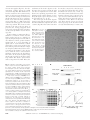
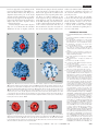
REPORTS
12. GenBank accession number X73298.
13. B. Baur, K. Fisher, K. Winter, K. J. Dietz, Plant
Physiol. 106, 1225 (1994).
14. M. Wada, personal communication.
15. Mutant cDNA sequences were obtained by reverse
transcription followed by PCR amplification with total
RNAs and NPH1-specific primers. PCR products
were purified with a QIAGEN PCR purification kit and
sequenced directly. Both strands of the entire cDNA
sequence for each mutant were examined for mutations, and each lesion was confirmed by two independent PCR experiments.
16. J. P. Khurana and K. L. Poff, Planta 178, 400 (1989).
17. Because nph1-2 is in the Estland ecotype, we also
sequenced an Estland wild-type cDNA for comparison.
18. An additional allele, nph1-3, is identical in sequence
to nph1-1.
19. S. Hill, S. Austin, T. Eydmann, T. Jones, R. Dixon,
Proc. Natl. Acad. Sci. U.S.A. 93, 2143 (1996).
20. S. I. Bibikov, R. Biran, K. E. Rudd, J. S. Parkinson, J.
Bacteriol. 179, 4075 (1997 ); A. Rebbapragada et al.,
Proc. Natl. Acad. Sci. U.S.A. 94, 10541 (1997 ).
21. W. A. Catterall, Science 242, 50 (1988); J. Tytgat, K.
Nakazawa, A. Gross, P. Hess, J. Biol. Chem. 268,
23777 (1993).
22. X. Li, J. Xu, M. Li, J. Biol. Chem. 272, 705 (1997 ).
23. M. Ahmad and A. R. Cashmore, Nature 366, 162
(1993); C. Lin, M. Ahmad, J. Chan, A. R. Cashmore,
Plant Physiol. 110, 1047 (1996).
24. I. B. Zhulin, B. L. Taylor, R. Dixon, Trends Biochem.
Sci. 22, 331 (1997 ); Z. J. Huang, I. Edery, M. Rosbash, Nature 364, 259 (1993).
25. E. Huala et al., data not shown.
26. Single-letter abbreviations for the amino acid residues are as follows: A, Ala; C, Cys; D, Asp; E, Glu; F,
Phe; G, Gly; H, His; I, Ile; K, Lys; L, Leu; M, Met; N,
Asn; P, Pro; Q, Gln; R, Arg; S, Ser; T, Thr; V, Val; W,
Trp; and Y, Tyr.
27. K. Meyer, M. P. Leube, E. Grill, Science 264, 1452
(1994).
28. J. J. Kieber, M. Rothenberg, G. Roman, K. A. Feldmann, J. R. Ecker, Cell 72, 427 (1993).
29. F. Kunst et al., Nature 390, 249 (1997).
30. P. Ballario et al., EMBO J. 15, 1650 (1996).
31. D. Leong, F. Pfeifer, H. W. Boyer, M. C. Betlach, J.
Bacteriol. 170, 4903 (1988).
32. J. W. Warmke and B. Ganetzky, Proc. Natl. Acad.
Sci. U.S.A. 91, 3438 (1994).
33. J. Ludwig et al., EMBO J. 13, 4451 (1994).
34. T. Kaneko et al., DNA Res. 3, 109 (1996).
35. M. H. Drummond and J. C. Wooton, Mol. Microbiol.
1, 37 (1987).
Alignment of Conduits for the
Nascent Polypeptide Chain in the
Ribosome-Sec61 Complex
Roland Beckmann,* Doryen Bubeck, Robert Grassucci,
Pawel Penczek, Adriana Verschoor, Gu¨nter Blobel,
Joachim Frank
An oligomer of the Sec61 trimeric complex is thought to form the protein-conducting
channel for protein transport across the endoplasmic reticulum. A purified yeast Sec61
complex bound to monomeric yeast ribosomes as an oligomer in a saturable fashion.
Cryo – electron microscopy of the ribosome-Sec61 complex and a three-dimensional
reconstruction showed that the Sec61 oligomer is attached to the large ribosomal subunit
by a single connection. Moreover, a funnel-shaped pore in the Sec61 oligomer aligned
with the exit of a tunnel traversing the large ribosomal subunit, strongly suggesting that
both structures function together in the translocation of proteins across the endoplasmic
reticulum membrane.
The
existence of a protein-conducting
channel (PCC) for protein transport across
the endoplasmic reticulum (ER) was proposed in 1975 (1). Electrophysiological experiments in 1991 provided the first direct
evidence for the existence of the PCC (2).
Moreover, fluorescently labeled nascent
chains in membrane-bound ribosomes remain in an aqueous environment sealed
from the cytoplasm and accessible to fluoR. Beckmann and G. Blobel, Howard Hughes Medical
Institute, Laboratory of Cell Biology, Rockefeller University, 1230 York Avenue, New York, NY 10021, USA.
D. Bubeck, R. Grassucci, A. Verschoor, Wadsworth Center, New York State Department of Health, Empire State
Plaza, Albany, NY 12201– 0509, USA.
P. Penczek and J. Frank, Wadsworth Center, New York
State Department of Health, and Department of Biomedical Sciences, State University of New York at Albany,
Empire State Plaza, Albany, NY 12201– 0509, USA.
*To whom correspondence should be addressed. E-mail:
[email protected]
rescence quenching from the lumen of the
ER (3). An aqueous pore with a diameter of
40 to 60 Å during cotranslational translocation is suggested by similar experiments
(4).
The Sec61 trimeric complex is a strong
candidate for the PCC of the ER in yeast
and mammalian cells (5, 6). The Sec61
complex provides the principal binding site
for ribosomes at the ER during protein
translocation (7, 8) and, together with other membrane proteins, is associated with
ribosomes after solubilization of rough microsomes with digitonin (6). A two-dimensional map of the purified Sec61 complex
obtained by electron microscopy has revealed a quasi-pentagonal, circular structure
with a central depression (9).
The three-dimensional (3D) structure of
monomeric ribosomes is currently known at
various resolutions for Escherichia coli (10),
www.sciencemag.org
36. G. Blanco, M. Drummond, P. Woodley, C. Kennedy,
ibid. 9, 869 (1993); R. Raina, U. K. Bageshwar, H. K.
Das, Mol. Gen. Genet. 237, 400 (1993).
37. D. Siddavattam, H. D. Steibl, R. Kreutzer, W. Klingmuller, Mol. Gen. Genet. 249, 629 (1995).
38. H. Allmeier, B. Cresnar, M. Greck, R. Schmitt, Gene
111, 11 (1992).
39. R. F. Smith and T. E. Smith, Protein Eng. 5, 35
(1992).
40. F. Gropp and M. C. Betlach, Proc. Natl. Acad. Sci.
U.S.A. 91, 5475 (1994).
41. F. Narberhaus, H.-S. Lee, R. A. Schmitz, L. He, S.
Kustu, J. Bacteriol. 177, 5078 (1995).
42. S. K. Crosthwaite, J. C. Dunlap, J. J. Loros, Science
276, 763 (1997 ).
43. We thank K. Meyer and W. Lukowitz for providing the
genomic library, J. R. Ecker for providing the cDNA
and YAC libraries, C. Somerville for assistance with
the AFLP technique, and P. Reymond for many helpful comments on the manuscript. This work was
funded by NSF grants MCB-9219256 and
IBN-9601164. This paper is Carnegie Institution of
Washington. Department of Plant Biology publication 1367.
2 September 1997; accepted 5 November 1997
wheat germ (11), and yeast (12). Among
the structural features recognized is a tunnel
that traverses the large ribosomal subunit
and has been considered a candidate for the
nascent chain conduit. Here, we present a
3D reconstruction of the ribosome-Sec61
complex.
For purification of the trimeric Sec61
complex (13) containing the Sec61a,
Sec61b, and Sec61g subunits (Sec61p,
Sbh1p, and Sss1p), a heptameric complex
(14) was isolated first with protein A–
tagged Sec63 protein, followed by elution of
the trimeric Sec61 complex with Triton
X-100 (Fig. 1A). To determine whether the
trimeric Sec61 complex could bind to ribosomes (15) in a membrane-free system, we
incubated the purified Sec61 complex with
ribosomes and analyzed the incubation mixture by sucrose density-gradient centrifugation (16). The Sec61 complex incubated
without ribosomes remained in the top fraction of the gradient. In the presence of
ribosomes, however, the Sec61 complex
migrated with ribosomes as determined
by immunoblotting (16) with antibodies
to Sec61b (anti-Sec61b) (Fig. 1B) and
Sec61a (anti-Sec61a) (17). In agreement
with the known salt sensitivity of the
Sec61-ribosome interaction, there was no
binding at 1 M KOAc (OAc, acetate) (17).
Incubation of a fixed amount of ribosomes
with increasing amounts of Sec61 complex
resulted in saturation of ribosome-binding
sites (Fig. 1, C and D). On the basis of the
amount of Sec61a and ribosomes, we estimate that, at saturation, two to four Sec61
trimers were bound per ribosome and that
the dissociation constant Kd is about 10
nM.
The ribosome-Sec61 complex formed
under saturating conditions was examined
by cryo– electron microscopy (18). In the
z SCIENCE z VOL. 278 z 19 DECEMBER 1997
2123
electron micrographs (Fig. 2A), the ribosome-Sec61 complex appears in random
orientations (Fig. 2B). This distribution allowed an artifact-free 3D reconstruction by
means of a 3D projection alignment procedure (19) with an existing reconstruction of
the ribosome from yeast (12) as a reference.
In side views of the ribosome (marked by
arrows in Fig. 2B), an ;100 Å–long ellipsoidal mass of density appears at the surface
of the large subunit. This location on the
ribosome is the same as the site where, in
projection, the exiting polypeptide chain
was located on both eubacterial and eukaryotic ribosomes by immuno–electron microscopy (20).
In the resulting reconstruction (Fig. 3),
which has a resolution of 26 Å (21), the
Sec61 complex appears as a slightly pentagonally shaped toroidal structure with an
outer diameter of 95 Å, an inner diameter
ranging from 15 to 35 Å depending on
depth, and an overall thickness of 40 Å
supported by a single stem attached to the
base of the large ribosomal subunit. A single
site of rigid attachment may facilitate lateral opening of the channel, at the opposite
site, to allow the release of nascent transmembrane segments into the lipid bilayer.
The surface of the Sec61 oligomer facing
the ribosome is parallel to the surface of the
large subunit. The distance between these
surfaces ranges from 15 to 20 Å. Hence, the
attachment of the Sec61 oligomer to the
ribosome does not appear to form the tight
seal that was implicated in nascent chain
fluorescence-quenching experiments. Sealforming proteins could be missing, or, more
likely, the signal sequence may be necessary
for seal formation (2, 3, 8, 22).
The central pore of the Sec61 oligomer
aligns precisely with an opening in the large
ribosomal subunit that represents the exit of
a tunnel (Fig. 3B). This tunnel runs from
Fig. 2. (A) Cryo–electron
micrograph showing a
field of yeast ribosomeSec61 complexes. Scale
bar, 200 Å. (B) Averaged projections of the
ribosome-Sec61 complex obtained by classification. Particles marked
with arrowheads show
the ribosome in side
view, with the Sec61
complex visible as a 100
Å–long mass lying parallel to the ribosome surface. Scale bar, 200 Å.
the interface canyon (12) to the lower portion of the large subunit (Fig. 3D). At the
threshold level chosen (23), a small segment of the tunnel is blocked because of the
limiting resolution, but it appeared to be
open at increased threshold levels (17). A
similar effect has been observed in the reconstruction of the 70S E. coli ribosome
(10). These tunnels, seen in cryo–electron
microscopy maps of the ribosome from E.
coli (10) and Saccharomyces cerevisiae (12),
A
B
Gradient fraction
2
3
2124
SCIENCE
z VOL. 278 z 19 DECEMBER 1997 z www.sciencemag.org
4
5
6
Anti-Sec61β
Anti-Sec61β
12
10
8
6
4
2
Fluorescence units
Amount of Sec61α bound to
ribosomes (µg)
Fig. 1. Purification and binding of the trimeric
A 1 2 3 4
B
Sec61 complex to ribosomes. (A) Purification of
1
the Sec61 complex. Crude nuclear envelopes
from Sec63prA cells were extracted with digito97
Sec61 complex
nin, and the digitonin extract was incubated with
66
IgG-Sepharose. The trimeric Sec61 complex was
eluted with Triton X-100 and analyzed by SDSSec61 complex
45
+ ribosomes
PAGE followed by Coomassie blue staining. Lane
Sec61α
1, crude nuclear envelopes (1/30,000); lane 2,
digitonin extract (1/30,000); lane 3, IgG-Sepha30
rose flow through (1/30,000); and lane 4, Triton
X-100 eluate (1/10). Ten to 15 micrograms of
Ribosomes
20
Sec61a [ by comparison with a bovine serum albumin standard ] were typically purified from 80 g
14
Sec61β,γ
of packed cells. Molecular mass is given in kilodaltons at the left. (B) The Sec61 complex binds to
ribosomes. The Sec61 complex (0.5 mg) was incubated with and without ribosomes (0.5 A260 ),
Bound
Unbound
C
D
and the incubation mixture was analyzed by su1 2 3 4 5 6 1 2 3 4 5 6
crose density-gradient centrifugation followed by
45
0.15
Sec61α
SDS-PAGE analysis and immunoblotting of gradient fractions. Lanes 1 to 6 represent top to bottom
fractions probed with a peptide antibody to
0.10
30
Sec61b. For the ribosomes, the Coomassiestained proteins were quantitated with NIH image
(arb. units, arbitrary units). (C) Binding of the
0.05
20
Sec61 complex to ribosomes is saturable. Increasing amounts of the Sec61 complex (0 mg of
14
Sec61a in lane 1 to 0.5 mg of Sec61a in lane 6)
were incubated with a fixed amount of ribosomes
(0.125 A260 ) and analyzed by density-gradient centrifugation followed by SDS-PAGE and SYPRO
Red staining. Asterisks denote Sec61a. (D) Quantitation of data in (C). The amount of bound Sec61a
was calculated according to the calibration shown in the inset.
Protein
(arb. units)
14
12
10
8
6
4
2
0.5
0.1
0.3
Amount of Sec61α (µg)
0.1
0.2
0.3
0.4
Amount of Sec61α (µg)
0.5
REPORTS
have been proposed as exit pathways of the
nascent polypeptide chain, although the evidence for that is indirect (20). The precise
alignment between the pore of the Sec61
oligomer and the tunnel (Fig. 3, C and D)
provides strong support for this hypothesis.
Conversely, this structural arrangement also
implies that the Sec61 oligomer indeed
constitutes the PCC.
Detailed analysis of the Sec61 oligomer
(Fig. 4, A to C) shows that the azimuthal
distribution of mass is irregular, with the
bulk of the mass lying on the side attached
to the stem. Again, as speculated above, the
thinner wall opposite the attachment stem
might facilitate lateral opening of the channel. There is a further asymmetry in the
Sec61 oligomer: the pore is funnel-shaped,
with a diameter of 15 Å at the lumenal site
of the ER, widening toward the ribosome,
so that a small vestibule with a diameter of
35 Å is formed. Consistent with the estimate from kinetic data, the measured volume of the Sec61 oligomer would accom-
modate two trimeric Sec61 complexes. The
reasons for the asymmetric appearance of
the ribosome-bound Sec61 oligomer are
presently not clear.
It is likely that the Sec 61 oligomer
bound to the ribosome represents the PCC
in its inactive and closed conformation.
Future structural analyses of an in vitro–
assembled complex containing a ribosome,
a nascent chain, and the Sec61 complex
will lead to information regarding the active state of the PCC.
REFERENCES AND NOTES
___________________________
A
B
40S
C
60S
D
40S
60S
Fig. 3. Three-dimensional reconstruction of the ribosome-Sec61 complex in a surface representation.
(A) Front view, with the Sec61 oligomer shown in red. (B) Front view, with the Sec61 oligomer shown as
transparent, to demonstrate the alignment of the Sec61 oligomer pore with the tunnel exit of the large
ribosomal subunit indicated with the yellow arrow. (C) Side view, with the Sec61 oligomer shown in red.
(D) Ribosome-Sec61 complex lying in the same orientation as in (C), cut along a plane that cross
sections the pore of the Sec61 oligomer and the ribosome tunnel. The arrow indicates the stem
connecting the ribosome with the Sec61 oligomer; the space between the two ribosomal subunits is
indicated by an asterisk. The ribosomal tunnel and its alignment with the Sec61 pore is indicated by a
broken yellow line. Scale bar, 100 Å.
A
B
C
Fig. 4. Three closeup views of the Sec61 oligomer. (A) Surface facing the ribosome. There is a vestibule
(diameter of 35 Å) formed by the funnel-like structure and the pore (diameter of 15 Å). (B) Surface facing
away from the ribosome. (C) View of the side opposite the attachment site. The ribosome would be
located underneath the channel. The wall opposite the attachment site is thinner and more irregular.
Arrows indicate the attachment site. Scale bar, 50 Å.
www.sciencemag.org
1. G. Blobel and B. Dobberstein, J. Cell Biol. 67, 835
(1975).
2. S. M. Simon and G. Blobel, Cell 65, 371 (1991); ibid.
69, 677 (1992).
3. K. S. Crowley, G. D. Reinhart, A. E. Johnson, ibid.
73, 1101 (1993); K. S. Crowley, S. Liao, V. E. Worrel,
G. D. Reinhart, A. E. Johnson, ibid. 78, 461 (1994).
4. B. D. Hamman, J.-C. Chen, E. E. Johnson, A. E.
Johnson, ibid. 89, 535 (1997 ).
5. R. J. Deshaies and R. Schekman, J. Cell Biol. 105,
633 (1987 ); C. J. Stirling, J. Rothblatt, M. Hosobuchi, R. Deshaies, R. Schekman, Mol. Biol. Cell 3, 129
(1992).
6. D. Go¨rlich, S. Prehn, E. Hartmann, K. U. Kalies, T. A.
Rapoport, Cell 71, 489 (1992); D. Go¨rlich and T. A.
Rapoport, ibid. 75, 615 (1993).
7. K. U. Kalies, D. Go¨rlich, T. A. Rapoport, J. Cell Biol.
126, 925 (1994).
8. B. Jungnickel and T. A. Rapoport, Cell 82, 261
(1995).
9. D. Hanein et al., ibid. 87, 721 (1996).
10. J. Frank et al., Nature 376, 441 (1995); H. Stark et al.,
Structure 3, 815 (1995).
11. A. Verschoor, S. Srivastava, R. Grassucci, J. Frank,
J. Cell Biol. 133, 495 (1996).
12. A. Verschoor, J. R. Warner, S. Srivastava, R. A.
Grassucci, J. Frank, Nucleic Acids Res., in press.
13. A yeast strain (Sec63prA) was constructed in which
the genomic copy of the gene encoding Sec63p was
tagged by COOH-terminal, in-frame integration of a
DNA fragment encoding for the immunoglobulin G
(IgG)– binding domains of protein A. The DNA fragment encoding the protein A gene and adjacent
HIS3 and URA3 markers was amplified by polymerase chain reaction with specific primers for SEC63
and a template plasmid as described [J. D. Aitchison, M. P. Rout, M. Marelli, G. Blobel, R. Wozniak, J.
Cell Biol. 131, 1133 (1995)]. A crude nuclear pellet
was prepared from a 36-liter culture as described (C.
Strambio-de-Castillia, G. Blobel, M. P. Rout, ibid., p.
19). Thirty milliliters of crude nuclei (one-fifth of the
preparation) was extracted by incubation with deoxyribonuclease I (20 mg/ml) and Heparin (1 mg/ml)
in buffer E [10 mM bis-tris-Cl (pH 6.5), 1 mM MgCl2,
10 mM KOAc, 1 mM dithiothreitol (DT T), and 0.5 mM
phenylmethylsulfonyl fluoride (PMSF)] for 20 min at
25°C and 40 min on ice. The crude nuclear envelopes were sedimented for 40 min at 145,000g at
4°C and extracted with 40 ml of buffer S [3% digitonin, 0.4 M sucrose, 10 mM triethanolamine-OAc (pH
7.5), 750 mM KOAc, 1.5 mM Mg(OAc)2, 0.5 mM
EDTA, and 1 mM DT T] for 30 min on ice. After pelleting by centrifugation of insoluble material for 35
min at 145,000g at 4°C, the extract was diluted with
1 volume of buffer D [0.4 M sucrose, triethanolamine-OAc (pH 7.5), 1.5 mM Mg(OAc)2, 1 mM
PMSF, and 1 mM DT T ] and incubated overnight with
1 ml of IgG-Sepharose (Cappel, Durham, NC) at 4°C.
The column was washed with 10 volumes of buffer
W [1% digitonin, 100 mM KOAc, Azolectin (0.5 mg/
ml), 10 mM triethanolamine-OAc (pH 7.5), 10% glycerol, 3 mM Mg(OAc)2, 0.5 mM DT T, and 1 mM
CaCl2], and the trimeric Sec61 complex was eluted
with buffer T [1% Triton X-100, 200 mM KOAc, 10
mM triethanolamine-OAc (pH 7.5), 10% glycerol, 3
mM Mg(OAc)2, 0.5 mM DT T, and 1 mM CaCl2]. The
z SCIENCE z VOL. 278 z 19 DECEMBER 1997
2125
14.
15.
16.
17.
18.
19.
20.
21.
identity of the proteins was confirmed by specific
antibodies to Sec61a (Sec61p) and Sec61b (Sbh1p)
(17 ).
S. Panzner, L. Dreier, E. Hartmann, S. Kostka, T. A.
Rapoport, Cell 81, 561 (1995)
For the purification of ribosomes, the yeast strain
DF5 was grown in 3.5 liters of yeast extract, peptone, and dextrose medium. At an optical density of
600 nm (OD600) of 1.0, the cells were washed with
water and incubated for 15 min at 25°C in 100 mM
tris-SO4 (pH 9.4) and 10 mM DT T. After homogenization by French press in buffer A [50 mM triethanolamine-OAc (pH 7.5), 50 mM KOAc, 5 mM MgCl2, 1
mM DT T, and 0.5 mM PMSF ], the homogenate was
centrifuged for 30 min at 100,000g at 4°C. The supernatant was layered over a continuous 10 to 40%
sucrose gradient in buffer A. After centrifugation for
4.5 hours at 200,000g at 4°C, the monomeric ribosomes were pooled according to the A254 profile.
The ribosomes were pelleted by centrifugation for
4.5 hours at 145,000g at 4°C, resuspended in water,
and frozen in liquid N2.
Purified Sec61 complex and ribosomes were incubated for 30 min on ice in a buffer containing 0.5% Triton
X-100, 100 mM KOAc, 5 mM triethanolamine-OAc
(pH 7.5), 5% glycerol, 1.5 mM Mg(OAc)2, 0.5 mM
DT T, and 0.5 mM CaCl2. To separate unbound
Sec61 complex from ribosome-bound Sec61 complex, we carried out gradient centrifugation using a 10
to 50% sucrose step gradient in 0.5% Triton X-100,
100 mM KOAc, 10 mM triethanolamine-OAc, 1 mM
DT T, and 3 mM Mg(OAc)2. After centrifugation for 60
min at 240,000g at 4°C, six fractions were collected
manually. For saturation assays, the first two fractions
were pooled as the unbound fraction and the following two as the bound fraction. Fractions were analyzed by SDS–polyacrylamide gel electrophoresis
(PAGE ) and stained with SYPRO Red. The amount of
protein was quantitated with the STORM system (red
fluorescence) and NIH image. For immunoblotting,
proteins were precipitated, separated on 10 to 20%
SDS-PAGE gradient gels, transferred to a nitrocellulose membrane, incubated consecutively with antiSec61a or anti-Sec61b and horseradish peroxidase–
conjugated donkey antibodies to rabbit, and detected
by ECL as described (Amersham).
R. Beckmann et al., data not shown.
Incubation to form the ribosome-Sec61 complex
was performed as described (16), and the mixture
was diluted with 4 volumes of water immediately
before it was applied to the grid. Grids for cryo –
electron microscopy were prepared as described [ T.
Wagenknecht, R. Grassucci, J. Frank, J. Mol. Biol.
199, 137 (1988); J. Dubochet et al., Q. Rev. Biophys.
21, 129 (1988)]. Micrographs were recorded under
low-dose conditions on a Philips EM 420, with 1.5mm defocus and magnification of 52,200 6 2% as
checked by a tobacco mosaic virus standard.
P. A. Penczek, R. Grassucci, J. Frank, Ultramicroscopy 53, 251 (1994).
C. Bernabeu, E. M. Tobin, A. Fowler, I. Jabin, J. A.
Lake, J. Cell Biol. 96, 1471 (1983).
Micrographs were checked for drift, astigmatism, and
presence of Thon rings by optical diffraction. Scanning was done with a step size of 25 mm corresponding to 4.78 Å on the object scale, on a Perkin-Elmer
PDS 1010 A microdensitometer. Particles were selected by an automated selection procedure that differed from the one previously described [K. R. Lata, P.
Penczek, J. Frank, Ultramicroscopy 58, 381 (1995)] in
that the particle candidates were compared directly
with the reference set of 87 quasi– evenly spaced projections (19) of an existing reconstruction of the ribosome from yeast (12). A total of 13,178 particles were
picked. The reconstruction was done with two independent approaches to obtain the orientations of the
projections. In the first approach, an existing reconstruction of the ribosome from yeast (12) was used as
a reference in the 3D projection alignment procedure
(19). In the second approach, an initial reconstruction
was obtained with the simultaneous minimization
technique [P. Penczek, J. Zhu, J. Frank, Ultramicroscopy 63, 205 (1996)]. In both cases, four steps of the
3D projection alignment procedure (19) were applied with a 2° angular interval. In each step, the
2126
SCIENCE
refined 3D structure was calculated with 70% of
the best matching particles (on the basis of the
value of the cross-correlation coefficient). Both reconstructions proved to be indistinguishable within
the measured resolution range. The final resolution,
estimated with the Fourier shell correlation with a
cutoff value at 0.5 [B. Bo¨ttcher, S. A. Wynne, R. A.
Crowther, Nature 386, 88 (1997 )], was 26 Å.
22. R. Gilmore and G. Blobel, Cell 42, 497 (1985).
23. In trying to gauge the correct threshold value, we
were led by two criteria: (i) We observed the structure
as the threshold was increased. There is normally a
“plateau,” a range of threshold values within which
the appearance (or the volume encompassed) varies
only slightly. (ii) Three-dimensional connectivity must
not be violated, which means that, in this case, we
could not choose a threshold, within the plateau
defined above, that makes the connecting rod disappear. Thus, the plateau was further narrowed.
These criteria were applied separately in the preparation of the 3D representations of the ribosome and
the channel. Because of the residual uncertainty in
the molecular boundaries, the measurements for
pore size and the distance between channel and
ribosome have an uncertainty of 25%.
24. We thank A. Fischer for the purification of the Sec61
complex; members of the Blobel lab and R. Agrawal
for discussions; S. Darst and A. Malhotra for discussions, support with the electron microscopy, and
assistance with the image processing; A. Heagle for
help with the illustrations; and the National Center for
Supercomputer Applications, University of Illinois at
Urbana-Champaign, for computing support. Supported by grants from NIH (1R01 GM29169) and
NSF (BIR 9219043) (to J.F.) and a fellowship of the
Deutsche Foschungsgemeinschaft (to R.B.).
26 September 1997; accepted 17 November 1997
Abscisic Acid Signaling Through Cyclic
ADP–Ribose in Plants
Yan Wu, Jennifer Kuzma, Eric Mare´chal,* Richard Graeff,
Hon Cheung Lee, Randy Foster, Nam-Hai Chua†
Abscisic acid (ABA) is the primary hormone that mediates plant responses to stresses
such as cold, drought, and salinity. Single-cell microinjection experiments in tomato were
used to identify possible intermediates involved in ABA signal transduction. Cyclic
ADP–ribose (cADPR) was identified as a signaling molecule in the ABA response and was
shown to exert its effects by way of calcium. Bioassay experiments showed that the
amounts of cADPR in Arabidopsis thaliana plants increased in response to ABA treatment
and before ABA-induced gene expression.
Plants
endure environmental challenges
such as drought, salinity, or cold by adjusting rather than escaping. These responses
are mediated by ABA (1), which through
unknown signals affects the regulation of
many genes (2–7). One signaling intermediary is calcium (Ca21) (8). ABA-mediated
increases in guard cell Ca21 levels lead to
stomatal closure (9). Ca21 can also induce
the expression of an ABA-responsive gene
in maize protoplasts (10).
Three regulators of Ca21 levels are inositol (1,4,5)-trisphosphate (IP3) (11), cyclic
adenosine 59-diphosphate ribose (cADPR),
and nicotinic acid adenine dinucleotide
phosphate (NAADP1) (12–15). The receptor for IP3 is known (11), whereas those
for cADPR and NAADP1 are not (15). A
putative receptor for cADPR is the ryanodine receptor (RyR) (15). NAADP1, a meY. Wu, J. Kuzma, E. Mare´chal, R. Foster, N.-H. Chua,
Laboratory of Plant Molecular Biology, Rockefeller University, 1230 York Avenue, New York, NY 10021– 6399,
USA.
R. Graeff and H. C. Lee, Department of Physiology, University of Minnesota, Minneapolis, MN 55455, USA.
*Present address: Laboratoire de Physiologie Cellulaire
Ve´ge´tale, Departement de Biologie Moleculaire et Cellulaire, Universite Joseph Fourier et CEA-Grenoble, 17 rue
des Martyrs, F-38054 Grenoble Cedex 9, France.
†To whom correspondence should be addressed. E-mail:
[email protected]
tabolite of nicotinamide adenine dinucleotide phosphate (NADP1) identified in
vitro, regulates a third Ca21 channel that
appears to be distinct from the IP3 and
cADPR receptors (14). cADPR can be
produced by ADP–ribosyl cyclase or by
CD38, a lymphocyte protein, both of
which use nicotinamide adenine dinucleotide (NAD1) as a precursor (16).
Both IP3 and cADPR elicit Ca21 release
from beet storage root vacuoles (17), and a
RyR-like activity, sensitive to cADPR, has
been detected in beet microsomes (18).
Here we demonstrate that cADPR is a likely in vivo intermediate of ABA signal transduction that exerts its effects by way of
intracellular Ca21 release.
We used microinjection to screen for
compounds that may be involved in ABA
responses. We studied the Arabidopsis
genes rd29A (also termed Iti78 and cor78)
(5), a desiccation-responsive gene (3), and
kin2 (also termed cor6.6) (5), a cold-inducible gene (2). Both genes are rapidly
induced by ABA, without requiring new
protein synthesis.
We microinjected 7- to 10-day-old etiolated hypocotyls of the phytochrome-deficient tomato mutant aurea (19, 20) with
rd29A-GUS, kin2-GUS (21), and potential
agonists and antagonists of ABA signal
z VOL. 278 z 19 DECEMBER 1997 z www.sciencemag.org