1
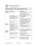
Analysis Procedures: Western blot
Troubleshooting the Western blot procedure
Problem: Chemiluminescent or chromogenic signal weak or not visible
Possible cause
Remedy
Poor isolation of tagged protein
Antibody too dilute
Use a different cell lysis procedure
Double the concentration of the Tag-specific
Antibody (and/or the Secondary Antibody)
Add more protein to gel.
1) Increase the electrical current and/or the
transfer time for the blot.
2) Be sure there are no air bubbles between the
membrane and gel during transfer.
Too little protein on the gel
Poor transfer of proteins from gel
to membrane
(Note: Check efficiency of transfer by silver
staining the remaining gel. There should be
minimal protein left in the gel.)
Wrong type membrane
Antibody incubation too short
Signal development time too short
Wash time too long or too stringent
Enzyme on antibody conjugate
inactivated by preservative
(Note: To check enzyme activity, dot
increasing dilutions of the antibody conjugate
onto a membrane and perform the visualization
reaction directly on the dilutions.)
For maximum signal, use PVDF membranes for
transfer.
Incubate the Tag-specific Antibody (and/or the
Secondary Antibody) with the membrane blot
for a longer time.
Double the development time.
1) Shorten the washing time.
2) Omit Tween 20 from the Wash Buffer.
If you use POD-conjugated antibodies, do not
use sodium azide in any Western blot reagent.
If you use AP-conjugated antibodies, do not
use a phosphate buffer in the washes following
the incubation with Secondary Antibody;
substitute a Tris buffer.
Make fresh dilution of substrate or start with a
different stock of substrate.
Substrate inactive
(Note: Check reagent on dot blot containing
dilutions of antibody conjugate with established
enzyme activity.)
Epitope tag sequence is not detectable due to:
• Proteolytic cleavage
Include protease inhibitors in lysis buffer.
• Low level of expression
Use alternative expression system or optimize
your expression system.
Insert multiple tag sequences into target protein
to increase avidity of antibody reaction.
• Premature translation termination
Use alternative insertion site within the target
resulting in loss of C-terminal
gene for the epitope tag sequence.
tag sequence
3A
3.10
CONTENTS
Analysis Procedures: Western blot
Problem: High background, additional bands on blot
Possible cause
Remedy
Antibody too concentrated
Decrease concentration of Tag-specific Antibody
(and/or Secondary Antibody) by half.
Increase washing times.
Leave blot membrane in substrate for a
shorter time.
For minimum background, use PVDF membranes for transfer.
Use nonfat dry milk (5% w/v) dissolved in
Reagent Diluent as Blocking Reagent.
(Caution: High concentrations of nonfat dry milk
may reduce specific signal as well as background.)
1) Use clean equipment, freshly prepared
buffers, and new membranes.
2) Always avoid touching membranes with bare
hands; use gloves and forceps.
Use an F(ab')2 fragment of a secondary antibody,
rather than an intact IgG.
Use direct detection with peroxidase-conjugated
monoclonal antibody to visualize tagged
proteins.
Reduce development time by half.
Wash time too short
Incubation of membrane with substrate
too long
Wrong type membrane
Blocking Reagent too dilute
Contaminated reagents or equipment
Secondary antibody binds untagged proteins
Heavy and light chains of primary antibody
visible on blot membrane (Indirect detection
procedures only)
Signal development time too long
3A
CONTENTS
3.11