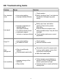
1
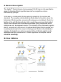
RayBio® Human/Mouse/Rat Ghrelin
Enzyme Immunoassay Kit
Catalog #: EIA-GHR, EIAM-GHR, EIAR-GHR
User Manual
Last revised December 1, 2015
Caution:
Extraordinarily useful information enclosed
ISO 13485 Certified
3607 Parkway Lane, Suite 100
Norcross, GA 30092
Tel: 1-888-494-8555 (Toll Free) or 770-729-2992, Fax:770-206-2393
Web: www.RayBiotech.com, Email: [email protected]
1
Table of Contents
Section
Page #
I.
Introduction
3
II.
General Description
4
III.
How It Works
4
IV.
Storage
5
V.
Reagents
5
VI.
Additional Materials Required
6
VII.
Reagent Preparation
A. Preparation of Plate and Anti-Ghrelin Antibody
B. Preparation of Biotinylated Peptide (Item F)
C. Preparation of Standards
D. Preparation of Positive Control
E. Preparation of Samples
F. Preparation of Wash Buffer and HRP-Strep
6
6
7
8
9
9
10
VIII.
Assay Procedure
10
IX.
Assay Procedure Summary
11
X.
Calculation of Results
A. Typical Data
B. Sensitivity
C. Detection Range
D. Reproducibility
E. Assay Diagram
12
12
12
12
12
13
XI.
Specificity
14
XII.
Select Publications
14
XIII.
Troubleshooting Guide
15
Please read the entire manual carefully before starting your experiment
2
I. Introduction
Obesity, which is characterized by excessive accumulation of adipose tissue in the
body, has become one of the greatest public health challenges. Obesity is not only
associated with health problems linked to increased weight-dependent pressure
overload on lung, joints and bones, but also a important risk factor for lifethreatening diseases such as cardiovascular diseases, type 2 diabetes and certain
cancers.
Ghrelin is synthesized as a preprohormone, and then proteolytically processed to
yield a 28-amino acid peptide. Synthesis of ghrelin occurs predominantly in
epithelial cells lining the fundus of the stomach, with smaller amounts produced in
the placenta, kidney, pituitary and hypothalamus.
Ghrelin has emerged as the first circulating hunger hormone. Ghrelin increases
food intake and thus fat mass by an action exerted at the level of the hypothalamus.
They activate cells in the arcuate nucleus that include the orexigenic neuropeptide
Y (NPY) neurons. Ghrelin-responsiveness of these neurons is both leptin and
insulin sensitive. Ghrelin also activates the mesolimbic cholinergic-dopaminergic
reward link, a circuit that communicates the hedonic and reinforcing aspects of
natural rewards, such as food.
Ghrelin levels in the plasma of obese individuals are lower than those in leaner
individuals except in the case of Prader-Willi syndrome-induced obesity. Those
suffering from the eating disorder anorexia nervosa have high plasma levels of
ghrelin compared to both the constitutionally thin and normal-weight controls.
These findings suggest that ghrelin plays a role in both anorexia and obesity.
Ghrelin levels are also high in patients who have cancer-induced cachexia.
3
II. General Description
The RayBio® Ghrelin Enzyme Immunoassay (EIA) Kit is an in vitro quantitative
assay for detecting Ghrelin peptide based on the competitive enzyme
immunoassay principle.
In this assay, a biotinylated Ghrelin peptide is spiked into the samples and
standards. The samples and standards are then added to the plate, where the
biotinylated Ghrelin peptide competes with endogenous (unlabeled) Ghrelin for
binding to the anti-Ghrelin antibody. After a wash step, any bound biotinylated
Ghrelin then interacts with horseradish peroxidase (HRP)-streptavidin, which
catalyzes a color development reaction. The intensity of the colorimetric signal is
directly proportional to the amount of captured biotinylated Ghrelin peptide and
inversely proportional to the amount of endogenous Ghrelin in the standard or
samples. A standard curve of known concentration of Ghrelin peptide can be
established and the concentration of Ghrelin peptide in the samples can be
calculated accordingly.
III. How It Works
4
IV. Storage
The entire kit may be stored at -20°C to -80°C for up to 6 months from the date of
shipment. For extended storage, it is recommended to store at -80°C. Avoid
repeated freeze-thaw cycles. For prepared reagent storage, see table below.
V. Reagents
Component
Size / Description
Storage / Stability
After Preparation
Ghrelin Microplate (Item A)
96 wells (12 strips x 8 wells) coated with
secondary antibody.
1 month at 4°C*
Wash Buffer Concentrate
(20X) (Item B)
25 ml of 20X concentrated solution.
1 month at 4°C
Standard Ghrelin Peptide
(Item C)
2 vials of Lyophilized Ghrelin Peptide. 1 vial is
enough to run each standard in duplicate.
Do not store and
reuse
Anti-Ghrelin Polyclonal
Antibody (Item N)
2 vials of Lyophilized anti-Ghrelin.
Do not store and
reuse
5X Assay Diluent B (Item E)
15 ml of 5X concentrated buffer. Diluent for both
standards and samples including serum, plasma,
cell culture media or other sample types.
1 month at 4°C
Biotinylated Ghrelin Peptide
(Item F)
2 vials of Lyophilized Biotinylated Ghrelin
Peptide, 1 vial is enough to assay the whole plate.
Do not store and
reuse
HRP-Streptavidin
Concentrate (Item G)
600 µl 50X concentrated HRP-conjugated
streptavidin.
Do not store and
reuse
Positive Control (Item M)
1 vial of Lyophilized Positive Control.
Do not store and
reuse
TMB One-Step Substrate
Reagent (Item H)
12 ml of 3,3,5,5'-tetramethylbenzidine (TMB) in
buffer solution.
N/A
Stop Solution (Item I)
8 ml of 0.2 M sulfuric acid.
N/A
*Return unused wells to the pouch containing desiccant pack, reseal along entire edge.
5
VI. Additional Materials Required
1.
2.
3.
4.
5.
6.
7.
8.
9.
10.
11.
Microplate reader capable of measuring absorbance at 450 nm
Precision pipettes to deliver 2 µl to 1 ml volumes
Adjustable 1-25 ml pipettes for reagent preparation
100 ml and 1 liter graduated cylinders
Absorbent paper
Distilled or deionized water
SigmaPlot software (or other software which can perform four-parameter
logistic regression models)
Tubes to prepare standard or sample dilutions
Orbital shaker
Aluminum foil
Plastic wrap
VII. Reagent Preparation
Keep kit reagents on ice during reagent preparation steps.
A. Preparation of Plate and Anti-Ghrelin Antibody
1. Equilibrate plate to room temperature before opening the sealed pouch.
2. Label removable 8-well strips as appropriate for your experiment.
3. 5X Assay Diluent B (Item E) should be diluted 5-fold with deionized or distilled
water.
4. Briefly centrifuge the anti-Ghrelin antibody vial (Item N) and reconsititute with
55 µl of 1X Assay Diluent B to prepare the antibody concentrate. Pipette up
and down to mix gently.
5. The antibody concentrate should then be diluted 100-fold with 1X Assay
Diluent B. This is your anti-Ghrelin antibody working solution, which will be
used in step 2 of Assay Procedure (Section VIII).
Note: The following steps may be done during the antibody incubation procedure
(step 2 of Assay Procedure)
6
B. Preparation of Biotinylated Ghrelin (Item F)
5. Briefly centrifuge the vial of Biotinylated Ghrelin (Item F) and reconstitute with
20 µl of ddH2O before use.
6. See the image below for proper preparation of Item F. Transfer the entire
contents of the Item F vial into a tube containing 10 ml of 1X Assay Diluent B.
This is your Working Stock of Item F. Pipette up and down to mix gently.
The final concentration of biotinylated Ghrelin will be 20 ng/ml.
a. Second Dilution of Item F for Standards: Add 2 ml of Working Stock Item
F to 2 ml of 1X Assay Diluent B. The final concentration of biotinylated
Ghrelin will be 10 ng/ml.
b. Second Dilution of Item F for Positive Control: Add 100 µl of Working
Stock Item F to 100 µl of the prepared Positive Control (Item M). (See
section D for Positive Control preparation) The final concentration of
biotinylated Ghrelin will be 10 ng/ml.
c. Second Dilution of Item F for samples: Add 125 µl of Working Stock Item
F to 125 µl of prepared sample (see section E for sample preparation).
This is a 2-fold dilution of your sample. The final concentration of
biotinylated Ghrelin will be 10 ng/ml.
7
C. Preparation of Standards
7. Label 6 microtubes with the following concentrations: 1000 ng/ml, 100 ng/ml,
10ng/ml, 1 ng/ml, 100 pg/ml and 0 pg/ml. Pipette 450 µl of biotinylated Ghrelin
Item F working solution (prepared in step 6a) into each tube, except the 1,000
ng/ml (leave this one empty).
It is very important to make sure the concentration of biotinylated Ghrelin is 10
ng/ml in all standards.
8. Briefly centrifuge the vial of Ghrelin Standard (Item C). Reconstitute with 10 µl
of ddH2O and briefly vortex if desired. Pipette 8 µl of Item C and 792 µl of 10
ng/ml biotinylated Ghrelin working solution (prepared in step 6a) into the tube
labeled 1000 ng/ml. Mix thoroughly. This solution serves as the first standard
(1,000 ng/ml Ghrelin standard, 10 ng/ml biotinylated Ghrelin).
9. To make the 100 ng/ml standard, pipette 50 µl of the 1000 ng/ml Ghrelin
standard into the tube labeled 100 ng/ml. Mix thoroughly.
10. Repeat this step with each successive concentration, preparing a dilution
series as shown in the illustration below. Each time, use 450 µl of biotinylated
Ghrelin and 50 µl of the prior concentration until the 100 pg/ml is reached. Mix
each tube thoroughly before the next transfer.
8
D. Positive Control Preparation
11. Briefly centrifuge the Positive Control vial (Item M) and reconstitute with 100 µl
of ddH2O.
12. Refer to step 6b. This is a 2-fold dilution of the Positive Control. The final
concentration of biotinylated Ghrelin should still be 10 ng/ml.
The Positive Control is a cell culture media sample that serves as a system control
to verify that the kit components are working. The resulting OD will not be used in
any calculations; if no positive competition is observed please contact RayBiotech
Technical Support. The Positive Control may be diluted further if desired, but be
sure the final concentration of biotinylated Ghrelin is 10 ng/ml.
E. Sample Preparation
13. If you wish to perform a 2-fold dilution of your sample, proceed to step 6c. If
you wish to perform a higher dilution of your sample, dilute your sample with
1X Assay Diluent B before performing step 6c.
EXAMPLE (to make a 4-fold dilution of sample):
a. Dilute sample 2-fold (62.5 µl of sample + 62.5 µl of 1X Assay Diluent B.).
b. Perform step 6c (125 µl of working solution Item F + 125 µl of sample
prepared above).
The total volume is 250 µl, enough for duplicate wells on the microplate.
It is very important to make sure the final concentration of the biotinylated Ghrelin is
10 ng/ml.
Note: Optimal sample dilution factors should be determined empirically, however you
may reference below for recommended dilution factors for serum: Human=4X
Mouse=4X Rat=2X.
If you have any questions regarding the recommendended dilutions you may contact
technical support at 888-494-8555 or [email protected].
9
F. Preparation of Wash Buffer and HRP
14. If Item B (20X Wash Concentrate) contains visible crystals, warm to room
temperature and mix gently until dissolved.
15. Dilute 20 ml of Wash Buffer Concentrate into deionized or distilled water to yield
400 ml of 1X Wash Buffer.
16. Briefly centrifuge the HRP-Streptavidin vial (Item G) before use.
17. Dilute the HRP-Streptavidin concentrate 50-fold with 1X Assay Diluent B.
VIII. Assay Procedure
1. Keep kit reagents on ice during reagent preparation steps. It is recommended
that all standards and samples be run at least in duplicate.
2. Add 100 µl of Anti-Ghrelin Antibody (Item N) (See Reagent Preparation step 3)
to each well. Incubate for 1.5 hours at room temperature with gentle shaking (1-2
cycle/sec). You may also incubate overnight at 4ºC.
3. Discard the solution and wash wells 4 times with 1X Wash Solution Buffer (200300 µl each). Washing may be done with a multichannel pipette or an automated
plate washer. Complete removal of liquid at each step is essential to good assay
performance. After the last wash, remove any remaining Wash Buffer by
aspirating or decanting. Invert the plate and blot it against clean paper towels.
4. Add 100 µl of each standard (see Reagent Preparation Section C), Positive
Control (see Reagent Preparation Section D) and sample (see Reagent
Preparation Section E) in appropriate wells. Be sure to include a blank well
(Assay Diluent only). Cover wells and incubate for 2.5 hours at room
temperature with gentle shaking (1-2 cycles/sec) overnight or at 4ºC.
5. Discard the solution and wash 4 times as directed in Step 3.
10
6. Add 100 µl of prepared HRP-Streptavidin solution (see Reagent Preparation
step 7) to each well. Incubate for 45 minutes at room temperature with gentle
shaking. It is recommended that incubation time should not be shorter or longer
than 45 minutes.
7. Discard the solution and wash 4 times as directed in Step 3.
8. Add 100 µl of TMB One-Step Substrate Reagent (Item H) to each well. Incubate
for 30 minutes at room temperature in the dark with gentle shaking (1-2
cycles/sec).
9. Add 50 µl of Stop Solution (Item I) to each well. Read at 450 nm immediately.
IX. Assay Procedure Summary
1. Prepare all reagents, samples and standards as instructed.
2. Add 100 µl anti-Ghrelin to each well. Incubate 1.5 hours at room temperature or
overnight at 4ºC.
3. Add 100 µl standard or sample to each well. Incubate 2.5 hours at room
temperature or overnight at 4ºC.
4. Add 100 µl prepared Streptavidin solution. Incubate 45 minutes at room
temperature.
5. Add 100 µl TMB One-Step Substrate Reagent to each well. Incubate 30 minutes
at room temperature.
6. Add 50 µl Stop Solution to each well. Read at 450 nm immediately.
11
X. Calculation of Results
Calculate the mean absorbance for each set of duplicate stands, controls, and
samples and subtract the blank optical density. Plot the standard curve using
SigmaPlot software (or other software which can perform four-parameter logistic
regression models), with standard concentration on the x-axis and percentage of
absorbance (see calculation below) on the y-axis. Draw the best-fit curve through the
standard points.
Percentage absorbance = (B-blank OD)/B 0-blank OD) where
B = OD of sample or standard and
B0 = OD of zero standard (total binding)
A. Typical Data
These standard curves are for demonstration only. A standard curve must be run with
each assay.
B. Sensitivity
The minimum detectable concentrations of Ghrelin is 161 pg/ml or 12.46 pM.
C. Detection Range
0.1-1,000 ng/ml
D. Reproducibility
Intra-Assay: CV<10%
Inter-Assay: CV<15%
12
E. Assay Diagram
Recommended Plate Layout:
Key:
Blank = Buffer Only
Total Binding = Biotin- Ghrelin only
Standard 1 = 1000 ng/ml
Standard 2 = 100 ng/ml
Standard 3 = 10 ng/ml
Standard 4 = 1 ng/ml
Standard 5 = 100 pg/ml
Pos Control = Biotin with Item M
13
XI. Specificity
Cross Reactivity: This EIA kit shows no cross-reactivity with any of the cytokines
tested: Nesfatin, Angiotensin II, NPY and APC.
XIV. Publications Citing This Product
1. Plum L., et al. The Obesity Susceptibility Gene Carboxypeptidase E Links FoxO1
2.
3.
4.
5.
6.
Signaling in Hypothalamic Pro–opiomelanocortin Neurons with Regulation of Food
Intake. Nat Med. 2009 Oct;15(10):1195-201. doi: 10.1038/nm.2026
Species: Mouse
Sample Type: Serum
Fung JN., et al. Expression and In Vitro Functions of the Ghrelin Axis in Endometrial
Cancer. Horm Cancer. 2010 Oct;1(5):245-55. doi: 10.1007/s12672-010-0047-1.
Species: Human
Sample Type: Cell Lysate
Ma X., Zhao Y., Wang Q., Wu L., Wang Z., et al. Plasma Ghrelin Concentrations Are
Negatively Correlated With Urine Albumin-to-Creatinine Ratio in Newly Diagnosed Type 2
Diabetes. The American Journal of Medical Sciences 2014 June. Epub Ahead of Print.
doi: 10.1097/MAJ.0000000000000297
Species: Human
Sample Type: Plasma
Han J., Kim H., Lee J., Choi M., Kim Y., Son C. Repeated Sense of Hunger Leads to the
Development of Visceral Obesity and Metabolic Syndrome in a Mouse Model . PLOS One
Published: May 30, 2014. DOI: 10.1371/journal.pone.0098276
Species: Rat
Sample Type: Serum
Liu R., Ma D., Li Y., Hu R., Peng Y., Wang Q. The anorexic effect of Ex4/Fc through GLP1 receptor activation in high-fat diet fed mice. Acta Biochimica et Biophysica Sinica. Epub
Ahead of Print 2014. DOI: 10.1093/abbs/gmu044.
Species: Mouse
Sample Type: Serum
Zhang S., et al. Ghrelin and obestatin plasma levels and ghrelin/obestatin prepropeptide
gene polymorphisms in small for gestational age infants. Journal of International Medical
Research published online 15 September 2014. DOI: 10.1177/0300060514533525
Species: Human
Sample Type: Plasma
For additional publications citing this product please contact technical support at 888494-8555 or [email protected].
14
XIII. Troubleshooting Guide
Problem
Cause
Solution
Inaccurate pipetting
Improper standard dilution
Check pipettes
Briefly centrifuge Item C and dissolve
the powder thoroughly by gently
mixing
Low signal
Improper preparation of
standard and/or
biotinylated antibody
Too brief incubation times
Inadequate reagent
volumes or improper dilution
Briefly spin down vials before
opening. Dissolve the powder
thoroughly.
Ensure sufficient incubation time;
assay procedure step 2 may be done
overnight
Check pipettes and ensure correct
preparation
Large CV
Inaccurate pipetting
Air bubbles in wells
Check pipettes
Remove bubbles in wells
High
background
Plate is insufficiently
washed
Contaminated wash buffer
Review the manual for proper wash.
If using a plate washer, ensure that
all ports are unobstructed.
Make fresh wash buffer
Improper storage of the
ELISA kit
Stop solution
Follow storage recomendations in
sections IV and V. Keep substrate
solution protected from light.
Add stop solution to each well before
reading plate
Poor standard
curve
Low sensitivity
15
RayBio® ELISA Kits
Over 2,000 ELISA kits available, visit www.RayBiotech.com/ELISA-Kits.html for
details.
This product is for research use only.
©2015 RayBiotech, Inc
16