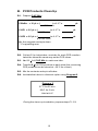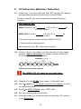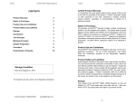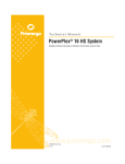
1
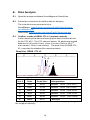
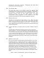
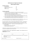
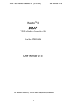
KRAS Codon 61 Mutation Analysis Reagents User Manual V1.4 Cat No. GP06 32 reactions 1 CONTENTS Introduction 4 Overview of MutectorTM Assay 5 Materials Provided 6 Materials Required 7 Equipment Required 7 DNA Sample Preparation 8 Sequencer Setup 8 Thermal Cycling Programs 9 MutectorTM Assay Protocol 10 A. PCR Amplification 10 B. PCR Product Clean Up 12 C. STA Reaction 13 D. Sample Loading 14 E. Data Analysis 15 F. Troubleshooting 17 Storage Upon receipt of the kit, store at –20oC until use. At this temperature the reagents are stable for 6 months. After first use, store all of reagents at 2-8oC and keep them protected from direct light. At this condition the reagents are stable for 1 month. 2 Notice to Purchaser The MutectorTM kit is provided as research use only, not for use in diagnostic procedures. The purchaser must determine the suitability of the product for their particular use. TRIMGEN DISCLAIMS ALL WARRANTIES W ITH RESPE CT TO THIS DOCUMENT, EXP RESSE D OR IMPLIE D, INCLUDI NG BUT NOT LIMITED TO THOSE OF MERCHANTABILITY OR FITNESS FOR A PARTICULAR PURPOSE. TO THE FULLEST EXTENT ALLOWED BY LA W, I N NO EVENT S HALL TRIMGE N BE LIABLE, W HETHE R IN CONTRACT, TORT, WA RRA NTY, OR UNDER A NY STATUTE OR ON A NY OTHER BAS IS FOR SPECIAL, INCIDE NTAL, INDIRE CT, P UNITIVE, MULTIPLE OR CONSE QUE NTIAL DAMAGES IN CONNE CTION WIT H OR ARI SING F ROM THIS DO CUMENT, INCLUDI NG BUT NOT LI MITED TO THE USE THEREOF, WHET HER OR NOT FORESEEABLE A ND W HETHE R OR NOT TRIMGE N IS ADVISED OF THE POSSIBILITY OF SUCH DAMAGES. Limited Product Warranty It is imperative that the users strictly adhere to this manual. Failure to do so will void TrimGen's guarantee of this product. TrimGen Corporation makes no other warranties of any kind, expressed or implied, including without limitation, warranties of merchantability or fitness for a particular purpose. License The purchase of MutectorTM kit includes a limited, nonexclusive license to use the kit. This license does not grant rights to reproduce or modify the MutectorTM kit for resale, or to use the MutectorTM kit to manufacture commercial products without written approval of TrimGen Corporation. No other license, expressed, implied or by estoppels is granted. Product Safety and Liabilities When working with the kit reagents, always wear a lab coat, disposable gloves, and protective goggles. TrimGen Corporation shall not be liable for any direct, indirect, consequential or incidental damages arising out of the misuse, the results of use, or the inability to use this product. Trademarks The trademarks mentioned herein are the property of TrimGen or their respective owners. TrimGen Corporation. All rights reserved. Information in this document is subject to change without notice. TrimGen GP06 KRAS Codon 61 Manual 10-2014 3 Introduction MutectorTM KR AS Codon 61 Mutation Analysis Reagents are designed to detect and differentiate the following 5 mutations occurring in codon 61 of the KRAS gene. Codon 61 mutations Gln Gln Gln Gln Gln 61 61 61 61 61 His Leu Arg Glu His Q61H Q61L Q61R Q61E Q61H (CAA (CAA (CAA (CAA (CAA >CAT) >CTA) >CGA) >GAA) >CAC) The mutation detection is performed in a single tube. Each kit provides reagents enough for 32 reactions. The assay’s products are analyzed on an Applied Biosystems Genetic Analyzer using fragment analysis software. The kit uses Shifted Termination Assay* (STA) technology to enrich the mutation signal and is able to accurately detect low -level somatic mutations. * Shifted Termination Assay (STA) Shifted Termination Assay is a proprietary technology that uses uniquely designed primers, mixtures of modified enzymes and specially synthesized nucleotides. STA technology extends primers by multiple bases to increase signal strength and fragment size, creating mutation peaks that are easily distinguished from wild type. The enriched mutation signals are then detected by fragment analysis. The STA technology can detect low-level mutations often missed by sequencing. Wild type x Mutation Wild type Mutant x STA reaction Fragment analysis 4 Overview of MutectorTM Assay PCR Amplification 1.5 hours* * Time varies by thermal cycler used PCR Product Clean-up 30 min STA reaction (Mutation detection) 40 min* * Time varies by thermal cycler used Sample Loading To Sequencer Wild type Capillary Electrophoresis Fragment analysis 25-40 min* Mutation * Time varies depending on the type of sequencer 5 Materials Provided: The MutectorTM KRAS Codon 61 Mutation Detection kit contains reagents enough for 32 tests. Reagents Quantity Description Master Mix 650 μl Master Mix Reagents for DNA amplification KRAS 61 PCR Primers 50 μl PCR primer mix for amplification of KRAS gene codon 61 C-UP1 20 μl Enzyme 1 for cleanup of PCR products C-UP2 20 μl Enzyme 2 for cleanup of PCR products C-UP Buffer 430 μl Buffer for C-UP reaction KRAS ST-61* 430 μl Pre-mixed STA reagents for detection of KRAS codon 61 mutations KRAS DP-61 80 μl Pre-mixed detection primers for KRAS codon 61 mutations KRAS CTL-61 60 μl Mutation controls for KRAS codon 61 Loading Buffer * 1000 μl Sample loading buffer with size standards * Light Sensitive: Keep these reagents protected from direct light. 6 Materials required: 0.2 ml PCR tubes (8-well strip tube) DS-32 Matrix Standard kit (Applied Biosystems Cat. No. 4345831). This kit is a one-time calibration to set up the correct spectral channels. This is required for all Mutector II assays. Equipment required: Thermal Cycler: Any type of thermal cycler with a 0.2 ml tube block is acceptable for performing the assay. Sequencer: Applied Biosystems Genetic Analyzer Instrument Data Collection Data Analysis Data Collection Softw are v 3.0 or v 3.1 GeneMapper® Softw are v 4.0 or v 4.1 3500 Data Collection Softw are v 1.0 GeneMapper® Softw are v 4.1 Genetic analy zer 3100 Genetic analy zer 3700 Genetic analy zer 3130 Genetic analy zer 3500 7 DNA Sample Preparation: Reagents for DNA preparation are not provided with the kit. Paraffin (FFPE) and fresh or frozen tissue samples TrimGen has developed the WaxFree DNA extraction kit especially for FFPE samples. The kit uses special resins that bind and remove PCR inhibitors in the tissue extracts, leaving all DNA or RNA fragments in the extract. This method recovers more DNA in comparison with other extraction methods. The kit has been validated in many laboratories using a variety of FFPE samples as well as fresh and frozen tissue samples. WaxFree’s simple procedure and high DNA yield ensures a PCR amplification success rate of > 95%. Product information: WaxFree TM DNA for 50 samples (Cat. WF-50) WaxFree TM DNA for 100 samples (Cat. WF-100) DNA concentration: When using a column or bead DNA extraction method, adjust the final concentration of extracted DNA to 20-80 ng /l When using TrimGen’s WaxFree DNA kit, follow the user manual to perform PCR reaction. Sequencer setup: First time users should set up the analysis program for the ABI sequencer (one time setup). After setup, the program can apply to all Mutector™ tests for data analysis. GeneMapper® Analysis Step I. GeneMapper® Setup www.trimgen.com/docs/PartI-GeneMapper-Setup.pdf Step II. Data Collection® Software Setup www.trimgen.com/docs/PartII-Data-Collection-Setup.pdf Step III. Data Analysis Using GeneMapper® www.trimgen.com/docs/PartIII-Data-Analysis-GeneMapper.pdf 8 Important Spectral calibration is required before running the test The sequencer needs to be calibrated with the DS-32 calibration kit (Applied Biosystems cat No. 4345831). This is a one-time calibration to set up spectral channels to collect the test results. Refer to the DS-32 Matrix standards kit to prepare the DS-32 matrix standards. Run a Matrix Standard Set DS-32 (5FAM, JOE, NED, ROX) to perform a spectral calibration. Thermal Cycling Programs: Program 1 (PCR) 1 cycle 94 oC 5 min 35 cycles 94 oC 30 sec 52 oC 30 sec 72oC 30 sec 1 cycle 72 oC 5 min Hold at 4 oC Program 2 (Clean-up) 37 oC 25 min 95 oC 5 min Hold at 4 oC Program 3 (EM reaction) 1 cycle 94oC 4 min 20 cycles 94 oC 20 sec 60oC 30 sec 70 oC 20 sec Hold at 4 oC 9 MutectorTM Assay Protocol: A. PCR Amplification Thaw all reagents and keep on ice. Spin down the reagents before use. A negative control (water) is recommended to run with samples each time. A.1. Prepare PCR Reaction Mix: l + 2*) x 1.1** = Master Mix = 18 x ( # of Samples KRAS 61 PCR Primers = 1 x ( + *2) x 1.1** = l # of Samples * For negative and positive sample controls. ** For pipetting error. Transfer entire volume of the reagents to one tube and gently mix (avoid bubble) the contents . This is the PCR Reaction Mix. A.2. Collect 0.2 ml PCR strip tubes and label the tubes as follows: Sample 1, 2, 3 …… Neg Pos 1 2 3 4 5 Neg: Negative Control Pos: Positive Control A.3. Transfer 19 l of PCR Reaction Mix into all of the tubes. A.4. Add 1 l of nuclease-free water to the “Neg” tube. A.5. Add 1 l of KRAS Codon 61 Positive Control to the “Pos” tube. 10 A.6. Add 1-2 l* of sample DNA (20-80 ng/l) to each sample tube. When using TrimGen WaxFree kit for paraffin sample DNA extraction, add 0.5-1 l* final extract to each sample tube. Add too much sample may cause an inhibition of PCR reaction. A.7. Place the PCR tubes in a thermal cycler and run Program 1. Program 1 1 cycle 94 oC 5 min 35 cycles 94 oC 30 sec 52 oC 30 sec 72 oC 30 sec 1 cycle 72 oC 5 min Hold at 4 oC Optional: The PCR products can be verified by agarose gel electrophoresis (5 l loading). The correct band size is 120 bp. The procedure can be temporarily stopped after Program 1. The PCR products can be stored at 4 oC for 2-3 days. During the PCR amplification process, prepare steps B1-B2. 11 B. PCR Products Clean Up B.1. Prepare C-UP Mix: C-Buffer = 10 μL x ( ) x 1.1** = μL # of PCR tubes C-UP1 = 0.5 μL x ( ) x 1.1** = μL # of PCR tubes C-UP2 = 0.5 μL x ( ) x 1.1** = μL # of PCR tubes Mix the reagents and spin down ** For pipetting error B.2. Collect 0.2 ml strip tubes, one tube for each PCR reaction. Label the tubes the same way as the PCR tubes. B.3. Add 11 l of C-UP Mix to each new tube. B.4. Transfer 6 l of PCR products to each tube (the remaining PCR products can be stored at –20C for re-test). B.5. Mix the contents and spin all tubes. B.6. Incubate the tubes in a thermal cycler using Program 2. Program 2 37 oC for 25 min 95 oC for 5 min Hold at 4 oC During the clean-up incub ation, prepare steps C1-C4. 12 C. STA Reaction (Mutation Detection) C.1. Collect one 2 ml tube and label with “ST”. Mi x the ST reagent and detection primers to make the pre-mixed ST Mix. The pre-mixed ST Mix can be prepared using the following formula: Pre- mixed ST Mix KRAS ST-61 = 11 x ( μL + 1*) x 1.1** = # of C-UP samples KRAS DP-61 = 2 x ( + 1*) x 1.1** = μL # of C-UP samples *One extra tube for mutant controls (KRAS CTL-61) ** Adjustment for pipetting error. Add reagents to the “ST” tube and mix gently. C.2. Collect 0.2 mL strip tubes, one tube for each C -UP treated sample. Add an extra tube for mutant controls (KRAS CTL61) and label the tubes as follows: Sample 1, 2, 3 …. CTL Neg Pos 1 3 2 4 5 Extra tube for m utant controls The KRAS CTL-61 must be run each time. C.3. Transfer 13 l of ST Mix (from step C.1) into each tube. C.4. Add 5l each of C-up treated controls and samples to their corresponding tube. C.5. Add 2l of KRAS CTL-61 to the “CTL” tube. C.6. Mix the contents and spin all tubes. C.7. Place the tubes into a thermal cycler and perform ST reaction using Program 3. 13 Program 3 1 cycle 94oC 4 min 20 cycles 94oC 20 sec 60oC 30 sec 70oC 20 sec Hold at 4 oC During the STA reaction, prepare step D1-D3. D. Sample Loading D.1. Add 15 µl of the Loading buffer to each well of a sequencer adapter plate. D.2. Transfer 5 µl of the ST products into each well and remove any bubbles in the well. D.3. Load the plate to sequencer and run the pre -set Data Collection Program (ref. page 8). 14 E. Data Analysis E.1. Open the analysis software GeneMapper or GeneScan. E.2. Follow the instructions to add the data for analysis. The instructions are provided online: GeneMapper: www.trimgen.com/docs/PartIII-Data-AnalysisGeneMapper.pdf GeneScan: www.trimgen.com/docs/PartIV-Genescan.pdf. E.3. Confirm results of KRAS CTL-61 (mutant controls) In the sample plot window (shows graphic data), find the results for the CTL-K61. The CTL shows 6 peaks. All peaks are located between 32-42 on the X-axis, zoom in on the X-axis to 25 (2 nd size marker) - 80 (6 th size marker). The peak size of KRAS CTL61 is used as the standard for sample analysis. Result for KRAS CTL-61 2 3 4 6 5 1 Peak # Peak Color Peak Size* 1 Red 36.83 Mutation Q61H (CAA >CAT) 2 Blue 37.48 Mutation Q61R (CAA >CGA) 3 Black 37.94 Mutation Q61H (CAA >CAC) 4 Red 38.20 Mutation Q 61L (CAA >CTA) 5 Blue 38.40 Mutation Q 61E (CAA >GAA) Interpretation 6 Black 39.51 Wild type *Peak size may vary slightly depending on instrument, polymer type and the length of capillary. 15 E.4. Sample analysis Zoom in on the X-a xis to 25 (2 nd size marker) –80 (6 th size marker). The wild type peak is observed in every sample . If the peak is not observed, it indicates that the DNA amplification failed (see troubleshooting section F.4.) or the sample is 100% mutant, such as mutant cell lines. If sample contains mutation(s), the mutation(s) will show as an additional peak(s). Compare the peak size and color with the KRAS CTL-61 panel. The peak size may be slightly shifted due to migration differences between capillary tubes (Compare the wild type peak of the sample with the wild type peak of KRAS CTL-61 to identify the migration shift). An y peak that does not match with the mutant controls will not be considered (see trouble shooting F.6.). Example of assay results Sample: FFPE sample, one section (1 x 0.5 cm, 10 µm). DNA extraction: WaxFree DNA kit. 1 μL extract w as used for assay. Mutation (red) Q 61L (CAA>CTA) Wild type (black) Mutation (black) Q61H (CAA>CAC) Wild type (black) Wild type (black) Mutation (blue) Q 61E (CAA > GAA) 16 F. Troubleshooting F.1. “Color leak-through” When the sample DNA concentration is too high, the ST reaction generates a strong fluorescent signal >5,000 rfu. Fluorescence spillover will occur. For example, the black peak of the wild type signal may be observed in the red and/or blue channels. This color spillover is caused by limitation of the instrument. The “leak-through” peak will have the exact same peak size as the original peak. Because the mutation peaks h ave different peak size, leak-through will not affect data analysis. F.2. The peak signal is too high The assay is set at a condition to detect mutations in a small sample, such as DNA extracted from fine needle aspiration (FNA) sample. For regular FFPE sample, the assay signal may be too high to analyze (peak height >8000 rfu, cannot see the top of the peak or the peak is highlighted with pink color). Diluting the final STA product with de-ionized water can efficiently reduce the signal and optimize the peak height. Do not dilute the assay reagents, it will cause improper enzymatic reaction and generate a miss call. Each laboratory has different PCR instrument(s), the signal intensity may vary among the laboratories, first time users should define the dilution factor (1-20 times dilution). Once the dilution factor is determined, the assay will have consistent results. F.3. Graphic data will not automatically show Check the raw data. If the signals from the sample and size standards are too low, the capillary tube may be blocked by a bubble. The sample needs to be re-loaded. When adding a sample to the loading plate, carefully add the sample to avoid bubbles. The ST products will compete with the size standard DNA to enter the capillary tube. If the sample signal is too strong and the size standard is too low, the software cannot detect the size standard correctly and the program will not show the graphic data. Diluting the final ST product with de-ionized water and reloading the sample will easily resolve this problem. The size standard may be miscalculated. Check the size standard and manually correct the size standard (see the 17 sequencer’s instruction manual). correction of the size standard. Reanalyze the data after F.4. No wild type peak The wild type peak is an internal control for sample DNA amplification; this peak should show in all samples. If the peak is not observed, it indicates that the PCR amplification failed. The possible causes could be poor DNA quality, low DNA concentration and/or existence of PCR inhibitors in the DNA sample (see page 8 for DNA sample preparation section). F.5. Background noise Normally, the background of the assay is low. When the peak signal is too strong (over 8000 rfu and highlighted with pink color), background noise may pull-up as peak. To resolve this issue, simply dilute the final ST product with de-ionized water and re-load the sample. F.6. A peak that does not match with any peak in Mutant Controls (CTL) If such peaks is detected, please contact our tech support for further analysis. In some circumstances, when the sample DNA concentration is too low or the PCR did not amplify DNA properly - an unusual peak will appear in a very different position (most of them are far from the wild type peak). Any peaks outside of the data interpretation zone (25-80 on x-axis) are not considered for analysis. F.7. Mutation peak cut-off For some samples, a small peak may be observed in one of the mutation positions. To verify the peak, you need to confirm the signal strength of the wild type peak. If the wild type peak is too high (cannot see the top of the peak and the peak is highlighted with pink color), your ST reaction is too strong and the small peak may be “pull up” from background noise. Follow F.2. to dilute the final product of the ST reaction with de-ionized water. After dilution, reload the sample. If you can see the top of the wild type peak, use the following calculation to identify the small peak: Ratio = (Area of mutant peak) / (Area of wild type peak) 18 If the ratio is larger than 0.06, the peak is determined to be a mutation peak (the ratio does not represent the percentage of the mutation present in the sample). Otherwise, the peak is a background pull-up and does not indicate the presence of a mutation in the sample. F.8 “Bumper peak” For some samples, there are peaks that show as a “bumper” (see figure below). Most of these peaks are background pull -up. The causes for the bumper peaks are over loading of the ST product. Refer to F.2. in the Troubleshooting to dilute the final ST product. “Bumper” peaks Mutation 19 Wild type The sample is over loaded