1
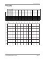
Procarta®Cytokine
Assay Kit
User Manual Specifically for measuring
Cytokines in Tissue or Cell Lysates
Panomics I Affymetrix, Inc.
Procarta Cytokine Assay Kit User Manual
Copyright
© Copyright 2009, Affymetrix, Inc. All rights reserved.
Trademarks
Procarta is a registered trademark of Affymetrix, Inc. xMAP and Luminex are registered trademarks of the Luminex Corporation. Bio-Plex is a registered
trademark of Bio-Rad Laboratories.
Citing Procarta in Publications
When describing a procedure for publication using this product, we would appreciate it if you would refer to it as the Procarta® Cytokine Assay Kit.
If a paper cites a Procarta product and is published in a research journal, the lead author(s) may receive a travel stipend for use at a technology conference
or tradeshow by sending a copy of the paper to our technical support group at [email protected] or via fax at (510) 818-2610.
Disclaimer
Affymetrix, Inc. reserves the right to change its products and services at any time to incorporate technological developments. This manual is subject to
change without notice.
Although this manual has been prepared with every precaution to ensure accuracy, Affymetrix,Inc. assumes no liability for any errors or omissions, nor for
any damages resulting from the application or use of this information.
Contents
About the User Manual . . . . . . . . . . . . . . . . . . . . . . . . . . . . . . . . . . . . . . . . . . . . . . . . . 5
Who Should Read this Manual. . . . . . . . . . . . . . . . . . . . . . . . . . . . . . . . . . . . . . . 5
What this Manual Covers . . . . . . . . . . . . . . . . . . . . . . . . . . . . . . . . . . . . . . . . . . . 5
Safety Warnings and Precautions . . . . . . . . . . . . . . . . . . . . . . . . . . . . . . . . . . . . 5
For More Information . . . . . . . . . . . . . . . . . . . . . . . . . . . . . . . . . . . . . . . . . . . . . . 5
About the Procarta Cytokine Assay Kit . . . . . . . . . . . . . . . . . . . . . . . . . . . . . . . . . . . . . 6
Procarta Cytokine Assay Defined. . . . . . . . . . . . . . . . . . . . . . . . . . . . . . . . . . . . . 6
Available Kit Formats . . . . . . . . . . . . . . . . . . . . . . . . . . . . . . . . . . . . . . . . . . . . . . 6
Procarta Cytokine Assay Kit Contents and Handling Conditions . . . . . . . . . . . . . . . . . . 6
Kit Contents and Storage . . . . . . . . . . . . . . . . . . . . . . . . . . . . . . . . . . . . . . . . . . . 6
Kit Handling . . . . . . . . . . . . . . . . . . . . . . . . . . . . . . . . . . . . . . . . . . . . . . . . . . . . . 6
Required Materials and Equipment Not Provided . . . . . . . . . . . . . . . . . . . . . . . . . . . . . 7
Equipment . . . . . . . . . . . . . . . . . . . . . . . . . . . . . . . . . . . . . . . . . . . . . . . . . . . . . . 7
Sample Type Specific Materials . . . . . . . . . . . . . . . . . . . . . . . . . . . . . . . . . . . . . . 7
Overview of Assay Workflow . . . . . . . . . . . . . . . . . . . . . . . . . . . . . . . . . . . . . . . . . . . . . 7
Set-Up and Operation of the Vacuum Manifold System . . . . . . . . . . . . . . . . . . . . . . . . 8
About Using the Vacuum Manifold . . . . . . . . . . . . . . . . . . . . . . . . . . . . . . . . . . . . 8
Sealing Filter Plates . . . . . . . . . . . . . . . . . . . . . . . . . . . . . . . . . . . . . . . . . . . . . . . 8
Setting Up and Calibrating the Manifold. . . . . . . . . . . . . . . . . . . . . . . . . . . . . . . . 8
Operating the Manifold . . . . . . . . . . . . . . . . . . . . . . . . . . . . . . . . . . . . . . . . . . . . . 9
Assay Preparation . . . . . . . . . . . . . . . . . . . . . . . . . . . . . . . . . . . . . . . . . . . . . . . . . . . . 10
About Preparing Samples . . . . . . . . . . . . . . . . . . . . . . . . . . . . . . . . . . . . . . . . . 10
Preparing Brain Tissue Lysates . . . . . . . . . . . . . . . . . . . . . . . . . . . . . . . . . . . . . 10
Preparing Lysates from Cultured Cells. . . . . . . . . . . . . . . . . . . . . . . . . . . . . . . . 10
Preparing Standards. . . . . . . . . . . . . . . . . . . . . . . . . . . . . . . . . . . . . . . . . . . . . . .11
Preparing 1X Wash Buffer . . . . . . . . . . . . . . . . . . . . . . . . . . . . . . . . . . . . . . . . . .11
Assay Procedure . . . . . . . . . . . . . . . . . . . . . . . . . . . . . . . . . . . . . . . . . . . . . . . . . . . . . 12
Assay Guidelines . . . . . . . . . . . . . . . . . . . . . . . . . . . . . . . . . . . . . . . . . . . . . . . . 12
Before You Start . . . . . . . . . . . . . . . . . . . . . . . . . . . . . . . . . . . . . . . . . . . . . . . . . 12
Performing the Assay . . . . . . . . . . . . . . . . . . . . . . . . . . . . . . . . . . . . . . . . . . . . . 12
Troubleshooting . . . . . . . . . . . . . . . . . . . . . . . . . . . . . . . . . . . . . . . . . . . . . . . . . . . . . . 16
Possible Problems and Recommended Solutions . . . . . . . . . . . . . . . . . . . . . . . 16
Contacting Panomics . . . . . . . . . . . . . . . . . . . . . . . . . . . . . . . . . . . . . . . . . . . . . . . . . . 17
Technical Help . . . . . . . . . . . . . . . . . . . . . . . . . . . . . . . . . . . . . . . . . . . . . . . . . . 17
For Additional Services . . . . . . . . . . . . . . . . . . . . . . . . . . . . . . . . . . . . . . . . . . . 17
Appendix I . . . . . . . . . . . . . . . . . . . . . . . . . . . . . . . . . . . . . . . . . . . . . . . . . . . . . . . . . . 18
Bead-Analyte Associations . . . . . . . . . . . . . . . . . . . . . . . . . . . . . . . . . . . . . . . . 18
Assay Performance . . . . . . . . . . . . . . . . . . . . . . . . . . . . . . . . . . . . . . . . . . . . . . . . . . . 19
Standards Comparison. . . . . . . . . . . . . . . . . . . . . . . . . . . . . . . . . . . . . . . . . . . . 19
Limit of Detection . . . . . . . . . . . . . . . . . . . . . . . . . . . . . . . . . . . . . . . . . . . . . . . . 19
Procarta Cytokine Assay User Manual
iii
Assay Cross Reactivity and Specificity . . . . . . . . . . . . . . . . . . . . . . . . . . . . . . . . 19
Assay Precision . . . . . . . . . . . . . . . . . . . . . . . . . . . . . . . . . . . . . . . . . . . . . . . . . 20
Assay Accuracy . . . . . . . . . . . . . . . . . . . . . . . . . . . . . . . . . . . . . . . . . . . . . . . . . 20
Sample and Blank Plate Layouts . . . . . . . . . . . . . . . . . . . . . . . . . . . . . . . . . . . . 21
About the User Manual
About the User Manual
Who Should Read Anyone that has purchased a Procarta Cytokine Assay Kit from Panomics to perform
this Manual quantitative, multiplexed measurement of Human, Mouse or Rat cytokines derived
from Tissue or Cell Lysates using the Luminex® Technology.
IMPORTANT Please note that we provide application specific manuals for cytokine
measurement from Cell Culture Supernatants and Serum/Plasma Samples. Our
most updated manuals can be obtained from our website.
]
What this Manual This manual provides recommendations and step-by-step procedures for the
Covers following:
♦
Sample and assay preparation
♦
Set up and operation of the vacuum manifold system
♦
Assay procedure
♦
Troubleshooting
Safety Warnings ! WARNING ! Though the general procedure is very similar to other
and Precautions Luminex cytokine assays, there are subtle differences due to the
approach we have taken in developing the assay to have an improved
workflow, so please ensure that you do not refer to other vendors
manuals and please read the supplied Panomics manual prior to
starting the assay.
CAUTION All chemicals should be considered potentially hazardous. We recommend that
this product and its components be handled by those trained in laboratory techniques and be
used according to the principles of good laboratory practice.
CAUTION This kit contains small quantities of sodium azide. Sodium azide reacts with lead
and copper plumbing to form explosive metal azides. When disposing, flush drains with a large
volume of water to prevent azide accumulation. Observe all state and local regulations for
disposal.
For More For information about the Procarta products mentioned in this manual, visit our
Information website at www.panomics.com.
Procarta Cytokine Assay User Manual
Page 5
About the Procarta Cytokine Assay Kit
About the Procarta Cytokine Assay Kit
Procarta Cytokine The Procarta Cytokine assays are multiplex immunoassays based on xMAP®
Assay Defined detection technology developed by Luminex. This bead based multiplex assay kit
can quantitatively measure multiple cytokines from as little as 25 µL of Tissue or Cell
Lysate in 3 hours with a sensitivity of less than 1 pg/ml/cytokine. For increased
sensitivity, the sample input can be increased to 100 µL of Tissue or Cell Lysate.
Available Kit The cytokine panels for Procarta Cytokine Assay Kit are available as:
Formats ♦ Standard pre-mixed panels
♦
“By Request”, user selected panels, provided in premixed and ready to use
format
Procarta Cytokine Assay Kits are available as single or ten plate 96-well formats and
contain all the reagents required to detect cytokines from cell culture supernatants.
For detection of cytokines in plasma or serum, standard diluent kits are sold
separately.
Procarta Cytokine Assay Kit Contents and Handling Conditions
Kit Contents and The Procarta Cytokine Assay Kit contains the following components. The kits are
Storage available as single plate or ten plate formats. Refer to the product insert for quantities
and details of components supplied.
Procarta Cytokine Kit components:
Component
Kit Handling ♦
Page 6
Storage
Reading Buffer
4 °C
10X Wash Buffer
4 °C
Detection Antibody, premixed
4 °C
Antigen Standards, premixed, lyophilized
4 °C
Streptavidin-PE (SAPE)
4 °C
Antibody Beads
4 °C
Filter Plate
4 °C
Plate Seals
4 °C
PCR 8 tube strip
4 °C
Store the entire kit at 4 °C
♦
Do not reuse or store resuspended antigen standards
♦
10X Wash Buffer and its 1X dilution can be stored at 4 °C or room temperature
for up to 6 months
Procarta Cytokine Assay User Manual
Required Materials and Equipment Not Provided
Required Materials and Equipment Not Provided
Equipment
Item
Source
Vacuum filtration system
Millipore (P/N MAVM0960R
and WP6111560)
Microplate shaker
Labline model 4625 or
equivalent with 3 mm orbit
Luminex or Luminex-based instrument
MiraiBio, Bio-Rad or other
Luminex instrument provider
Sample Type When preparing Tissue Lysates for cytokine measurements, you will need to order
Specific Materials the Procarta Lysis Buffer.
Catalog Number
PC6002
Contents
Procarta Lysis Buffer
Size
20 mL
Overview of Assay Workflow
Step 1.
Prepare Samples and Reagents
a. Collect and prepare samples
b. Prepare antigen standards
c. Prepare 1X Wash Buffer
Step 2.
Pre-wet Filter Plate
a. Let sit for 5 min
b. Filter
Step 3.
Add Antibody Beads
a. Filter
b. Wash once
c. Filter
Step 4.
Add samples/standards
a. Incubate 30 min shaking, 20-25 C
b. Wash/filter three times
Step 5.
Add Detection Antibody
a. Incubate 30 min shaking, 20-25 C
b. Wash/filter three times
Step 6.
Add Streptavidin-PE
a. Incubate 30 min shaking, 20-25 C
b. Wash/filter three times
Step 7.
Procarta Cytokine Assay User Manual
Read plate
Page 7
Set-Up and Operation of the Vacuum Manifold System
Set-Up and Operation of the Vacuum Manifold System
About Using the This topic describes how to set up and use the Millipore vacuum manifold. This
Vacuum Manifold includes how to calibrate the pressure and important guidelines that will help to
ensure good assay reproducibility.
We recommend that you set up and calibrate the manifold before you start the assay
to ensure the assay is performed without interruption.
Sealing Filter ♦
Plates
Lay a Plate Seal over the Filter Plate and roll a 5 mL serological pipet (or
equivalent) over the Plate Seal to seal the Filter Plate.
This ensures adequate plate sealing while avoiding any leakage due to capillary
action.
IMPORTANT To avoid Filter Plate leakages, do not seal Filter Plates using a rubber roller (or
equivalent) as they apply significant pressure resulting in leakage.
♦
Seal all unused wells with an enclosed Plate Seal to ensure proper vacuum
pressure.
Setting Up and To set up and calibrate the manifold:
Calibrating the
Step
Action
Manifold
1
Set up the Filter Plate vacuum manifold as shown below. Follow the
manufacturer’s manual for details.
2
Calibrate the vacuum pressure using the Filter Plate:
a. Place the Filter Plate on top of the manifold.
b. Turn on the vacuum.
c. Press the corners of the Filter Plate to form a tight seal.
d. Set the pressure to 2–3 mm of Hg.
IMPORTANT If the vacuum is too high, beads will be trapped on the filter.
Page 8
Procarta Cytokine Assay User Manual
Set-Up and Operation of the Vacuum Manifold System
Operating the To operate the manifold:
Manifold
Step
1
Action
Once the vacuum is set correctly, remove the Filter Plate.
Check vacuum calibration periodically. As a general guideline, 200 μL of solution
should take approximately 8-10 seconds to clear the well of a Filter Plate.
2
For all filtration steps, turn the Filter Plate vacuum manifold on, transfer the Filter
Plate to the vacuum manifold and then filter the solution. Avoid splashing and
cross-contamination of wells during all wash steps.
IMPORTANT During filtration, maintain the vacuum between 2–3 mm of Hg.
Higher vacuum settings may result in Capture Bead loss.
IMPORTANT Do not allow the Filter Plates to air-dry following washes.
Immediately add the next component following each filtration step.
IMPORTANT We recommend peforming all of the washes steps next to the
manifold to minimize the amount of time that the beads are exposed to air.
3
Break the vacuum immediately after each solution has been completely filtered
from all wells (approximately 8-10 seconds).
Note
Wells typically filter at different rates.
4
Place the Filter Plate back on the Filter Plate .
5
Following the last wash in each series, blot the bottom of the Filter Plate
thoroughly with a paper towel to remove traces of 1X Wash Buffer. Avoid touching
the bottom of the Filter Plate with your fingers or to the bench during
manipulations and immediately move to the next step to ensure that the beads are
in the appropriate buffered solution.
Procarta Cytokine Assay User Manual
Page 9
Assay Preparation
Assay Preparation
About Preparing This instruction manual is specifically intended for the application of measuring
Samples cytokines from Tissue or Cell Lysates. We provide application specific manuals for
measuring cytokines from Serum, Plasma and Cell Cultured Supernatants. You can
visit our website to download our latest manuals.
Examples are provided below for measuring cytokines derived from brain and
cultured cells. Other tissues can be used as a source of starting material, but these
protocols have been provided as examples. For other tissue specific protocols,
please call or email our technical support staff.
Preparing Brain Brain Tissue Lysates
Tissue Lysates
Step
Action
1
Animals are rapidly transcardially perfused with ice cold saline containing heparin
to remove the blood.
2
The animal is then decapitated, the brain removed rapidly and 8mm carotid slices
(or any size designed) cut and put into ice cold PBS
3
Regions of interest are excised from the slices. These tissue chunks are approx
2x2x8mm. The bigger of tissue size, the better, as long as same size for every
sample, including treated and untreated. The size can go up to 10mg
4
The tissue chunks are immediately put into ice cold Procarta lysis buffer which
contain protease inhibitors (150 μl per tissue chunk; we keep each chunk
separate), and left on ice until all regions of interest are excised. 150 μL of Procarta
Lysis Buffer can be used for up to 10 mg of tissue.
5
The tissue is then homogenized in a dounce homogenizer until there are no more
tissue clumps (approx. 30- 40 strokes)
6
Centrifuge at 14,000 rpm (bench top centrifuge) for 10 minutes at 4° C.
7
The supernatant is transferred to a new tube and is ready for use. Store the
samples at -80°C if they are to be used at a later date.
Preparing Lysates Cell Culture Lysates
from Cultured
Step
Action
Cells
1
Adherent Cells
Preparation is for 1x10^6 to 1x10^7 cells grown in a 10 cm plate. Trypinsinze the
cells and remove the cells using a cell scraper. Transfer the cells to a 15 ml conical
tube and centrifuge at 500 x g at 4° C for five minutes. Carefully aspirate the
supernatant and re-suspend the cells in 5 mls of ice cold PBS. Repeat this step
using 5 mls of ice cold PBS and centrifuge again at 500 x g at 4° C. Completely
remove all of the PBS.
Suspension Cells
Preparation is for 1x10^6 to 1x10^7 cells grown in a flask. Transfer cells to a 15
ml conical tube and spin at 500 x g at 4° C for five minutes. Carefully aspirate the
supernatant and re-suspend the cells in 5 mls of ice cold PBS and spin again at
500 x g at 4° C. Completely remove all of the PBS.
Page 10
2
Add 200 μL of ice cold Procarta Cell Lysis Buffer, pipette up and down several
times and then incubate on ice for 5 minutes.
3
Transfer entire contents to a 1.5 ml microcentrifuge tube
Procarta Cytokine Assay User Manual
Assay Preparation
Cell Culture Lysates
Step
Action
4
Centrifuge at 14,000 rpm (bench top centrifuge) for 10 minutes at 4° C.
5
The supernatant is transferred to a new tube and is ready for use. Store the
samples at -80°C if they are to be used at a later date.
Preparing To prepare standards:
Standards
Step
Action
1
Reconstitute the premixed lyophilized protein standard with 250 μL of Procarta
Lysis Buffer
2
Vortex gently for 10 seconds and incubate on ice for 5 minutes. Transfer the entire
contents to the first tube of the PCR 8-tube strip provided.
3
Prepare serial dilutions of the premixed standard as indicated below.
a. Using the 8 tube strip provided, add the following
b. Add 75 μL of Procarta Lysis Buffer to tubes 2-8. Follow the table below to
create the dilutions. Use a P200 pipette to transfer and mix the 25 μL
standard from tube to tube.
c. Store tube strip on ice until ready to use. When transferring the standards
to the to beads in the filter plate, we recommend using a multi-channel
pipette.
d. See Plate Map for example serial dilutions and blank
Preparing 1X Wash
Buffer
Step
Tube
Number
Procarta Lysis
Buffer
(μL)
Antigen Standard
(μL)
Final Analyte
Concentration in the
Assay (pg/mL)
1
—
250
10,000
2
75
25 from Tube 1
2500
3
75
25 from Tube 2
625
4
75
25 from Tube 3
156
5
75
25 from Tube 4
39
6
75
25 from Tube 5
9.76
7
75
25 from Tube 6
2.4
8
75
25 from Tube 7
0.6
Blank
100 per well
Procarta Lysis Buffer
0
Action
1
Vortex the 10X Wash Buffer to dissolve all the salts.
2
Mix 20 mL of the 10X Wash Buffer with 180 mL deionized, sterile water.
Note 1X Wash Buffer can be stored at 4 °C or room temperature for up to 6
months.
Procarta Cytokine Assay User Manual
Page 11
Assay Procedure
Assay Procedure
Assay Guidelines IMPORTANT For optimal assay performance and consistent results, please read these
guidelines before proceeding with the assay.
♦
Change the pipet tips after every transfer and avoid creating bubbles when
pipetting.
♦
Follow the guidelines in “Set-Up and Operation of the Vacuum Manifold System”
on page 8.
♦
During the incubation steps, cover the Filter Plate assembly with aluminum foil to
prevent photobleaching of the fluorescent beads.
♦
Bring all buffers to room temperature before setting up the assay. Store the
detection antibody, antibody beads, Streptavidin-PE, and reconstituted
standards on ice during the assay.
Before You Start Make sure the Luminex or Luminex-based instrument is turned on at least 30 minutes
before you intend to read your plates.
Performing the To perform the assay:
Assay
Step
1
Action
Columns 1 and 2 are typically used for duplicate standards. Mark the sample wells
you want to use.
A blank plate layout is provided in the appendix for recording your plate layout.
2
Seal the un-used wells of the plate with a Plate Seal provided in the kit before
starting the assay.
3
Pre-wet the Filter Plate:
a. Add 150 μL of Reading Buffer to each non-sealed well.
b. Incubate 5 minutes at room temperature.
c. Remove the Reading Buffer with vacuum filtration. Reading Buffer should
clear wells within 3–5 seconds. If outside this range, see “Set-Up and
Operation of the Vacuum Manifold System” on page 8.
IMPORTANT Do not invert the plate.
4
Add the Antibody Beads:
a. Vortex the premixed Antibody Beads for 30 seconds at room temperature.
b. Add 50 μL of Antibody Beads to each unsealed well.
c. Remove buffer with vacuum filtration.
IMPORTANT Do not invert the plate.
5
Wash the Antibody Beads:
a. Add 150 μL/well of 1X Wash Buffer and remove with vacuum filtration.
IMPORTANT Do not invert the plate.
b. Blot the bottom of the Filter Plate thoroughly with paper towels to remove
residual buffer.
6
Page 12
Add 75 μL/well of Procarta Lysis Buffer to only the Standard Wells.
Procarta Cytokine Assay User Manual
Assay Procedure
To perform the assay: (continued)
Step
Action
7
Add 25 μL/well of standards marked on the plate layout sheet. For the sample, add
100 μL/well of Lysate. For the blank samples, add 100 μL/well of Procarta Lysis
Buffer. Seal the plate gently using the Plate Seal. If you are using 25 μL or 50 uL of
Lysate, you will need to add 75 or 50 uL of Procarta Lysis Buffer for a total of 100
μL for each sample well.
8
Incubate and wash the Filter Plate:
a. Completely wrap the Filter Plate with aluminum foil.
b. Shake for 30 minutes at 500 rpm at room temperature. Optionally, you can
incubate overnight without shaking at 4 °C, after the 30 minute shaking
incubation step.
c. Carefully remove the Plate Seal to avoid splashing the plate contents.
d. Remove solution with vacuum filtration.
IMPORTANT Do not invert the plate.
e. When washing the plate, we recommend using multi-channel pipette and a
large enough plastic reservoir for the wash buffer. We also recommend
stationing the wash buffer reservoir next manifold to minimize the amount
of time that the beads are exposed to air.
f.
9
Wash the plate three times with 150 μL/well of 1X Wash Buffer. After the 3rd
wash, blot the bottom of the Filter Plate with paper towels.
Add premixed Detection Antibodies:
a. Add 25 μL/well of the Detection Antibody.
b. Seal the Filter Plate with a new Plate Seal.
c. Completely wrap the Filter Plate with aluminum foil.
d. Shake for 30 minutes at 500 rpm at room temperature.
10
Remove the solution with vacuum filtration and wash the plate:
a. Carefully remove the Plate Seal to avoid splashing the plate contents.
b. Remove solution with vacuum filtration.
IMPORTANT Do not invert the plate.
c. Wash the plate three times with 150 μL/well of 1X Wash Buffer. After the 3rd
wash, blot the bottom of the Filter Plate with paper towels.
11
Add Streptavidin–PE:
a. Vortex the Streptavidin–PE.
b. Add 50 μL/well of Streptavidin-PE.
c. Seal the Filter Plate with a new Plate Seal.
d. Completely wrap the Filter Plate with aluminum foil.
e. Shake for 30 minutes at 500 rpm at room temperature.
12
Remove the solution with vacuum filtration and wash the plate:
a. Carefully remove the Plate Seal to avoid splashing the plate contents.
b. Remove solution with vacuum filtration.
IMPORTANT Do not invert the plate.
c. Wash the plate three times with 150 μL/well of 1X Wash Buffer. After the 3rd
wash, blot the bottom of the Filter Plate with paper towels.
Procarta Cytokine Assay User Manual
Page 13
Assay Procedure
To perform the assay: (continued)
Step
13
Action
Prepare plate for analysis on a Luminex instrument:
a. Add 120 μL/well of the Reading buffer.
b. Seal the Filter Plate with a new Plate Seal.
Note The Filter Plate can be wrapped with aluminum foil and stored for up to 48
hours before proceeding. However, delay in reading the plate may result in
decreased sensitivity for some analytes.
c. Shake for 5 minutes at 500 rpm at room temperature or until the Luminex
instrument is available.
d. Remove the Plate Seal before analyzing on the Luminex instrument.
Page 14
Procarta Cytokine Assay User Manual
Assay Procedure
To perform the assay: (continued)
Step
14
Action
Analyze the plate following the respective operation manual for the Luminex or
Luminex-based
instrument.
Software
Sample
Size
DD Gate
Timeout
Bead
Events/Be
ad Region
Luminex 100/
50 μL
7,500-15,000
25 sec
50 or 100
50 μL
4335-10,000
45 sec
50 or 100
50 μL
2,000-15,000
25 sec
50 or 100
50 μL
4,335-10,000
25 sec
50 or 100
IS100 v 2.318
Bioplex/
Bio-Rad
Miraibio/
Hitachi
Starstation/
ACS
When the Luminex beads are injected into the flow cell, a small percentage of the
beads will have a tendency to clump and go through the flow cell as doublets. The
DD Gate or Doublet Discriminator Gate will allow for discrimination of the doublets
from the singlet beads. When setting the DD Gate, you can follow the appropriate
settings from the table above. However, for the best results, you should adjust the
DD Gate and center the gates around the largest peak which is the singlet beads.
You can adjust the gates when processing the first sample.
The Bio-Plex® Suspension Array System allows calibration using Low or High
sensitivity settings. Perform the sensitivity selection during calibration, using
predetermined values of CAL2 RP1 target, as provided by Bio-Rad. Using the RP1
Low target value will provide results comparable to those obtained from the
Luminex 100. Using the RP1 High target value may increase detection sensitivity
for low cytokine protein concentrations but sacrifices part of the linear dynamic
range for high concentrations. We recommend RP1 Low target value for BioPlex.
The number of beads events per region can be set to 50 or 100. Please note that
the total time to measure 100 events vs 50 events will take longer.
IMPORTANT Check to ensure the probe height in the Luminex instrument is
adjusted appropriately for the Filter Plate.
IMPORTANT We recommend that you calibrate the Luminex or Luminex-based
instrument each day the assay is run.
IMPORTANT If there is a malfunction of the machine or software during the run
or there was a problem with the system, the plate can be re-processed on the
Luminex machine. Remove the plate from the machine and vacuum filter the plate.
Resuspend the beads in Reading Buffer and shake for 5 minutes then re-read. The
plate may take longer to read since there will be less beads in the plate.
Procarta Cytokine Assay User Manual
Page 15
Troubleshooting
Troubleshooting
Possible Problems
and
Recommended
Solutions
Observation
Possible Cause
Recommended Action
Filter plate leakage
Vacuum pressure too high
Adjust the vacuum pressure
as recommended in “Set-Up
and Operation of the
Vacuum Manifold System”
on page 8.
Filter Plate is misaligned (at
an angle) during
incubation/processing
Set the Filter Plate on a flat,
level surface during
incubation/processing.
Leakage from capillary
action
After each vacuum step,
blot the bottom of the Filter
Plate using paper towels or
absorbent paper.
Plate Seal applied using too
much force
Lay Plate Seal on the plate
and gently roll a 5 mL
serological pipet over the
plate to seal.
Samples and antigen
standards were not stored
on ice
Prepare the samples and
standards on ice before
setting up the assay.
Bottom of the Filter Plate is
not dry
After each vacuum step,
blot the bottom of the Filter
Plate using paper towels or
absorbent paper.
Contamination from
re-using the Plate Seal
Use a new Plate Seal for
each incubation step.
The high concentration
standards have high
variability.
After adding the standards
and samples, it is very
important that any excess
standards are removed
during the wash step
Contamination from Wash
Buffer
Be careful not to splash
Wash Buffer during wash
steps into adjacent wells.
The machine was calibrated
using the high PMT (CAL2
High RP1) and the top 3
standards have similar MFI
values
Calibrate the machine using
the CAl2 Low RP1 target
value
High CV
Limited Dynamic Range
when using the BioPlex
software
Page 16
Procarta Cytokine Assay User Manual
Contacting Panomics
Observation
Possible Cause
Recommended Action
Low bead count
Volume of bead solution is
too low
Add 120 μL/well Reading
Buffer and shake for 5
minutes to resuspend beads
before reading in the
Luminex instrument.
Beads are clumping
Vortex the bead solution
well before using in the
assay.
Vacuum pressure too high
Use 2–3 mm Hg vacuum
pressure.
Filter ruptured due to
excess vacuum time
Do not use the vacuum over
10 seconds in any of the
steps.
Dyes contained in the beads
are photo-bleached from
overexposure to light
Store bead solution and the
assay plate in the dark
Reader is clogged
Follow the instructions in
the Luminex instrument user
documentation.
Standards not reconstituted
and diluted correctly
Prepare fresh antigen
standards following the
instructions in “Preparing
Standards” on page 11.
Beads or reagents expired
Verify the expiration date of
the kit.
Beads stuck to the bottom
of the plate
Make sure the plate is
agitated at 500 rpm for the
recommended time and for
at least 5 minutes before
reading.
Reagents expired
Verify the expiration date on
the kit.
Samples and antigen
standards were not stored
on ice
Prepare the samples and
standards on ice before
setting up the assay.
Low signal or sensitivity
Poor recovery
Contacting Panomics
Technical Help For technical questions, contact our technical support group by telephone at
866-317-2626 or by email at [email protected], or visit our website
www.panomics.com for an updated list of FAQs and product support literature.
For Additional For information about Panomics products or for ordering information, contact your
Services Regional Sales Manager, or visit our website at www.panomics.com.
Procarta Cytokine Assay User Manual
Page 17
Appendix
Appendix
Bead-Analyte
Associations
The following table may not be the most up to date current bead-analyte
associations setting for your Luminex instrument. Please refer to your
product insert for analytes included in your kit, especially if you have a
custom order or “By Request” assay.
Bead
Human
Bead
Human
Bead
24
Mouse
Adiponectin
Bead
Rat
73
CRP
66
Adiponectin
22
IP10
54
CRP
74
I-TAC
8
CD120B
52
Eotaxin
84
EGF
6
Leptin
52
Eotaxin
46
G-CSF
17
ENA78
51
MCP - 1
46
G-CSF
36
GM-CSF
52
Eotaxin
13
MCP - 3
44
GM-CSF
21
ICAM
55
FGF - basic
80
MIF
43
IFM-gamma
43
IFN-gamma
46
G-CSF
11
MIG
18
IL - 1 alpha
18
IL-1 alpha
44
GM-CSF
8
MMP- 1
19
IL - 1 beta
19
IL-1 beta
61
GRO alpha
7
MMP- 2
20
IL - 2
20
IL-2
79
HGF
53
MMP- 3
21
IL - 3
34
IL-4
73
ICAM - 1
47
MIP - 1 beta
25
IL - 4
27
IL-5
48
IFN alpha
67
MIP - 3 alpha
26
IL - 5
25
IL-6
43
IFNy
12
MIP - 1 alpha
27
IL - 6
35
IL-10
62
IL - 1 alpha
83
MMP - 7
38
IL - 9
32
IL-12(p40)
18
IL - 1 beta
72
MMP - 8
28
IL - 10
44
IL-13
63
IL - 1ra
9
MMP - 9
33
IL - 12(p40)
28
IL-17
19
IL - 2
78
MMP - 12
34
IL - 12(p70)
57
IL-18
20
IL - 4
64
MMP - 13
36
IL - 13
37
KC
21
IL - 5
38
NGF
35
IL - 17
6
Leptin
25
IL - 6
14
PAI - 1
41
IL-23
54
MCP - 1
26
IL - 7
37
PDGFBB
22
IP10
51
MCP - 3
27
IL - 8
42
RANTES
37
KC
47
MIP-1 alpha
28
IL - 10
86
Resistin
6
Leptin
38
NGF
33
IL - 12(p40)
75
SAA
54
MCP - 1
42
RANTES
34
IL - 12(p70)
85
sCD40L
51
MCP - 3
29
TGF beta
35
IL - 13
77
sE-selectin
47
MIP - 1 alpha
33
TNF alpha
65
IL - 15
29
TGF beta
42
RANTES
26
V-CAM
36
IL - 17
45
TNF alpha
7
Srankl
56
VEGF
39
IL - 17F
32
TNF beta
29
TNF beta
57
IL-18
88
VCAM - 1
45
TNF alpha
41
IL-23
56
VEGF
56
VEGF
*TGF-beta must be used as a single plex assay because the sample must be
prepared in an acidic buffer.
Page 18
Procarta Cytokine User Manual- Cell Culture Supernatants
Assay Performance
Assay Performance
Standards The following table contains conversion factors for Procarta Antigen Standards for
Comparison which World Health Organization (WHO) standards were available for comparison.
Procarta Antigen Standards, Human
Analyte
WHO Lot Number
100 Procarta pg/mL =
IL-1β
86/680
11.9 IU/mL
IL-4
88/656
15.6 IU/mL
IL-8
89/520
1.91 IU/mL
IL-10
93/722
3.91 IU/mL
IL-12(p70)
95/544
11.9 IU/mL
IL-13
94/622
1.33 IU/mL
IFNγ
88/606
29.4 IU/mL
GM-CSF
88/646
20.3 IU/mL
TNFα
88/786
147.7 IU/mL
Procarta Antigen Standards, Mouse
Analyte
WHO Lot Number
100 Procarta pg/mL =
IL-1α
93/672
580.8 IU/mL
IL-1β
93/668
99.8 IU/mL
IL-2
93/566
67.3 IU/mL
IL-3
91/662
141.0 IU/mL
IL-4
91/656
8.2 IU/mL
IL-6
93/730
97.6 IU/mL
GM-CSF
88/532
151.5 IU/mL
TNFα
91/658
92.5 IU/mL
Limit of Detection The Limit of Detection (LOD) for human, mouse and rat cytokines from cell culture
supernatants, serum, and plasma samples is less than 1 pg/ml. The LOD is defined
as the antigen concentration with a Median Fluorescent Intensity (MFI) greater than
or equal to the sum of the background MFI plus two standard deviation
Assay Cross Antibody cross reactivity is negligible and does not exceed 5%. Cross reactivity was
Reactivity and verified by examining the non-specific signal from each bead after:
Specificity ♦ A single antigen was added
♦
All antigens, except the antigen matching the bead being evaluated, was added
Procarta Cytokine Assay User Manual
Page 19
Assay Performance
Assay Precision The following tables present typical intra-assay and inter-assay coefficients of
variation (CVs) for human samples from cell culture supernatants, plasma, and
serum. The average intra-assay and inter-assay CV is less than 10% based on the
analysis of 4 replicates.
Assay Accuracy Accuracy is defined as the percentage of analyte recovered from samples spiked
with a known quantity. The following table represents typical spike recovery results
based on spiking 3 different amounts (40, 400, and 4000 pg/mL) of analyte into
cell-culture supernatants, serum, and plasma samples from human and mouse. All
samples had low or undetectable amount of analyte present before the spike. Each
sample type had 4 replicates.
The spike recovery was 80–120%. The percent spike recovery is defined as the
observed Median Fluorescent Intensity (MFI) divided by the expected MFI times 100.
Page 20
Procarta Cytokine Assay User Manual
Assay Performance
Sample and Blank
Plate Layouts
Standards
Samples
1
pg/mL
2
pg/mL
3
4
5
6
7
8
9
10
11
12
20,000
20,000
Blank
Blank
8
8
16
16
24
24
32
32
B
5,000
5,000
1
1
9
9
17
17
25
25
33
33
C
1250
1250
2
2
10
10
18
18
26
26
34
34
D
313
313
3
3
11
11
19
19
27
27
35
35
E
78
78
4
4
12
12
20
20
28
28
36
36
F
19.5
19.5
5
5
13
13
21
21
29
29
37
37
G
4.9
4.9
6
6
14
14
22
22
30
30
38
38
H
1.2
1.2
7
7
15
15
23
23
31
31
39
39
!
"
#
$
%
&
'
(
Procarta Cytokine Assay User Manual
Page 21