1
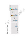
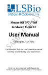
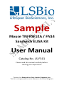
Sample Mouse TNFRSF12A / FN14 Sandwich ELISA Kit User Manual Catalog No. LS-F301 Please read this manual carefully before starting your experiment. This kit is for Research Use Only. Not for Diagnostic Use. This kit is not approved for use in humans or for clinical diagnosis. Assay Specifications......................................................... 1 Intended Use................................................................... 2 Assay Principle................................................................. 2 Assay Principle Image...................................................... 3 Kit Components............................................................... 4 Kit Storage....................................................................... 4 Other Required Supplies.................................................. 4 Sample Collection............................................................ 5 Sample Collection Notes.................................................. 7 Reagent Preparation........................................................ 8 Sample Preparation......................................................... 9 Standard Preparation.................................................... 10 Reagent Preparation Notes........................................... 11 Assay Procedure............................................................ 12 Assay Procedure Notes.................................................. 13 Assay Procedure Summary............................................ 15 Calculation of Results.....................................................16 Troubleshooting Guide.................................................. 17 Troubleshooting Guide (continued)...............................18 A SSAY S P E C I F I C A T I O N S Target: TNFRSF12A / FN14 Synonyms: TNFRSF12A / FN14, TNFRSF12A, tumor necrosis factor receptor superfamily, member 12A, CD266 antigen, CD266, FGFinducible 14, FN14, Tweak-receptor, TWEAKR Specificity: Recombinant and natural Mouse TNFRSF12A / FN14. [Add cross-reactivity data] Sample Types: This kit is recommended for use with Mouse serum, plasma, and tissue homogenates. Use with other sample types is not supported. Detection Range: [To be determined] Sensitivity (Lower Limit of Detection): [To be determined] Performance Characteristics: InterAssay CV <[To be determined]% IntraAssay CV=<[To be determined]% 1 I NTENDED U S E This Sandwich ELISA (Enzyme Linked Immunosorbent Assay) kit is intended for the in vitro quantitative determination of Mouse TNFRSF12A / FN14 concentrations in serum, plasma, and tissue homogenates. Use with other sample types is not supported. ASSAY P RINCIPLE This assay is based on the sandwich ELISA principle. Each well of the supplied microtiter plate has been pre-coated with a target specific capture antibody. Standards or samples are added to the wells and the target antigen binds to the capture antibody. Unbound Standard or sample is washed away. A biotin-conjugated detection antibody is then added which binds to the captured antigen. Unbound detection antibody is washed away. An Avidin-Horseradish Peroxidase (HRP) conjugate is then added which binds to the biotin. Unbound Avidin-HRP conjugate is washed away. A TMB substrate is then added which reacts with the HRP enzyme resulting in color development. A sulfuric acid stop solution is added to terminate color development reaction and then the optical density (OD) of the well is measured at a wavelength of 450 nm ± 2 nm. The OD of an unknown sample can then be compared to an OD standard curve generated using known antigen concentrations in order to determine its antigen concentration. 2 A SSAY P R I N C I P L E I M A G E 3 K IT C O M P O N E N T S Component Quantity Coated 96-well Strip Plate Standard (Lyophilized) Assay Diluent (5x) Detection Antibody HRP-Streptavidin Conjugate (800x) Wash Buffer (20x) TMB Substrate Stop Solution Plate Sealers 1 2 vials 1 vial x 15 ml 2 vials 1 vial x 200 µl 1 vial x 25 ml 1 vial x 12 ml 1 vial x 8 ml 4 K IT S T O R A G E The unopened kit may be stored for up to 6 months at 2°-8°C or 1 year at -20°C. Once reconstituted, Standard should be stored at -20°C/-80°C. Avoid freeze thaw cycles. Once opened, all other reagents can be stored up to 1 month at 2°-8°C. Once individual reagents are opened it is recommended that the kit be used within 1 month. Unused Strip Plate wells should be stored at 2°-8°C in a sealed bag containing desiccant in order to minimize exposure to moisture. Do not use the kit beyond its expiration date. O THER R E Q U I R E D S U P P L I E S Microplate reader with 450nm wavelength filter High-precision pipette and sterile pipette tips Eppendorf tubes 37°C incubator Deionized or distilled water Absorbent paper 4 S AMPLE C O L L E C T I O N This assay is recommended for use with Human in serum, plasma, and tissue homogenates. Use with other sample types is not supported The sample collection protocols below have been provided for your reference. Breast Milk - Centrifuge samples for 20 minutes at 1000×g to remove particulates. Collect the supernatant for assaying. Cell Lysates - Collect and pellet the cells by centrifugation and remove the supernatant. Wash the cells 3 times with PBS* then resuspend in PBS*. Lyse the cells by ultrasonication 4 times. Alternatively freeze the cells to -20°C and thaw to room temperature 3 times. Centrifuge at 1500×g for 10 minutes at 2 - 8°C to remove cellular debris. Collect the supernatant for assaying. Erythrocyte Lysates - Centrifuge whole blood for 20 minutes at 1000×g to pellet the cells and remove the supernatant. Wash the cells 3 times with PBS* then resuspend in PBS*. Freeze (-20°C)/thaw (room temperature) the cells 3 times. Centrifuge at 5,000×g for 10 minutes at 2-8°C to remove cellular debris. Collect the supernatant for assaying. Erythrocyte lysates must be diluted with Sample Diluent before running. Animal and Plant Tissue Supernatants – Using a mortar and pestle, grind the tissues to a powder with liquid nitrogen. Resuspend the powder in 3x sample volume of sample extraction buffer (10% TCA) and incubate overnight at -20°C. Centrifuge at 8000rpm for 1h at 4°C to collect precipitated protein and decant the supernatant. Add the same volume of ice cold 100% acetone, centrifuge at 8000rpm for 15min at 4°C, then dry vacuum deposition in reserve. Add lysis buffer (2.7g urea, 0.2g CHAPS add dH20 to 5ml), incubate at room temperature for 30 minutes, then centrifuge at 8000rpm for 15min at 4°C. Collect the supernatant for assaying. Plasma - Collect plasma using EDTA or heparin as an anticoagulant. Centrifuge samples for 15 minutes at 1000×g at 2–8°C within 30 minutes of collection. Collect the supernatant for assaying. Platelet-Poor Plasma - Collect plasma using EDTA as an anticoagulant. Centrifuge samples for 15 minutes at 1000×g at 2–8°C within 30 minutes of collection. It is recommended that samples should be centrifuged for 5 10 minutes at 10,000×g for complete platelet removal. Collect the supernatant for assaying. Sperm and Seminal Plasma – Allow semen to liquefy at room temperature or 37°C. After liquefaction, centrifuge at 2,000×g for 10-15 minutes. Collect seminal plasma supernatant for assaying. Wash the precipitated protein 3 times with PBS* then resuspend in PBS*. Lyse the cells by ultrasonication then centrifuge at 2,000×g for 10-15 minutes. Collect the supernatant for assaying. Serum - Use a serum separator tube and allow samples to clot for 2 hours at room temperature or overnight at 4°C before centrifugation for 20 minutes at approximately 1000×g. Collect the supernatant for assaying. Tissue Homogenates – Because preparation methods for tissue homogenates vary depending upon tissue type, users should research tissue specific conditions independently. The following is one example only. Rinse tissues in PBS* to remove excess blood and weigh before homogenization. Finely mince tissues and homogenize them in 5-10mL of PBS*with a glass homogenizer on ice. Lyse the cells by ultrasonication or freeze (-20°C)/thaw (room temperature) 3 times. Centrifuge homogenate at 5000×g for 5 minutes. Collect the supernatant for assaying. Urine - Aseptically collect the first urine of the day (mid-stream), voided directly into a sterile container. Centrifuge to remove particulate matter and collect the supernatant for assaying. Cell culture supernatants, cerebrospinal, follicular, and lung lavage fluids, saliva, sweat, tears, and other biological fluids - Centrifuge samples for 20 minutes at 1000×g to remove particulates. Collect the supernatant for assaying. * 1xPBS (0.02mol/L pH7.0-7.2) 6 S AMPLE C O L L E C T I O N N O T E S 1. LSBio recommends that samples are used immediately upon preparation. Alternatively, samples stored at 2-8°C should be used within 5 days. For long-term storage sample aliquots should be prepared and stored at -20°C if used within 1 month, or -80°C if used within 6 months. Long term storage can result in protein degradation and denaturation, which may result in inaccurate results. 2. Avoid repeated freeze/thaw cycles for all samples. 3. In the event that a sample type not listed above is intended to be used with the kit, it is recommended that the customer conduct validation experiments in order to be confident in the results. 4. Due to chemical interference, the use of tissue or cell extraction samples prepared by chemical lysis buffers may result in inaccurate results. 5. Due to the factors including cell viability, cell number or sampling time, samples from cell culture supernatant may not be detected by the kit. 6. Samples should be brought to room temperature (18-25°C) before performing the assay without the use of extra heating. 7. Sample concentrations should be predicted before being used in the assay. If the sample concentration is not within the range of the standard curve, users must determine the optimal sample dilutions for their particular experiments. 8. LSBio is responsible for the quality and performance of the kit components but is NOT responsible for the performance of customer supplied samples used with the kit. 7 R EAGENT P R E P A R A T I O N Bring all reagents to room temperature (18-25°C) before use. 1x Assay Diluent: Prepare 75 ml of 1x Assay Diluent by diluting the supplied 15 ml of 5x Assay Diluent concentrate with 60 ml of deionized or distilled water. Assay Diluent can be stored at 4°C once prepared. 1x Wash Buffer: If crystals have formed in the concentrate, warm to room temperature and mix it gently until crystals have completely dissolved. Prepare 400 ml of Working Wash Buffer by diluting the supplied 20 ml of 20x Wash Buffer Concentrate with 380 ml of deionized or distilled water. Wash Buffer can be stored at 4°C once prepared. Detection Antibody Concentrate and Working Solution: Briefly spin down the Detection Antibody and add 100 µl of 1x Assay Diluent to prepare the Detection Antibody Concentrate. The Detection Antibody Concentrate can be stores at 4°C for 5 days. Calculate the volume of Detection Antibody Working Solution needed for your particular experiment and prepare that volume by diluting the Detection Antibody Concentrate 80-fold (1:80) with 1x Assay Diluent. HRP-Streptavidin Working Solution: Gently mix the stock solution before use. Calculate the volume of HRP-Streptavidin Working Solution needed for your particular experiment and prepare that volume by diluting the HRP-Streptavidin Conjugate Solution 800-fold (1:800) with 1x Assay Diluent. Prepared HRP-Streptavidin working solution must be prepared fresh for each experiment and cannot be stored. TMB Substrate Solution: Using sterile techniques remove the needed volume of TMB Substrate Solution for the number of wells you are planning to run. Dispose of unused TMB Substrate Solution rather than returning it to the stock container. 8 S AMPLE P R E P A R A T I O N Please predict the concentration of your samples before assaying. The resulting optical density (OD) values of your sample must fall within the OD readings of the standard curve in order for the calculated antigen concentration to be accurate. A dilution series of each sample may be necessary. Running duplicate wells for each sample is recommended. All samples should be diluted with 1x Assay Diluent. [List of any specific sample preparation details.] 9 S TANDARD P R E P A R A T I O N The following are instructions for the preparation of a Standard dilution series which will be used to generate the standard curve. The standard curve is then used to determine the concentration of target antigen in unknown samples (see the Calculation of Results section). The following will prepare sufficient volumes to run the Standard dilution series in duplicate. Standard dilutions should be used immediately once prepared. Standard Stock Solution (x Units): Reconstitute 1 tube of lyophilized Standard with x ml of Sample Diluent. Incubate at room temperature for 10 minutes with gentle agitation (avoid foaming). D1 (x Units): D2 (x Units): D3 (x Units): D4 (x Units): D5 (x Units): D6 (x Units): D7 (x Units): Pipette a µl of Stock Standard into x µl of Sample Diluent Pipette 250µl of D1 into 250µl of Sample Diluent Pipette 250µl of D2 into 250µl of Sample Diluent Pipette 250µl of D3 into 250µl of Sample Diluent Pipette 250µl of D4 into 250µl of Sample Diluent Pipette 250µl of D5 into 250µl of Sample Diluent Pipette 250µl of D6 into 250µl of Sample Diluent Zero Standard (0 Units): Use Sample Diluent alone. 10 R EAGENT P REPARATION N O T E S 1. It is highly recommended that standard curves and samples are run in duplicate within each experiment. 2. Once resuspended, standard should be used immediately or placed in long term storage at -20/-80°C. 3. In order to avoid repeat freeze thaws, prepare one-time use aliquots of standard for long term storage at -20/-80°C. 4. All solutions prepared from concentrates are intended for one-time use. Do not reuse solutions. 5. Do not prepare Standard dilutions directly in wells. 6. Prepared Reagents may adhere to the tube wall or cap during transport; centrifuge tubes briefly before opening. 7. All solutions should be gently mixed prior to use. 8. Reconstitute stock reagents in strict accordance with the instructions provided. 9. To minimize imprecision caused by pipetting, ensure that pipettes are calibrated. Pipetting volumes of less than 10μL is not recommended. 10. Substrate Solution is easily contaminated; sterility precautions should be taken. Substrate Solution should also be protected from light. 11. Do not substitute reagents from one kit lot to another. Use only those reagents supplied within this kit. 12. Due to the antigen specificity of the antibodies used in this assay, native or recombinant proteins from other manufacturers may not be detected by this kit. 11 A SSAY P R O C E D U R E Bring all reagents and samples to room temperature without additional heating and mix thoroughly by gently swirling before pipetting (avoid foaming). Prepare all reagents, working standards, and samples as directed in the previous sections. 1. Add 100 μl of Standard, Blank, or Sample per well, cover with a plate sealer, and incubate for 2.5 hours at room temperature or overnight at 4°C with gentle shaking. 2. Aspirate the liquid from each well and wash 4 times. Wash by adding approximately 300 μl of Wash Buffer using a squirt bottle, multi-channel pipette, manifold dispenser or automated washer. Allow each wash to sit for 1-2 minutes before completely aspirating. After the last wash, aspirate to remove any remaining Wash Buffer then invert the plate and tap against clean absorbent paper. 3. Add 100 μl of Detection Antibody Working Solution to each well and gently agitate to ensure thorough mixing. Incubate for 1 hour at room temperature. 4. Aspirate and wash the wells as outlined in step 2. 5. Add 100 μl of HRP-Streptavidin Working Solution to each well and incubate for 45 minutes at room temperature with gentle shaking. 6. Aspirate and wash the wells as outlined in step 2. 7. Add 100 μl of Substrate Solution to each well and incubate for 30 minutes at room temperature in the dark with gentle shaking. Monitor periodically until optimal color development has been achieved. 8. Add 50 μl of Stop Solution to each well and record the total development time. The blue color will change to yellow. If color change does not appear uniform, gently tap the plate to ensure thorough mixing. The Stop Solution should be added to wells in the same order and timing as the substrate solution. 9. Determine the optical density (OD value) of each well immediately using a microplate reader set to 450 nm. 12 A SSAY P R O C E D U R E N O T E S 1. ELISA Plate: Keep appropriate numbers of strips for 1 experiment and remove extra strips from microtiter plate. Removed strips should be placed in a sealed bag containing desiccant and stored at 4°C. 2. Solutions: To avoid cross-contamination, change pipette tips between additions of each standard, between sample additions, and between reagent additions. Also, use separate reservoirs for each reagent. 3. Applying Solutions: All solutions should be added to the bottom of the ELISA plate well. Avoid touching the inside wall of the well. Avoid foaming when possible. 4. Assay Timing: The interval between adding sample to the first and last wells should be minimized. Delays will increase the incubation time differential between wells, which will significantly affect the experimental accuracy and repeatability. For each step in the procedure, total dispensing time for addition of reagents or samples should not exceed 10 minutes. 5. Incubation: To prevent evaporation and ensure accurate results, proper adhesion of plate sealers during incubation steps is necessary. Do not allow wells to sit uncovered for extended periods of time between incubation steps. Do not let wells dry out at any time during the assay. Strictly observe the recommended incubation times and temperatures. 6. Washing: Proper washing procedure is critical. Insufficient washing will result in poor precision and falsely elevated absorbance readings. Residual liquid in the reaction wells should be patted dry against absorbent paper during the washing process. Do not put absorbent paper directly into the reaction wells. 7. Controlling Substrate Reaction Time: After the addition of the TMB Substrate, periodically monitor the color development. Stop color development before the color becomes too deep by adding Stop Solution. Excessively strong color will result in inaccurate absorbance reading. 8. Reading: The microplate reader should be preheated and programmed prior to use. Prior to taking OD readings, remove any residual liquid or fingerprints from the underside of the plate and confirm that there are no bubbles in the wells. 13 9. Reaction Time Control: Control reaction time should be strictly followed as outlined. 10. Stop Solution: The Stop Solution contains an acid, therefore proper precautions should be taken during its use, such as protection of the eyes, hands, face, and clothing. 11. Mixing: During incubation times, the use of a micro-oscillator at low frequency is recommended. Sufficient and gentle mixing is particularly important in producing reliable results. 12. Kits from different batches may be a little different in detection range, sensitivity, and color developing time. Please perform the experiment exactly according to the supplied instructions. 13. Due to inter- and intra-assay variability, it is recommended that appropriate carry-over controls be included between assays. 14. Prior to running valuable samples, LSBio recommends that the user run a preliminary experiment using the supplied controls in order to validate the assay. 15. To minimize external influence on the assay performance, operational procedures and lab conditions (such as room temperature, humidity, and incubator temperature) should be strictly controlled. It is also strongly suggested that the whole assay is performed by the same operator from the beginning to the end. 16. The kit should not be used beyond the expiration date on the kit label. 14 A SSAY P R O C E D U R E S U M M A R Y Prepare all reagents, samples and standards. Add 100 μl standard or sample to each well and incubate for 2.5 hours at room temperature or overnight at 4°C. Aspirate and wash 4 times. Add 100 μl prepared Detection Antibody and incubate for 1 hour at room temperature. Aspirate and wash 4 times. Add 100 μl prepared HRP-Streptavidin Solution and incubate for 45 minutes at room temperature. Aspirate and wash 4 times. Add 100 μl Substrate Solution and incubate for 30 minutes at 37°C. Add 50 μl Stop Solution. Read immediately at 450nm. 15 C ALCULATION OF R E S U L T S Average the duplicate readings for each standard, control, and sample and subtract the average zero standard optical density. Create a standard curve by reducing the data using computer software capable of generating a four parameter logistic (4-PL) curve-fit. As an alternative, construct a standard curve by plotting the mean absorbance for each standard on the x-axis against the concentration on the y-axis and draw a best fit curve through the points on the graph. The data may be linearized by plotting the log of the target antigen concentrations versus the log of the O.D. and the best fit line can be determined by regression analysis. Use of a commercial software program such as CurveExpert is recommended for performing this calculation. This procedure will produce an adequate but less precise fit of the data. If samples have been diluted, the concentration read from the standard curve must be multiplied by the dilution factor. Concentration (Units) Typical Data: The following standard curve is an example only and should not be used to calculate results for tested samples. A new standard curve must be generated for each set of samples tested. [Standard Curve Image] Optical Density 16 TROUBLESHOOTING GUIDE Problem Possible Cause Solution Poor standard curve Inaccurate pipetting Check pipettes Improper standard dilution Briefly spin the vial of standard and dissolve the powder thoroughly by a gentle mix. Wells not completely aspirated Completely aspirate wells between steps. Too brief incubation times Ensure sufficient incubation time. Incorrect assay temperature Use recommended incubation temperature. Bring substrate to room temperature before use. Inadequate reagent volumes Check pipettes and ensure correct preparation. Low signal Improper dilution Deep color but low value Plate reader settings not optimal Verify the wavelength and filter setting in the plate reader. Open the Plate Reader ahead to preheat. 17 T R O U B L E S H O O T I N G G UIDE ( C O N T I N U E D ) Problem Possible Cause Solution Large CV Inaccurate pipetting Check pipettes High background Concentration of detector too high Use recommended dilution factor. Plate is insufficiently washed Review the manual for proper washing instructions. If using a plate washer, check that all ports are unobstructed. Contaminated wash buffer Make fresh wash buffer. Improper storage of the ELISA kit All the reagents should be stored according to the instructions. Stop solution not added Stop solution should be added to each well before measurement. Low sensitivity 18 Important Note: During shipment, small volumes of product will occasionally become entrapped in the seal of the product vial. We recommend briefly centrifuging the vial to dislodge any liquid in the container's cap prior to opening. Warning: This reagent may contain sodium azide and sulfuric acid. The chemical, physical, and toxicological properties of these materials have not been thoroughly investigated. Standard Laboratory Practices should be followed. Avoid skin and eye contact, inhalation, and ingestion. Sodium azide forms hydrazoic acid under acidic conditions and may react with lead or copper plumbing to form highly explosive metal azides. On disposal, flush with large volumes of water to prevent accumulation. Returns, Refunds, Cancelations: Any problems with LifeSpan products must be reported to LifeSpan within 10 days of product receipt. The customer must obtain written authorization from LifeSpan before returning items. To request that goods be returned, please contact LifeSpan Technical Support. If an error by LifeSpan BioSciences results in shipment of an incorrect order, LifeSpan will, at its option, either ship a replacement order at no charge, or credit the customer's account for the original product shipped in error. Returns and cancelations may be subject to a 30% restocking fee. Conditions & Warranty: All LifeSpan products are intended for Research Use Only and are not for use in Human therapeutic or diagnostic applications. The information supplied with each product is believed to be accurate, but no warranty or guarantee is offered for the products, because the ultimate conditions of use are beyond LifeSpan's control. The information supplied with each product is not to be construed as a recommendation to use this product in violation of any patent, and LifeSpan will not be held responsible for any infringement or other violation that may occur with the use of its products. Under no event will LifeSpan be responsible for any loss of profit or indirect consequential damage, including, but not limited to, personal injuries resulting from use of these products. LifeSpan's liability to any user of Products for damages that do not result from any fault of the user, will be limited to replacement of the Product(s) only, and in no event shall LifeSpan's liability exceed the actual price received by LifeSpan for the Product(s) at issue. LifeSpan shall not be liable for any indirect, special, incidental or consequential damages. LIFESPAN FURTHER DISCLAIMS ANY AND ALL EXPRESS AND IMPLIED OR STATUTORY WARRANTIES WITH RESPECT TO THE PRODUCTS, INCLUDING BUT NOT LIMITED TO ANY IMPLIED WARRANTIES OF MERCHANTABILITY, FITNESS FOR A PARTICULAR PURPOSE. LifeSpan disclaims any and all responsibility for any injury or damage which may be caused by the fault of the user. For Research Use Only. Not approved for use in Rats or for clinical diagnosis. 2401 Fourth Avenue Suite 900 Seattle, WA 98121 Tel: 206-464-1554 Fax Toll Free (North America): 866-206-6909 Fax International Or Local: 206-577-4565 [email protected] 19