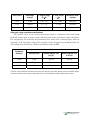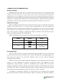
1
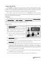
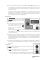
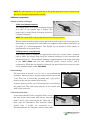
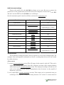
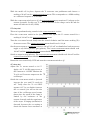
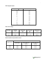
OC-725C Manual, Rev. 050816.2
Warner Instruments
Oocyte Clamp Amplifier
Model OC-725C
Warner Instruments
1125 Dixwell Avenue, Hamden, CT 06514
(800) 599-4203 / (203) 776-0664
(203) 776-1278 - fax
OC-725C Manual, Rev. 050816.2
Table of Contents
NOMENCLATURE.................................................................................................................................... 4
Text conventions ..................................................................................................................................... 4
CONTROL DESCRIPTION...................................................................................................................... 5
Front panel .............................................................................................................................................. 5
Voltage electrode.................................................................................................................................. 5
Bath electrodes ..................................................................................................................................... 6
Clamp section ....................................................................................................................................... 6
Commands............................................................................................................................................ 7
Current electrode ................................................................................................................................. 7
Rear panel ............................................................................................................................................... 8
Additional components .......................................................................................................................... 9
Voltage recording headstages .............................................................................................................. 9
Bath headstage ..................................................................................................................................... 9
Current electrode cable........................................................................................................................ 9
Model cell........................................................................................................................................... 10
Comments.............................................................................................................................................. 10
Connecting to line power ................................................................................................................... 10
High voltage outputs .......................................................................................................................... 10
USING THE MODEL MEMBRANE ..................................................................................................... 10
Initial instrument settings .................................................................................................................... 11
Test procedures..................................................................................................................................... 11
Offset controls .................................................................................................................................... 11
Voltage electrode test ......................................................................................................................... 12
Buzz .................................................................................................................................................... 12
Current electrode test......................................................................................................................... 12
DC clamp test ..................................................................................................................................... 13
AC clamp test ..................................................................................................................................... 13
SETUP........................................................................................................................................................ 15
Pipettes .................................................................................................................................................. 15
Electrode holders .................................................................................................................................. 15
Bath probe............................................................................................................................................. 16
Electrode placement and grounding ................................................................................................... 16
Bath clamp electrode placement ........................................................................................................ 16
Single oocyte setup with indirect ground ........................................................................................... 17
OC-725C Manual, Rev. 050816.2
Single oocyte setup with direct ground .............................................................................................. 17
Dual ooctye set-up.............................................................................................................................. 17
Cable connections ................................................................................................................................. 18
Resting position of the controls ........................................................................................................... 19
Using the gain select ............................................................................................................................. 19
Other gain range selection considerations.......................................................................................... 20
A PROCEDURE FOR RECORDING FROM OOCYTES .................................................................. 21
Initial electrode placement................................................................................................................... 21
Voltage electrode placement ................................................................................................................ 21
Current electrode placement ............................................................................................................... 22
Impaling the cell ................................................................................................................................... 22
Clamping the cell .................................................................................................................................. 23
Clamping high conductance cells ........................................................................................................ 23
Unclamping the cell .............................................................................................................................. 23
Removing the electrodes ...................................................................................................................... 23
SPECIAL CIRCUMSTANCES ............................................................................................................... 24
High side current measuring ............................................................................................................... 24
Configuration ..................................................................................................................................... 24
Optional voltage headstage .................................................................................................................. 24
COMMENTS AND RECOMMENDATIONS ....................................................................................... 26
Membrane damage............................................................................................................................... 26
Repeated recordings............................................................................................................................. 26
Electrophysiology ................................................................................................................................. 26
APPENDIX................................................................................................................................................ 28
Specifications - Equipment is intended to be operated in a controlled laboratory environment ........... 28
Gain telegraph outputs......................................................................................................................... 29
Gain select settings ............................................................................................................................... 29
Noise from bath clamp feedback resistor ........................................................................................... 29
References ............................................................................................................................................. 30
Certifications......................................................................................................................................... 31
Certifications......................................................................................................................................... 31
4
OC-725C Manual, Rev. 050816.2
The model OC-725C Oocyte clamp is designed for two-electrode, whole-cell voltage clamping
of Xenopus oocytes, as well as for other large cells and cell structures such as squid axons.
The instrument has several features making it ideal for these purposes.
Unique Features
==========================================================
High Voltage Compliance: The OC-725C
combines high AC and DC gains and a
voltage compliance of ±180 V to insure
fast, nonsaturating clamp performance
under nearly any condition. The AC clamp
gain is variable up to 2000. An additional
DC gain of 106 may be employed for high
conductance cells or leaky oocytes.
Two clamp speeds are available. The Slow
mode is used for screening oocytes or for
applications not requiring rapid response
times. The Fast mode is used for accurate
voltage clamping of fast whole cell
currents. Response time in the Fast mode
is 350 µs (10-90% rise time) when
applying a 100 mV step to a model cell.
Bath Clamp Headstage: The current
measuring range of the OC-725C bath
clamp headstage is extended at both ends
by a 3-position range multiplier. This
allows smaller currents to be amplified to
usable levels while larger currents (up to
1 mA) can be recorded without saturation.
Dual Oocyte Studies: Studies involving
two oocytes in a common bath requires
two clamp amplifiers. Traditional bath
clamp headstages cannot provide effective
clamping because they cannot separate
the individual currents from the combined
currents appearing in the bath. The
OC-725C solves this problem by the
application of an internal switch
permitting measurement of the current in
series with the current electrode instead
of in the bath. Additionally, an optional
differential voltage headstage is available
which subtracts the voltage drop across
the series resistance in the bath.
Voltage Headstage Probe: The voltage
measuring headstage is a single-ended,
high-impedance probe. Its small size,
convenient mounting rod, and two meter
cable make for easy attachment to a
micropositioner. Warner electrode holders
having a 2 mm jack mount directly onto
the headstage.
Voltage and Current Meters: Independent
meters provide simultaneous displays of
membrane voltage Vm and membrane
current Im. To assure proper impalement
of the current electrode, the current meter
displays membrane potential, Ve, from the
current electrode before the clamp circuit
is activated.
=======================================================
The unique design of the bath clamp
eliminates the need for series resistance
compensation. It provides an accurate
measurement of bath current by creating
a virtual ground in the bath while
simultaneously
clamping
the
bath
potential at zero.
Additional Features
Buzz controls for each electrode aid in
penetration of cell membranes with a
minimum of leakage.
Electrode Test for voltage and current
electrodes.
Capacity Compensation
voltage input.
for
the
Vm
Overload Alarm serves as a reminder
when the feedback amplifier reaches its
maximum output voltage, a condition
which could result in damage to the
oocyte.
DC Offsets for voltage and current
electrodes.
Warner Instruments
A Harvard Apparatus Company
4
OC-725C Manual, Rev. 050816.2
NOMENCLATURE
Text conventions
This manual refers to amplifier controls at four functional levels; operational sections,
control blocks, specific controls within a block, and settings of specific controls. To minimize
the potential for confusion, we have employed several text conventions which are specified
below. Since our goal is to provide clarity rather than complexity, we welcome any feedback
you may wish to provide.
¾ Warner Instrument product numbers are presented using a bold type.
¾ References to instrument panel control blocks are specified using UNDERLINED SMALL CAPS.
(e.g., COMMANDS, CLAMP)
¾ References to specific controls within a block are specified using
CAPS. (e.g., MODE SWITCH, DC GAIN)
NON-UNDERLINED SMALL
¾ Finally, references to individual control settings are specified in italic type. (e.g., slow,
fast, 100 mV)
¾ Special comments and warnings are presented in highlighted text.
Any other formatting should be apparent from context.
THIS EQUIPMENT IS NOT DESIGNED NOR INTENDED
FOR USE ON HUMAN SUBJECTS
Warner Instruments
A Harvard Apparatus Company
5
OC-725C Manual, Rev. 050816.2
CONTROL DESCRIPTION
The OC-725C is comprised of three functional channels: A high impedance voltage
sensing channel with capacity compensation and input offset to measure membrane
potential, a current sensing channel with bath clamp to clamp the bath and measure the
membrane current, and a high voltage amplifier to deliver the clamping current.
The complete voltage clamp system consists of the OC-725C, the voltage recording
probe with electrode holder, the current sensing bath probe with silver wire electrodes, and
the current cable with electrode holder.
Front panel
The instrument front panel is divided into six control blocks titled VOLTAGE ELECTRODE
[Vm], BATH ELECTRODES [Im], CLAMP, COMMANDS, and CURRENT ELECTRODE. The instrument rear
panel has BNC connectors for GAIN TELEGRAPH OUTPUT and Ve x10 output, an ALARM on/off
switch, and binding posts for CIRCUIT and CHASSIS GROUND.
Voltage electrode
The
the
control block contains
VOLTAGE PROBE CONNECTOR, the VOLTAGE
METER, the Vm OFFSET control, the ELECTRODE
TEST and BUZZ pushbuttons, and the Vm OUTPUT
BNC.
Controls
for
NEGATIVE
CAPACITY
COMPENSATION are also located in this block.
The
The
VOLTAGE ELECTRODE
reports the membrane
voltage (Vm) with a full scale range of
±199.9 mV.
VOLTAGE METER
VOLTAGE PROBE CONNECTOR
is a 7-pin DIN connector for attachment of the voltage
probe to the instrument.
control is a 10-turn potentiometer providing up to ±200 mV at the
VOLTAGE PROBE input for offset of membrane junction potentials.
The Vm
OFFSET
The Vm ELECTRODE TEST control is used to determine the internal resistance of the
voltage probe. When the pushbutton is depressed, a constant 10 nA current is
passed through the voltage electrode producing a voltage drop of 10 mV/MΩ of
probe resistance. The measured potential is displayed by the meter or reported at
the Vm x10 output BNC (at 100 mV/MΩ).
The Vm BUZZ push button facilitates penetration of the voltage electrode by producing a
10 kHz square wave at the pipette tip.
The Vm x10 OUTPUT BNC reports the membrane voltage in mV multiplied by 10.
Warner Instruments
A Harvard Apparatus Company
6
OC-725C Manual, Rev. 050816.2
NEGATIVE CAPACITY COMPENSATION (-C) has been added to the OC-725C allowing for its use
as an electrometer in intracellular measurements. Input capacitance up to 45 pF
can be neutralized using the two associated controls. A lit LED indicates the active
status of this circuit.
In general, negative capacity compensation is not useful for oocyte clamp
applications since clamp speed is a function of (1) the current electrode resistance,
(2) the RC time constant of the oocyte (typically 1 MΩ in parallel with 0.5 μF), and
(3) the compliance voltage of the clamp current.
Bath electrodes
The BATH ELECTRODES control block contains
the BATH PROBE connector, the CURRENT METER,
the GAIN SELECT switches, and the I MONITOR
output BNC’s.
The
reads the voltage (Ve) of
the current electrode when the CLAMP
MODE SELECTOR switch is off (see CLAMP
section). A lit LED indicates voltage
readings in mV.
CURRENT METER
When in clamp mode (CLAMP MODE SELECTOR switch set to slow or fast), the CURRENT
METER displays the current (Im) sensed by the bath electrode. A lit LED indicates
current readings in µA.
Instrument gain is set by the two GAIN SELECT controls. Gain is selected by a 7-position
GAIN SELECTOR switch, which ranges from 0.1 to 10 in 1-2-5 steps, and a 3-position
toggle switch which selects the gain multiplier (x0.1, x1.0 and x10). LED’s indicate
the gain multiplier selection. The combination of these controls allows gain to be
set from 0.01 to 100.
Current outputs are available from the I MONITOR BNC at full bandwidth (~10 kHz) and
from the I MONITOR FILTERED BNC which is filtered at 1 kHz by an integral 4-pole
Bessel filter.
Clamp section
The CLAMP control block contains the
the GAIN and DC GAIN controls.
The
MODE SELECTOR
switch as well as
switch selects for slow and fast clamp
speeds, or for off. These choices are described below.
CLAMP MODE SELECTOR
off - In the off position, the clamp amplifier is disconnected from
the current electrode. The voltage difference between the
Warner Instruments
A Harvard Apparatus Company
7
OC-725C Manual, Rev. 050816.2
current electrode and the bath electrode [Ve, mV] is read on the METER in the
BATH ELECTRODES control block. This information is also available at the Ve x10
output BNC on the rear of the instrument.
All CLAMP controls are disabled by turning the CLAMP MODE SELECTOR
switch to the off position. Be sure to switch this control to the off position before
NOTE:
handling electrodes!
slow - The slow clamp mode is useful for screening of oocytes or where high clamp
speeds are not required. The slow clamp speed is approximately 0.5 ms when
measured with the model membrane (1 MΩ shunted with 0.47 μF). In this
mode, measured currents are displayed on the BATH ELECTRODES METER in units
of μA.
fast - Most oocyte clamping is performed in the fast mode. The clamp speed is
limited by the resistance of the current electrode and the oocyte membrane
capacitance. Therefore, the current electrode resistance must be kept as low as
possible to obtain the fastest clamp speeds. Currents are read on the BATH
ELECTRODES METER in units of μA. Fast clamp speeds are ~350 μs when
measured with the model cell as described above.
The
control is a single turn potentiometer which varies the full-bandwidth openloop gain from 0 to 2000. A high DC GAIN (106) can be switched in with the DC GAIN
toggle switch to provide a hard clamp when passing large currents from high
expression oocytes.
GAIN
Commands
The COMMANDS control block contains the
IN÷10 input BNC.
HOLD
controls and
COMMAND
controls - HOLD potential is set with the DIGITAL POTENTIOMETER
thumbwheel and RANGE toggle switch. Ranges are ±99 mV and
±198 mV depending on the scale multiplier selected (x1.0 or
x2.0). Signal polarity, or off is selected with the associated toggle
switch.
HOLD
÷10 input BNC- Command signals from an external
generator or computer connected to this input are attenuated by
10. Maximum input is ±10 V.
COMMAND IN
Current electrode
The CURRENT ELECTRODE block includes the Ve OFFSET, ELECTRODE TEST, and Ve BUZZ
controls. This section also contains the OVER VOLTAGE indicator and a DIN connector for
the current electrode.
Warner Instruments
A Harvard Apparatus Company
8
OC-725C Manual, Rev. 050816.2
Ve
- With a range of ±200 mV (center zero), this
control is used to adjust the offset voltage of the
current electrode. Use this control to establish a zero
reference before impaling the oocyte. Once the oocyte
has been pierced, the resting potential can be read
from Ve x10 output BNC or on the current meter (BATH
ELECTRODES section).
OFFSET
- A voltage proportional to the resistance of
the current electrode [10 mV/MΩ] will be displayed on
the meter by depressing the ELECTRODE TEST push
button when the CLAMP MODE SELECTOR switch is in the off position.
ELECTRODE TEST
Ve
- The Vm BUZZ pushbutton facilitates penetration of the voltage electrode by
producing a 10 kHz square wave at the pipette tip.
BUZZ
LED - If the voltage at the current electrode exceeds ±160 V, the OVER
VOLTAGE lamp will light. An alarm will also sound when the rear panel ALARM
switch is in the on position.
OVER VOLTAGE
Rear panel
The line power connector and fuse are located on the rear panel. Operating voltage is
specified on the MODEL/SERIAL NUMBER sticker applied to the rear of the instrument. The rear
panel also contains Ve x10 and GAIN TELEGRAPH output BNCs, the ALARM switch and instrument
GROUNDS.
NOTE: Verify that the instrument is wired for the proper voltage before connecting the line
cord.
The Ve x10 output BNC monitors the voltage of the current electrode (x10) when the
MODE SELECTOR switch is off
The
CLAMP
output BNC provides a DC voltage indicating the gain setting of the
instrument. The output varies from 0.2 to 2.6 volts in 200 mV steps as shown in the
appendix.
GAIN TELEGRAPH
ALARM
switch - Activates or deactivates the OVER VOLTAGE current electrode audible alarm.
GROUNDS -
Both CIRCUIT and CHASSIS grounding posts are located on the rear panel. CHASSIS is
common with the instrument enclosure and connected to earth through the power line
cord. A shorting link allows for interconnection of the two grounds. In most
experimental setups, separating the grounds will result in minimizing 50/60 Hz signal
interference from ground loops. However, trial and error will determine the best
results.
Warner Instruments
A Harvard Apparatus Company
OC-725C Manual, Rev. 050816.2
9
NOTE: For safe operation, the ground pin on the power plug must not be removed and
the use of "cheater" plugs must be avoided.
Additional components
Voltage recording headstages
7250V PROBE (Standard Version)
The voltage probe is an active headstage housed
in a 1.25 x 5 cm cylinder (dia. x length). The
probe body is nickel plated and epoxy sealed for
corrosion resistance.
NOTE: The outer shell is electrically driven at the input potential.
Warner microelectrode holders with 2 mm jacks mate directly to the input pin on the
probe body. A mounting block and handle are supplied and facilitate attachment of
the probe to a micromanipulator. The handle can be mounted either axially or
perpendicular to the probe body.
7255DI DIFFERENTIAL PROBE (Optional)
This voltage probe is designed for applications where two oocytes share a common
bath or where the voltage drop across the solution resistance is to be measured and
subtracted from Vm. The headstage housing is approximately 2 cm longer than that
of the 7250V PROBE and has two additional inputs; CIRCUIT GROUND and V
DIFFERENTIAL. When the two inputs are shorted, the probe functions exactly the same
as the standard single ended 7250 PROBE.
Bath headstage
The BATH PROBE is housed in a 2.8 x 3.5 x 4.2 cm aluminum
enclosure. Inputs are two 1 mm pin jacks labeled I SENSE and
I OUT. The case is electrically grounded and a pin jack is
located on the side for connecting to shields.
A plastic plate with two screw mounting slots is attached to
the probe base. The BATH PROBE connects to the control unit
with a 6 pin connector
Current electrode cable
A two meter shielded cable is supplied with a 2 mm
pin jack on one end to mate with the an electrode
holder, and a 3-pin connector on the other end to
mate with the instrument. The electrode holder
should have a handle for mounting to a
micromanipulator. An example is shown to the right.
Warner Instruments
A Harvard Apparatus Company
10
OC-725C Manual, Rev. 050816.2
Model cell
The model cell supplied with the OC-725C is useful as a training aid and as a
calibration and test device. It has connections for the voltage and current probes and to
the bath clamp allowing all aspects of the amplifier’s function to be tested.
Comments
Connecting to line power
The model OC-725C is supplied with a 3-conductor power cord. One conductor provides
a connection between the instrument housing and the earth ground. Safe operation of
the instrument will be assured provided that the ground circuit in the power outlet is
wired correctly and is connected to earth.
NOTE: If the ground pin of the power cord is removed for any reason the instrument
chassis must be directly connected to earth ground using a separate heavy (14 gauge or
larger) ground wire.
High voltage outputs
When handling the current electrode cable, be sure to set the
clockwise and the CLAMP MODE switch to off.
GAIN CONTROL
fully
CAUTION!: The current clamp is capable of high power output (10 mA @ ±180 V) and
can cause serious injury if not properly handled.
USING THE MODEL MEMBRANE
The model cell supplied with the OC-725C can be used for two purposes. First, novice
users will find it a convenient tool for gaining experience in the operation of the instrument.
Additionally, it is a convenient tool for trouble shooting since the function of the
instrument can be quickly checked.
A schematic of the
model cell is shown to the
right.
The
oocyte
is
represented by a 1 MΩ
resistor shunted by a 0.47
μF capacitor. The voltage
and current electrodes are
each represented by 1 MΩ
resistances and the bath
probes are represented by
the 10 kΩ resistors.
to bath
clamp
10k
10k
1M
to voltage
probe
1M
to current
cable
1M
0.5 uF
Warner Instruments
A Harvard Apparatus Company
11
OC-725C Manual, Rev. 050816.2
Initial instrument settings
Connect the model cell to the OC-725C as shown on its cover. Be sure to connect the
ground wire to the ground mini-jack on the side of the bath probe. Connect the Vm x10 BNC
and the I MONITOR BNC on the OC-725C to an oscilloscope.
Set the instrument panel controls as follows and turn the POWER SWITCH on.
Control
Control block
Setting
off
POWER SWITCH
Vm OFFSET
VOLTAGE ELECTRODE
Center of rotation
(approximately 5 turns)
OUTPUT GAIN
BATH ELECTRODES
0.1 V/µA
GAIN SELECT
BATH ELECTRODES
x1.0
CLAMP MODE switch
CLAMP
off
DC GAIN toggle
CLAMP
out
GAIN
CLAMP
CCW to detent off
HOLD POTENTIAL
COMMANDS
00 mV
HOLD POTENTIAL MULTIPLIER
COMMANDS
x1.0
HOLD POLARITY
COMMANDS
off
Ve OFFSET
CURRENT ELECTRODE
Center of rotation
(approximately 5 turns)
Test procedures
In the following testing procedures, allow a tolerance of ±1% on the readings taken. For
example, if the test response is indicated as 100 mV, a reading from 99.0 to 101.0 mV would
be within tolerance.
Offset controls
Vm
OFFSET (VOLTAGE ELECTRODE
section): The full range of this control is ±200 mV. This can be
verified by rotating the control first fully clockwise and then fully counterclockwise
while observing the VOLTAGE ELECTRODE METER. The displayed readings will indicate off
scale at the extremes of the control’s manipulation since the meter is only capable of
displaying ±199.9 mV.
Vm x10 output BNC (VOLTAGE ELECTRODE section): This output can be monitored using an
oscilloscope. The reported voltage will swing between ±2 V as the Vm OFFSET control is
Warner Instruments
A Harvard Apparatus Company
12
OC-725C Manual, Rev. 050816.2
manipulated throughout its full range. Set the Vm OFFSET to 0.0 reading on the meter
and verify that the Vm x10 reading on the scope also reads 0 V.
Ve
control is tested in the same
manner as the Vm OFFSET control. This control is adjustable when the CLAMP MODE switch
is set to off. The voltage of the Ve OFFSET is read from the BATH ELECTRODE METER when
the CLAMP MODE switch is set to off.
OFFSET
control (CURRENT
ELECTRODE
section): The Ve
OFFSET
Ve x10 output BNC: This BNC is located on the rear panel of the instrument and reports
the setting of the Ve OFFSET control when the CLAMP MODE switch is set to off.
Return all controls to their initial settings when done with this test.
Voltage electrode test
In actual practice, the voltage electrode test is used prior to entering the cell and indicates
the resistance of the electrode. When used in conjunction with the model cell, it measures
both the electrode and membrane resistance (2 MΩ). This test is performed using controls
in the VOLTAGE ELECTRODE section.
With the model cell in place, depress the Vm ELECRODE TEST pushbutton and observe a
reading of 20 mV on the VOLTAGE ELECTRODE METER. This corresponds to a 2 MΩ reading
at a calibrated response of 10 mV/MΩ.
On the oscilloscope, the Vm x10 output will read 200 mV which is x10 the applied test
voltage. Since the test current is being passed through 1 MΩ (Rm), the I MONITOR output
will indicate 1 mV, which corresponds to 10 nA of current.
With the CLAMP MODE switch set to off, the BATH ELECTRODE METER monitors Ve (voltage at the
current electrode). In this case, Ve will be a measure of the voltage across Rm and the
meter will indicate 10 mV (1 MΩ). The Ve x10 output BNC on the rear panel can also be
checked to see that it reads 100 mV (meter reading x10).
Buzz
This test is performed using controls in the VOLTAGE ELECTRODE section.
Set the oscilloscope sensitivity to 5 V/div and depress the BUZZ pushbutton while monitoring
the Vm x10 output. A 10 kHz square wave of approximately 24 V p-p will be generated
as long as the button is depressed.
Current electrode test
This test is performed using controls in the CURRENT ELECTRODE section.
Warner Instruments
A Harvard Apparatus Company
13
OC-725C Manual, Rev. 050816.2
With the model cell in place, depress the Ve ELECRODE TEST pushbutton and observe a
reading of 20 mV on the CURRENT ELECTRODE METER. This corresponds to a 2 MΩ reading
at a calibrated response of 10 mV/MΩ.
With the CLAMP MODE switch set to off, the BATH ELECTRODE METER monitors Ve (voltage at the
current electrode). In this case, Ve will be a measure of the voltage across Rm and the
meter will indicate 10 mV (1 MΩ).
DC clamp test
This test is performed using controls in the CLAMP ELECTRODE section.
Place the CLAMP MODE switch in the fast position and adjust the Vm
reading of 100 mV on the VOLTAGE ELECTRODE METER.
OFFSET
control for a
Turn the GAIN control on and slowly turn the control clockwise until the meter reading (Vm)
decreases to zero. The CURRENT ELECTRODE METER should read -0.10 μA.
Set the HOLD POTENTIAL (COMMANDS section) to 100 mV (50 mV on thumbwheel and MULTIPLIER
toggle at x2). Select positive (pos) polarity. The VOLTAGE ELECTRODE METER should read
100 mV and the CURRENT ELECTRODE METER should read 0.00 μA.
Switch to negative (neg) polarity. The VOLTAGE ELECTRODE METER should read 100 mV and the
CURRENT ELECTRODE METER should read 0.2 μA
Return the GAIN control fully CCW and turn the CLAMP MODE switch to off.
AC clamp test
Adjust the Vm OFFSET control to 0.0 V.
Apply a 0.8 V, 100 Hz square wave to
the COMMAND IN ÷10 BNC. Monitor the
Vm x10 and I MONITOR outputs on the
oscilloscope.
Switch the CLAMP MODE switch to fast and
increase the GAIN until Vm reads 80
mV. Verify that the Vm x10 BNC
reports 0.8 V. As you further increase
the GAIN control, you will see the rise
time of the oscilloscope trace become
faster since the speed of the clamp is
limited by the resistance of the
current electrode and the capacitance
of the oocyte. If ringing (oscillation) is
observed, decrease the GAIN setting to
obtain the fastest clean waveform as
Warner Instruments
A Harvard Apparatus Company
OC-725C Manual, Rev. 050816.2
14
shown on the previous page. (The current signal (I MONITOR) shown in the figure
displays the high current spikes required to charge the oocyte capacitance.)
Set the HOLD control to a reading of 50 mV and switch the POLARITY toggle to pos. The square
wave will be displaced 50 mV in the positive direction. Switching the polarity to neg
will produce a -50 mV offset.
Warner Instruments
A Harvard Apparatus Company
OC-725C Manual, Rev. 050816.2
15
SETUP
The following instructions are designed to guide the user, step-by-step, through a
typical recording session involving a Xenopus oocyte. It is assumed that the user is already
familiar with the techniques of Xenopus oocyte excision and microinjection (for a review of
those techniques, see Colman, 1984). It is also assumed that the user has some familiarity
with the basic circuitry of a two-electrode voltage clamp (for review, see Hille, 1984).
Pipettes
Microelectrodes can be made using the same glass (tubing and dimensions) as those
used for a typical patch pipette and are usually filled with 3 M sterile filtered KCl. Unlike
the pipettes used as patching electrodes, microelectrode pipettes do not require fire
polishing nor coating with Sylgard. They will need to be broken off, however, to a relatively
large diameter to insure a fast response time by the clamp.
For the voltage electrode, the pipette tip should be broken back to an O.D. of 3-5 μm.
The current electrode pipette should be broken back to an O.D. of 7-9 μm. The resistances of
these pipettes should be about 2 MΩ and 1 MΩ (or less), respectively.
When installed, the current electrode pipette should be shielded from the voltage
electrode and that shield should be grounded to the circuit ground. This can be
accomplished by wrapping the current pipette with aluminum foil or by mounting a metal
screen or plate between the two pipettes. In either configuration, the shield can be
grounded by connecting it to the "ground" mini-jack on the side of the bath probe. When
using the aluminum foil method, care must be taken to prevent the foil from touching the
surface of the bath solution at the bottom end of the pipette or the silver electrode wire at
the top end.
Electrode holders
Voltage Electrode - The voltage recording electrode holder uses a silver wire for the
electrical coupling between the pipette and holder connector. Any silver wire contacting the
KCl solution in the pipette must be chlorided to reduce junction potentials (see Chloriding
Warner Instruments
A Harvard Apparatus Company
OC-725C Manual, Rev. 050816.2
16
Procedure in Appendix). The pipette should contain just enough KCl so that approximately
1/2 inch of the chlorided wire is submerged. The pipette/holder assembly is attached
directly to the voltage headstage prior to mounting in a micropositioner.
NOTE: Do not fill the voltage holder with KCl solution.
Current Electrode - The current recording electrode also uses a silver wire for coupling.
In an manner analogous to the voltage electrode, the current electrode wire must be
chlorided prior to assembly and use. The pipette/holder assembly is mounted in a
micropositioner with the mounting rod supplied.
NOTE: Do not fill the current holder with KCl solution.
Bath probe
The bath clamp is designed to maintain a virtual ground in the oocyte perfusate. The
bath probe should be positioned so that the silver electrode wires can be inserted into the
recording chamber or into the agar bridge wells. Sticky wax or tape is usually sufficient to
secure the unit when positioned on a flat surface or, alternatively, the unit can be held in
place on a separate stand. The bath probe electrodes should also be chlorided before use, as
described above.
Electrode placement and grounding
Three drawings (shown on pages 19 and 20) have been included to illustrate the
various ways a bath circuit can be configured. Most applications involve only a single oocyte
and Figures A and B illustrate these setups. Figure C shows a setup for recording from 2
oocytes in a common bath with the use of dual clamps
NOTE: Cable routing must be performed with care. Bundle cables together rather than
routing them individually and keep them as far as possible from sources of 50/60 Hz
interference (e.g., line cords, transformers, etc.).
Bath clamp electrode placement
Proper placement of the bath electrodes (Iout and Isense) is important for obtaining optimum
performance. The Isense electrode (or the agar bridge associated with it) should be placed as
close to the oocyte as possible since this point is the virtual ground node, and on the same
side as the voltage recording electrode. The Iout electrode (or the agar bridge associated with
it), on the other hand, can be placed at a greater distance from the oocyte and should be on
the same side as the current electrode
It is recommended that the user not directly expose the electrode wires to the perfusate if
the recording session is to last for more than a few minutes. Instead, agar bridges should be
employed to provide a circuit between these electrodes and the bath. This protects the cell
membrane from the potential adverse effects of the silver wire.
Warner Instruments
A Harvard Apparatus Company
OC-725C Manual, Rev. 050816.2
17
Single oocyte setup with indirect ground
Single oocyte studies are best accomplished with the setup shown in Figure A. This method
uses the bath clamp headstage to establish the bath ground and is preferred for two
reasons: (1) current readings with the bath clamp will have the lowest noise level, and (2)
properly placed bath clamp electrodes will negate the need for series resistance
compensation.
Single oocyte setup with direct ground
Applications where use of the bath clamp is not suitable (such as those with a very long
solution path to ground) can be configured using the alternate method of directly grounding
the bath as shown in Figure B. In this configuration, current is read from the "high side" of
the current output leg.
This method also requires the use of the optional DIFFERENTIAL VOLTAGE HEADSTAGE. Two
disadvantages are: (1) The noise levels of the current signal measured in the "high side" is
approximately double that obtained with a bath clamp, and (2) high levels of clamp current
could produce a substantial voltage drop across the solution series resistance.
NOTES: The oocyte clamp has no provisions for compensating the voltage drop described
above. The differential voltage electrode should be placed close to the oocyte and in the
current path between the Vm electrode and ground.
Dual ooctye set-up
Dual oocyte setup is accomplished using two clamps as illustrated in Figure C. Both
clamps must be configured to read current from the "high side" as described in the section
titled HIGH SIDE CURRENT MEASURING (see page 26) and each clamp must be equipped with
the optional DIFFERENTIAL VOLTAGE HEADSTAGE.
Warner Instruments
A Harvard Apparatus Company
OC-725C Manual, Rev. 050816.2
18
Cable connections
Bath clamp headstage: After positioning the probe as described above, connect it to the
BATH PROBE socket (BATH ELECTRODES section).
Voltage electrode headstage: The high impedance probe for recording membrane potential
should be mounted on a micro-manipulator and connected to the VOLTAGE PROBE socket
(VOLTAGE ELECTRODE section).
High voltage current electrode: The holder should be mounted on a micro-manipulator and
the cable connected to the I ELECTRODE socket (CURRENT ELECTRODE section).
Command potential: If a computer or external generator is used for controlling the clamp
command potential, its signal should be connected to the front panel COMMAND IN ÷10
input (COMMANDS section).
External monitoring: To monitor the microelectrode’s potentials on an oscilloscope,
computer or a chart recorder, the following connections should be made:
Vm - Membrane potential may be recorded from the Vm x10 connector (VOLTAGE
ELECTRODE section).
Ve - The voltage of the current electrode can be monitored from the Ve connector on the
REAR PANEL.
The output will be the same as that reported on the CURRENT ELECTRODE METER,
showing the voltage across the current electrode when the CLAMP SELECTOR switch is
in the off position. (Recall that the meter indicates the current (Im) when the CLAMP
SELECTOR switch is in either the slow or fast position).
Warner Instruments
A Harvard Apparatus Company
19
OC-725C Manual, Rev. 050816.2
Im - The current signal is available from the I
(BATH ELECTRODES section).
MONITOR
and I
MONITOR FILTERED
outputs
Gain telegraph – Automatic monitoring of the Im gain can be achieved by connecting the
rear panel GAIN TELE BNC to the appropriate input on your analog-to-digital converter.
Power: The power cord should be connected to a properly grounded AC receptacle with the
line voltage specified on the instrument nameplate (REAR PANEL).
Resting position of the controls
To begin, set the instrument controls to the following positions:
Control
Section
Setting
off
POWER
I MONITOR output
BATH ELECTRODE
1 V/µA
GAIN SELECT
BATH ELECTRODE
x1
HOLD
COMMANDS
0 mV
POLARITY toggle
COMMANDS
off
MODE SELECT
CLAMP
off
CLAMP DC GAIN toggle
CLAMP
out
CLAMP GAIN
CLAMP
off (fully CCW)
ALARM
REAR PANEL
off or on (as desired)
Turn POWER on.
Using the gain select
A wider range of bath current (Im) measurements is now possible with the addition of
the GAIN SELECT toggle switch located above the GAIN control. The switch has 3 positions;
x0.1, x1 and x10. Resistance values shown below each LED indicate the bath clamp
feedback resistor used for the current measurement.
The chart on the next page shows the effect of the range selection on the Im output.
Note also that range selection changes the sensitivity of the current meter. Typically,
measured currents will fall in the x1 range. The lower and higher ranges are intended to
cover those applications where currents are beyond the x1 range. Currents below 1 µA
should be monitored in the x10 range. Large currents above 100 µA require the x0.1 range.
Since there is overlap in the ranges, the current being measured may be monitored in one of
two ranges in which case the choice may be made on the basis of noise or clamp speed.
Warner Instruments
A Harvard Apparatus Company
20
OC-725C Manual, Rev. 050816.2
Gain select
Headstage
resistor
Im output range
(V/μA)
Im max output
(μA)
Maximum meter
reading
x0.1
10 kΩ
0.01 - 1.0
10 - 1000
199.9 μA
x1.0
100 kΩ
0.1 - 10
1 - 100
19.99 μA
x10
1 MΩ
1.0 - 100
0.1 - 10
1.999 μA
Other gain range selection considerations
The intrinsic noise of the current measuring circuit is a function of the bath clamp
feedback resistor with a larger resistor offering lower noise and greater signal resolution.
For comparisons, the following measurements were made with a shorted input, with the
standard 0.5 μF capacitance model cell, and with a 0.22 μF capacitance modified model cell.
All readings were recorded at 1 kHz (8-pole Bessel) and are RMS.
Noise
Feedback
Resistor
Shorted Input
Standard Model Cell
(0.5 µF)
Modified Model Cell
(0.22 µF)
10 kΩ
75 pA
6.0 nA
4.4 nA
100 kΩ
28 pA
5.5 nA
4.4 nA
1 MΩ
22 pA
5.0 nA
4.0 nA
A lower value feedback resistor increases the speed of the bath clamp and can handle larger
currents without saturating, important when recording from high expression oocytes.
Warner Instruments
A Harvard Apparatus Company
21
OC-725C Manual, Rev. 050816.2
A PROCEDURE FOR RECORDING FROM OOCYTES
Initial electrode placement
1)
Make sure that the bath electrodes are submerged in the chamber (or in the agar
bridge wells with the agar bridges completing the circuit to the bath) and the oocyte is
stable on the chamber floor.
2)
Install the voltage and current pipettes onto their respective holders but do not yet
place them in the chamber bath solution.
Voltage electrode placement
3)
Advance the voltage recording electrode into the bath. The VOLTAGE ELECTRODE
will indicate (in mV) the potential between the electrode and the bath.
4)
If there is no voltage reading and you are sure that the pipette tip is in the bath
solution, perform the following checks:
METER
a) make sure that all cables are connected properly.
b) inspect the voltage electrode to see if there is a bubble in the pipette which will
cause an open circuit.
5)
Using the Vm OFFSET control, adjust the VOLTAGE ELECTRODE potential to read 0 mV. If the
junction potential of the voltage electrode can not be adjusted to 0 mV, the electrode
holder may be at fault. (See Electrode Holders, page 17.)
6)
To test the resistance of the voltage electrode pipette, depress the ELECTRODE TEST
button. This passes a 10 nA current across the voltage electrode. The VOLTAGE
ELECTRODE METER will display the resulting potential in mV. The resistance of the
electrode can be easily calculated by dividing the current into the potential. The
resulting answer will be expressed in Ω. For example, if the electrode test indicates
that a potential of 25.0 mV is produced by the 10 nA test current, then
R=
V 25 mV
=
= 2.5 x10 6 Ω = 2.5 MΩ
I
10 nA
NOTES:
a)
A simpler calculation is to divide the voltage readout by 10 and append the units of
MΩ. (e.g., 25 mV = 2.5 MΩ)
b) The calculated resistance value may vary widely from pipette to pipette but should
be less than 4 MΩ for the voltage electrode.
Warner Instruments
A Harvard Apparatus Company
OC-725C Manual, Rev. 050816.2
22
Current electrode placement
7)
Advance the current electrode until the tip is in the chamber bath solution. Adjust Ve
OFFSET for a zero reading on the CURRENT ELECTRODE METER. This will establish a null
reference allowing the resting potential to be directly read.
8)
With the
switch in the off position, the resistance of the CURRENT
ELECTRODE pipette is tested in the same manner as the VOLTAGE ELECTRODE. Pressing the
Ve ELECTRODE TEST pushbutton will cause a 10 nA current to be passed across the
CURRENT ELECTRODE. The resulting voltage (in mV) will be displayed on the METER in the
BATH ELECTRODE section. From that value, the resistance of the pipette can be calculated
exactly as described above (i.e., divide the readout by 10 to get the resistance in MΩ).
Since the current electrode has a larger diameter, its resistance should be less than
that of the voltage electrode (about 1.0 MΩ or less).
9)
If no voltage display is present during the electrode test procedure and you’re sure that
the electrode is contacting the bath, perform the following checks:
CLAMP SELECTOR
a) Make sure that all cables are connected properly.
b) Check to see that the aluminum shield around the current electrode pipette (if
used) is not touching the bath solution or the electrode wire.
c) Check the current electrode to see if there is a bubble in the pipette causing an open
circuit.
Impaling the cell
10) Recheck the VOLTAGE ELECTRODE METER to verify that the potential is correctly offset to
read 0.0 mV and readjust the Vm OFFSET control if needed.
11) Advance the VOLTAGE ELECTRODE until its tip is slightly depressing the plasma
membrane of the cell and depress the Vm BUZZ pushbutton. This will produce a 1 V, 10
kHz oscillation at the voltage electrode, disrupting the membrane and causing the tip
of the electrode to impale the cell with no further movement of the micro-manipulator
(this technique will work best with "fresh" oocytes, i.e., 1 or 2 days post-excision). If the
buzz technique fails to cause electrode penetration, further advance the voltage
electrode until it "pops" through the membrane.
12) The potential across the membrane will now be displayed on the
METER.
VOLTAGE ELECTRODE
13) Now, advance the current electrode until its tip is slightly depressing the plasma
membrane of the cell and depress the Ve BUZZ pushbutton. Similar to the voltage
electrode BUZZ, the current electrode BUZZ produces a 1 V, 10 kHz oscillation across the
current electrode. This disrupts the cell membrane and causes the tip of the electrode
to impale the cell with no further movement of the micro-manipulator. Once again, if
Warner Instruments
A Harvard Apparatus Company
23
OC-725C Manual, Rev. 050816.2
the BUZZ technique fails to cause penetration, further advance the current electrode
until it "pops" through the membrane.
Clamping the cell
14) Activate the clamp by switching the CLAMP MODE switch to either the slow or fast mode.
NOTE: slow mode is useful for initial screening.
control clockwise as far as possible without illuminating the
VOLTAGE LED (located in the CURRENT ELECTRODE section).
15) Adjust
CLAMP GAIN
OVER
NOTE: If the ALARM signal switch on the back panel is on, an alarm will sound when the
OVER VOLTAGE LED illuminates.
16) The clamped membrane potential can now be observed over time or it can be
manipulated by selecting the desired polarity and amplitude with the controls located
in the COMMANDS section. Alternatively, you can control the COMMAND voltage externally
from a computer by leaving the POLARITY toggle switch in the off position and connecting
the appropriate analog output from your computer DAC to the COMMAND IN ÷10 BNC
connector. (See Cable Connections, page 20.)
17) Depending on the amplitude of the response you wish to record, you may adjust the
instrument GAIN to a higher or lower position. The CURRENT ELECTRODE METER should now
be displaying the current (in µA) that is delivered to hold the cell at the designated
command potential.
Clamping high conductance cells
18)
mode (CLAMP section) may be required to clamp high conductance (low
resistance) cells. This condition will be evidenced by the inability of the instrument to
maintain a DC holding potential (to within 1% or better of the set value) and the
maximum instrument gain is not sufficient to provide a hard clamp. DC GAIN mode
provides an additional DC gain greater than 106 while the AC gain remains at 2000
maximum for stability.
DC GAIN
Unclamping the cell
19) To unclamp the cell, turn the GAIN control (CLAMP section) fully counter-clockwise to the
detent off position. This will also disengage the DC GAIN.
NOTE: If the control is not fully off and the
DC GAIN
is left on, the preparation will not be
unclamped.
Removing the electrodes
20) It is very important that the CLAMP GAIN be returned to the off position (fully counterclockwise to click off) as described above and the CLAMP MODE selector switch be placed
in the off position before removing the current electrode from the cell.
Warner Instruments
A Harvard Apparatus Company
OC-725C Manual, Rev. 050816.2
24
Failure to perform the above steps will overload the feedback amplifier (due to the
large current generated when the membrane resistance between the current electrode
and the bath virtual ground goes to zero). This will damage the oocyte! For this reason
we recommend that the user enable the audible overload alarm to provide a warning
when the potential for such damage exists.
SPECIAL CIRCUMSTANCES
High side current measuring
In studies of single oocytes, current is monitored by the bath clamp headstage.
Experiments involving two oocytes in a common bath (such as gap junction studies)
requires monitoring currents from each oocyte. This is done in the current output leg (in
series with and ahead of the current electrode).
Two disadvantages of monitoring the current in this manner exist.
a) The noise level of this signal is higher. However, this is usually not a serious
problem since currents are typically in the μA range.
b) The voltage drop across the solution resistance (from oocyte to bath ground)
becomes an error voltage since it is not subtracted out as when the bath clamp
headstage is used. This problem is overcome by using the optional DIFFERENTIAL
VOLTAGE HEADSTAGE.
Configuration
The OC-725C current measuring circuit can be changed to the high side current
measuring mode by setting a dip switch on the main circuit board.
1)
First disconnect the power cord from the wall.
2)
Remove the two screws at the rear of the top cover and it off.
3)
Locate dip switch S10 on the circuit board. For normal operation S4 is on and all other 7
switches are off.
4)
To convert to high side current measuring mode, turn S4 off and turn S2, S5, and S7 on.
All other switches should remain in the off position.
Optional voltage headstage
The optional DIFFERENTIAL INPUT VOLTAGE RECORDING HEADSTAGE (Model 7255DI) is used in
applications where the bath clamp headstage cannot be effectively employed. Two examples
are (1) situations where the solution path from oocyte to ground is very long and (2) when
recording from two oocytes in a common bath.
Two 1 mm input jacks are located on the side of the headstage, V DIFF and GND. A
shorting jumper is supplied and is used for normal single-ended recording. For double-
Warner Instruments
A Harvard Apparatus Company
25
OC-725C Manual, Rev. 050816.2
ended recording, the jumper is removed and a V
jack as shown below.
DIFF ELECTRODE
is connected to the V
DIFF
If a shield between the voltage and clamp current electrodes is used, it should be
connected to the headstage ground.
Warner Instruments
A Harvard Apparatus Company
26
OC-725C Manual, Rev. 050816.2
COMMENTS AND RECOMMENDATIONS
Membrane damage
Recording from the same cell at a later time requires that the cell remain healthy
during the interim incubation. The less damage done to the membrane during handling and
impaling the cell, the happier it will be. Use of the BUZZ function should help minimize the
trauma from electrode penetration. Membrane damage can be further reduced by properly
isolating the experimental platform from vibration. Finally, hydraulically driven micromanipulators will also reduce membrane damage while the electrodes are in the cell.
Repeated recordings
Most recording sessions will involve repeating the above steps several times with many
different cells. Unless there is a concern about contamination of the bath solution by
something carried over from previous experiments, the pipettes can also be used repeatedly.
They should be free of debris and should have approximately the same resistance as they
had in the previous recording. A significantly higher resistance could indicate that the
pipette is partially plugged with cellular debris.
Make the following control settings before the next recording is carried out:
Control
Section
Setting
POLARITY
COMMAND
off
MODE SELECT
CLAMP
off
GAIN
CLAMP
0
(fully CCW)
Electrophysiology
If you are well versed in setting up electrophysiological equipment, you can safely skip
over the rest of this section. If, however, this is your initiation into electrophysiology (as it
may well be for some of you molecular biologists), then you may find the following
recommendations helpful.
While the whole cell electrophysiological configuration is more forgiving than the patch
clamp, it is still important to minimize mechanical motion. The platform for your
experimental setup, therefore, should be mechanically well isolated. This will reduce
leakage around the electrodes, making the clamp more effective and reducing noise in your
recordings. The latter is especially important when recording responses of certain ligandgated channels where membrane potential changes may only be a few mV.
In addition to mechanical isolation, the setup must also be isolated from external
electrical noise sources. These include motors, lamps, and wiring. The platform should be
Warner Instruments
A Harvard Apparatus Company
OC-725C Manual, Rev. 050816.2
27
shielded from these sources of electrostatic radiation with a Faraday cage. All equipment
within the Faraday cage should be grounded to the rear panel instrument circuit ground
and is best achieved by connecting everything (including the cage) to a ground bus within
the cage. Then only one wire is run from the setup to the instrument ground.
You will need to mount the voltage recording electrode headstage and the current
injecting electrode on micro-manipulators. They need not be hydraulically driven but such
drives will minimize the damage to the cell during and after penetration, and will make for
better seals around the electrodes. Another advantage gained by reducing membrane
damage by electrodes is to enhance the possibility of making subsequent recordings from
the same cell. These suggestions are also important for minimizing mechanical noise in the
recorded data
You will need a dissecting scope for viewing the placement of the electrodes. Anything
more powerful than 40x will just get in the way. The light source for your scope should be
DC and may need to be IR and UV filtered if you plan to use it during recording.
Minimally, the recording chamber can be a stable surface on which the oocyte will not
roll around. A disposable petri dish with a piece of nylon mesh on the bottom has been
successfully used for this purpose. The dish can be stabilized by a holder or by some wax
placed around its perimeter. The diameter of the dish needs to be large enough to
accommodate the oocyte and the two bath electrodes. The walls of the recording chamber
should be low enough to not interfere with electrode placement. Perfusion of the chamber
can be accomplished using a gravity fed system. Perfusate from the dish can be evacuated
using gentle vacuum. Use as small an aperture as possible to avoid disturbing the surface
of the perfusate in the dish.
NOTE: Specialized equipment suitable for oocyte studies (oocyte chambers, perfusion
systems, a Faraday cage, a noise-free DC lamp, and micro-manipulators) are available from
Warner Instruments.
Although it is not required, a computer can be employed to control the command
voltage. Acquisition and display of data is also usually handled by computer. The OC-725C
is fully compatible with all commercially available software packages designed for
electrophysiological research.
Finally, a microelectrode puller is necessary for making appropriately sized voltage and
current electrodes. Usually, the microinjection pipette puller can also be used to make
microelectrodes. You will need use of a microscope to break off the pipette tips.
Warner Instruments
A Harvard Apparatus Company
28
OC-725C Manual, Rev. 050816.2
APPENDIX
Specifications - Equipment is intended to be operated in a controlled laboratory environment
Voltage recording channel (Vm)
Input Impedance
5 x10¹¹ Ω, shunted by 3 pF
Output Resistance
100 Ω
Vm OFFSET
± 200 mV at V probe input
Noise*
50 μV RMS at 1 kHz
Electrode Test
10 mV/MΩ
Negative Capacity
0 - 45 pF
Vm Meter Range, full scale
± 199.9 mV
Bath electrode channel (Im)
Ve OFFSET
± 200 mV
Noise*
Im Clamp (clamp on)
5.5 nA RMS at 1 kHz [x1 range]
Open loop (clamp off)
28 pA RMS at 1 kHz [x1 range]
I MONITOR
0.01 - 100 V/μA in 3 ranges, 7 steps per range
I MONITOR FILTERED (4-pole Bessel])
Same as above, filtered at 1 kHz
Gain Telegraph
0.2 - 2.6 VDC in 0.2 V steps
Meter Ranges (full scale)
Ve (clamp off)
± 199.9 mV
Im (x0.1 range)
± 199.9 μA
Im (x1.0 range)
± 19.99 μA
Im (x10 range)
± 1.999 μA
Current electrode channel (Ve)
Compliance Voltage
± 180 V
Alarm
± 160 V
Gain
Variable
0 - 2000 AC/DC
DC
6
1 x10 DC, switch selected
Electrode Test
10 mV/MΩ
Commands
Hold (internal)
± 198 mV in 2 ranges
External input (attenuated by 10)
1 V in = 0.1 V command
Maximum external input
10 V
Power requirements
100 -130 or 220-240 VAC, 50/60 Hz
Dimensions
Enclosure
9 x 42 x 25 cm (H x W x D)
Voltage Headstage
1.25 x 5 cm (dia. x length); with 1.8 m cable
Mounting Handle
4.8 mm x 6.3 cm (dia. x length)
Bath Headstage
2.8 x 3.5 x 4.2 cm (H x W x D); with 1.8 m cable
* All noise measurements made with an 8-pole Bessel filter.
Warner Instruments
A Harvard Apparatus Company
29
OC-725C Manual, Rev. 050816.2
Gain telegraph outputs
Im Output (V/μA)
Gain Telegraph
0.01
0.2 V
0.02
0.4 V
0.05
0.6 V
0.1
0.8 V
0.2
1.0 V
0.5
1.2 V
1
1.4 V
2
1.6 V
5
1.8 V
10
2.0 V
20
2.2 V
50
2.4 V
100
2.6 V
Gain select settings
Gain select
Headstage
Im output range
Im max output
Maximum meter
resistor
(V/μA)
(μA)
reading
x0.1
10 kΩ
0.01 - 1.0
10 - 1000
199.9 μA
x1.0
100 kΩ
0.1 - 10
1 - 100
19.99 μA
x10
1 MΩ
1.0 - 100
0.1 - 10
1.999 μA
Noise from bath clamp feedback resistor
Noise
Feedback Resistor
Shorted Input
Standard Model Cell
Modified Model Cell
(0.5 µF)
(0.22 µF)
10 kΩ
75 pA
6.0 nA
4.4 nA
100 kΩ
28 pA
5.5 nA
4.4 nA
1 MΩ
22 pA
5.0 nA
4.0 nA
Warner Instruments
A Harvard Apparatus Company
OC-725C Manual, Rev. 050816.2
30
References
Colman, A. (1984). Translation of eukaryotic messenger RNA in Xenopus oocytes.
Transcription and Translation, eds. B.D. Hames and S.J. Higgins (IRL Press, Oxford).
Ch. 10
Hille, B. (1984). Ionic Channels of Excitable Membranes. Sinauer (Sunderland, MA). Ch. 2.
Zhou, J., Potts, J.F., Trimmer, J.S., Agnew, W.S. and Sigworth, F.J. (1991). Multiple gating
modes of the µI sodium channel, Neuron 7: 775-785
Warner Instruments
A Harvard Apparatus Company
31
OC-725C Manual, Rev. 050816.2
Certifications
Declaration of Conformity
CE MARKING (EMC)
Application of Council Directive: 89/336/EEC
Standards To Which Conformity
Is Declared:
EN55022 Class A
EN61000-3-2
EN61000-3-3
EN50082-1:1992
EN61000-4-2
EN61000-4-3
ENV50204
EN610000-4-4
EN610000-4-8
EN610000-4-11
Manufacturer’s Name:
Warner Instruments, LLC
Manufacturer’s Address:
1125 Dixwell Avenue
Hamden, CT 06514
Tel: (203) 776-0664
Equipment Description:
Instrument Amplifier
Equipment Class:
ITE-Class A
Model Numbers:
OC-725C
I the undersigned, hereby declare that the equipment specified
above, conforms to the above Directive(s) and Standard(s).
Place: Hamden, Connecticut USA
Signature:
Full Name: Burton J. Warner
Position: President
Warner Instruments
A Harvard Apparatus Company
32
OC-725C Manual, Rev. 050816.2
Declaration of Conformity
CE MARKING (LVD)
Application of Council Directive: 73/23/EEC
Standards To Which Conformity Is
Declared:
EN61010-1:1993
Manufacturer’s Name:
Warner Instruments, LLC
Manufacturer’s Address:
1125 Dixwell Avenue
Hamden, CT 06514
Tel: (203) 776-0664
Equipment Description:
Equipment Class:
Instrument Amplifier
Safety requirements for electrical
equipment for measurement and
laboratory use
Class I
Model Numbers:
OC-725C
I the undersigned, hereby declare that the equipment specified
above, conforms to the above Directive(s) and Standard(s).
Place: Hamden, Connecticut USA
Signature:
Full Name: Burton J. Warner
Position: President
Warner Instruments
A Harvard Apparatus Company
OC-725C Manual, Rev. 050816.2
33
Warner Instruments
A Harvard Apparatus Company