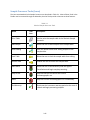
1
Mycobacteria Identification System Operating Manual (Version 6.2B) MIDI, Inc. 125 Sandy Drive Newark, DE 19713 USA June 2013 Tel: (302) 737-4297 Fax: (302) 737-7781 Email: [email protected] www.midi-inc.com Trademarks Sherlock is a U.S. registered trademark of MIDI, Inc. All other trademarks are the property of their respective owners. This manual may not be copied, photocopied, reproduced, translated, or converted to any form, electronic, mechanical, or otherwise, without the prior written consent of MIDI, Inc. Copyright © 2013 MIDI, Inc. All rights reserved. First Printing: February 2002 Second Printing: June 2013 Part Number: 2082 (06/2013) MIDI, Inc. 125 Sandy Dr. Newark, DE 19713 Sherlock Software and Libraries LICENSE AGREEMENT AND LIMITATIONS OF WARRANTY IMPORTANT: Please carefully read this License Agreement before installing the software contained in this package. The right to use this MIDI, Inc. software product is granted only on the condition that the Customer agrees to the terms of this license. For installation to continue, you must agree to the license terms. If you do not accept the license terms, installation will be cancelled. In return for payment of a one-time license fee for this software product, the Customer receives from MIDI, Inc. a license to use this product subject to the following terms and conditions: 1. The product may be used without time limit on one personal computer or workstation. 2. The customer may not modify this software, security ID module or copies of them in any way. 3. To use this product on additional computers, the user must contact MIDI about additional licenses and security ID modules. 4. Purchase of this license does not transfer any right, title or interest in the software product to the Customer, except as specifically set forth in this license agreement. 5. The Customer shall not use this software product to create and distribute competing products, including but not limited to identification libraries, methods, or software. The Customer may use this software product to create libraries and methods for use on MIDI supplied systems within the Customer’s organization. 6. The license may not be transferred to another person or organization without the express written consent of MIDI, Inc. 7. The Customer is on notice that this software product is protected by copyright laws. 8. With prior written approval, MIDI may grant other license rights. 9. By installing the software, the Customer signifies acceptance of this license agreement. 10. This agreement is subject to the laws of the United States of America and the jurisdiction of the U.S.A. courts. MIDI, Inc. makes no warranty of any kind with regard to this software product, including, but not limited to, the implied warranties of merchantability and fitness for a particular purpose. MIDI, Inc. shall not be liable for errors contained herein or for incidental or consequential damages (including lost profit) in connection with the furnishing, performance, or use of this material, whether based on warranty, contract, tort, or any other legal theory. Table of Contents CHAPTER 1 – GETTING STARTED ......................................................................................................................... 1-1 OVERVIEW................................................................................................................................................................ 1-1 Hardware and Software Installation ................................................................................................................ 1-1 Agilent Technologies HPLC ............................................................................................................................... 1-2 Computer and Software ................................................................................................................................... 1-3 INTENDED USE .......................................................................................................................................................... 1-3 Limitations........................................................................................................................................................ 1-3 OVERVIEW OF SHERLOCK OPERATION ............................................................................................................................ 1-4 CHAPTER 2 – PREPARING EXTRACTS ................................................................................................................... 2-1 OVERVIEW................................................................................................................................................................ 2-1 CULTURE GROWTH .................................................................................................................................................... 2-1 SAMPLE PREPARATION ................................................................................................................................................ 2-2 Harvesting Cultures and Saponification ........................................................................................................... 2-2 Extraction ......................................................................................................................................................... 2-4 Derivatization ................................................................................................................................................... 2-4 HPLC SOLVENTS ....................................................................................................................................................... 2-4 REAGENTS ................................................................................................................................................................ 2-5 PRECAUTIONS ........................................................................................................................................................... 2-6 ACKNOWLEDGEMENTS ................................................................................................................................................ 2-7 REFERENCES ............................................................................................................................................................. 2-7 CHAPTER 3 – ANALYZING SAMPLES .................................................................................................................... 3-1 OVERVIEW................................................................................................................................................................ 3-1 Loading the Automatic Liquid Sampler ............................................................................................................ 3-1 Loading Samples Into the Tray ......................................................................................................................... 3-1 Analyzing Samples............................................................................................................................................ 3-1 Calibration Standard ........................................................................................................................................ 3-2 SAMPLE PROCESSOR CONFIGURATION ........................................................................................................................... 3-3 Output Tab ....................................................................................................................................................... 3-3 Report Parameters Tab .................................................................................................................................... 3-5 Instrument Parameters Tab ............................................................................................................................. 3-6 THE SAMPLE PROCESSOR............................................................................................................................................. 3-7 The Sample Processor Menu Bar ...................................................................................................................... 3-7 The Sample Processor Tool Bar ........................................................................................................................ 3-9 Adding Samples and Calibrations ................................................................................................................... 3-10 Editing the Sample Table ................................................................................................................................ 3-11 Starting a Batch.............................................................................................................................................. 3-12 Prematurely Stopping a Batch ....................................................................................................................... 3-13 Sample Processor Tools (Icons) ...................................................................................................................... 3-15 CHAPTER 4 – INTERPRETING SHERLOCK REPORTS ............................................................................................... 4-1 OVERVIEW................................................................................................................................................................ 4-1 Chromatographic Report .................................................................................................................................. 4-1 i Sherlock Composition Report ........................................................................................................................... 4-2 Calibration Reports .......................................................................................................................................... 4-6 Quality Control ................................................................................................................................................. 4-8 SHERLOCK LIBRARY SEARCH ......................................................................................................................................... 4-8 Interpreting the Library Search ........................................................................................................................ 4-9 Similarity Index ................................................................................................................................................. 4-9 Sherlock Comparison Charts........................................................................................................................... 4-10 INTERPRETATION GUIDELINES ..................................................................................................................................... 4-11 Interpretation of Library Groups .................................................................................................................... 4-11 Visual Confirmation ........................................................................................................................................ 4-13 CHAPTER 5 – SHERLOCK COMMANDCENTER ...................................................................................................... 5-1 OVERVIEW................................................................................................................................................................ 5-1 Sherlock Terminology ....................................................................................................................................... 5-1 SAMPLE SELECTION .................................................................................................................................................... 5-2 Basic Sample Selector Mode ............................................................................................................................ 5-3 Advanced Sample Selector Mode ..................................................................................................................... 5-5 PRINTING SAMPLE INFORMATION ............................................................................................................................... 5-12 CONFIGURING THE PRINTER ....................................................................................................................................... 5-15 CORRECTING DATA ENTRY ERRORS.............................................................................................................................. 5-16 VIEWING INSTALLED METHODS .................................................................................................................................. 5-16 Displaying Method Information ..................................................................................................................... 5-17 Printing Method Parameters.......................................................................................................................... 5-18 VIEWING INSTALLED LIBRARIES ................................................................................................................................... 5-19 Printing Library Information ........................................................................................................................... 5-21 COMMANDCENTER TOOLS AND VIEWS (ICONS) ............................................................................................................. 5-22 CHAPTER 6 – ROUTINE MAINTENANCE ............................................................................................................... 6-1 OVERVIEW................................................................................................................................................................ 6-1 Data Storage .................................................................................................................................................... 6-1 SHERLOCK UTILITIES ................................................................................................................................................... 6-2 Data Backup ..................................................................................................................................................... 6-2 Data Restore .................................................................................................................................................... 6-4 SHERLOCK TOOLBOX ................................................................................................................................................... 6-6 Install Methods ................................................................................................................................................ 6-8 Install Libraries ................................................................................................................................................. 6-9 Offload Methods .............................................................................................................................................. 6-9 Offload Libraries ............................................................................................................................................. 6-10 ROUTINE MAINTENANCE OF THE HPLC ........................................................................................................................ 6-11 Replacing the Purge Valve Frit ....................................................................................................................... 6-11 CHAPTER 7 – TROUBLESHOOTING ....................................................................................................................... 7-1 OVERVIEW................................................................................................................................................................ 7-1 For MIDI Technical Support: ............................................................................................................................. 7-1 INSTALLATION PROBLEMS ............................................................................................................................................ 7-2 Cannot Install Software .................................................................................................................................... 7-2 ii Problems Attempting to Run First Set of Samples ............................................................................................ 7-2 CHROMATOGRAPHIC PROBLEMS ................................................................................................................................... 7-2 Calibration Messages ....................................................................................................................................... 7-3 Sample Messages ............................................................................................................................................. 7-4 SAMPLE PREPARATION ERRORS .................................................................................................................................... 7-5 HARDWARE PROBLEMS ............................................................................................................................................... 7-6 APPARENT SOFTWARE PROBLEMS ................................................................................................................................. 7-6 PERFORMANCE QUALIFICATION TABLE ........................................................................................................................... 7-7 APPENDIX A - EQUIPMENT AND CONSUMABLES ................................................................................................A-1 OVERVIEW................................................................................................................................................................ A-1 APPENDIX B - MYCOBACTERIA LIBRARY ENTRIES ................................................................................................ B-1 OVERVIEW................................................................................................................................................................ B-1 APPENDIX C - PROCEDURAL NOTES .................................................................................................................... C-1 EVAPORATION OF LIQUIDS IN TEST TUBES AND VIALS. ........................................................................................................ C-1 APPENDIX D - HPLC PERFORMANCE QUALIFICATION TABLE .............................................................................. D-1 APPENDIX E - PERFORMANCE CHARACTERISTICS ................................................................................................ E-1 OVERVIEW................................................................................................................................................................ E-1 iii Chapter 1 – Getting Started Overview The Sherlock Mycobacteria Identification System (SMIS), developed and marketed by MIDI, Inc., Newark, DE, USA analyzes and identifies microorganisms isolated in pure culture on a solid growth medium. Sherlock uses a sample preparation procedure and high performance liquid chromatography (HPLC) to yield qualitatively and quantitatively reproducible mycolic acid composition profiles. This sophisticated chromatographic system has been developed to be used by microbiologists; thus, chromatographic experience is not essential for operation. The Sherlock software calibrates and monitors the system to ensure proper functioning. Mycolic acids extracted from unknown mycobacteria are automatically quantified and identified by the Sherlock software to determine the mycolic acid composition. The mycolic acid profile is then compared to a “library” (of profiles of reference strains of mycobacteria) stored in the computer to determine the identity of the unknown. Unknown samples may be identified to the species or to the species-complex group. The species-complex will typically be organisms that have high DNA similarities and/or are similar in clinical significance. Examples are the Mycobacterium avium/intracellulare and M. tuberculosis/bovis/africanum complexes. Warning: The SMIS should be considered a dedicated mycobacterial identification system. Any use of the HPLC component for other applications will invalidate the system for its intended purpose! Hardware and Software Installation This manual is intended to contain all information necessary for successful operation of the Sherlock system. It is assumed that a qualified MIDI technician has properly installed and connected all the components of your chromatographic system. Specific hardware installation procedures are not included in this manual. There is a hardware installation checklist in the Installation Qualification that should be verified by the MIDI representative and by the customer. Consult the Agilent manuals accompanying the individual components for more information involving operation of the hardware without use of Sherlock. The MIDI Sherlock software package includes an Installation Qualification that will guide the MIDI service person or the user through the installation of the Sherlock software. The installation instructions are specific to your hardware and computer operating system configuration and must be followed carefully. The signed and completed Installation Qualification also serves as proof that proper installation procedures were followed for GLP purposes. Your software is shipped with a Security Module that must be connected to the computer’s USB port. The Sherlock software and libraries are locked to the serial number of the module and will not operate without it. 1-1 Note: If your computer requires service, or is replaced, be sure to retain the Security Module and connect it to any replacement system. Loss of the security module may require the purchase of new Sherlock software! Agilent Technologies HPLC The Sherlock Mycobacteria Identification Software can be used only with the Agilent Technologies 1100 and 1200 series HPLCs. A computer with compatible version of the Agilent Technologies ChemStation software installed is also required. For specific information and requirements regarding any of these hardware components, refer to the accompanying Installation Qualification included with your system. Figure 1-1 Agilent HPLC 1200 Series This manual assumes that a qualified MIDI representative has properly installed and connected all the components of your chromatographic unit during a standard installation. Environmental Considerations The chromatographic unit will operate within temperatures of 4-40°C (39-104°F) and < 95% relative humidity (See Chapter 1 of each Agilent HPLC component manual for detailed environmental information); however, an environment comfortable for human habitation (reasonably constant temperature and humidity conditions) is recommended for optimum performance and instrument lifetime. Avoid exposure to corrosive substances and dust. Do not use the HPLC column for other applications. Warning: Column damage may occur if the peltier heater is at 70°C and there is no solvent flow through the column. 1-2 Computer and Software Sherlock runs on an IBM-compatible personal computer. The computer should be considered to be a dedicated instrument controller. All Sherlock files are located in the top-level directory named C:\SHERLOCK. MIDI tests Sherlock software with Dell computers and Hewlett-Packard printers. Minimum requirements: Windows 7 is recommended (Windows XP /SP3 is supported) A computer that fully supports the version of Windows (Preferred minimum computer 1.5 GHz, 2 GB RAM, and 20 GB free disk space) Agilent Technologies ChemStation that is compatible with the version of Windows and the HPLC model and revision (prefer B.04.03 or higher). CD-ROM drive 1024x768 Minimum Color Display Resolution LAN interface Dedicated printer Intended Use The MIDI Sherlock Mycobacteria Identification System is intended to aid in the identification of M. tuberculosis and differentiation from other mycobacteria species through the analysis of mycolic acids derived from cultured bacterial samples, using high performance liquid chromatography (HPLC) performed on the Agilent 1100 and 1200 Series HPLCs, along with Sherlock pattern recognition software. The system is used, along with other identification methods, to identify mycobacteria that have been isolated from clinical specimens by traditional culturing techniques. Following observation of growth on the solid medium, identification of the mycobacterial isolate is done with the MIDI Sherlock Mycobacteria Identification System. Results should be interpreted in conjunction with other laboratory observations and procedures. Limitations Sherlock can identify only those microorganisms for which mycolic acid composition profiles of a representative number of correctly named reference strains have been determined and entered into the Mycobacteria Library. The library entries have been determined by analyzing reference strains grown under controlled culture conditions. These culture conditions and sample preparation procedures must be followed. The Sherlock Mycobacteria Identification System has been shown to reliably differentiate the M. tuberculosis complex from other mycobacteria. Definitive identification of Mycobacterium other than tuberculosis (MOTT) requires use of additional laboratory testing to identify clinically significant organisms. Sherlock may be useful in conjunction with probe hybridization, biochemical testing, and other laboratory observations. The ability of Sherlock to correctly identify MOTT not listed in the database has not been evaluated (See Appendix E). 1-3 Overview of Sherlock Operation The Sherlock system is completely controlled by the computer. After sample extracts have been prepared (discussed in Chapter 2 - Preparing Extracts), the labeled sample vials are inserted into the automatic liquid sampler tray. Information about each sample in the tray is entered into the Sherlock Sample Table using the computer keyboard (Chapter 3). When the information about each sample has been entered into the Sample Table, a batch can be started. A batch is a run of one or more sample extracts on the HPLC. When a batch is started from the Sample Processor, the following occurs: The sample’s Method (instrument setpoints, calibration instructions, and sample tray location) is downloaded to a ChemStation file. ChemStation sets the HPLC parameters and controls the injection by the automatic liquid sampler. The automatic sampler selects the sample vial for the calibration mix. The sampler controller causes the injection of a small portion of the extract into the solvent flow stream of the HPLC. The C18 (reverse phase) column installed in the HPLC column-compartment separates the mycolic acids present in the mix as they pass through the column to the detector. The mycolic acid sample preparation (Chapter 2) converts the mycolics into fluorescent derivatives. As the compounds pass through the fluorescence detector, the fluorescent tags are excited by 345-nm wavelength light and this causes emission of light at 425- nm that is quantitatively detected by a photo-multiplier tube in the detector. The amount of light emitted is related to the concentration of the tagged compound and the time of elution is related to the chromatographic properties (structure) of the compound. This signal is stored in the ChemStation data file. The plotted and integrated signals, called the chromatogram, are formatted by ChemStation and printed by Sherlock. When the run is complete, the retention time, peak width and response of each peak are transmitted from ChemStation to Sherlock for processing. Peaks in the chromatogram are identified by mycolic acid Equivalent Carbon Length (ECL) value (name). When peak naming is complete, Sherlock searches the library to identify the unknown organism. The library search uses both the peak name and the peak percentage amount to match with known profiles stored in the library. Following the library search, the computer prints the Composition Report, which includes the peak naming, chromatogram plot, and library classification results. Each library entry is a computer-generated composite of the reference strains of each species or subspecies group of organisms, taking into consideration strain-to-strain and experimental variability. Reference strains were cultured and processed under carefully controlled conditions. The computer system automatically sets the operating parameters of the chromatographic unit. There is no need to manually enter the HPLC parameters. The system will automatically recalibrate within a preset interval. You are permitted to enter sample information into the Sample Table while samples are being processed. This allows for continuous operation of your system. Note: The figures in this manual show the method and library versions at the time the screen shots were taken. The version installed on your system may be newer. 1-4 Chapter 2 – Preparing Extracts Overview The mycolic acids of mycobacteria are part of the structure of the cell wall (Figure 2-1). To release the compounds from the cell wall and to make the samples safe to work with, the harvested cells are saponified in an autoclave. Figure 2-1 Mycobacteria Cell Wall The mycolic-acids are converted to free acids by lowering the pH, extracted into chloroform to remove materials not soluble in organic solvents and derivatized by a fluorescent derivatizing agent. Because the mycobacteria are killed by the saponification procedure, the procedure is safe from a microbiology standpoint from the first step forward. Culture Growth The mycobacteria cultures typically are grown on solid medium such as Middlebrook 7H10 (or 7H11) at 35-37°C, until visible growth is noted. Some species of mycobacteria may normally be grown under different conditions and the database has been constructed to reflect this. Examples would be the culturing of M. marinum and M. haemophilum at 30°C, with the latter organism being grown on chocolate agar rather than on Middlebrook . Examination of cultures under a dissecting microscope may aid in detection of potentially mixed cultures of the slow-growing organisms. 2-1 Warning: Observe Biosafety Level III practices while working with viable cultures. An approved biological safety cabinet must be used! Sample Preparation There are three steps required to prepare samples to be run on the HPLC, and these are outlined in the following sections. Figure 2-2 gives a detailed graphical version of these sample preparation steps. Harvesting Cultures and Saponification 1. Obtain a sufficient number of 13 x 100 mm screw cap tubes for the sample batch. Each batch should contain a known strain as a positive control and a negative control. For a positive control use M. gordonae ATCC 14470. For a negative control use Candida albicans ATCC 60193. Carefully inspect each tube for defects and proper cap fit. Discard any defective tubes/caps. 2. Label tubes with appropriate identifier using a laboratory-marking pen that will withstand autoclave conditions. 3. Add 1.0ml of Saponification Reagent (Reagent 1) to each tube using a repeat dispenser. 4. Harvest growth from the agar plate into the tube containing the saponification reagent. Only a very small amount of cells is needed therefore an amount of cells barely visible on the end of a sterile applicator stick or disposable plastic bacteriological loop is sufficient. It is better to work with isolated colonies. 5. Securely tighten a teflon-lined cap onto each tube. 6. Vortex each tube for 5-10 seconds. 7. Place autoclave indicator tape on the rack containing the tubes. 8. Autoclave for 30-60 minutes at 121°C with slow exhaust. 9. Turn on heat block. 10. Prepare a sufficient quantity of potassium bicarbonate coated vials if necessary. Note: After autoclaving the tubes are safe to work with at the laboratory bench or a chemical fume hood. 2-2 Figure 2-2 Sample Preparation 2-3 Extraction 1. Remove tubes from autoclave and allow them to cool to ambient temperature. 2. Add 1.8ml of Acidification Reagent (Reagent 2) with the repeat dispenser. Cap the tubes and invert as necessary (2-3x) to obtain thorough mixing. Allow the tubes to stand for 5 minutes. 3. Uncap tubes and add 1.5ml of chloroform (Reagent 3) using the repeat dispenser. 4. Recap each tube and vortex vigorously for 30-60 sec. Visually inspect each tube to insure full vortex. 5. After phase separation, transfer the bottom (chloroform) layer to a clean 13 x 100 mm borosilicate glass tube. Note: It may be necessary to briefly centrifuge the tube if an emulsion is present. The tube must be labeled with the corresponding label number from the original 13 x 100mm tube. 6. Evaporate the chloroform from each tube. See Procedural Note 1.0 for instructions using the Pierce Reacti Therm or Procedural Note 2.1 for the Labconco system. 7. Cool the tubes to ambient temperature. Derivatization 1. Add 200ul Derivatizing Reagent (Reagent 4) with dispenser. 2. Gently swirl the tube to dissolve its contents. 3. Transfer the mixture from each tube into a correspondingly labeled amber vial (2 ml size) which has had the interior precoated with 100ul of 2% Methanolic Potassium Bicarbonate (Reagent 5). 4. Swirl the vial gently by hand to assure mixing of the contents. 5. Heat the vials uncapped for 10 minutes at 60±2°C. 6. Slowly evaporate the contents of each vial. (See the Appendix C procedural notes for the MIDI Evaporator, Pierce Reacti Therm, or Labconco System.) 7. Remove the vials and cool to ambient temperature. 8. Add 500uL of isopropyl alcohol (IPA) (Reagent 6) and cap the vial. 9. Swirl to totally dissolve the contents and mix the sample extract. 10. To a tapered insert vial containing the internal standards (MIDI Part # 1600-A) add 50 ul of the sample extract. 11. Dispense the extract along the walls of the insert to ensure that the dried internal standards are totally solubilized. 12. Once dispensed, thoroughly mix by slowly pumping the solution up and down in the pipette. 13. After capping the vials, the samples are ready for HPLC analysis. HPLC Solvents Solvent A: Methanol, Shelf life = 3 years in unopened container (1 yr. if opened). Danger: Flammable, toxic if consumed. Solvent B: IPA, Shelf life = 3 years in unopened container (1 yr., if opened). Danger: Flammable, toxic if consumed. 2-4 Reagents Reagent 1: Saponification Reagent. Shelf life = 1 year. Potassium Hydroxide: 250 g Deionized Water: q.s. to 500 ml with continuous stirring. Danger: Reagent is extremely caustic. Wear eye protection. Observe splash precautions. Substantial heat is released during reagent preparation that may cause the mixture to boil, bump, or steam. Prepare reagent ONLY in a BOROSILICATE GLASS flask inside a chemical fume hood. Prepare by slowly adding KOH pellets to 200ml dIH2O until totally dissolved. Continue adding H2O until the final volume is 500 ml. Note: Protect from air (absorbs CO2). Reagent 2: Acidification Reagent. Shelf life = 1 year. To prepare, slowly add 250 ml concentrated HCL to 250 ml deionized water with continuous stirring. Danger: Reagent is extremely corrosive. Wear eye protection. Observe splash precautions. Substantial heat is released during preparation that may cause the mixture to boil, bump, or steam. Prepare reagent ONLY in a BOROSILICATE GLASS flask inside a chemical fume hood. Add acid slowly to water. Never add water to acid. Reagent 3: Chloroform. Shelf life 1 year in unopened container, 6 months if opened. Danger: Toxic Reagent 4: Derivatizing Reagent. Shelf life = 6 months (in amber container and stored in the dark). Dissolve 100-mg of 4-bromomethyl-6,7-dimethoxycoumarin (F.W. = 299.1) and 100-mg of 18-crown-6 ether (F.W.= 264.3) in 100-ml of chloroform. Danger: toxic, lacrimator Reagent 5: 2% Methanolic Potassium Bicarbonate Solution. Shelf life = 1 year. Dissolve 2.0 g of potassium bicarbonate (KHCO3) in 100ml of 50% aqueous methanol (50 ml methanol; 50 ml deionized water). Prepare vial by adding 100ul of Reagent 5 to a clean, amber, 2 ml autosampler vial. Dry the contents of the vial completely. See Procedural Note 1.0 for the Pierce Reacti Therm System or Procedural Note 2.3 for the Labconco system. The coated vials may be prepared in batches and stored at room temperature in a dried covered condition (Shelf Life = 6 months). Reagent 6: Sample Extract Diluent. HPLC grade isopropyl alcohol (ACS certified preferred). Shelf life = 3 years in unopened container (1 yr if opened). Warning: Flammable 2-5 Precautions Extracts may be stored refrigerated and in the dark for 7 days prior to analysis. Mycolic acids of rapid growers tend to deteriorate more quickly than those of slow growers. The Reagent 4 (the Derivatizing Reagent) should be stored in an amber container in the dark for maximum shelf life. The extraction/derivatization procedure may be stopped for as much as 24-hours following the autoclaving-saponification step. Do not reuse any glassware. Use of plastic autosampler vials or plastic inserts may cause problems with back-pressure elevation of the HPLC and should not be used. The isopropanol in the sample diluent may dissolve some of the plastic components and carry that into the methanol/isopropanol mobile phase, where it is less soluble and thus may come out of solution, plugging the plumbing and damaging the column. Repeating pipetters make dispensing the reagents safer and more reproducible. The dispensers should be well primed before use in order to expel air bubbles in the pickup and delivery tubes. This is especially important with low-volume chloroform solution dispensers. Avoid pumping the dispensers too fast since air bubbles can be introduced that lead to inaccurate reagent delivery. When pipetting or dispensing, tilt tubes or dispenser tip, or pipet so that reagent runs smoothly down the side of the tube. Samples must be completely dry after evaporation steps for subsequent reactions to be efficient. The use of 200-μl sample inserts in the autosampler vials allows easy injection by the system and permits reuse of the vials. Vial caps may also be reused if they are the screw-on type and the septum is replaced for each reuse. All work with alkali, acids, and solvents must be performed in an approved explosion-resistant chemical fume hood. Eye protection and other personal protective equipment must be worn during all work with alkali, acids, and solvents. 2-6 Acknowledgements The MIDI extraction protocol was adapted from the methodology established at the Texas Department of Health, Bureau of Laboratories, Mycobacteriology/Mycology Branch. References Richard J. Wallace, Jr., Barbara A. Brown-Elliott, Leslie Hall, Glenn Roberts, Rebecca W. Wilson, Linda B. Mann, Christopher J. Crist, Sher H. Chiu, Robbie Dunlap, Maria J. Garcia, J. Todd Bagwell, and Kenneth C. Jost, Jr. Clinical and Laboratory Features of Mycobacterium mageritense. 2002. J. Clin. Microbiol. 40:2930-2935. Brown, B. A., B. Springer, V. Steingrube, R. W. Wilson, G. E. Pfyffer, M. J. Garcia, M. C. Menendez, K. C. Jost, Jr., S. H. Chiu, G. O. Onyi, E. C. Bottger, and R. J. Wallace, Jr. 1999. Description of Mycobacterium wolinskyi and Mycobacterium goodii, two new rapidly growing species related to Mycobacterium smegmatis and associated with human wound infections: a cooperative study from the International Working Group on Mycobacterial Taxonomy. Int. J. Syst. Bacteriol. 49:1493-1511. 2-7 Chapter 3 – Analyzing Samples Overview The previous chapter discussed the preparation of HPLC-ready extracts from cell cultures. This chapter continues with the actual HPLC processing of the samples. In this chapter we will look at: Loading the Automatic Liquid Sampler Sample Processor Configuration Sample Processing Under Computer Control. Loading the Automatic Liquid Sampler The Automatic Liquid Sampler 1100 and 1200 Series Agilent HPLC consists of a sample tray, a robotic arm for moving the sample vial from the tray into position to be sampled, and a sampling syringe mechanism. Figure 3-1 Automatic Liquid Sampler Loading Samples into the Tray The first position in the sample tray (numbered “1”) should hold a vial containing the Calibration Standard. Positions with higher numbers should contain sample vials that correspond to their numbers in the sample table. There must not be empty spaces in the sample tray unless those positions are designated as “Empty” in the Sherlock sample table. NOTE: This manual generally refers to the physical container as a vial. The Sherlock software generally refers to the position in the tray as the bottle number. Analyzing Samples When the operator clicks on the Start Batch tool in the Sherlock Sample Processor Toolbar, the system will begin a conditioning process that includes a warm up period and a “blank run”, or analysis in which no sample is injected. As a result, the solvent gradient is performed. This conditioning is helpful in enabling the system to equilibrate so that the analysis of the calibration is not only successful, but 3-1 provides optimal times for the peak naming table to use in naming peaks in subsequent samples. At the completion of the blank run, the robotic arm will pick up the vial in position 1 (Calibration Std.), move it to a space between the injection syringe (which has raised to accept the vial) and the syringe seat. The syringe will then pick up the amount of sample designated by the Sherlock method. The sample vial is returned to its original position and the syringe moves down into the syringe seat, causing injection of the sample into the flowing stream of carrier solvents. The entire sample is put into the analysis and the syringe is continuously flushed by solvent during the entire analysis. This virtually eliminates carry-over of sample to the next analysis. Calibration Standard The calibration standard is used for the first two injections of a batch and is reanalyzed after every 10th sample injection to ensure system stability. When a calibration analysis is completed, the computer checks the results against the Sherlock Method’s Peak Naming Table for a specific number of peaks and a pattern of retention times. The Peak Naming Table contains the expected retention time for each peak in the calibration analysis. Deviations from the expected values may result in a failure to calibrate with a warning message to the user. If the system fails to calibrate on the first attempt, it will try once more to calibrate. If the failure is due to the reference peaks falling outside expected time “windows”, consult the Troubleshooting Section (starting on page 7-2). A second function of the Calibration Standard is to provide reference retention times for the mycolic acids. These retention times are used to calculate Equivalent Carbon Length (ECL) values by which peaks in subsequent analyses are named. The system calculates how much the calibration analysis has deviated from the expected retention times and reports the Root Mean Square (RMS) fit error. If a calibration run is invalid due to a high RMS fit error, Sherlock will print a message to warn the user and will repeat the calibration analysis. If the system fails to calibrate after two consecutive attempts, the error message will be repeated and the sample batch aborted. Consult the Troubleshooting Chapter (starting on page 7-2). The Calibration Standard is shipped dry in glass inserts. An order contains instructions for use and storage of the Calibration Standard. The expiration date of the Calibration Standard is stated on the package. It should be stored dry, in the dark and at ca. 4°C. The Calibration Standard should be solubilized by addition of 80-μl of IPA. If the calibration Standard is not completely used in a batch of analyses, it may be recapped with a fresh cap and stored in the dark at ca. 4°C for up to one month. Before reuse, the calibration Standard should be brought to ambient temperature to assure complete solution of all mycolic acids. Each bottle placed into the sample tray must be logged (entered) into the computer’s Sample Table. This procedure is discussed in the Sample Processor section (see page 3-7). Before starting a sample batch, be certain that the descriptions logged into the Sample Table match the bottles in the sample tray. 3-2 Sample Processor Configuration Sample Processor Configuration is accessed from the Sherlock Toolbox Icon on the desktop, or through the Programs/Sherlock group under the Windows Start button (other Toolbox items will be described in Chapter 7- Routine Maintenance). This screen has three tabs and controls where Sherlock results are sent and stored, important instrument control parameters, and the level of detail that report parameters are recorded. The Update and Exit button will save all changes made to the window, while Cancel and Exit button will not save any changes. Figures 3-2a, 3-2b, and 3-2c show the three configuration tabs. Figure 3-2a Sample Processor Configuration Screen – Output Tab Output Tab Results to Printer/ Results to File These sections determine how Sherlock Reports are stored while a batch is running. Checking the RESULTS TO PRINTER checkbox will print Sherlock Reports after each sample and calibration is run. MIDI suggests this option so results are immediately available. The Margin menu controls the margins for the text when printing during a batch run or from CommandCenter. The options are ¼ , ½, and ¾ inch. If you need to put your reports in a 3-ring binder, choose the ¾ inch option to avoid loss of text from a hole punch. Checking the SAVE RESULTS TO FILE checkbox will send Sherlock reports to a formatted text file in Rich Text Format (RTF). The file name is chosen automatically when the option is chosen. The user can print the files later using MS Word®. 3-3 To print the files in MS Word: Open MS Word. Open file in C:/ Sherlock/Results /xxxxxxxxx.rtf. You can print the entire file or choose the data you want to print. Note: Each report will be in a separate file. Data Storage This controls the storage location of data in the Sherlock/DATA directory. There are eight storage locations, DATA and DATA1 through DATA7. This is where data are accessed for further manipulation by CommandCenter functions. Data can be transferred to other data volumes within this directory or transferred from this directory to the archival system of the customer’s choice. Data File Suffix Sets the letter used as the suffix on Sherlock data file names. Typically this is set to “A.” For sites with multiple Sherlock systems, a different suffix is used for each system. Figure 3-2b Sample Processor Configuration Screen – Report Parameters Tab 3-4 Report Parameters Tab This section controls the amount of sample identification detail that will appear on a Sherlock Sample or Calibration Report that is generated from the Sample Processor. Reports that are generated from CommandCenter have their own control. The menu choices are: No reports Brief No report is generated Will print only the sample name and warning message Profile Full-page report, listing all peaks in the chromatogram Brief with The sample name, warning Classify messages, match to the library Profile with Full-page report, listing all Classify peaks in the chromatogram and match to the library. MIDI strongly suggests using the Profile with Classify option for routine operation. Note that all raw data is stored in a separate file in Sherlock. Through CommandCenter, Sherlock can generate a more or less detailed report at any time desired by the operator. The Number of Charts field refers to Comparison Charts. These graphical plots show how the unknown sample profile compares with library search results. There can be up to four comparison charts printed. See Chapter 4 for a more detailed explanation of Comparison Charts. The Include Chromatogram checkboxes determine if the chromatogram plot will be printed. MIDI recommends that these be printed. The Print Pre-Sample File and Print Post-Sample File checkboxes define a custom header and footer for the reports. The header might contain the laboratory’s name and address. The footer might contain a signoff line for report approvals. If Generate Summary Report at End of Batch is checked, a list of samples with identifications is printed. 3-5 Figure 3-2c Sample Processor Configuration Screen – Instrument Tab Instrument Parameters Tab This section will not need to be amended, if your instrument was configured by a MIDI technician during the Installation Qualification. Note: Contact MIDI if the parameters don’t match exactly as shown in Fig. 3-2c. For Sherlock configuration settings, the 1100 Series HPLC and the 1200 Series are equivalent. 3-6 The Sample Processor The Sample Processor is accessed from the Sample Processor Icon on the desktop. Sherlock uses the Sample Processor as a Sample Table and as a link with the Agilent ChemStation. The top menu bar and toolbars allow manipulation of the Sample Table (Figure 3-3). The middle windowpane gives Sherlock and ChemStation status information, currently running sample status and the data storage location. The bottom section is the Sample Table, where the Calibration Standard and sample identification information is logged. Figure 3-3 Sherlock Sample Processor Screen Menu Bar Tool Toolbar Current State of Sample Table Status Area Sample Table The Sample Processor Menu Bar File / Print Allows printing the current sample table, optionally including result information. The Sample Table can be printed at any time, even when running samples. The table is printed as soon as the printer is available. File / Exit Allows exiting the Sample Processor. If the Sample Processor is running samples, it is strongly recommended that the user abort or stop the batch before exiting. Exiting the Sample Processor while samples are running will leave the system in an unstable state. 3-7 Table – Clear Options Each of the table clear options removes samples from the Sample Table by erasing the sample identification number and name, and changing the bottle type to EMPTY. When the system is running samples, the editor will not allow the removal of CALIB, or RUNNING bottles from the table. Table / Clear All Samples Removes all Queued or Done samples, but not calibration entries. Table / Clear If Done Removes samples that have already been analyzed (bottles with a status of DONE). Table / Reset Table Removes all samples and calibration entries, and establishes a new calibration entry. Table / Clear Current Bottle Removes information about the currently selected bottle. Note: Table Clear functions must be used with caution as there is no Undo command in Sherlock and there is no way to save a sample table. Table / Set Auto ID Number The Auto ID Number feature should be used to assign and increment the Sequence Number (Seq #) field automatically for each sample run. Each sample will then have a unique identifying number. Sequence numbers determine the order that samples will be run in (but see “Stat” below for a way to override this behavior). By default this number will also be used as the ID number. The user may change the ID numbers for samples as needed. The numbers don’t have to be unique. The ID numbers are saved with the sample in the Sherlock data file. Note: The Sequence # (Seq #) is not alterable. Batch / Start Batch This function will start the ChemStation automatically and begin sample processing. This function is only available when the Sample Processor is not currently running samples. A calibration is always the first sample analyzed. Batch / Stop at end of run Stops the batch after the chromatographic analysis of the current sample is completed and the reports are printed. Batch / Abort Batch Stops the current analysis immediately. Note that the current analysis is lost. Note also that since ChemStation takes a few minutes to load completely, to prevent potential problems with ChemStation it is prudent to wait until ChemStation has completely loaded before Aborting. 3-8 View / Reset Resets the sample table, showing all the columns. Use this option if resizing columns and want to view library search results during a batch. The Sample Processor can show results of sample runs in the sample table. The right-most column of the table can show the similarity index for the library entry with the highest match achieved by that sample. If there are multiple high matches, the similarity index is preceded by a plus sign ("+"). You should always review the sample's report for proper interpretation of the results. View / Reset No Results Resets the sample table, hiding results columns. Use this option if resizing columns and want to return to a standard state. This view is especially useful for initially setting up a sample table. The Sample Processor Tool Bar Note: A description of all the Sample Processor icons is given in Table 3-1 at the end of the chapter. Add Samples Before putting a sample bottle into the sample tray, the proper identification information must be logged into the corresponding bottle number in the Sample Table. The sample bottle type can be a calibration sample (Calib) or a sample extract (Samp) to be processed for identification. In addition, the bottle type can be labeled as Stat, which indicates a priority sample that will be run next (after any required calibrations), Blank for the negative control, QC for a positive control, or Empty. This places the system in a mode that simplifies adding new samples to the sample table. When the Add Samples tool is pressed, the Sample Table editor automatically advances to the first empty bottle position. The sample type, method, and status will be entered automatically by the system based on the previous sample’s information, while the user will enter sample name and pertinent sample information (Figure 3-4). The Seq # is automatically incremented and displayed. Note: The first sample in the analysis should be the QC negative control (C. albicans). Under the “Type” column, select Blank as the type. The second should be a positive control with type QC selected. All subsequent specimens will automatically default to Samp. This is the only correct way to add new samples to the Sample Processor. The Add Samples tool will change to Done Adding tool while the user is adding samples to the table (Figure 3-4). Press this tool to complete adding samples. See details in the following Adding Samples and Calibrations section. 3-9 Figure 3-4 Sample Processor – Add Samples Mode Lock Table / Unlock Table This tool toggles the table between locked and unlocked. When the Sample Table is locked, no changes can be made inadvertently to the table. When adding or editing samples, the table must be unlocked. Print Table This tool is identical in function to the File / Print menu item. Start Batch This tool is identical in function to the Batch / Start Batch menu item. Stop At End This tool is identical to the Batch / Stop at end of run menu item. Abort This tool is identical to the Batch / Abort Batch menu item. Adding Samples and Calibrations Every Sample Table must contain a calibration entry. Sherlock will automatically calibrate twice prior to sample analysis and will recalibrate the system after every 10 samples. Samples may be added before or during the processing of samples by Sherlock. When the Add Samples tool is selected, the Sample Table editor automatically advances to the first empty bottle position. The next sequence number will be incremented automatically by the system. The 3-10 status of the new sample is set to QUEUED. Enter 1 to 42 characters for the sample identification in the NAME field. There must be at least one character entered as a sample name. Strike the [Enter] key to advance to the next empty bottle position. To stop adding samples, press the Done Adding tool; the table can also be locked by clicking the Lock Table tool to ensure no inadvertent changes are made. Figure 3-5 Editing the Sample Table Editing the Sample Table To change an existing entry in the Sample Table, Unlock the table, click on the field to change, and select the choice from that drop down menu or type in the change (Figure 3-5). Sample Table entries can be edited at any time, even while samples are being run on the LC. Samples listed with a status of RUNNING are protected from edits. It is a good idea to Lock the table when finished editing. The pencil icon on the left side of the screen indicates that changes are outstanding (next to bottle 6 in Figure 3-5). Changing the Type Click on the Type field to select the choice for priority: CALIB A calibration bottle. There must be at least one calibration logged into the Sample Table. If necessary to maintain calibration, a calibration will run before next sample, even before a STAT sample. Its Sequence Number field will automatically be set to 1 and cannot be changed. SAMPLE Normal priority. STAT High priority sample. STAT samples are processed before ordinary SAMPLES. QC Indicates a positive control sample. BLANK Negative control bottle or a bottle that contains no sample. EMPTY No bottle in this sample tray position. 3-11 Changing the Status The status of the selected sample is either QUEUED, DONE, or RUNNING. To change from QUEUED to DONE or vice versa, click on the status field and choose from the drop down menu. Samples that have a status of RUNNING cannot be changed. If the user wants to process samples out of order, they can use the STAT function. Removing items from the Sample Table If the table is not Locked, samples can be removed from the Sample Table by scrolling to the entry and choosing any of the Table/Clear options from the menu bar. Record Keeping Enter enough data about the sample into the “Name” field so that anyone can go back a year later and know what was run. If the growth conditions were not standard for the Method being used, enter the nonstandard conditions in the name field. Also, enter any unusual observations that may aid in interpreting results. Consistency in the name is extremely important for record keeping as well as for cataloging samples. It is advantageous to organize name fields so that the user can look at groups of samples of interest. To compare groups of entries it will be necessary to create a system for making groups and subgroups very early in the data collection process. The name of a sample consists of two sections separated by an open parenthesis sign “(”. A total of 42 characters may be entered. We suggest using UN- in front of unknown samples. The second section of the name begins with an open parenthesis sign “(”, and allows the user to enter discrete information about a sample, such as the patient identification number, type of media, age, etc. It is helpful to use the initials or some personal identifier for the person logging the sample into the sample table. That person is verifying that the samples are placed in the correct positions on the auto sampler tray. It is not necessary to close the parenthesis or use capital letters or hyphens after the open parenthesis. Once it “sees” the left parenthesis, the software looks no further for cataloging functions. While the user will be able to view this information in any file listing, it is for personal use only and the software does not use it. Starting a Batch Sherlock is ready to analyze samples when the following are achieved: Verified that the proper printer parameters are in use, and that the printer has paper. Note: See Page 3-3, if immediate printing of results is not desired. Confirmed that each sample has a unique sequence number and logged the sample information into the Sample Table. Loaded the sample tray with the samples and the Calibration Standards corresponding to the Sample Table entries. Confirmed that ChemStation is not open or running. Verify that all instrument modules are on and in a ready state. Verify that there is sufficient solvent in the solvent bottles. 3-12 When the above steps have been taken, the system is ready to start. To start a batch, press on the Start Batch tool in the Toolbar. If prompted, make any necessary adjustments to the Sample Table. If no adjustments are needed, click on Start Batch. The system will prompt the user to warm up the system. Select “Yes”. The system analyzes the Calibration Standard (two runs at the beginning of the batch), and if the system is working properly will proceed through the samples in the Sample Table. After all samples have been analyzed, the software automatically shuts off the pumps, detector, and heater column modules of the HPLC, and closes the ChemStation software. While the system is running samples, other commands are still active. Click on the Sample Table to make additions or edits to the sample table. Note: The Running sample may not be edited. Prematurely Stopping a Batch Ordinarily, once Sherlock has started a sample batch, all samples will be analyzed and the system shutdown as described above. However, if it becomes necessary to stop a batch before all samples are analyzed, there are two options– Stop at End and Abort. The Stop at End tool replaces the Start Batch tool after a run has been started. Click on the Stop at End tool if it is necessary to stop the operation of the system at the end of the current chromatographic run. This will complete the analysis in progress, print the results, and then stop the batch. The Abort tool also appears in the Toolbar after a run has been started. Click on the Abort tool to stop the operation of the system any point before completion of the batch. This will immediately stop analysis in progress and then stop the batch. The Abort tool should be used with caution. If the sample injection has occurred, any non-eluted compounds will remain on the chromatographic column. If there is sample left on the column, a blank must be run to clear the column of any residual sample. This is typically done automatically at the beginning of the next batch (see Analyzing Samples, page 3-1). 3-13 In addition to the Toolbar icons, two Sherlock menu options describing the options for correctly terminating a batch are also available in the menu bar; Click on the Batch / Stop at end of run menu item to complete the analysis in progress, print the results, and then stop the batch. Click on the Batch / Abort Batch menu item to immediately stop the analysis in progress and stop the batch. Caution: It is not possible to stop the batch correctly using the ChemStation Abort function from the ChemStation menu bar. The LC will stop the run and return to a standby status but Sherlock will not be able to proceed with the batch or communicate with ChemStation and will be difficult to close. It is then necessary to completely exit back to the “Windows” screen and then click on the Sherlock Sample Processor to restart the analysis. It may be necessary to use the Windows Task Manager (Ctrl/ Alt/ Delete/ Task Manager/ Applications) and end the Sherlock application, to completely exit back to the Windows screen and then click on the Sherlock Sample Processor Icon to restart the analysis. Precaution: Never Abort Sherlock while ChemStation is loading. There will be time after ChemStation is loaded and before the first injection to halt the Sample Processor. If the ChemStation exits abnormally and/or Sherlock “locks up”, you should reboot your computer to clear any remaining errors in Windows. Failure to reboot will most likely result in another lockup. 3-14 Sample Processor Tools (Icons) The icons associated with the Sample Processor are described in Table 3-1. Most of these Tools in the Toolbar can be accessed through the Menu Bar, but the Tools provide a shortcut to these features. Table 3-1 Sherlock Sample Processor Tools Name Tool Icon Description Print Table Used to print the sample table in the Sherlock Sample Processor. Add Sample Allows the user to add samples to the sample table. Done Adding Press this button when done adding samples to the sample table. Lock Table Allows the user to lock the sample table from editing. Unlock Table Enables the user to unlock the sample table. Start Batch Allows the user to automatically load the Agilent ChemStation and begin sample processing. Stop at End Stops the batch at the end of the current chromatographic run. Abort Stops the current chromatographic run immediately. End Warmup Terminate the instrument warmup period at the start of a batch and begin processing samples. 3-15 Chapter 4 – Interpreting Sherlock Reports Overview Your Sherlock system reports analysis results in two forms: A Chromatographic Report produced by ChemStation A Sherlock Composition Report, including a library search. The primary component of the Chromatographic Report is the chromatogram, a visual plot or trace of the electronic signal generated by the fluorescence detector as mycolic acids of the sample elute from the column. The raw data of the chromatogram is stored in a ChemStation file and can be reintegrated on-screen and reprinted if desired. Sherlock stores a file containing all peak retention times, heights, peak widths, and a pointer to the chromatogram plot. The Sherlock CommandCenter can reprint the chromatogram plot. Note: Earlier versions of Sherlock did not save a pointer to the chromatogram plot; therefore, it is not available for reprinting by Sherlock. The Sherlock Composition Report comprises a Mycolic Acid Composition Report, a Library Search Report, and (optional) Comparison Charts and chromatogram plot. The Composition Report contains the mycolic acid composition of the organism. The Library Search Report lists the results of comparing the mycolic acid composition to the Sherlock Library. Comparison Charts may also be requested. These are plots comparing the mycolic acid composition of the unknown to the most similar (up to four) library entries. Comparison charts can be printed by one of two ways: either with the Sample Processor report, using the Sherlock Sample Processor Configuration screen (Figure 3-2b) or by specifying this option in the Sherlock CommandCenter Print Preview and Print dialog boxes (Figure 5-12 & 5-14). Chromatographic Report The ChemStation is used to accumulate raw data and to develop the chromatogram. In routine use of Sherlock, the chromatogram with the peak times plotted by the ChemStation normally need not be evaluated. However, you should be familiar with the contents of the plotted chromatogram to confirm proper operation of the system. Using the Sample Table and the Calibration Sequence parameters specified in the method, the Sherlock software selects the sample scheduled for injection. The chromatogram heading lists the injection time and date, bottle number and the sample name as it was entered in the Sample Table name field. The sample ID number appears in the Sample Name fields and within the data file name. Once the sample is injected, the ChemStation plots the signal from the fluorescence detector of the HPLC, creating the chromatogram as shown in Figure 4-1. Mycolic acids in the sample are separated by the column and identified by the retention time of each peak. Retention times are measured to a resolution of 0.001 minute. (Figure 4-1 chromatogram is typical of M. tuberculosis complex). 4-1 Figure 4-1 HPLC Chromatogram Plot 8.101 FLD1 A, Ex=345, Em=425 (C:\SHERLOCK\RAW\E00719.433\A01645A3.D) 7.818 LU 8.368 100 80 8.631 7.538 60 9.841 10.026 9.152 6.977 6.672 6.359 6.472 6.098 5.730 5.373 4.877 5.084 4.545 4.677 4.151 3.736 4.010 3.538 3.277 3.106 20 8.867 7.261 40 0 3 4 5 6 7 8 9 10 min Sherlock Composition Report The peak retention time, width and height data from the ChemStation are transmitted to Sherlock data files at the end of each run. The data are processed, peaks are assigned names (based on ECL values, not structural names), the mycolic acid pattern for the sample is compared to the library and a report is printed. An example report is shown on the following page (Figure 4-2). General Information As determined by the user in the Sample Processor Configuration tool, the computer stores the chromatographic data in files on the selected data volume (see Chapter 5 for a suggested protocol for selection of data volume). For each batch of samples, a file name is automatically assigned by the computer and is printed at the top of the Composition Report. The heading identifies the sample with the sample ID number, bottle number, sample name and the date and time of the analysis. If the data have been edited, the date and time of the last edit is also printed. 4-2 Figure 4-2 Sherlock Composition Report Volume: DATA File: E026196.94A Type: Samp Bottle: 10 Created: 6/19/2002 7:32:29 PM Samp Ctr: 12 Method: MYCOLC1 ID Number: 2246 Sample ID: UN-MYCO (1004 4WK GO Profile: RT 2.665 2.838 3.017 3.184 5.571 5.930 6.298 6.523 6.592 6.816 7.096 7.371 7.648 7.929 8.194 8.462 8.697 8.988 9.463 9.681 9.859 ------------------------------------- Response 3406 2799 8922 1215 660 742 1537 3104 3113 10131 35798 115990 228230 307522 264531 129343 32646 9733 535 700 575 1215 742 1537 3104 10131 35798 115990 228230 307522 264531 129343 32646 Ar/Ht 0.088 0.091 0.097 0.093 0.131 0.190 0.177 0.110 0.096 0.157 0.130 0.123 0.117 0.112 0.119 0.121 0.140 0.151 0.163 0.138 0.202 ------------------------- ECL 36.413 38.175 40.000 41.699 64.427 67.881 71.426 73.591 74.252 76.385 79.056 81.688 84.312 86.977 89.450 91.951 94.214 97.000 101.535 103.617 105.324 ------------------------------------- ECL Deviation: 0.131 Total Response: 1134562 Percent Named: 100.00% Peak Name ECL 40.000 standard Sum In Feature 1 ECL 64.625 Sum In Feature 9 Sum In Feature 10 Sum In Feature 11 ECL 74.350 Sum In Feature 12 Sum In Feature 13 Sum In Feature 14 Sum In Feature 15 Sum In Feature 16 Sum In Feature 17 Sum In Feature 18 Sum In Feature 19 ECL 97.000 standard Summed Feature 1 Summed Feature 9 Summed Feature 10 Summed Feature 11 Summed Feature 12 Summed Feature 13 Summed Feature 14 Summed Feature 15 Summed Feature 16 Summed Feature 17 Summed Feature 18 Summed Feature 19 Percent ---------0.11 0.06 0.07 0.14 0.27 0.27 0.89 3.16 10.22 20.12 27.10 23.32 11.40 2.88 ------------0.11 0.07 0.14 0.27 0.89 3.16 10.22 20.12 27.10 23.32 11.40 2.88 Comment1 < min rt < min rt ECL deviates 0.000 ECL deviates 0.099 ECL deviates -0.198 ECL deviates 0.168 ECL deviates 0.039 ECL deviates 0.162 ECL deviates -0.098 ECL deviates 0.107 ECL deviates 0.046 ECL deviates 0.074 ECL deviates -0.028 ECL deviates -0.088 ECL deviates -0.160 ECL deviates -0.306 ECL deviates -0.109 ECL deviates 0.000 > max rt > max rt > max rt Reference ECL Shift: 0.034 Total Named: 1134562 Total Amount: 1134562 Comment2 Reference 0.015 ECL 41.600 ECL 67.713 ECL 71.387 ECL 73.429 index ECL 76.278 index ECL 79.010 index ECL 81.614 index ECL 84.340 index ECL 87.065 index ECL 89.610 ECL 92.257 index ECL 94.323 Reference -0.046 Number Reference Peaks: 2 Matches: Library MYCAG1 1.02 Sim Index 0.906 Entry Name Mycobacterium-tuberculosis complex (TB,bovis,africanum,microti) 4-3 A table containing the mycolic acid profile for the sample follows the general information. The columns of this table are: RT, the retention time, is used to calculate the ECL value. Response, for HPLC, peak height is used rather than peak area as it is more reliable. Ar/Ht, is the calculated area divided by the height. This is an estimate of the peak width. ECL, is the Equivalent Carbon Length of the mycolic acid. It standardizes the elution time across instruments and columns. Peak Name, a number such as ECL 92.527 is the interpolated carbon length between the two internal standards. Percent, the relative amount of each named peak. Comment 1, gives a peak match to the expected position in the naming table. Comment 2, is additional information about peak naming or peak ECL drift. Peak Names and Amounts Each peak from the chromatographic analysis is listed by retention time (RT), Response, and area/height ratio (Ar/Ht). Also, included in the composition report is the ECL, which is a linear interpolation of each peak’s retention related to the preceding calibration standard and to the two internal standard reference peaks. Sherlock software compares the ECL of each peak in the analysis with the expected ECL of the mycolic acids in the Peak Naming Table. The mycolic acid “name” (this is the assigned ECL value) is printed in the Peak Name column. Peaks that do not correspond to ECL values of known mycolic acids are unnamed and are not used in the library search or identification. Due to the great diversity of isomers and the difficulty of reproducibly resolving all compounds, most peaks will be listed as Summed Feature. Summed Features are created by summing adjacent mycolic acid peaks into a single, more reliable sum. The very distinctive patterns of the mycolic acids allows use of summed features while still giving great precision in matching to the stored library of mycolic acid profiles in the library. The two comment columns are used to provide additional information about each peak that may help troubleshoot chromatographic issues. Note: ECL 40.000 and 97.000 peaks are used as internal standards and thus no response is assigned to these peaks. Sherlock refers to them as “Zero Features.” Summary Section The Summary Section of the Composition Report is useful to troubleshoot the system. Several of the performance measures, listed below, are checked by the system during operation, and warning messages are printed if limits are exceeded. ECL Deviation, the error between the actual ECL values and the expected ECL values. It is a measure of the chromatographic accuracy for naming peaks. Reference ECL Shift, the ECL drift calculated for designated Reference Peaks. It is a measure of the chromatographic stability. 4-4 Number Reference Peaks, a count of the number of reference peaks found in the sample. For the MYCOLC1 method it should be two (the ECL 40.000 standard and ECL 97.000 standard). Total Response, the sum of the Response column values for all peaks, excluding peaks that are outside the RT of interest (“< min rt” and “> max rt”) and “zero feature” peaks. Zero feature peaks are assigned peak names but are not included in the calculation of percentages (e.g. ECL 40.000 and ECL 97.000 in Figure 4-2). Total Named, the sum of the Response column values for all named peaks, excluding “zero feature” peaks (see Total Response). Percent Named, 100 * (Total Named) / (Total Response). Total Amount, equivalent to Total Named for the MYCOLC1 method. It is used as the denominator for calculating the values in the Percent column. Composition Report Messages Messages are printed on the Composition Report to help you evaluate the analysis. Each composition report has a Name field and two Comment fields that are used to convey specific information about individual peaks. The most common messages are those that describe or identify chromatographic features. These messages are listed below with a brief description. See Chapter 7 - Troubleshooting for more information and potential solutions. “Detector Overload” The fluorescence detector reaches saturation at about 500,000 luminosity units (LU). (Note: Sherlock uses a multiplier of 1,000 for LU designation.) Additional signal then is lost and peak height is incorrectly truncated at ca. 480,000 LU. To avoid this problem, the Sherlock method of analysis causes rejection of any analysis containing a peak with a height greater than 450,000 LU, thus giving a margin of safety for the analyses. The software will print out a warning message on the report and may automatically re-inject a smaller volume of the original sample (2ul). The report will be labeled with the letters “RR” (rerun) in the parenthetical portion of the name field with the same Seq #. “Minimum Response” If the sample injected results in a “Total Response” of less than 20,000, the “Minimum Response” warning message will be printed on the report. The system will automatically reanalyze the sample with injection of four times the amount of the initial injection (20ul). The report will be labeled with the letters “RR” (rerun) in the parenthetical portion of the name field. “> max ar/ht” Indicates that the indicated peak width is wider than the expected value for good mycolic acid peaks. The operator may choose to reanalyze the sample and accept the report if no error messages appear. If the >max ar/ht report still occurs, it may be necessary to confirm the identification by other techniques or to re-extract the sample. “< min rt” The peak elutes before the ECL 40 Internal Standard, so it is not identified as a mycolic acid of interest. The peak is not used in the library search and thus has no negative impact on naming. 4-5 “>max rt” The peak elutes after the ECL 97 Internal Standard so it is not identified as a mycolic acid of interest. The peak is not used in the library search and thus has no negative impact on naming of the organism. “Reference” This peak (an internal standard) is used as a reference to adjust the ECL values of the other peaks. The difference (± in ECL units) between the expected and measured ECL is given. “Index” Index peaks are those peaks used to calculate ECL values. They occur in the Cal standard. “Sum in Feature…” The ECL value of the peak corresponds to one of the mycolic acids that cannot be resolved reliably from another mycolic acid by the chromatographic conditions used. This acid comprises a portion of a Summed Feature. The total percentage of all the mycolic acids that are grouped as one feature are printed at the end of the mycolic acid composition list. “Summed Feature” The total percentage of those peaks identified as a member of a summed feature is given at the end of the Composition listing. Calibration Reports Calibration analyses are automatically run according to the Calibration Sequence parameters specified in the method as described in Chapter 3 Analyzing Samples. When a calibration analysis is scheduled, the computer checks the results against the Peak Naming Table for a specific number of peaks and a pattern of retention times. If the analysis results in a report with peak data outside of the tolerance range set for the calibration, a warning is printed on the message line of the report and the calibration is repeated. A second failure results in the system shutting down and requires corrective measures (See Chapter 7). Calibration Standard The calibration standard contains 16 compounds prepared by MIDI for use in the calibration standard. The first and last peaks of the chromatographic profile of the calibration standard are the same as the internal standards. The calibration standard is used to: 1) assign the ECL values of peaks in the unknown samples, 2) check the chromatographic performance of the system and 3) allow the “LC Adjust” algorithm to make all systems using the MIDI HPLC Mycobacteria Identification System software achieve highly similar results. The “ECL” values of the calibration standard are those by which peaks in subsequent analyses are named. The system calculates how much the calibration analysis has deviated from the expected relative retention times and reports the Root Mean Square (RMS) fit error. If a calibration run is invalid due to a high RMS fit error; Sherlock will print a message to warn the user and then will repeat the calibration analysis. If the system fails to calibrate after two consecutive attempts, the error message will be repeated and the sample batch aborted. The user should consult Troubleshooting (Chapter 7). 4-6 Figure 4-3 Sherlock Calibration Report of Acceptable Analysis Volume: DATA File: E026196.94A Samp Ctr: 2 Type: Calib Bottle: 1 Method: MYCOLC1 Created: 6/19/2002 5:02:35 PM Sample ID: Cal Std. for Method MYCOLC lot#104121 ID Number: 1 Profile: GOOD PEAK MATCHING: PEAK POSITION MATCHING ERROR (RMS) IS 0.0004. RT 3.016 3.300 3.636 4.179 4.519 5.060 5.423 6.508 6.807 7.094 7.367 7.655 7.942 8.500 8.747 8.993 ---------------------------------------- Response 8141 6845 5605 6274 3778 5664 5181 6395 14061 7612 7052 9942 6890 4548 13662 7534 12450 6274 3778 5664 5181 6395 14061 7612 7052 9942 6890 4548 13662 Ar/Ht 0.098 0.155 0.123 0.109 0.112 0.103 0.102 0.107 0.105 0.117 0.118 0.110 0.106 0.127 0.118 0.119 --------------------------- ECL 40.000 42.884 46.069 51.275 54.537 59.526 63.000 73.429 76.278 79.010 81.614 84.340 87.065 92.257 94.654 97.000 ---------------------------------------- Peak Name ECL 40.000 standard Sum In Feature 1 Sum In Feature 1 Sum In Feature 5 Sum In Feature 6 Sum In Feature 7 Sum In Feature 8 Sum In Feature 11 Sum In Feature 12 Sum In Feature 13 Sum In Feature 14 Sum In Feature 15 Sum In Feature 16 Sum In Feature 18 Sum In Feature 19 ECL 97.000 standard Summed Feature 1 Summed Feature 5 Summed Feature 6 Summed Feature 7 Summed Feature 8 Summed Feature 11 Summed Feature 12 Summed Feature 13 Summed Feature 14 Summed Feature 15 Summed Feature 16 Summed Feature 18 Summed Feature 19 Percent ---6.61 5.41 6.06 3.65 5.47 5.01 6.18 13.58 7.35 6.81 9.60 6.66 4.39 13.20 ---12.03 6.06 3.65 5.47 5.01 6.18 13.58 7.35 6.81 9.60 6.66 4.39 13.20 Comment1 Comment2 Peak match -0.0006 Peak match 0.0008 Peak match 0.0003 Peak match 0.0002 Peak match -0.0004 Peak match 0.0004 Peak match -0.0006 Peak match 0.0006 Peak match 0.0004 Peak match -0.0001 Peak match -0.0002 Peak match 0.0004 Peak match -0.0003 Peak match 0.0002 ECL 42.884 index ECL 46.069 index ECL 51.275 index ECL 54.537 index ECL 59.526 index ECL 63.000 index ECL 73.429 index ECL 76.278 index ECL 79.010 index ECL 81.614 index ECL 84.340 index ECL 87.065 index ECL 92.257 index ECL 94.654 index Total Response: 103510 Total Named: 103510 Percent Named: 100.00% Total Amount: 119184 Profile: GOOD PEAK MATCHING: PEAK POSITION MATCHING ERROR (RMS) IS 0.0004. Matches: Library MYCAG1 1.02 Sim Index 0.898 Entry Name _Calibration mix for mycobacteria 4-7 Acceptable Calibration Figure 4-3 represents an acceptable calibration report. The key parameters (blue italicized) on the calibration report that should be recorded on the Performance Qualification Table (See Appendix D) are the 40.000 and 97.000 ECL Peak Retention Time (RT), the Similarity Index (Sim Index), Total Response, and Peak Position Matching Error (RMS). Tracking of these parameters will give an indication of the acceptable performance of the system. The expected range for each parameter is given in parenthesis in the corresponding column on the Performance Qualification table. Parameters that are out of the expected range give an indication that action may need to be taken to continue proper performance. The user should consult Troubleshooting (Chapter 7). Note: The match against the library for the calibration mix, as measured by the Similarity Index (SI), is for reference only. It is desired that the SI will be above 0.500; however, since each batch of calibration mix will have variance in relative size of peaks, the actual SI may vary significantly. As long as the other calibration parameter specifications are met and the positive and negative control specifications are met, an SI < 0.500 will not affect subsequent sample runs. Quality Control Each batch of samples should contain both a positive and negative control as described in Chapter 2, Harvesting Cultures and Saponification. Use M. gordonae ATCC 14470 for the positive control and C. albicans ATCC 60193 for the negative control. C. albicans does not produce mycolic acids and thus is primarily used to measure reagent purity. Incubate both on Middlebrook 7H10/11 media at 35-37°C. The same plate can be repeatedly harvested for up to 1-2 months for M. gordonae and 1-2 weeks for the C. albicans. Routinely subculture to maintain working cultures. M. gordonae should be subcultured once every month, and C. albicans once each week. For each batch of runs, record critical parameters from the process controls on the Performance Qualification Table (see Appendix D). The positive control must name as M. gordonae at a similarity index (SI) value >0.600. A SI value of <0.600 for M. gordonae suggests improper processing of samples or possibly a mixed culture. Record the Total Named response and the Percent Named result on the Performance Qualification Table. These results are for reference only. The result for negative control C. albicans should not match any library entry and must have the Total Response < 1000. If the negative control has a total response > 1000, reagent contamination is suspected. If the M. gordonae SI < 0.600 or the C. albicans Total Response > 1000, the source of the problem should be identified and corrected. Subsequently, the sample cultures should be re-extracted (along with positive and negative controls) per Chapter 2, Preparing Extracts, and re-analyzed. Sherlock Library Search Once a microorganism has been cultured, processed, and properly analyzed by Sherlock, its mycolic acid composition is matched with those of known organisms that are stored in the Mycobacteria Library. The Library profiles have been carefully developed to take into account strain-to-strain and experimental variation. 4-8 The library search is rapid. The naming of the unknown is available within seconds of the completion of the HPLC chromatographic analysis. The Sherlock Library Search Report lists the most likely matches to the unknown composition, and provides a similarity index for each match. Interpreting the Library Search If the search results in more than one possible match, the suggested identities are listed in order of descending similarity index (Figure 4-4). Figure 4-4 The Library Search Results Matches: Library MYCAG1 1.02 Sim Index 0.725 0.437 Entry Name Mycobacterium-simiae Mycobacterium-lentiflavum/triplex Similarity Index Some microbiology identification systems present results as a “probability” percentage. Thus, the system may report a 98% probability for the identification of an isolate. The basic assumption behind a “probability” assignment is that species are well-defined groups of organisms with little variation in how they perform certain biochemical tests. Since comparisons have traditionally been made between two or more biochemical test systems, the comparisons are simply how well the systems perform similar enzyme assays. Even when the naming is incorrect, the “probability” of the identification may be quite high and may be “confirmed” using a similar enzyme assay system. The technique used by the Sherlock system is based on a “Similarity Index”. The Similarity Index is a numerical value that expresses how closely the mycolic acid composition of an unknown compares with the mean mycolic acid composition of the strains used to create the library entry listed as its match. The database search presents the best matches and associated similarity indices. This value is a softwaregenerated calculation of the distance, in multi-dimensional space, between the profile of the unknown and the mean profile of the closest library entry. Thus, it is not a “probability” or percentage, but an expression of the relative distance from the population mean. An exact match of the mycolic acid makeup of the unknown and the mean of the library entry would result in a Similarity Index of 1.000. As each mycolic acid varies from the mean percentage, the Similarity Index will decrease in proportion to the cumulative variance between the composition of the unknown and the library entry. 4-9 Figure 4-5 Comparison Chart Sherlock Comparison Charts A visual representation of the results of the library search is (optionally) given after the listings of the best possible matches and corresponding Similarity Indices. You can select 0, 1, 2, 3, or 4 charts as an option when searching libraries. An example of the comparison chart of an unknown extract with a library is shown in Figure 4-5. All mycolic acids and summed features (a grouping of mycolic acids) found in the extract and the library entry are listed in elution order on the left side of the chart. A scale of percentages is printed on the bottom of the chart. For each acid, the horizontal bar gives a ±2 standard deviation window around the mean. The library entry mean-value for a mycolic acid or summed feature is identified with a vertical line. A blue oval is placed on the line opposite the name indicating the amount of that acid or summed feature in the sample of interest. Examination of the chart may give the user a better understanding of the quality of the match than the Similarity Index; however, it must be remembered that the Similarity Index has been calculated using all the features and their cross correlation terms. The relationship of acids to each other (cross correlation terms) is not evident in this type of chart. The Similarity Index assumes that species of microorganisms have normal Gaussian distribution (the classic “bell shaped curve”) and that the mean of the population in any group of traits (e.g., mycolic acid 4-10 percentages) characterizes the group. Most of the population falls somewhere near the mean, but individuals will differ in composition and thus may show considerable variance from the mean. In Figure 4-5, the perfect mean percentage for all mycolic acids in a single species entry (no variance on any mycolic acid) is indicated by the vertical line that crosses the center of each bar. The Similarity Index for a strain that falls on this line is 1.000. As the variance increases, the strain falls further and further from the line, and the Similarity Index drops. As you can see in Figure 4-6, in the Mycolic Library, a strain with a Similarity Index of 0.660 falls three standard deviations in cumulative variation from the mean. The cumulative variance of the mycolic acid composition of an unknown strain can best be visualized by looking at the chart of the mycolic acid composition of that unknown and observing the distance from the mean of each mycolic acid. Interpretation Guidelines Use the following guideline when interpreting the Similarity Index (SI). Strains with an SI of 0.500 or higher with a separation of 0.200 between the first and second choice SI are considered good library comparisons. Strains that have an SI of an unknown below 0.500 or with an unacceptably close second choice should be considered indeterminate, and a confirmatory test should be used. Precaution: All interpretations, especially those not M. tuberculosis, should be visually compared (using chromatograms). Identifications of MOTT (regardless of SI) should be determined using Sherlock findings along with the results of other laboratory testing and observations. Figure 4-6 Population Distribution S.I. = 1.000 S.I. = 0.950 S.I. = 0.830 S.I. = 0.660 Interpretation of Library Groups Unknown samples may be identified to the species level or to the “species-complex group”. The species-complex will typically be organisms having high DNA similarities and/or are similar in clinical 4-11 significance. Examples are the complexes of Mycobacterium aurum/vaccae, M. avium complex, M. lentiflavum/triplex and the Mycobacterium tuberculosis complex that includes M. tuberculosis, M. bovis, M. africanum, and M. microti. With the exception of M. bovis BCG, the organisms in the TB complex cannot readily be distinguished by mycolic acid analysis, by DNA homology or by DNA sequencing. The Mycobacterium-MAC complex includes the species M. avium, M. intracellulare and M. scrofulaceum. Subgroups A and B are predominantly M. avium, subgroup C is predominantly M. intracellulare and Mycobacterium-MAIS complex is predominantly M. scrofulaceum. For definitive speciation, DNA probes or biochemical tests may be used with the mycolic acid identification aiding in the choice of test. Since treatment following identification of any one of the complexes would be the same, there is commonly little value in identification to the species level. The Mycobacterium abscessus/chelonae and Mycobacterium chelonae/abscessus groups contain organisms that cannot currently be reliably distinguished at the species level by mycolic acid analysis. An identification report listing of Mycobacterium chelonae/abscessus would suggest that the organism is more likely M. chelonae rather than M. abscessus and is intended to aid in confirmation of identification when a secondary test (e.g. biochemical test) is used to help determine the species of the unknown. Other similar “groups” are M. fortuitum/peregrinum and M. peregrinum/fortuitum, M. nonchromogenicum/terrae and M. terrae/nonchromogenicum. The M. bovis BCG (not 35737) entry will reliably distinguish BCG strains (other than American Type Culture Collection strain 35737) from the M. tuberculosis complex. The ATCC 35737 strain will give an indeterminate answer between the two library entries and requires orthogonal testing to differentiate. A Roman numeral I or II following a species name indicates a subgroup of mycolic acid pattern within a species. Although there is no taxonomic significance to such subgroups, such designation may be useful to infection control personnel. Both M. haemophilum and M. marinum contain the growth condition of 30°C temperature in parentheses following the name field and that M. haemophilum should be grown on chocolate agar. 4-12 Visual Confirmation An additional feature of the Sherlock Software allows the user to perform a visual comparison of the HPLC pattern to a known “reference” chromatogram. As samples are analyzed by the Sample Processor, Sherlock prints visual comparisons like that shown in figure 4-7. The visual comparison is printed for samples that have a top match to a library entry with an SI of 0.400 or higher. The chromatogram will be aligned using the internal standard peak times and the reference chromatogram will be scaled to the same area as the current analysis. The reference chromatogram is printed as a mirror image immediately below the current analysis for easy visual comparison. Figure 4-7 Visual Confirmation 4-13 Chapter 5 – Sherlock CommandCenter Overview To facilitate regenerating reports and data retention, Sherlock stores data from calibration and sample runs into Sherlock data files, which are then stored into data volumes (labeled DATA, DATA1,…DATA7) on the hard disk. You can add additional data volumes as well (eg. DATA8, DATA9, etc.). Typically, you will collect data from recent batch runs into the DATA volume and later organize it into other volumes (described in Chapter 6). Sherlock saves the raw chromatographic data in the Sherlock RAW folder. Sherlock shares the hard disk with the Agilent ChemStation and any other applications that are on the computer. A minimum of 20 gigabytes of free disk space available for installation and operation is recommended. The Sherlock CommandCenter is the heart of the Sherlock software. Instructions on how to use the software for viewing stored sample and calibration data, regenerating reports, and correcting data entry errors are presented in this chapter. The chapter first illustrates how to select and view sample data using either the Basic Sample or Advanced Sample Selector modes. Then, the chapter describes how to view installed methods and libraries. Chapter 6 describes how to install and update methods and also covers general maintenance functions, such as how to backup and restore data. Chapter 6 outlines the Sherlock disk structures and how to remove old data to maintain the free disk space needed for reliable operation. Note: The Sherlock CommandCenter is icon intensive and descriptions of all the icons used in the CommandCenter are given in Tables 5-1 through 5-4 at the end of this chapter. Sherlock Terminology The Sherlock software uses some terminology that you should be familiar with to better follow the manual text and understand the operation of the software. Figure 5-1 illustrates some of the commonly used terminology with the Sherlock interface. All of the Tools in the Toolbar can be accessed through the Menu Bar, but the Tools provide a shortcut to these features. By clicking on any of the Column Headers, you can sort that column in alphabetical or numerical order. Also, when the mouse cursor is placed over any horizontal or vertical resizing bar within the CommandCenter window, the cursor changes shape to one of the following: , or 5-1 . You can then resize this field. Figure 5-1 Sherlock CommandCenter Terminology Current View Tool Column Header Menu Bar Toolbar Sequence (selected) View Taskbar Data Volume Message Line (no message showing) Tab Windowpane (Samples View has 3 Panes) Button Sample Selection The Sherlock Command Center contains tools that allow you to view and print sample and calibration data stored on the hard disk. Double click the Sherlock CommandCenter Icon on the desktop to start the CommandCenter application. Figure 5-2 shows the CommandCenter main screen. The CommandCenter always calls up the view from the last time it was used. If it is not in the Samples View as shown in Figure 5-2, click the Samples View in the Taskbar on the left hand side of the CommandCenter application window. If the Samples View is not visible, click the button in the Taskbar to make it visible and then click on it. 5-2 Figure 5-2 Sherlock CommandCenter Main Screen In the Toolbar, the Select Style dropdown should be in the Sample mode (default). This mode is the Basic Sample Selector mode, and is recommended for customers new to the Sherlock software. An Advanced Sample Selector mode (discussed later in this chapter) is available, and is recommended for customers who are more familiar with the Sherlock software. In the bottom right windowpane, you can edit the fields in white. Basic Sample Selector Mode To better understand the operation of the Sherlock system, a demonstration will be run with real-world data. For this example, the sample data of interest is in DATA. Clicking on the plus sign next to DATA expands it to show the list of Sherlock data files (the plus sign will change to a minus sign, Figure 5-3). 5-3 Figure 5-3 Volume DATA Expanded to Show Individual Samples Each Sherlock data file contains the data from all the sample and calibration runs in that batch. Figure 5-3 shows the expanded list of files in DATA. To find the data file(s) of interest, click on the box(es) to the left of the file name (scroll down if necessary). In this example, the data file E026196.94A is selected (Figure 5-4). The data file name is constructed from the year, month, day, and time (Eyymddt.ttA) when the batch started. The file names sort in order from oldest to newest. The upper right-hand windowpane then lists the samples in this data file. The 12th sample is selected (highlighted). The details of the 12th sample are shown in the lower right-hand windowpane. The General tab shows the Sample ID and its ID Number, along with other tracking information. The Sample ID is the identifying information (e.g. a patient identifier) that was entered in the Sherlock Sample Processor screen when the sample was entered into the system. The Advanced Sample Selector mode could have been used to select this sample data. This is described in the next section. 5-4 Figure 5-4 Data File E026196.94A Selected with the 12th Sample Highlighted Advanced Sample Selector Mode Sherlock CommandCenter also allows you to search for sample data using the Advanced Sample Selector mode. In this mode, you can search for data files by any combination of the following: Data Volume Sherlock Data Files Method ID Field Prefixes To better understand the operation of the advanced sample selector, the previous sample (12th sample in Figure 5-4) will be located using this mode. To get to the Advanced Sample Selector mode from the Samples View (Figure 5-1), the down arrow on the Select Style Tool, drops down to Advanced. The screen should then look similar to Figure 5-5. 5-5 is pressed and the user Figure 5-5 Advanced Sample Selector Mode In the Advanced Sample Selector, you can first select samples by choosing complete data volumes and/or individual data files. To select a complete data volume, click on the box directly to the left of the data volume name, DATA. To expand a data volume, in order to choose individual data files, click on the plus sign to the left of the data volume. The plus sign will change to a minus sign, and the data files in that data volume will be displayed. In this example, one data file from DATA was chosen (Figure 5-6). The data file selected (E026196.94A) is displayed in the Files and Samples List (bottom right windowpane). Note: You can click on the Apply button in the bottom windowpane at any time in the Advanced Sample Selector to view all the samples in the current query. These samples will appear in the top windowpane (see Figure 5-8). 5-6 Figure 5-6 Advanced Sample Selector – Choosing Data Files You can then select samples by choosing the method under which the sample was run. To choose a method, click on the Methods tab in the bottom windowpane. In this example (Figure 5-7), the Methods tab was pressed and the MYCOLC1 method was chosen. This allows for a narrower search of Sherlock data files chosen, by only including samples in the data files that were run under the MYCOLC1 method. 5-7 Figure 5-7 Advanced Selector – Choosing Methods You can finally narrow the sample search by selecting specific sample ID field prefixes. To choose a prefix, click on the Prefixes tab in the bottom windowpane. The bottom left windowpane of Figure 5-8 shows the sample ID prefixes associated with the query done in the previous two steps. In this example, several of the prefixes were chosen (checked boxes). These prefixes are then displayed in the bottom right windowpane. Pressing the Apply button in the bottom windowpane provides a preview of this data set (the samples in the query are displayed in the top windowpane). 5-8 Figure 5-8 Advanced Selector – Choosing Prefixes You can then view the details of the samples from the query by clicking on the OK button on the bottom windowpane or by clicking the Detail Tool in the Toolbar. Figure 5-9 illustrates what the sample detail screen looks like from the example query. The highlighted sample is the same sample as in Figure 5-4. Note: Figure 5-4 and 5-9 are similar, as both allow you to view sample details. The only difference is that in Figure 5-4 the Basic Sample Selector was used to choose the sample, and in Figure 5-9 the Advanced Sample Selector was used to choose the sample. From this point on the Advanced Sample Selector screens will be used to demonstrate the operation of the Sherlock CommandCenter. The Advanced Sample Selector screens are the same, except that they don’t show the sample windowpane on the left hand side as shown in Figure 5-4. Otherwise, the following operations are the same for both selectors. 5-9 Figure 5-9 Advanced Sample Selector – Viewing Sample Details Selecting the Profile tab in the bottom windowpane displays the list of peaks (or compounds) found in the selected sample (Figure 5-10). This is the same information that can be printed as the samples are run with the Sample Processor. You can also view the raw ChemStation data at his point by clicking on the Raw Data tab in the bottom windowpane. 5-10 Figure 5-10 Displaying the Mycolic Acid Profile for the 12th Sample (continuation from Figure 5-4) Selecting the Matches tab displays the sample name as identified from a search of the library (or libraries) specified in the method (see Figure 5-11). In this example, the MYCAG1 library was searched for an organism match. As a result, this sample is matched to the Mycobacterium tuberculosis complex library entry with extremely high confidence (Similarity Index = 0.906). Selecting the Chromatogram tab displays the chromatogram plot for the selected sample. It appears as shown in figure 4-1. Note that the plot will not be displayed for samples analyzed with older versions of Sherlock that did not save the plot image. Selecting the Comments tab displays user comments that are associated with the sample. The Set Comment button, available in this tab, provides a mechanism for adding and editing the comment. The comment can be printed in the report header (see Figure 5-12a). The Mgr Comments tab will be empty unless the optional Sherlock ERS module is installed. This function is described in the separate ERS User’s Manual. 5-11 Figure 5-11 Matches against the MYCAG1 Library for the 12th Sample Printing Sample Information Clicking the Print Preview Tool in the Toolbar allows you to preview the results before committing them to paper. Figures 5-12a and 5-12b show the dialog box for Sample Print Preview Options. This dialog box allows you to select how much information is printed. It also allows you to store data as an RTF file, which can be opened in Microsoft Word®. If the Print Chromatogram checkbox is selected, the chromatogram plot will be included in the report. Note: Earlier versions of Sherlock did not save a pointer to the chromatogram plot; therefore, it is not available for reprinting by Sherlock. In this example, the general information for Sample Ctr # 12 in Figure 5-11 (highlighted), along with the profile and library match result will be printed. Clicking the OK button in the dialog box of Figure 5-12 results in the Print Preview window appearing on the computer screen (Figure 5-13). Note: Printing all the samples for a large sample list can yield a very large report. Consider just printing the match results and turning off “Each sample on a new page” to minimize paper usage. 5-12 Figure 5-12a Sample Print Preview Options Dialog Box – General Tab Figure 5-12b Sample Print Preview Dialog Box – Chromatogram Tab 5-13 The navigation tools on the left-hand side of Figure 5-13 are used to navigate the preview pages. You can also set the page range to be printed. For example, if there were multiple pages, you could enter any page range(s) into the Print Page Range field. Clicking the Print button will then send the designated pages to the printer. If save to RTF was selected in Figure 5-12a, the View RTF button will launch an appropriate viewer (e.g. Microsoft Word) to display the file. Figure 5-13 The Print Preview Window To print the profile information and match results directly to the printer, use the Print Tool in the Toolbar. The same options as in Figure 5-12 are available in this dialog box (Figure 5-14), with one exception: The Print to Current Printer selection (Figure 5-14) is not in the Sample Print Preview Options dialog box (Figure 5-12). 5-14 Figure 5-14 Printing Results for the Current Highlighted Sample Configuring the Printer Use the Setup Printer tool (on the file menu dropdown) to select and configure the printer for hardcopy output. Clicking this tool displays the standard Print Setup dialog box shown in Figure 5-15. In this example, The LaserJet 1010 is selected as the printer. If there is more than one printer installed on the computer, designate which printer will be used in this dialog box. This dialog box can also be used to choose between a Landscape or Portrait mode of printing. Clicking the Properties button displays the printer specific configuration dialog. See the documentation and help files supplied by the printer manufacturer for information about the Properties dialog box. 5-15 Figure 5-15 The Print Setup Dialog Box Correcting Data Entry Errors Sherlock allows you to correct entry errors made when samples were logged into the system via the Sherlock sample table. You can correct errors in any of the white fields shown in Figure 5-4 (bottom right windowpane) or Figure 5-9 (bottom windowpane). If either of these fields are edited, the Modified field will be set to the date and time of the modification. The sample will be permanently marked as modified. Printed reports created from a modified entry will be marked with the date and time that they were edited. Viewing Installed Methods Sherlock contains tools that allow you to view and print method information. The system does not allow you to edit the methods supplied by MIDI, Inc. The ability to view methods is provided mainly for troubleshooting, with the help of MIDI’s technical support. Double click the Sherlock CommandCenter Icon on the desktop to start the Sherlock CommandCenter application. Figure 5-16 shows the Methods View. CommandCenter reverts to the view from the last time it was used. If it is not in the Methods View as shown in Figure 5-16, click the Methods View in the Taskbar on the left hand side of the CommandCenter application window. If the Methods View is not visible, click the button in the Taskbar to make it visible and then click on it. 5-16 Figure 5-16 Viewing Installed Methods Displaying Method Information The top windowpane lists the methods found on the system. Click on the method row in the top windowpane to select a method. In the current example, the MYCOLC1 method is selected (highlighted). Details for this selected method are shown in the bottom windowpane. Note: None of the bottom windowpane information can be edited. In Figure 5-17, the Naming Table tab is selected to show the MYCOLC1 peak naming table. Clicking on other tabs, such as Sample Control will display other details for the method. 5-17 Figure 5-17 Viewing the MYCOLC1 Peak Naming Table Printing Method Parameters To print the method parameters, click the Print Tool in the Toolbar. The Method Print Options dialog box will then appear (Figure 5-18). You can then choose the portions of the method to print by checking the appropriate boxes and clicking the OK button. The method information can also be saved to an RTF file, which can be opened in Microsoft Word. The Print Preview Tool also be used to view the same method information on the computer screen. 5-18 in the Toolbar can Figure 5-18 Method Print Options Dialog Box Viewing Installed Libraries Sherlock contains tools that allow you to view and print library information. The system does not allow you to edit the libraries supplied by MIDI, Inc. The ability to view libraries allows you to determine the list of species a particular library can recognize. Deviations from standard growth conditions are also described. If the library does not contain a particular species, then the system will not be able to name that species. Double click the Sherlock CommandCenter Icon on the desktop to start the Sherlock CommandCenter application. Figure 5-19 is the Libraries View. The CommandCenter application remembers the view from the last time you used it. If it is not in the Libraries View as shown in Figure 5-19, click the Libraries View in the Taskbar on the left hand side of the CommandCenter application window. If the Libraries View is not visible, click the button in the Taskbar so that it is visible and click on it. Figure 5-19 shows the MYCAG1 library in the Choose Lib Tool, and the dropdown arrow can be used to choose another installed library. Initially, just the general library information is shown in the top windowpane. To view the list of library entries, click the Entries tab in the top windowpane as shown in Figure 5-20. The list of entries for that library is displayed. 5-19 Figure 5-19 Viewing Installed Libraries The bottom windowpane displays information for the entry selected in the top windowpane. The graphic in the bottom windowpane in Figure 5-20 gives the typical percentages and ranges for each feature (mycolic acid or summed feature) in the selected library entry. The boxes show a ±2-sigma range for each feature in the data used to build the library entry. The vertical line through the middle is the mean for the library entry data. 5-20 Figure 5-20 Selecting and Displaying Library Entries Printing Library Information Selected library information can be sent to the printer or viewed on the screen the same as was done for methods. Clicking on the Print Tool will bring up a dialog box similar to Figure 5-21. You can then choose the portions of the library to print by checking the appropriate boxes and clicking the OK button. The library information can also be saved to an RTF file, which can be opened in Microsoft Word. The Print Preview Tool can also be used to view the same library information on the computer screen. 5-21 CommandCenter Tools and Views (Icons) The icons associated with the CommandCenter are described in Tables 5-1 through 5-4. All of the Tools in the Toolbar (Tools) can be accessed through the Menu Bar, but the Tools provide a shortcut to these features. Table 5-1 Samples View – Basic Selector Name Sherlock Samples View Icon Print Sample Information Description Clicking this icon will open up the Sherlock Samples view, where the user can view and edit sample data. Allows the user to print sample information for the number of samples desired to the printer or to a Rich Text Format (RTF) file. Allows the user to preview sample information directly on the computer screen. Print Preview Sort The user can sort the sample table by any of the headers in the sample table. Save Any changes made to the sample table (changes are made in the bottom right window pane) can be saved. Table 5-2 Samples View – Advanced Selector Name Select Samples Detail Icon Description In the Advanced Selector View, allows the user to toggle back to the sample selection mode from the detail mode. In the Advanced Selector View, allows the user to view the details for the samples in the current query. 5-22 Table 5-3 Methods View Name Sherlock Methods View Print Method Information Print Preview Save Create Method Delete Method Icon Description Clicking this icon will open up the Sherlock Methods view, where the user can view the entry information for each method. Allows the user to print information pertaining to each method to the designated printer or to a Rich Text Format (RTF) file. Allows the user to preview method information directly on the computer screen. Any changes made to a custom method can be saved (changes can only be made with the Library Generation Software upgrade installed). Allows the user to create a custom method based on a previously installed method (Only available with the library generation software installed). The user can delete an active method. 5-23 Table 5-4 Libraries View Name Sherlock Libraries View Print Library Information Print Preview Icon Description Clicking this icon will open up the Sherlock Libraries view, where the user can view the entry information from each library. Allows the user to print information pertaining to each library to the designated printer or to a Rich Text Format (RTF) file. Allows the user to preview library information directly on the computer screen. Train Not active without Library Generation Software license. Calculates and updates library entries. New Entry Not active without Library Generation Software license. Adds an entry to a custom library. Search Library Save Undo General Chart The user can locate specific entries based on up to three features (specific mycolic acid or summed feature) and the values of those features. Not active without Library Generation Software license. Save changes to a custom library. Not active without Library Generation Software license. Undo last change to a custom library. Allows the user to view general library information. Not active without Library Generation Software license. Tools to explore the quality of library entries. Top View Enables the user to view the top windowpane of library information. Both View Allows the user to view both windowpanes of library information. Bottom View Allows the user to view the bottom windowpane of library information. 5-24 Chapter 6 – Routine Maintenance Overview Routine maintenance of your Sherlock HPLC Mycobacteria identification system comprises the following: Data backup, removal and storage. Changing the purge valve frit. To prevent loss of data in the event of a computer hard drive crash, it is important to regularly back up data to a more permanent storage system. Sherlock provides a Windows-based program to move batch data out of the Sherlock DATA directories. The only user performed routine maintenance procedure is the changing of the purge valve frit. A technician trained by MIDI should perform all other maintenance tasks. Data Storage When a Sherlock batch is run, data is stored in two directories. The raw ChemStation chromatogram and integration results are stored in the C:\SHERLOCK\RAW directory while the Sherlock data used to create profiles is stored in selected C:\SHERLOCK\DATA directories. The information needed to manipulate Sherlock files in the future is in the C:\SHERLOCK\DATA directory. The raw chromatographic data, including the chromatographic plot image, is stored in the C:\SHERLOCK\RAW directory. Without the contents of the RAW directory, the Sherlock CommandCenter cannot reprint chromatograms on reports. For this reason, the C:\SHERLOCK\RAW directory should be included in a backup strategy. It is possible to backup C:\SHERLOCK\DATA files using Windows Explorer, the details of which will not be discussed here. Sherlock provides Backup and Restore functions that allow the user to copy files between Sherlock DATA volumes and backup storage. Note: As an alternative to the backup and restore procedures described below, MIDI strongly recommends that the entire C:\SHERLOCK directory be routinely copied to an external USB disk or to a network share using standard Windows file management procedures. In the event of a computer failure, MIDI’s Technical Support staff can use this backup to assist in the complete recovery of data, methods, and libraries. 6-1 Sherlock Utilities Data Backup Double click the Sherlock CommandCenter Icon on the desktop to start the CommandCenter application. CommandCenter reverts to the view from the last time it was used. If it is not in the Backup View as shown in Figure 6-1, click the button in the Taskbar, then click the Backup View in the Taskbar, so that [Backup] appears in the Current View. The Source Volume label is displayed in the middle windowpane (the default is DATA). The Sherlock data files in this volume will be displayed in the bottom windowpane. Figure 6-1 Data Backup Screen To access other Source Volumes, select a data volume from the drop-down menu (click on the down arrow). Sherlock data files can be selected for backup in sevaral ways: All files in the Data volume can be selected by clicking on the Select All Tool 6-2 in the Toolbar. Individual files may be chosen by clicking the small box to the left of the file name. The Invert Selection Tool All files in the data volume can be unselected by clicking the Clear Selections Tool will reverse the files that have been selected. . Once the data files are selected, make sure a backup device is available and click on the Backup Files Tool . Specify the device and folder in the Target Volume field to backup Sherlock data files. To choose a Target Volume other than the A:, click the button. A screen like Figure 6-2 will appear, and you can then select a new location for the Target Volume. After selecting a new location, press the OK button and this new target will appear in the Target Volume field. Figure 6-2 Browse for Folder As the files are copied into the backup volume, the selection box(s) will clear. If the backup volume becomes full, Sherlock will ask the user how to proceed. The program will prompt the user with an “overwrite” confirmation box if the selected files already exist in the volume. Any files that were not copied will remain checked. The user can rename data files in the Backup View by selecting the files of interest (one at a time) and then pressing the Rename Files Tool press the Delete Files Tool . To delete any data file(s), choose the files(s) of interest and . 6-3 Also under the File menu, you can select Exit to exit the Sherlock software. To go to another CommandCenter view, click the button and select the view of interest. Data Restore Double click the Sherlock CommandCenter Icon on the desktop to start CommandCenter application. CommandCenter reverts to the view from the last time it was used. If it is not in the Restore view as shown in Figure 6-3, click button in the Taskbar, then click the Restore View in the Taskbar, so that [Restore] appears in the Current View. The Source Volume label is displayed in the middle windowpane (the default is A:\). To change this, click the the Source Volume field and choose data files as was done in Figure 6-2. button or anywhere in Figure 6-3 Data Restore Screen The Sherlock data files contained on the disk or directory will appear in the bottom windowpane. To access other Target Volumes, select a data volume from the drop-down menu (click on the down arrow). The default Target Volume location is DATA, but you can select the down arrow to select other target volumes to restore the data to. Data files can be selected in several ways: 6-4 All data files in the Data volume can be selected by clicking on the Select All Tool in the Toolbar. Individual data files may be chosen by clicking the small box to the left of the file name. The Invert Selection Tool All data files in the data volume can be unselected by clicking the Clear Selections Tool will reverse the data files that have been selected. . Once the data files are selected, click on the Restore Files Tool . As the files are copied onto the hard drive, the selection box(s) will clear. The program will prompt the user with an “overwrite” confirmation box if the selected files already exist on the data volume. Any files that were not copied will remain checked. In the Data Restore View, the Refresh Tool data directory if new media is inserted. allows the user to update the Under the File menu, you can select Exit to exit the Sherlock software or to go to another CommandCenter view, click the button and select the view of interest. The icons associated with various Tools in Sherlock Utilities are described in Table 6-1. Table 6-1 Utilities Associated Tools Select All In the Backup or Restore View, allows the user to choose all the Sherlock data files. Clear Selections In the Backup or Restore View, allows the user to deselect all the Sherlock data files. Invert Selections In the Backup or Restore View, allows the user to invert the current selections. Backup Files In the Backup View, pressing this icon enables the user to backup the selected data files to the target volume. Rename Files Allows the user to rename a selected file in the Backup View. Delete Files In the Backup View, this allows the user to delete any of the selected Sherlock data files. Refresh Directory In the Restore View, this feature is used when the user changes media and wants to refresh the directory. 6-5 Sherlock Toolbox The Sherlock Toolbox contains ancillary tools that are used to manage the workings of the Sherlock system. These tools, as well as all of the icons associated with these tools are described in Table 6-2. The following tools are described in more detail: Install and Offload. To access the Toolbox, double click on the Toolbox Icon on the desktop. A window will appear similar to Figure 6-4. Some tools are not available for this application and can be ignored (Figure 6-5). Figure 6-4 Sherlock Toolbox Figure 6-5 Unavailable Tools 6-6 Table 6-2 Toolbox Associated Tools Name Sample Processor Configuration Activate Methods and Libraries Install Icon Description Allows for adjustment of the instrument configuration and Sample Processor output parameters. Installs MIDI Inc.’s factory copies of the methods and libraries ordered by the customer. Allows the user to install methods and/or libraries from any location, including network stores. Offload Allows the user to copy current methods and/or libraries to any location, including network stores. Select All In the Install or Offload tool, allows the user to select all the libraries or methods in the bottom left windowpane. In the Install or Offload tool, allows the user to deselect all the libraries or methods in the bottom left windowpane. In the Install or Offload tool, this feature is used when the user changes media and needs to refresh the directory. This feature is used in the Offload View to delete the libraries and/or methods from the destination directory in the bottom right windowpane. A feature for use with High Performance Liquid Chromatography (HPLC) that aids in moving the reference peaks to acceptable time windows. Clear Selections Refresh Directory Empty Directory LC Adjust 6-7 Install Methods The user will occasionally need to install custom methods. To install methods, double click on the Toolbox Icon on the desktop and then click on the Install Tool in the Toolbox. The Install Methods screen should look like Figure 6-6. Make sure that the Install Methods tab is selected. The default directory to install from will be the A:\ directory. Click on the Browse button to search for methods to install from another location. These methods can be on the hard disk, removable media or Local Area Network (LAN). Choose the methods of interest and click the Install Tool the selected methods. Figure 6-6 Install Methods 6-8 again to install Install Libraries The user will occasionally need to install custom libraries. To install libraries, double click on the Toolbox Icon on the desktop and then click on the Install Tool in the Toolbox. The Install Libraries screen should look like Figure 6-7. Make sure that the Install Libraries tab is selected. The default directory to install from will be the A:\directory. Click on the Browse button to search for libraries to install from another location. These libraries can be on the hard disk, removable media or Local Area Network (LAN). Choose the libraries of interest and click the Install Tool selected libraries. again to install the Figure 6-7 Install Libraries Offload Methods The user will occasionally want to offload custom methods (make a copy of a method). To offload a method, double click on the Toolbox Icon on the desktop and then click on the Offload Tool in the Toolbox. The Offload Methods screen should look like Figure 6-8. Make sure that the Offload Methods tab is selected. Next, click on the Browse button to search for a destination for a copy of the method. The destination path can be on the hard disk, removable media or Local Area Network (LAN). In this example, the MYCOLC1 method is offloaded to the A:\ directory. The LC settings will be remembered with this technique. 6-9 Figure 6-8 Offload Method Offload Libraries The user will occasionally want to offload custom libraries (make a copy of a library). To offload a library, double click on the Toolbox Icon on the desktop and then click on the Offload Tool in the Toolbox. The Offload Libraries screen should look like Figure 6-9. Make sure that the Offload Libraries tab is selected. Next, click on the Browse button to search for a destination for the copy of the library. The destination path can be on the hard disk, removable media or Local Area Network (LAN). In this example, the MYCAG1 Library is offloaded to the A:\ directory. 6-10 Figure 6-9 Offload Libraries Routine Maintenance of the HPLC Note: The instructions below are for your convenience and are not intended to replace those provided by MIDI through a service contract, through MIDI training courses, or appearing in Agilent supplied service and maintenance documentation. Replacing the Purge Valve Frit When is this required When the pump piston seals are replaced or when contaminated or blocked. Blockage may be indicated by increased back-pressure of the system during routine operation. It is good policy, as part of preventive maintenance, to change the frit after each series of ca. 500 analyses. Tools required Wrench, ¼ inch Wrench, 14 mm Forceps (tweezers) Part required PTFE frit, from Agilent Technologies, (pack of 5) 01018-22707 6-11 Preparation Switch off the pump and remove the front cover of the pump assembly. Steps in replacement of the frit (For complete details, see the Agilent CD ROM on maintenance of the HPLC) 1. Disconnect the waste tubing. Take care for leaking solvents due to hydrostatic pressure. 2. Using the ¼ inch wrench, disconnect the outlet capillary at the purge valve. 3. Using the 14-mm wrench, unscrew the purge valve and remove it. 4. Carefully remove the plastic cap with the gold seal from the purge valve. 5. Using the forceps, remove the frit (Figure 6-10). 6. Insert the new frit into the purge valve. 7. Replace the cap with the gold seal. Note: Check the gold seal, replace if badly deformed. 8. Insert the purge valve into the pump head and turn until it is hand tight. 9. Position the valve with the waste outlet pointed down. 10. Tighten the purge valve using the 14-mm wrench so the waste outlet is pointed down. 11. Replace the outlet capillary and the waste tubing. 12. Open the purge valve (turn the knob counterclockwise two turns) and operate the solvent pump for 1 minute to flush the new frit, and then close the purge valve. Figure 6-10 Replacing Purge Valve Frit 6-12 Chapter 7 – Troubleshooting Overview Sherlock is an easy-to-use system. Except for the time required to carry out routine maintenance procedures (Chapter 6), Sherlock should operate with little “downtime”. The software usually prints messages to alert you when the system is unable to function properly. These messages may appear in a message box on the computer screen or on the final composition report. The easiest way to ensure dependable system functioning is to be certain to follow the sample preparation instructions in Chapter 2 very carefully. If reagent preparation, sample extraction procedures and routine maintenance protocols are followed, instrument downtime can be kept to a minimum and reproducible sample identifications will be obtained. This chapter discusses the most common sources of problems. Reading this chapter before problems arise will result in prevention and easy trouble-shooting of problems should a complication occur. If you encounter difficulty, the MIDI Technical Support staff is available to assist you with all aspects of the system. When contacting MIDI for technical support it is helpful if you have the following information ready: MIDI Support Contract number. Hardware model number (e.g. 1100) Computer Specifications (e.g. Windows 7) ChemStation version number (e.g. B.04.03) Sherlock version number (e.g. 6.2B or higher) If applicable, record of the exact text of all software error messages, including the dialog box title (this can be printed). You can fax a copy of your Calibration Chromatogram and Sample Composition Report to MIDI for evaluation and assistance. (This typically saves time). For MIDI Technical Support: Tel: (302) 737-4297 Monday-Friday 8 AM - 5 PM EST Fax: (302) 737-9035 Email: [email protected] 7-1 Installation Problems This section discusses typical problems encountered with the software installation process. Cannot Install Software If you are having trouble loading software on your system: Make sure you are following the instructions in the Installation Qualification. The IQ is very detailed with instructions that are specific for hardware and software configurations. The IQ is located on the Sherlock CD and can be printed. Problems Attempting to Run First Set of Samples Invalid Security module or module not in place Consult your Installation Qualification. The security module, which was included with your software purchase, must be installed before Sherlock will operate. “No Samples to Run” This usually indicates a problem with the Sample Table entries. Confirm that samples were added to the Sample Table properly using the ADD function. Confirm that all samples and Calibrations have a Sample ID number and are Queued. Verify that there is a Calibration (Calib) for each Method of queued samples. Refer to the examples in Chapter 3. Note: Calibration samples must be specified as type Calib in the sample table and will have their ID Num set to 1. “Cannot Connect to Chemstation” This indicates that Sherlock and ChemStation are not communicating. Several things may cause this. Confirm that the Installation Qualification was followed. You should have been able to run a sample using only ChemStation using the Checkout method that is supplied on the Sherlock system CD. Confirm that ChemStation is installed on the C: drive. Confirm that ChemStation (Instrument 1) is closed before starting a Sherlock batch. Chromatographic Problems If your Sherlock fails to calibrate, or fails to name peaks, there are several potential causes, but most are predictable. In this section, we discuss how chromatographic problems manifest themselves in Sherlock reports. Sherlock has been designed to accept and search mycolic acid fluorescent extracts only if specific chromatographic requirements are met. Since the system relies on the qualitative and quantitative results of a single analysis for identification of mycobacteria, several thresholds are set to assure the quality of the analyses. If results are outside the acceptable tolerance windows, error messages are printed in the Composition Report to assist you in troubleshooting the system. Usually these problems 7-2 are identified by Sherlock during the initial calibration runs. Figure 7-1 shows a chromatogram of a good calibration run. Figure 7-1 Good Calibration Run Chromatogram 8.932 7.586 6.739 5.695 5.346 4.821 8.686 12.5 4.503 3.995 15 3.689 3.418 17.5 8.150 7.313 20 9.176 7.869 7.030 FLD1 A, Ex=345, Em=425 (C:\SHERLOCK\RAW\E00904.479\A01926A2.D) LU 5.002 10 7.5 5 2.5 0 3 4 5 6 7 8 9 10 min Calibration Messages Sherlock is trained to recognize the Calibration Mix. The calibration analyses require a specific number of mycolic acid peaks with a set pattern (retention times). With each calibration analysis, the following messages may be printed: “Good peak matching: peak position matching error (RMS) is 0.XXXX” The normal deviation from the best-fit (RMS = root mean square) is less than 0.0060. This message indicates that the values were within allowable tolerance. Large deviations from the expected calibration values result in one of the error messages that follow. “Not enough good peaks to match expected calibration mix” One or more peaks were rejected or are missing. Verify that the proper Calibration Standard bottle was in the correct tray position. Repeated use of a Calibration bottle may have allowed evaporation of the solvent. “Cannot match peaks to expected relative positions in calibration mix” Compare with past calibration analyses for excessive retention time drift. The LC ADJUST program (Figure 7-2) in the Sherlock Toolbox folder can be used for a semi-automatic adjustment of the solvent gradient. Contact MIDI Technical Support. 7-3 “Bad peak matching: peak position matching error (RMS) is 0.XXXX” The system will reject a calibration if the RMS is above 0.0150. The LC ADJUST program in the Sherlock Toolbox folder can be used for a semi-automatic adjustment of the solvent gradient. This calculates the changes in the solvent gradient necessary to move the reference peaks to acceptable positions and then offers a “help” box that will automatically implement the changes by clicking on the [OKAY] button. If the changes in solvent concentrations fall outside logical ranges, the HPLC Adjust algorithm will issue a warning and offer to change the pump flow rate and the solvent gradient. Contact MIDI Technical Support. Precaution: The requirement to change the pump flow rate suggests chromatography problems that may soon require maintenance. Figure 7-2 LC Adjust Screen Sample Messages “Total response less than 20,000. Concentrate and re-run” Extracts that are too dilute will not result in valid searches since the smaller mycolic acid peaks will not be detected. Usually this occurs when the isolate is a very slow grower and too few cells were harvested. The system may automatically reanalyze the sample injecting four times the original injected amount (20ul). If many samples give this error, there may be a problem with the HPLC or the extraction/derivatization protocol. “Column overload: A peak’s response is greater than 450,000. Dilute and re-run” Peaks with heights greater than 450,000 Luminosity Units (LU) cause saturation of the fluorescence detector and thus the height assigned to them will be inaccurate. This is detected by the Sherlock software and the sample may be automatically reanalyzed by injecting only 2ul. If this occurs commonly, you are harvesting too many cells and should reduce the amount put into the tube. 7-4 “PERCENT NAMED IS LESS THAN 85%” “Percent named is less than 85%” This error usually occurs when there are many unnamed peaks. This may suggest retention time drift or other failure of the chromatographic method. Re-run the sample or re-extract. “Review report comments” This message may occur when the peaks do not have the correct symmetrical shape (i.e. too broad). Possible Solutions: If there is just one obviously large, wide peak, re-run the sample. There may have just been a random “hiccup “ of the system. This should not happen on a frequent basis. If this happens on a frequent basis, you have chromatography problems. Contact MIDI Technical Support. Library search results in “No matches found in MYCAG1” This indicates the Sherlock parameters have determined that the sample is so different from all of the entries in the database that it will not show a Similarity Index. This may be an “acceptable answer” in that it will tell you that you do not have this species in the database, or you may have a mixed culture and the Sherlock software has detected this fault. By following the steps below, you may be able to find the closest entry in the Library. This may help you choose which confirmation test to perform on the culture to make a proper ID of the organism in question. To run a “NO MATCH” sample to find the closest match via Comparison Charts or to reclassify a previously run sample against an updated Library, follow the instructions in Chapter 5 – Sherlock CommandCenter. Sample Preparation Errors Low similarity indices or “NO MATCH” library results may be caused by mixed cultures or deviations in the sample preparation procedure. If low (less than 0.5) similarity index values are frequent: Verify that the media preparation and culture conditions are as specified in Chapter 2. Use Positive and Negative Controls. The easiest way to catch problems with reagent or sample extract preparation is using a positive and a negative sample control with each batch of samples. If sufficient acidification reagent is not used, the mycolic acids may not be effectively derivatized. The amount of cells in the extraction tube will affect the total height. Too few cells may not yield enough mycolic acids for a reliable comparison to the database. In this case, a warning will be printed on the profile concerning the TOTAL RESPONSE being less than 20,000. Contact MIDI Technical Support for additional assistance. 7-5 Hardware Problems All hardware problems should be reported to MIDI Technical Support (see page 7-1). MIDI will coordinate with the Customer onsite service, if necessary, through Agilent Technologies. The following conditions are examples of potential hardware problems relating to the HPLC. Elevated backpressure This most commonly results from the plugging of the frit in the purge valve. Another source may be in plugging of the small diameter tubes between the injector and the column. Solvent leaks The system is designed to minimize need to touch the HPLC plumbing, but if it is necessary to do so, trained personnel should do this. Each module has sensors to detect gross leaks. Pump seals wear out This could result in erratic flow or pressure. This should not be a frequent problem, but the seals eventually wear out and should be replaced by a person trained in maintenance of the HPLC. Instructions are on the Agilent CD ROM covering maintenance. Missing bottles in the sample tray This causes the system to shut down after a time out period. Apparent Software Problems Regardless of the cause, these are often resolved by exiting from the Sherlock and ChemStation software and shutting down the computer and restarting. 7-6 Performance Qualification Table The purpose of the Performance Qualification Table (Performance Qualification Table, Appendix D) is to help the User determine normal operating conditions for their system. Tracking certain key parameters will help alert the User to any potential problems before an error condition occurs. Expected ranges of key system and QC organism parameters are given in the Performance Qualification Table. A value outside the expected range will not necessarily create a system error, however, it is an indication that the system is not operating in its optimal condition. Listed below are out of range conditions and potential troubleshooting steps. 40.000 / 97.000 ECL Peak Retention Time Out of Range This is an indication of excessive system drift. Run the LC Adjust tool (see page 7-4), which will make the appropriate adjustments to bring the retention times back into the expected range. If this does not bring the retention times back into the expected range, try a different calibration standard bottle and rerun the calibration. If the 40 and 97 ECL peak retention times are still out of range, contact MIDI Technical Support. Calibration Standard SI < 0.500 The calibration standard SI is for reference only. It is desirable that it be 0.500 or greater. However, as long as the other calibration parameter specifications, including the positive control, are within range, a calibration SI < 0.500 will not affect system performance. Calibration Standard Total Response < 20,000. This is an indication of a calibration standard problem. Check the level of the calibration standard and replace with fresh calibration standard, if necessary. If total response is still low, contact MIDI Technical Support. Calibration Standard Peak Matching Error (RMS) > 0.0060. This is an indication of excessive system drift. Run the LC Adjust tool (see page 7-4), which will make the appropriate adjustments to bring the retention times back into the expected range. If this does not bring the retention times back into the expected range, try a different calibration standard bottle and rerun the calibration. If the RMS value is still out of range, contact MIDI Technical Support. C. albicans Total Response > 1,000. This is an indication of a contamination problem, most likely with one or more reagents. The C. albicans culture plate may also be mixed. Check the plate for contamination. Re-streak C. albicans for purity, if necessary, re-extract and re-run. If the source of contamination is not found, contact MIDI Technical Support. M. gordonae SI < 0.600. This is an indication of a potential sample preparation problem or a mixed culture. Check the culture plate for contamination and/or sample preparation errors. Re-streak M. gordonae for purity, if necessary, re-extract and re-run. If the source of the low SI is not found, contact MIDI Technical Support. 7-7 Appendix A - Equipment and Consumables Overview Some additional equipment is needed for proper sample preparation for the Sherlock Mycobacteria Identification System. These items are listed in Table A-1 to aid in setting up and operating the laboratory. Most items should be readily available from your standard laboratory supplies providers. Substitutions may be made for the recommended items, but you should assure that the materials are equivalent. Other items are nonessential, but may be useful to increase the ease of the procedure or to improve reproducibility. Consult MIDI if you have any questions. Table A-1 Sherlock Additional Equipment (US Sources) ITEM SOURCE PART NUMBER 0.5-5ml BrandTech Dispensette bottle-top dispensers, Brand Tech Scientific # 4701131 (for Reagents 1 and 2) 0.5-5ml BrandTech Dispensette organic bottle-top dispenser, Brand Tech Scientific # 4731131 (for Reagent 3) MicroZippette 100/250 µL, Cat # 268-005N (for Reagent 4 and 5) 72-place tube holder (13mmx100 mm tube rack) Pasteur Pipette Bulb (24) Pasteur Pipettes 5 ¾ Borosilicate Glass Tubes - Threaded End (13x100mm) - 1000 count Phenolic Caps Borosilicate Glass Tubes (13x100mm) - 1000 count Brinkman Eppendorf Series Pipetters 10l - 100l epitips Reloads: 2 µL to 200 µL, refill 10 x 96 Brinkman Eppendorf Series Pipetters 100l – 1ml epitips Reloads: 50 µL to 1000 µL, refill 10 x 96 Vial rack – 50 Spaces Multi-tube Vortexer (120 V) Fisher 13-688-227 Fisher 13-688-270 VWR Fisher Fisher Fisher Fisher Fisher Fisher Fisher Fisher Fisher Fisher Fisher VWR 53410-004 14-793-14 03-448-26 13-678-20B 14-959-35C 14-930-15D 14-961-27 05-402-48 05-403-41 05-402-50 05-403-43 03-375-9 58816-115 MIDI N2 Evaporator MIDI MIDI-VAP Pierce Pierce Pierce Pierce Contact Vendor Contact Vendor Contact Vendor Contact Vendor Labconco Contact Vendor Labconco Contact Vendor Extraction/Sample Preparation -- OR -Pierce Reacti-therm Heating Module* Pierce Reacti-vap Evaporator* Pierce Reacti-Block S-1 Pierce Reacti-Block A-1 (Optional) *Consult Vendor for Additional Size configurations -- OR -Labconco Rapid-Vap Vacuum/vortex heat evaporator with lid heater and corrosion-proof vacuum pump Labconco Sample Block A-1 Table A-2 Sherlock Consumables Ordering Information ITEM SOURCE PART NUMBER Fisher Fisher Fisher Fisher Fisher Fisher Aldrich Sigma A452-4 A451-4 A144-500 C606-1 P250-500 P184-500 30, 145-0 C5515 Restek Restek Restek Restek Agilent MIDI Local Vendor 21143 21776 21154 24498 01018-22707 COLUMN L VWR Local Vendor Thomas Scientific MIDI MIDI 26668-007 Fisher Fisher B21174x BB21388 ATCC ATCC 14470 60193 Chemicals Methanol, case (HPLC Grade, ACS Certified) 2-Propanol, case (HPLC Grade, ACS Certified) HCL (.5L, concentrated 36% – 38%) (ACS Grade) Chloroform (1L) (HPLC Grade, ACS Certified) Potassium Hydroxide (KOH 500g) (ACS Grade) Potassium Hydrogen Carbonate (KHCO3 500g) (ACS Grade) 4 - (Bromomethyl) – 6,7 dimethoxycoumarin (F.W. = 299.1) 18-crown-6-ether (F.W. = 264.3) Glass/Plastic/Other 2.0ml Amber Vials (1000 pack) 250 l Glass insert with spring (100 pack) 2.0 ml Clear Vials (100 pack) Short Screw Caps with Liners (1000 pack) Purge Valve Frit (5) MIDI Certified, SB-C18 Zorbax Column 4.6mmx75mm 3.5m pore size N2 tank/regulator or Air tank/regulator (Note: Only required with the Pierce Evaporating System) Gas Drying Unit (Note: Only required with the Pierce Evaporating System) Chemical Fume Hood or Ductless Fume Hood Sterile Applicators (1000) for harvesting MIDI Calibration Standard MIDI Internal Standard 1132H05 1500-A 1600-A Media Middlebrook and Cohn 7H10 Agar (BD # 221174) Lowenstein Jensen Slants (BD # 221388) Quality Control Organisms Mycobacterium gordonae Candida albicans A-2 Appendix B - Mycobacteria Library Entries Overview The following pages contain a list of the organisms currently in the Sherlock Mycobacteria Library. Representative chromatograms for each entry are available upon request from MIDI. The chromatograms are intended for visual comparisons as an aid to confirmatory identification of those species of mycobacteria being identified by additional tests. Note: The chromatograms are not intended to be used as sole criteria for identification of species of mycobacteria. Many mycolic acid pattern variants of some species of mycobacteria may occur and these chromatograms are not inclusive of all patterns, only of the ones seen in the MIDI laboratory during creation of the database. For optimal comparisons, align the internal standards of the unknown with those of the reference strains. The Sherlock Mycobacteria Identification System has been shown to reliably differentiate the M. tuberculosis complex from other mycobacteria. Definitive identification of MOTT requires use of additional laboratory testing to identify clinically significant organisms. Sherlock may be useful in conjunction with probe hybridization, biochemical testing, and other laboratory observations. The ability of Sherlock to correctly identify MOTT not listed in the database has not been evaluated (See Appendix E). B-1 Table B-1 Sherlock Mycobacteria Library Entries Mycobacterium-abscessus/chelonae Mycobacterium-asiaticum Mycobacterium-aurum/vaccae Mycobacterium-bovis BCG (not 35737) Mycobacterium-celatum Mycobacterium-chelonae/abscessus Mycobacterium-flavescens I Mycobacterium-flavescens II Mycobacterium-fortuitum/peregrinum Mycobacterium-gordonae I Mycobacterium-gordonae II Mycobacterium-haemophilum I(30C, chocolate) Mycobacterium-haemophilum II(30C, chocolate) Mycobacterium-haemophilum III (30C, chocolate) Mycobacterium-interjectum Mycobacterium-intermedium Mycobacterium-kansasii Mycobacterium-lentiflavum/triplex Mycobacterium-MAC A (avium/intracellulare) Mycobacterium-MAC B(avium/intracellulare) Mycobacterium-MAC C (intracellulare/avium) Mycobacterium-MAIS complex(scrofulaceum/avium/intracellulare) Mycobacterium-malmoense Mycobacterium-marinum(30C) Mycobacterium-mucogenicum I Mycobacterium-mucogenicum II Mycobacterium-neoaurum Mycobacterium-nonchromogenicum/terrae Mycobacterium-peregrinum I/fortuitum Mycobacterium-peregrinum II/fortuitum Mycobacterium-simiae Mycobacterium-szulgai I Mycobacterium-szulgai II Mycobacterium-terrae/nonchromogenicum I Mycobacterium-terrae/nonchromogenicum II Mycobacterium-thermoresistible Mycobacterium-triviale Mycobacterium-tuberculosis complex(TB,bovis,africanum,microti) Mycobacterium-xenopi Mycobacterium-xenopi II B-2 Appendix C - Procedural Notes Evaporation of liquids in test tubes and vials. Evaporation of liquids in 13 x 100mm tubes or 2 ml sampler vials is performed with either a heat block air/N2 evaporator (e.g. MIDI or Pierce) or a Labconco RapidVap evaporator. See Appendix A for ordering information. The objective of the evaporation is to completely dry the contents of the container without splattering onto the sides of the container. Consult the vendor operation manual for specific usage information. The settings should be adjusted to dry the samples without causing boil-over and within the time frame indicated in the procedure. Slight adjustments may be needed depending on the number of samples processed. Consult MIDI for advice with your particular model if necessary. 1. MIDI Evaporator 1.1. Use a clean, dedicated needle for each sample. 1.2. Set Temperature to 60°C. A right-angle thermometer is provided with the unit. 1.3. A heating block with 10 slots for 13x100 tubes and 10 slots for 2mL GC vials is provided. 1.4. Turn on air/N2 to obtain a flow high enough to rapidly evaporate the liquid in the tube/vial but not so high that the liquid is splattered in the tube/vial. There should be a gentle dimpling of the surface of the liquid. 1.5. Position the evaporator head until the needle tips are approximately 25-50 mm above the surface of the tube/vial contents. 1.6. Monitor the liquid volume and remove the tube/vial when dry as evidenced by a slight residue. 2. Pierce Reacti-Therm/Reacti-Vap 2.1. Use a clean, dedicated needle for each sample. 2.2. Set Temperature to 60°C. 2.3. For 13x100 tubes use the S-1 heating block. 2.4. For 2 ml sampler vials, use the A-1 heating block. 2.5. Turn on air/N2 to obtain a flow high enough to rapidly evaporate the liquid in the tube/vial but not so high that the liquid is splattered in the tube/vial. There should be a gentle dimpling of the surface of the liquid. 2.6. Position the evaporator head until the needle tips are approximately 25-50 mm above the surface of the tube/vial contents. 2.7. Monitor the liquid volume and remove the tube/vial when dry as evidenced by a slight residue. Note: If the MIDI or Pierce unit is used with house compressed air, a gas drying unit packed with The desiccant contains a color moisture indicator. Change desiccant in the unit when the color of the moisture indicator changes from blue to red. Regenerate desiccant by drying overnight in a hot air oven at 120-170°C. C-1 3. Labconco RapiVap 3.1. 13 x 100 tubes 3.1.1. Temperature: 85°C 3.1.2. Vortex: 35 % full speed 3.1.3. Vacuum: (Full) 3.1.4. Time: (20 min) 3.2. Sample vials 2.0 ml 3.2.1. Temperature: 60°C 3.2.2. Vortex: 15 % full speed 3.2.3. Vacuum: (Full) 3.2.4. Time: (10 min) Note: Use old 2 ml vials in the heating block as spacers for the sample vials. This allows easy access for retrieving the sample vials. 3.3. KHCO3 Vials 3.3.1. Temperature: 60°C 3.3.2. Vortex: 12 % full speed 3.3.3. Vacuum: (Full) 3.3.4. Time: (30 min) Note: Use old 2 ml vials in the heating block as spacers for the sample vials. This allows easy access for retrieving the sample vials. 4. Cleaning MIDI Evaporator and Pierce ReactiTherm Needles 4.1. Daily: Wipe the tips with a KimWipe or lint free cloth moistened with isopropanol. 4.2. Weekly: 4.2.1. Soak needles in warm tap water for ~10 minutes. 4.2.2. Drain and repeat 2x. 4.2.3. Drain and soak in HPLC grade methanol or isopropanol for ~ 10 minutes. 4.2.4. Drain and repeat 2x. 4.2.5. Soak briefly in chloroform and allow to air dry. Caution: Perform only in a vented fume hood and wear protective gloves. C-2 Appendix D - HPLC Performance Qualification Table Mobile Phase Date Neg. Control C. albicans (60193) Calibration (second run) Pos. Control M. gordonae (14470) Baseline Offset1 Starting 1 Pressure ECL 40 RT ECL 97 RT SI Total Response RMS Named Response Named Response % Named SI (FRO) (FRO) (2.800 – 3.600 ) (8.000 – 10.000) (FRO) (>20000) (<.0060) (< 1000) (FRO) (FRO) (>.600) Note: Key calibration action limits are shown in parentheses in each column. Positive and negative control specification values are in parentheses and bolded. If either fails, the source of the problem should be identified and corrected, and sample cultures should be re-extracted (along with positive and negative controls) per Chapter 2, Preparing Extracts, and re-analyzed. FRO = For reference only RT = Retention Time (minutes) 1 nd Consult ChemStation Log or note from on screen display. Use values from 2 calibration run. D-1 Initials Appendix E - Performance Characteristics Overview Table E-1 summarizes an evaluation of the ability of the Sherlock Mycobacteria Identification System to identify routine clinical isolates of Mycobacterium spp. Each of 3 participating laboratory sites was asked to analyze all suspect mycobacterial isolates submitted to them during the evaluation period (3/02-7/02). Thus the distribution of species tested is reflective of the frequency of occurrence during that period. Some additional isolates included CAP proficiency test samples and archived, infrequently encountered species. Table E-2 provides a measure of the interlaboratory reproducibility for a diverse sampling of mycobacterial species. Each of the 3 participating laboratory sites was sent a set of blind-coded specimens consisting of well-characterized ATCC strains. M. tuberculosis was not sent as a result of safety issues. Following onsite training by MIDI personnel, each laboratory cultured the specimens under the standard conditions described in the MIDI protocol and analyzed on their respective system. All sample identifications from the participating laboratories were in agreement with the ATCC strain designation. Only 2 out of 36 specimens produced indeterminate results (i.e. first choice SI < 0.500, or second choice SI within 0.200 of first choice SI). The Sherlock Mycobacteria Identification System has been shown to reliably differentiate the M. tuberculosis complex from other mycobacteria. Definitive identification of MOTT requires use of additional laboratory testing to identify clinically significant organisms. Sherlock may be useful in conjunction with probe hybridization, biochemical testing, and other laboratory observations. The ability of Sherlock to correctly identify MOTT not listed in the database has not been evaluated. E-1 Table E-1: Comparison of Sherlock Mycobacteria Identification System with phenotypic, biochemical, and molecular tests for identification of Mycobacterium species. Species Identified by Reference Methods No. of Isolates No. (%) a Definitive 95% Confidence Interval Low High M. abscessus/chelonae group 17 17 (100) 89 M. avium complex 50 49 (98) 92 d No. (%) b Indeter. No. (%) c Discrep. 100 0 0 100 1 (2) 0 M. bovis BCG 2 2 (100) * * 0 0 M. flavescens 3 2 (67) * * 1 (33) 0 M. fortuitum group 18 15 (83) 62 99 3 (17) 0 M. gordonae 23 21 (91) 76 100 2 (9) 0 M. haemophilum 4 3 (75) * * 1 (25) 0 M. kansasii 15 13 (87) 64 100 2 (13) 0 M. marinum 2 2 (100) * * 0 0 M. mucogenicum 2 1 (50) * * 1 (50) 0 M. neoaurum 1 1 (100) * * 0 0 M. simiae complex 4 4 (100) * * 0 0 M.nonchromogenicum/terre 10 10 (100) 81 100 0 0 M. thermoresistible 1 1 (100) * * 0 0 M. tuberculosis complex 81 80 (99) 95 100 1 (1) 0 M. xenopi 2 2 (100) * * 0 0 235 223 (95) 92 98 12 (5) 0 Totals a b c d e e Definitive Identification = Similarity Index (SI) >0.500, and second choice SI >0.200 from first choice. Indeterminate Identification = SI < 0.500, or second choice SI within 0.200 of first choice SI. Discrepancies = False Positive or False Negative result by SMIS. Too few samples to calculate confidence interval. Precaution: Since these results were compiled (9/02), 2 strains of the rare isolate, M. kubicae, which currently is not a library database entry, were incorrectly identified as M. tuberculosis. One additional strain provided the correct designation of indeterminate. This species can be readily differentiated from M. tuberculosis by its bright yellow color and comparison to the reference chromatogram. E-2 Table E-2: INTERLABORATORY REPRODUCIBILITY DATA Sample Identification (ATCC #) Name SI Name M. malmoense (28571) M. malmoense .927 M. malmoense .877 M. malmoense .877 M. xenopi (19250) M. xenopi .662 M. xenopi .881 M. xenopi .907 M. fortuitum (6841) M. celatum (51130) M. intracellulare (13950) Lab 1 M. nonchromogenicum (19530) M. scrofulaceum (19981) M. avium (25291) M. simiae (25275) M. kansasii (12478) 1 2 1 SI 2 Lab 3 Name 1 SI .976 M. fortuitum/peregrinum .480 M. peregrinum II/fortuitum .610 M. peregrinum I/fortuitum .795 M. peregrinum II/fortuitum .320 M. fortuitum/peregrinum .562 M. celatum .946 M. celatum .850 M. celatum .870 M. MAC C (intracellulae/avium) .936 M. MAC C (intracellulae/avium) .840 M. MAC C (intracellulae/avium) .824 M. MAC B (avium/intracellulare) .694 M. MAC B (avium/intracellulare) .816 - - - M. MAC A (avium/intracellulare) .625 - - M. terrae/nonchromogenicum I .913 M. nonchromogenicum/terrae .780 M. terrae/nonchromogenicum I M. nonchromogenicum/terrae .691 M. terrae/nonchromogenicum I .702 - M. nonchromogenicum/terrae .893 M. nonchromogenicum/terrae .951 M. nonchromogenicum/terrae - M. terrae/nonchromogenicum I .580 - .903 M. MAIS complex (scrofulaceum/avium/intracellulare) .735 .885 M. MAC A (avium/intracellulare) .868 M. MAIS complex (scrofulaceum/avium/intracellulare) M. MAIS complex (scrofulaceum/avium/intracellulare) M. MAC A (avium/intracellulare) - M. gordonae (14470) Lab 2 M. fortuitum/peregrinum M. terrae (15755) 1 .940 .841 M. MAIS complex (scrofulaceum/avium/intracellulare) M. MAIS complex (scrofulaceum/avium/intracellulare) .690 M. MAC A (avium/intracellulare) .788 - M. MAC B (avium/intracellulare) .641 M. MAIS complex (scrofulaceum/avium/intracellulare) M. MAC B (avium/intracellulare) .895 .958 - .847 .703 M. gordonae I .925 M. gordonae I .882 M. gordonae I .934 M. simiae .830 M. simiae .696 M. simiae .738 M. lentiflavum/triplex .543 M. kansasii .926 M. kansasii .959 - - M. kansasii Similarity Index Value Indeterminate (first choice SI < 0.500, or second choice SI within 0.200 of first choice SI) E-3 .870 2