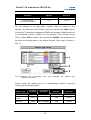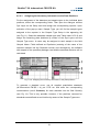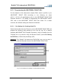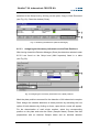
1
RealArt™ M. tuberculosis
TM PCR Kit
Quantitative
in vitro Diagnostics
for use with the ABI PRISM®
Sequence Detection Systems
(Applied Biosystems)
User Manual
February 2004
RealArt™ M. tuberculosis TM PCR Kit
Table of Contents
1.
Contents ......................................................................... 1
2.
Storage ........................................................................... 2
3.
Additionally Required Materials and Devices.............. 2
4.
General Precautions...................................................... 3
5.
Pathogen Information.................................................... 3
6.
Principle of Real-Time PCR........................................... 4
7.
Product Description ...................................................... 4
8.
Protocol .......................................................................... 5
8.1
DNA Isolation ................................................................................ 5
8.2
Internal Control.............................................................................. 6
8.3
Quantitation ................................................................................... 7
8.4
Preparing the PCR ........................................................................ 8
8.5
Programming the ABI PRISM® SDS ........................................... 13
8.5.1
EN040220
8.5.2
Programming the ABI PRISM® 7000 SDS ..................... 13
8.5.1.1
Pre-Settings for Creating a New File ............................................13
8.5.1.2
Creating/Selecting the Detectors ..................................................14
8.5.1.3
Assigning the Necessary Information to the Plate Positions.........16
8.5.1.4
Creating the Temperature Profile .................................................17
8.5.1.5
Saving the PCR Run ....................................................................18
8.5.1.6
Starting the PCR Run ...................................................................18
Programming the ABI PRISM® 7700 SDS ..................... 19
8.5.2.1
Pre-settings for Creating a New File.............................................19
8.5.2.2
Selecting the Fluorescence Dyes and Assigning the Sample
Type .............................................................................................19
8.5.2.3
Assigning the Necessary Information to the Plate Positions.........21
8.5.2.4
Creating the Temperature Profile .................................................22
8.5.2.5
Important Additional Settings........................................................23
RealArt™ M. tuberculosis TM PCR Kit
8.5.3
9.
8.5.2.6
Saving the PCR Run ....................................................................24
8.5.2.7
Starting the PCR Run ...................................................................24
Programming the ABI PRISM® 7900HT SDS ................ 25
8.5.3.1
Pre-Settings for Creating a New File ............................................25
8.5.3.2
Creating/Selecting the Detectors ..................................................26
8.5.3.3
Assigning the Necessary Information to the Plate Positions.........27
8.5.3.4
Creating the Temperature Profile .................................................28
8.5.3.5
Saving the PCR Run ....................................................................30
8.5.3.6
Starting the PCR Run ...................................................................30
Data Analysis ............................................................... 31
10. Troubleshooting .......................................................... 36
11. Specifications .............................................................. 37
11.1 Sensitivity .................................................................................... 37
11.2 Specificity .................................................................................... 38
11.3 Precision...................................................................................... 41
11.4 Robustness ................................................................................. 43
11.5 Reproducibility............................................................................. 43
11.6 Diagnostic Evaluation.................................................................. 43
12. Product Use Limitations.............................................. 43
13. Explanation of Symbols .............................................. 44
RealArt™ M. tuberculosis TM PCR Kit
RealArt™ M. tuberculosis TM PCR Kit
for use with the ABI PRISM® 7000, 7700 and 7900HT Sequence Detection
Systems (Applied Biosystems)♦.
Attention: The RealArt™ M. tuberculosis TM PCR Kit may neither be used in
combination with the GeneAmp® 5700 SDS nor with the 384 plate format of
the ABI PRISM® 7900HT SDS.
1. Contents
Labelling
and contents
Art. No. 5511-01
24 reactions
Art. No. 5511-02
48 reactions
Art. No. 5511-03
96 reactions
Blue
M. tuberculosis
TM Master
2 x 12 rxns
4 x 12 rxns
8 x 12 rxns
Yellow
M. tuberculosis
TM Mg-Sol
1 x 400 µl
1 x 400 µl
1 x 400 µl
Red
M. tuberculosis
TM QS 1¤
3 x 10⁴ cop/µl
1 x 200 µl
1 x 200 µl
1 x 200 µl
Red
M. tuberculosis
TM QS 2¤
3 x 10³ cop/µl
1 x 200 µl
1 x 200 µl
1 x 200 µl
Red
M. tuberculosis
TM QS 3¤
3 x 10² cop/µl
1 x 200 µl
1 x 200 µl
1 x 200 µl
Red
M. tuberculosis
¤
TM QS 4
1
3 x 10 cop/µl
1 x 200 µl
1 x 200 µl
1 x 200 µl
Green
M. tuberculosis
TM IC¤
1 x 1000 µl
1 x 1000 µl
1 x 1000 µl
White
Water
(PCR grade)
1 x 1000 µl
1 x 1000 µl
1 x 1000 µl
¤
♦
QS
IC
=
=
Quantitation Standard
Internal Control
Applied Biosystems is a registered trademark and ABI PRISM® is a trademark of
Applera Corporation or its subsidiaries in the US and/or certain other countries.
1
RealArt™ M. tuberculosis TM PCR Kit
2. Storage
The components of the RealArt™ M. tuberculosis TM PCR Kit should be
stored at -20°C and are stable until the expiry date stated on the label.
Repeated thawing and freezing (> 2 x) should be avoided, as this may reduce
the sensitivity. If the reagents are to be used only intermittently, they should
be frozen in aliquots. Storage at +4°C should not exceed a period of five
hours.
3. Additionally Required Materials and Devices
Disposable powder-free gloves
DNA isolation kit (see 8.1 DNA Isolation)
Lysozyme mix (see 8.1 DNA Isolation)
Pipettes (adjustable)
Sterile pipette tips with aerosol barrier
Vortex mixer
Desktop centrifuge with rotor for 2 ml reaction tubes
Centrifuge with rotor for microtitre plates (optional)
96-well reaction plate/reaction tubes for optical measurement with
corresponding optical closing materials♦ (see 8.4 Preparing the
PCR)
96-well two-part retaining rack for use with optical reaction tubes
(96-Well Tray/Retainer Set, Cat. No. 403 081, Applied Biosystems),
see 8.4 Preparing the PCR
Compression pad for use with optical adhesive covers (Optical Cover
Compression Pads, Cat. No. 4 312 639, Applied Biosystems), see
8.4 Preparing the PCR
Applicator for sealing of the reaction plates using optical adhesive
covers (Adhesive Seal Applicator Kit, Cat. No. 4 333 183, Applied
Biosystems)
♦
The use of reaction tubes for optical analyses with domed caps is only permitted with the
ABI PRISM® 7700 SDS and requires an adjustment of the exposure time (see
8.5.2 Programming the ABI PRISM® 7700 SDS, 8.5.2.5 Important Additional Settings).
2
RealArt™ M. tuberculosis TM PCR Kit
ABI PRISM® 7000, 7700 or 7900HT SDS (Applied Biosystems)
Attention: A valid calibration of the pure dyes (Pure Spectra Component File)
and the background (Background Component File) is necessary when putting
the instruments into operation.
4. General Precautions
The user should always pay attention to the following:
Use pipette tips with filters.
Extract and store positive material (specimens, controls and
amplicons) separately from all other reagents and add it to the
reaction mix in a spatially separated facility.
Thaw all components thoroughly at room temperature before starting
an assay.
When thawed, mix the components and centrifuge briefly.
Work quickly on ice or in the cooling block.
5. Pathogen Information
Mycobacteria are distributed worldwide. It is estimated that one third of the
world’s population is infected with Mycobacterium tuberculosis, the pathogen
of tuberculosis. About eight million new infections and about three million
deaths are reported each year. Countries in the third world are largely
affected, but due to resistance development and the importation of infections,
tuberculosis incidents occur increasingly often in industrialized nations. The
homeless, drug addicted and immunocompromised persons are particularly at
risk of infection. Transmission of the bacteria occurs via aerosols, with an
increased risk of infection in cases of active tuberculosis. Tuberculosis is a
chronic, cyclic disease. In the primary stage, mainly parts of the lungs and the
associated lymph nodes are affected. Depending on the immune status of the
patient, however, mycobacteria can colonize in the entire lung and other
organs. In cases of immunosuppression the infection may spread from these
sites and, even years later, can be reactivated (recrudescent). Signs of such a
3
RealArt™ M. tuberculosis TM PCR Kit
reactivation in the lungs include chronic fever, weight loss, night sweats and
coughing. Other symptoms depend on the affected organs.
6. Principle of Real-Time PCR
Pathogen diagnosis by the polymerase chain reaction (PCR) is based on the
amplification of specific regions of the pathogen genome. In real-time PCR,
the amplified product is detected via fluorescent dyes. These are usually
linked to oligonucleotide probes which bind specifically to the amplified
product. Monitoring the fluorescence intensities during the PCR run (i. e. in
real-time) allows the detection and quantitation of the accumulating product
without having to re-open the reaction tubes after the PCR run.
7. Product Description
The RealArt™ M. tuberculosis TM PCR Kit constitutes a ready-to-use system
for the detection of DNA of all members of the M. tuberculosis complex
(M. tuberculosis, M africanum I + II, M bovis, M bovis BCG, M. microti,
M. canettii) using polymerase chain reaction (PCR) in the ABI PRISM® 7000,
7700 and 7900HT Sequence Detection System (Applied Biosystems). The
M. tuberculosis TM Master contains reagents and enzymes for the specific
amplification of a 163 bp region of the mycobacterial genome. The amplicon is
detected by measuring the FAM fluorescence in the ABI PRISM® SDS. In
addition,
the
RealArt™ M. tuberculosis TM PCR Kit
contains
a
second
heterologous amplification system to identify possible PCR inhibition. This is
detected as an Internal Control (IC) by measuring the JOE fluorescence. The
detection limit
of
11.1 Sensitivity)
the
is
analytical
not
M. tuberculosis
reduced.
External
complex
positive
PCR
(see
controls
(M. tuberculosis TM QS 1 - 4) are supplied which allow the determination of
the pathogen load. For further information, please refer to section
8.3 Quantitation.
4
RealArt™ M. tuberculosis TM PCR Kit
8. Protocol
8.1 DNA Isolation
Before the DNA isolation, large sample volumes or strongly acidic samples
must first be concentrated or neutralized, respectively. For the analysis of
sputum, we recommend a NALC-NaOH decontamination; stomach fluids
should be neutralized with phosphate buffer. After a final centrifugation, the
bacteria pellet can be used for the following DNA isolation (for more details
contact our technical support at [email protected]).
Various manufacturers offer DNA isolation kits. Sample volumes for the
DNA isolation procedure depend on the protocol used. Please carry out the
DNA isolation according to the manufacturer’s instructions. The following
isolation kits are recommended:
Sample Material
Sputum, BAL,
bronchial secretion,
CSF, stomach
fluid, peritoneal
punction
Nucleic Acid
Isolation Kit
Catalogue
Number
Manufacturer
Carrier
DNA/RNA
PureArt™ DNA
Mini Kit (50)♦
90204-50
artus
not included
QIAamp DNA
Mini Kit (50)
51 304
QIAGEN
not included
Attention: It is important to follow the instructions in Protocol D (Protocols for
bacteria) described in the QIAGEN Kit User Manual. We recommend a
digestion with lysozyme (lysozyme mix: 20 mg/ml lysozyme, 20 mM Tris-HCl,
2 mM EDTA, 1.2 % Triton®) for at least one hour. The final DNA elution should
be performed once using 100 µl AE buffer.
If the selected isolation kit does not contain carrier DNA/RNA, please
note that the addition of carrier RNA (RNA-Homopolymer Poly(A),
Amersham Biosciences) at a concentration of 10 µg/ml lysis buffer to the
sample/lysis buffer mixture is strongly recommended for extraction of
nucleic acids from cell free body fluids and material low in DNA/RNA
content.
♦
Produced by QIAGEN GmbH, D-40724 Hilden for artus GmbH. Legal manufacturer:
artus GmbH, D-22767 Hamburg.
5
RealArt™ M. tuberculosis TM PCR Kit
When using isolation protocols with ethanol-containing washing buffers,
please carry out an additional centrifugation step before the elution to
remove any remaining ethanol. This prevents possible inhibition of PCR.
The RealArt™ M. tuberculosis TM PCR Kit should not be used with
phenol-based isolation methods.
Important: The Internal Control of the RealArt™ M. tuberculosis TM PCR Kit
can be used directly in the isolation procedure (see 8.2 Internal Control).
8.2 Internal Control
An Internal Control (M. tuberculosis TM IC) is supplied. This allows the user
both to control the isolation procedure and to check for possible
PCR inhibition. For this application, add the Internal Control to the isolation at
a ratio of 0.1 µl per 1 µl elution volume. For example, using the QIAamp DNA
Mini Kit (QIAGEN) the DNA is eluted in 100 µl AE buffer. Hence, 10 µl of the
Internal Control should be added initially. The quantity of IC used depends
only on the elution volume. Please note that the IC should be added to the
mixture of lysis buffer and sample material. Please do not add the IC to the
lysis buffer or sample material directly!
The IC can optionally be used exclusively to check for possible
PCR inhibition. For this application, add 0.5 µl of the IC and 2 µl
M. tuberculosis TM Mg-Sol per test mixture directly to 13 µl M. tuberculosis
TM Master. For each PCR reaction use 15 µl of the Master Mix produced as
described above♦, then add 10 µl of the purified sample. If you are preparing a
PCR run for several samples please increase the volume of the
M. tuberculosis TM Master, the M. tuberculosis TM Mg-Sol and the Internal
Control according to the number of samples (see 8.4 Preparing the PCR).
♦
The volume increase caused by adding the IC is neglected when preparing the PCR
assay. The sensitivity of the detection system is not impaired.
6
RealArt™ M. tuberculosis TM PCR Kit
8.3 Quantitation
The enclosed quantitation standards (M. tuberculosis TM QS 1 - 4) are treated
as previously purified samples and the same volume is used (10 µl). To
generate a standard curve in an ABI PRISM® Sequence Detection System, all
four quantitation standards should be used and defined as standards with
specification of the corresponding concentrations (see 8.5 Programming the
®
ABI PRISM SDS). The import of standard curves from previous runs is not
possible with the ABI PRISM® 7000, 7700 and 7900HT SDS software.
Attention: The quantitation standards are defined as copies/µl. The following
equation has to be applied to convert the values determined using the
standard curve into copies/ml of sample material:
Result (copies / ml)
=
Result (copies / µl) × Elution Volume (µl)
Sample Volume (ml)
If a concentration of the sample material was performed by e. g. centrifugation
prior to nucleic acid extraction, please enter the entire initial sample volume in
the above equation.
7
RealArt™ M. tuberculosis TM PCR Kit
8.4 Preparing the PCR
Prepare the number of required reaction tubes or a 96-well reaction plate for
the scheduled reactions. Recommended materials are listed in the following
table:
Article
Description
Catalogue
Number
Manufacturer
Retaining
Rack♦
Compression
Pad
96-well
optical
reaction
plate
96-Well Optical
Reaction Plate
4 306 737
Applied
Biosystems
no
-
Optical
adhesive
cover
Optical
Adhesive
Covers
4 311 971
Applied
Biosystems
-
yes
Optical
reaction
tubes
ABI PRISM®
Optical Tubes,
8 Tubes/Strip
4 316 567
Applied
Biosystems
yes
-
Optical
reaction
tubes
MicroAmp®
Optical Tubes
N8010933
Applied
Biosystems
yes
-
Optical
cap (flat)
ABI PRISM®
Optical Caps,
8 Caps/Strip
4 323 032
Applied
Biosystems
-
no
Attention: The use of reaction tubes for optical analyses with domed caps is
only permitted with the ABI PRISM® 7700 SDS and requires an adjustment of
the exposure time (see 8.5.2 Programming the ABI PRISM® 7700 SDS,
8.5.2.5 Important Additional Settings).
When preparing the PCR reaction, please make sure that at least one
quantitation standard per PCR run as well as one negative control (Water,
PCR grade) are performed in parallel. To generate a standard curve, use all
supplied quantitation standards (M. tuberculosis TM QS 1 - 4) for each PCR
run. Before each use, all reagents need to be thawed completely, mixed (by
repeated up and down pipetting or by quick vortexing) and centrifuged briefly.
♦
If the two-part retaining rack is used, it is necessary to open the reaction tubes when
inserting them into and removing them from the rack. In order to avoid contamination
due to this procedure, please use the lower part of the retaining rack only.
8
RealArt™ M. tuberculosis TM PCR Kit
If you want to use the Internal Control to monitor the isolation procedure
and to check for possible PCR inhibition, the IC has already been added to
the isolation (see 8.2 Internal Control). In this case, please use the following
pipetting scheme (for a schematic overview see Fig. 1):
1. Preparation of
Master Mix
2. Preparation of
PCR assay
Number of samples
1
12
M. tuberculosis TM Master
13 µl
156 µl
M. tuberculosis TM Mg-Sol
2 µl
24 µl
M. tuberculosis TM IC
0 µl
0 µl
Total volume
15 µl
180 µl
Master Mix
15 µl
15 µl each
Sample
10 µl
10 µl each
Total volume
25 µl
25 µl each
If you want to use the IC exclusively to check for PCR inhibition, the IC
must then be added directly to the M. tuberculosis TM Master. In this case,
please use the following pipetting scheme (for a schematic overview see
Fig. 2):
1. Preparation of
Master Mix
Number of samples
1
M. tuberculosis TM Master
13 µl
156 µl
M. tuberculosis TM Mg-Sol
2 µl
24 µl
0.5 µl
6 µl
M. tuberculosis TM IC
Total volume
2. Preparation of
PCR assay
15.5 µl
12
♦
186 µl♦
Master Mix
15 µl♦
15 µl each♦
Sample
10 µl
10 µl each
Total volume
25 µl
25 µl each
Pipet 15 µl of the Master Mix into each required reaction tube or well of the 96well reaction plate. Subsequently add 10 µl of the eluate from the
DNA isolation. Mix the solution well by repeated up and down pipetting. Close
the reaction tubes with the corresponding caps or, alternatively, when using a
96-well reaction plate, with an optical adhesive cover (optical adhesive covers,
Applied Biosystems). Centrifuge the reaction tubes (in a storage rack for PCR
♦
The volume increase caused by adding the IC is neglected when preparing the PCR
assay. The sensitivity of the detection system is not impaired.
9
RealArt™ M. tuberculosis TM PCR Kit
tubes) or the 96-well reaction plate in a centrifuge with a microtitre plate rotor
for 30 seconds at 1780 x g (4000 rpm) in order to collect the prepared reaction
volume in the bottom of the tubes or wells. If such a centrifuge is not at your
disposal, please make sure that both the Master Mix and the sample volume
are pipetted to the bottom of the tubes or wells. Store the prepared reactions
at +4°C until the ABI PRISM® SDS Instrument is programmed (see
8.5 Programming the ABI PRISM® SDS) and subsequently transfer them into
the instrument.
Attention:
When using optical reaction tubes in combination with optical caps,
always insert a retaining rack (96-Well Tray/Retainer Set, Applied
Biosystems) into the instrument (ABI PRISM® 7000, 7700 and 7900HT
SDS). If the two-part retaining rack is used, it is necessary to open the
reaction tubes when inserting them into and removing them from the rack.
In order to avoid contamination due to this procedure, please use the
lower part of the retaining rack only.
Using 96-well optical reaction plates in combination with optical adhesive
covers requires covering by a compression pad (Optical Cover
Compression Pads, Applied Biosystems).
10
RealArt™ M. tuberculosis TM PCR Kit
Addition of the Internal Control to the Purification Procedure
Purification
+ 0.1 µl IC per 1 µl
Elution Volume
2 µl
Mg-Sol*
10 µl Purified Sample*
13 µl
RealArtTM
Master*
15 µl Master Mix*
Optical Reaction
Plate / Tube
ABI PRISM® SDS
Fig. 1:
Schematic workflow for the control of both the purification
procedure and PCR inhibition.
*
Please make sure that the solutions are thawed completely,
mixed well and centrifuged briefly.
11
RealArt™ M. tuberculosis TM PCR Kit
Addition of the Internal Control into the RealArt™ Master
2 µl
Mg-Sol*
Purification
0.5 µl IC*
13 µl
RealArtTM
Master*
15.5 µl Master Mix*
10 µl Purified Sample*
15 µl Master Mix*
Optical Reaction
Plate / Tube
ABI PRISM® SDS
Fig. 2:
Schematic workflow for the control of PCR inhibition.
*
Please make sure that the solutions are thawed completely,
mixed well and centrifuged briefly.
12
RealArt™ M. tuberculosis TM PCR Kit
8.5 Programming the ABI PRISM® SDS
The software of the ABI PRISM® 7000, 7700 and 7900HT Sequence Detection
Systems (SDS) requires some additional information before starting the PCR
run. The programming procedures of the instruments, however, differ
considerably from each other, which is why they are treated in separate
chapters as follows.
®
8.5.1 Programming the ABI PRISM 7000 SDS
For the detection of the M. tuberculosis complex DNA, create a profile on your
®
ABI PRISM 7000 SDS according to the following six steps (8.5.1.1 - 8.5.1.6).
All specifications refer to the ABI PRISM® 7000 SDS Software Version 1.0.1.
For programming details of the ABI PRISM® 7000 SDS please refer to the
ABI PRISM® 7000 SDS User Guide. For a better overview, the software
settings are framed in bold black.
8.5.1.1
Pre-Settings for Creating a New File
Select the item New from the File menu on the ABI PRISM® 7000 SDS and
program the following initial settings for the new file (Fig. 3). A backup
template (SDS Template [*.sdt]) is available from the Template list or by
selection using the Browse function (see 8.5.1.5 Saving the PCR Run).
Confirm your settings (OK).
13
RealArt™ M. tuberculosis TM PCR Kit
Fig. 3: Pre-settings for creating a new file (New Document).
8.5.1.2
Creating/Selecting the Detectors
With the help of the submenu Detector Manager from the Tools menu, assign
the corresponding dyes to the file. For the detection of M. tuberculosis
complex DNA as well as the Internal Control by means of the
RealArt™ M. tuberculosis TM PCR Kit, the reporters/quenchers listed in the
following table are to be defined:
Detection
Reporter
Quencher
M. tuberculosis complex DNA
FAM
none
Internal Control
(M. tuberculosis TM IC)
JOE
TAMRA
To create these detectors, select the option File (bottom left of the Detector
Manager) and subsequently the option New.
14
RealArt™ M. tuberculosis TM PCR Kit
Fig. 4: Creating
the
M. tuberculosis
complex
specific
detector
(Detector Manager).
For
the
detection
of
Fig. 5: Creating the IC specific detector
(Detector Manager).
M. tuberculosis
complex
DNA
define
the
reporter/quencher combination FAM/none in the now appearing window. For
the detection of the Internal Control, select the combination JOE/TAMRA (as
shown in Fig. 4 and Fig. 5). By confirming the input data (OK), return to the
Detector Manager. Mark the newly created detectors and transfer each
selection to the Well Inspector by clicking on the option Add to Plate
Document (see Fig. 6). Close the window (Done).
Fig. 6: Selecting the detectors (Detector Manager).
15
RealArt™ M. tuberculosis TM PCR Kit
8.5.1.3
Assigning the Necessary Information to the Plate Positions
Open Well Inspector from the View menu to find the detectors selected under
8.5.1.2 (see Fig. 7).
Fig. 7: Assigning the necessary information to the plate positions (Well Inspector).
Mark the plate positions reserved for the detection of M. tuberculosis complex
DNA. Assign the selected detectors to these positions by activating the Use
option of both detectors, upon which a tick will appear. For the denomination
of each single reaction, select the corresponding position on the plate and
enter the name (Sample Name). Please note that reactions with an identical
Sample Name and an identical detector assignment will be identified as
replicates by the software and will be averaged with respect to the quantified
pathogen load. Then select the corresponding function (Task) for each sample
type according to the following table:
Sample Type
Function
(Task)
Concentration
(Quantity)
Reporter
Quencher
Sample
Unknown
-
FAM
none
NTC
-
FAM
none
Standard
see 1. Contents
FAM
none
Non-template
control
Standard
To generate a standard curve, use all supplied quantitation standards
(M. tuberculosis TM QS 1 - 4) per PCR run and enter the corresponding
concentrations (see 1. Contents) for each standard (Quantity). Please note
16
RealArt™ M. tuberculosis TM PCR Kit
that for a PCR run with the RealArt™ M. tuberculosis TM PCR Kit, ROX has to
be set as a passive reference (Passive). The equal distribution of the ROX
dye in all PCR preparations of a lot by mixing of the M. tuberculosis TM
Master guarantees the recognition and calculation of tube-to-tube variations
(fluorescence differences between several PCR preparations) by means of the
Sequence Detection Software (normalization).
8.5.1.4
Creating the Temperature Profile
To create a temperature profile, switch from the Setup level to the Instrument
level in the software. Enter the temperature profile specific for the detection of
M. tuberculosis complex DNA according to Fig. 8. In order to remove the 50°C
step stored in the pre-settings, mark it with the left mouse button
simultaneously holding the shift key, and subsequently delete it by using the
backspace key. Make sure that the reaction volume for the detection of
M. tuberculosis complex DNA using the RealArt™ M. tuberculosis TM PCR Kit
is set to 25 µl. The option 9600 Emulation should be activated, the presettings of the Auto Increment should remain unchanged (Auto Increment:
0.0°C, 0.0 Seconds).
Fig. 8: Creating the temperature profile.
17
RealArt™ M. tuberculosis TM PCR Kit
8.5.1.5
Saving the PCR Run
Save the settings (Setup) as a template in order to make use of them again
later in a modified or unchanged form. By saving the Template file as SDS
Template (*.sdt) in the Template Directory (Local Disk [C:]\Program Files\
ABI PRISM 7000\Templates, created by Applied Biosystems) this file may be
selected directly in the New Document window from the Template drop-down
list. Copies stored in other folders have to be opened via Browse. Before
starting the PCR run, save it again as an SDS Document (*.sds), in order to
guarantee the saving of the data that will be collected during the course of the
PCR.
8.5.1.6
Starting the PCR Run
Start the PCR run by selecting the option Start from the menu item Instrument
or the field Start on the Instrument level.
18
RealArt™ M. tuberculosis TM PCR Kit
®
8.5.2 Programming the ABI PRISM 7700 SDS
For the detection of M. tuberculosis complex DNA, create a profile on your
ABI PRISM®
7700
SDS
according
to
the
following
seven
(8.5.2.1 - 8.5.2.7). All specifications refer to the ABI PRISM
®
steps
7700 SDS
Software Version 1.9.1. For details of programming the ABI PRISM® 7700
SDS, refer to the ABI PRISM® 7700 SDS User`s Manual. For a better
overview, the software settings are framed in bold black.
8.5.2.1
Pre-settings for Creating a New File
Select the item New Plate from the File menu on the ABI PRISM® 7700 SDS
and program the following initial settings for the new file (Fig. 9). Confirm the
pre-settings (OK).
Fig. 9: Pre-settings for creating a new file (New Plate).
8.5.2.2
Selecting the Fluorescence Dyes and Assigning the Sample
Type
With the help of the Sample Type Setup (Setup level: Sample Type/Sample
Type Setup) assign the corresponding detector dyes and the corresponding
sample type to the file. For the detection of M. tuberculosis complex DNA as
well as the Internal Control by means of the RealArt™ M. tuberculosis TM
PCR Kit, the reporters/quenchers listed in the following table are to be
defined:
19
RealArt™ M. tuberculosis TM PCR Kit
Detection
Reporter
Quencher
M. tuberculosis complex DNA
FAM
TAMRA
Internal Control
(M. tuberculosis TM IC)
JOE
TAMRA
For the analysis of M. tuberculosis complex DNA by means of the
RealArt™ M. tuberculosis TM PCR Kit, select the reporter dye FAM as given
in the table. This applies to standards (STND) and samples (UNKN) as well as
to non-template controls (UNKN). For the analysis of the Internal Control
(IPC+), define JOE as reporter. As quencher set TAMRA. The assignment of
the dyes and sample types in the window Sample Type Setup is shown in
Fig. 10.
Fig. 10: Selecting the fluorescence
(Sample Type Setup).
dyes
and
assigning
the
sample
type
Please assign the sample type to a corresponding function (Acronym)
according to the following table:
Function
(Acronym)
Concentration
(Quantity)
Reporter
Quencher
Sample
UNKN
-
FAM
TAMRA
Non-template
control
UNKN
-
FAM
TAMRA
Standard
STND
see 1. Contents
FAM
TAMRA
Sample Type
20
RealArt™ M. tuberculosis TM PCR Kit
8.5.2.3
Assigning the Necessary Information to the Plate Positions
For the assignment of the detectors and sample types to the individual plate
positions, select the corresponding fields. Then open the dialogue window
Dye Layer on the Setup level and assign the corresponding reporter. Upon
activation of the pop-up menu Sample Type, you will find the sample types
assigned to the reporter in the Sample Type Setup in the appearing list
(see Fig. 11). Select the adequate sample type (see Table under 8.5.2.2) and
assign the remaining plate positions by means of the Dye Layers and the
Sample Type menu. A name may be assigned to each sample in the field
Sample Name. Fields defined as Replicates (entering of the name of the
reference sample into the Replicate column) are averaged by the software
with respect to the quantified pathogen load and the standard deviation will be
calculated.
Fig. 11/12: Assigning the necessary information to the plate positions.
To generate a standard curve, use all supplied quantitation standards
(M. tuberculosis TM QS 1 - 4) per PCR run and enter the corresponding
concentrations (see 1. Contents) for each standard into the field Quantity
(see Fig. 12). This is only possible, however, if the positions reserved for
standards were defined as such before by means of the Sample Type menu.
21
RealArt™ M. tuberculosis TM PCR Kit
8.5.2.4
Creating the Temperature Profile
To create a temperature profile, switch to the Thermal Cycler Conditions menu
on the Setup level. Enter the temperature profile special for the detection of
M. tuberculosis complex DNA according to Fig. 13. Make sure that the
reaction volume for the detection of M. tuberculosis complex DNA by using the
RealArt™ M. tuberculosis TM PCR Kit is set to 25 µl. The pre-settings of the
Ramp times and of the Auto Increment remain unchanged (Ramp Time: 0:00,
Auto Increment: 0.0°C, 0.0 Seconds).
Fig. 13: Creating the temperature profile.
In addition, the Thermal Cycler Conditions menu contains the option Show
Data Collection. By selecting this option the window depicted in Fig. 14 is
opened. Each ramp and each plateau temperature shows a Data Collection
icon, which illustrates the data being collected at this stage of the run.
Remove all symbols except the one for the Annealing-Extension step
(Stage2/Step2) in order to exclude unnecessary fluorescence measurements.
The total run time and the data quantity will thus be kept at a minimum.
22
RealArt™ M. tuberculosis TM PCR Kit
Fig. 14: Data collection.
8.5.2.5
Important Additional Settings
For the setting of the exposure time (excitation of the fluorescence dyes) as
well as for the selection of the Pure Spectra/Background files, switch from the
Setup level to the Analysis level. Select the activated sub-item Advanced
Options to be found in the Instrument menu under Diagnostics. Adjust the
settings according to Fig. 15. By inactivating the optional function Spectra
Components (Analysis) the actual calibration files stored in the file Spectra
Components at the moment of data generation are automatically used when
reevaluating already analysed runs. For an analysis of previous runs by using
newly entered Spectra Components, activate these two fields. Please note
that for a PCR run with the RealArt™ M. tuberculosis TM PCR Kit, ROX has to
be set as a passive reference (Reference). The equal distribution of the ROX
dye in all PCR preparations of a lot by mixing of the M. tuberculosis TM
Master guarantees the recognition and calculation of tube-to-tube variations
(fluorescence differences between several PCR preparations) by means of the
Sequence Detection Software (normalization).
Attention: When using 96-well reaction plates for optical measurements in
combination with optical adhesive covers or optical reaction tubes with flat
23
RealArt™ M. tuberculosis TM PCR Kit
caps, the exposure time is ten milliseconds. If you use optical reaction tubes
with domed caps, you have to adjust this exposure time to 25 milliseconds.
Fig. 15: Important additional settings (Advanced Options).
8.5.2.6
Saving the PCR Run
Save the settings (Setup) as a template in order to make use of them again
later in a modified or unchanged form. For this purpose store the file in the
Stationary File Format. Before starting a newly programmed PCR run, please
save it again in the Normal File Format in order to guarantee the saving of
data that will be collected during the course of the PCR.
8.5.2.7
Starting the PCR Run
Start the PCR run by selecting the option Run from the menu item Instrument
or the field Run on the Analysis level.
24
RealArt™ M. tuberculosis TM PCR Kit
®
8.5.3 Programming the ABI PRISM 7900HT SDS
For the detection of M. tuberculosis complex DNA, create a profile on your
ABI PRISM®
7900HT
SDS
according
to
the
following
(8.5.3.1 - 8.5.3.6). All specifications refer to the ABI PRISM
®
six
steps
7900HT SDS
Software Version 2.1. For details of programming the ABI PRISM® 7900HT
SDS, refer to the ABI PRISM® 7900HT SDS User Guide. For a better
overview, the software settings are framed in bold black.
8.5.3.1
Pre-Settings for Creating a New File
Select the item New from the File menu on the ABI PRISM® 7900HT SDS and
program the following initial settings for the new file (Fig. 16). A backup
template (ABI PRISM® SDS Template Document [*.sdt]) is available from the
Template list or by selection using the Browse function (see 8.5.3.5 Saving
the PCR Run). Confirm the pre-settings (OK).
Attention: The RealArt™ M. tuberculosis TM PCR Kit may not be used in
combination with the 384 plate format of the ABI PRISM® 7900HT SDS.
Fig. 16: Pre-settings for creating a new file (New Document).
25
RealArt™ M. tuberculosis TM PCR Kit
8.5.3.2
Creating/Selecting the Detectors
With the help of the submenu Detector Manager from the Tools menu
(alternatively:
Setup
level/Add
Detector
select
function),
assign
the
corresponding detector dyes to the file. For the detection of M. tuberculosis
complex DNA as well as the Internal Control by means of the
RealArt™ M. tuberculosis TM PCR Kit, the reporters/quenchers listed in the
following table are to be defined:
Detection
Reporter
Quencher
M. tuberculosis complex DNA
FAM
Non Fluorescent
Internal Control
(M. tuberculosis TM IC)
JOE
TAMRA
To create these detectors, select the option New (bottom left of the Detector
Manager).
Fig. 17: Creating
the
M. tuberculosis
complex
specific
detector
(Detector Manager).
Fig. 18: Creating the IC specific detector
(Detector Manager).
For the detection of M. tuberculosis complex DNA, define the reporter/
quencher combination FAM/Non Fluorescent in the now appearing window.
For the detection of the Internal Control, select the combination JOE/TAMRA
(as shown in Fig. 17 and Fig. 18). By confirming the input data (OK), return to
the Detector Manager. Mark the newly created detectors and transfer each
26
RealArt™ M. tuberculosis TM PCR Kit
selection to the Setup level by clicking on the option Copy to Plate Document
(see Fig. 19). Close the window (Done).
Fig. 19: Selecting the detectors (Detector Manager).
8.5.3.3
Assigning the Necessary Information to the Plate Positions
After having closed the Detector Manager (Done) the detectors selected under
8.5.3.2 are found on the Setup level (Well Inspector) listed in a table
(see Fig. 20).
Fig. 20: Assigning the necessary information to the plate positions.
Mark the plate positions reserved for the detection of M. tuberculosis complex
DNA. Assign the selected detectors to these positions by activating the Use
option of both detectors by clicking on them, upon which a cross will appear.
For the denomination of each single reaction, select the corresponding
position on the plate and enter the name (Sample Name). Please note that
preparations with an identical Sample Name and an identical detector
27
RealArt™ M. tuberculosis TM PCR Kit
assignment will be identified as replicates by the software and will be
averaged with respect to the quantified pathogen load. Then select the
corresponding function (Task) for each sample type according to the following
table:
Sample Type
Function
(Task)
Concentration
(Quantity)
Reporter
Quencher
Sample
Unknown
-
FAM
Non Fluorescent
NTC
-
FAM
Non Fluorescent
Standard
see 1. Contents
FAM
Non Fluorescent
Non-template
control
Standard
To generate a standard curve, use all supplied quantitation standards
(M. tuberculosis TM QS 1 - 4) per PCR run and enter the corresponding
concentrations (see 1. Contents) for each standard (Quantity). Please note
that for a PCR run with the RealArt™ M. tuberculosis TM PCR Kit, ROX has to
be set as a passive reference (Passive Reference). The equal distribution of
the ROX dye in all PCR preparations of a lot by mixing of the
M. tuberculosis TM Master guarantees the recognition and calculation of tube-
to-tube
variations
(fluorescence
differences
between
several
PCR
preparations) by means of the Sequence Detection Software (normalization).
8.5.3.4
Creating the Temperature Profile
To create a temperature profile, switch from the Setup level to the Instrument
level in the software. Enter the temperature profile specific for the detection of
M. tuberculosis complex DNA according to Fig. 21. Make sure that the
reaction volume for the detection of M. tuberculosis complex DNA by using the
RealArt™ M. tuberculosis TM PCR Kit is set to 25 µl. The option 9600
Emulation should be activated, the pre-settings of the Ramp time and the Auto
Increment should remain unchanged (Ramp Time: 0:00, Auto Increment:
0.0°C, 0.0 Seconds).
28
RealArt™ M. tuberculosis TM PCR Kit
Fig. 21: Creating the temperature profile.
In addition, the Instrument level contains the option Data Collection. By
selecting this option the window depicted in Fig. 22 is opened. Each ramp and
each plateau temperature shows a Data Collection Icon, which illustrates the
data being collected at this stage of the run. Remove all symbols except the
one for the Annealing-Extension step (Stage2/Step2) by clicking on them in
order to exclude unnecessary fluorescence measurements. The total run time
and the data quantity will thus be kept at a minimum.
29
RealArt™ M. tuberculosis TM PCR Kit
Fig. 22: Data collection.
8.5.3.5
Saving the PCR Run
Save the settings (Setup) as a template in order to make use of them again
later in a modified or unchanged form. By saving the Template file as an
®
ABI PRISM SDS Template Document (*.sdt) in the Template Directory
([D:]\Program Files\
Applied Biosystems\SDS 2.1\Templates,
created
by
Applied Biosystems) this file may be selected directly in the New Document
window from the Template list. Copies stored in other folders have to be
opened via Browse. Before starting the PCR run, save it again as an
ABI PRISM® SDS Document (*.sds) in order to guarantee the saving of the
data that will be collected during the course of the PCR.
8.5.3.6
Starting the PCR Run
Start the PCR run by selecting the option Start from the menu item Instrument
or the field Start on the Analysis level.
30
RealArt™ M. tuberculosis TM PCR Kit
9. Data Analysis
A valid calibration of the dyes (Pure Spectra Component File) and the
background (Background Component File) is necessary when putting the
instruments into operation. These calibration files are required for the exact
calculation of the results as follows:
All interfering signals generated by the instruments which influence the
measurement are eliminated by the Sequence Detection Software of the
ABI PRISM® Sequence Detection Systems by means of the Background
Component File.
Furthermore, interferences appear during multicolour analyses between the
emission spectra of the single fluorescence dyes. The software of the
ABI PRISM® SDS compensates these interferences by calculations using the
spectrum data of the individual dyes stored in the Pure Spectra Component
File. The software uses the same file for the assignment of the fluorescence
data comprising the entire measurable spectrum collected in the course of the
PCR to the programmed detectors. The fluorescence data of the individual
dyes are subsequently divided by the value obtained for the signal of the
passive reference (ROX) in order to account for tube-to-tube variations
(fluorescence differences between several PCR preparations). The thus
normalized signals may then be evaluated by means of the Amplification Plot.
The calibration files used for the evaluation of a PCR run are automatically
stored with the saving of the run. If no calibration files are installed, please
create these files according to the instructions given in the ABI PRISM® SDS
User Guide/Manual.
If more than one RealArt™ TM PCR system is integrated in your PCR run
(note temperature profile!), these assays should be analysed separately.
Samples with an identical Sample Name and an identical detector assignment
will automatically be identified as replicates by the ABI PRISM® 7000 and
7900HT SDS Software and will be averaged with respect to the quantified
pathogen load.
31
RealArt™ M. tuberculosis TM PCR Kit
The following results are possible:
1. A FAM fluorescence signal is detected.
The result of the analysis is positive: The sample contains DNA of
one or more members of the M. tuberculosis complex.
In this case, the detection of a JOE fluorescence signal (Internal Control) is
dispensable, since high initial concentrations of M. tuberculosis complex DNA
(positive FAM fluorescence signal) can lead to a reduced or absent fluorescence
signal of the Internal Control (competition).
2. No FAM fluorescence signal is detected. At the same time, a JOE
fluorescence signal from the Internal Control appears.
In the sample no DNA of members of the M. tuberculosis complex is
detectable. It can be considered negative.
In the case of a negative M. tuberculosis complex PCR the detected signal of the IC
rules out the possibility of PCR inhibition.
3. Neither a FAM fluorescence signal nor a JOE fluorescence signal is
detected.
No diagnosis can be concluded.
Information regarding error sources and their solution can be found in
10. Troubleshooting.
Examples of positive and negative PCR reactions are given in Fig. 23 / 24
(ABI PRISM® 7000 SDS), 25 / 26 (ABI PRISM® 7700 SDS) and 27 / 28
(ABI PRISM® 7900HT SDS).
32
RealArt™ M. tuberculosis TM PCR Kit
Fig. 23: Detection of the quantitation standards (M. tuberculosis TM
QS 1 - 4) by measurement of a FAM fluorescence signal
(ABI PRISM® 7000 SDS). NTC: non-template control.
Fig. 24: Detection of the Internal Control (IC) by measurement of a
JOE fluorescence signal (ABI PRISM® 7000 SDS) in parallel
with a simultaneous amplification of the quantitation standards
(M. tuberculosis TM QS 1 - 4). NTC: non-template control.
33
RealArt™ M. tuberculosis TM PCR Kit
Fig. 25: Detection of the quantitation standards (M. tuberculosis TM
QS 1 - 4) by measurement of a FAM fluorescence signal
(ABI PRISM® 7700 SDS). NTC: non-template control.
Fig. 26: Detection of the Internal Control (IC) by measurement of a
JOE fluorescence signal (ABI PRISM® 7700 SDS) in parallel
with a simultaneous amplification of the quantitation standards
(M. tuberculosis TM QS 1 - 4). NTC: non-template control.
34
RealArt™ M. tuberculosis TM PCR Kit
Fig. 27: Detection of the quantitation standards (M. tuberculosis TM
QS 1 - 4) by measurement of a FAM fluorescence signal
(ABI PRISM® 7900HT SDS). NTC: non-template control.
Fig. 28: Detection of the Internal Control (IC) by measurement of a
JOE fluorescence signal (ABI PRISM® 7900HT SDS) in
parallel with a simultaneous amplification of the quantitation
standards (M. tuberculosis TM QS 1 - 4). NTC: non-template
control.
35
RealArt™ M. tuberculosis TM PCR Kit
10. Troubleshooting
No FAM fluorescence signal with positive controls (M. tuberculosis TM
QS 1 - 4):
Incorrect programming of the ABI PRISM® Sequence Detection Systems.
Repeat the PCR with corrected settings.
Weak or no signal of the Internal Control (JOE fluorescence signal) and
simultaneous absence of a FAM fluorescence signal for the specific
M. tuberculosis complex PCR:
The PCR conditions do not comply with the protocol.
Repeat the PCR with corrected settings.
The settings used for the data analysis under Options (Extension Phase
Data Extraction) do not correspond to the settings of the Data Collection
(see ABI PRISM® 7700 SDS, 8.5.2.4 Creating the Temperature Profile).
Analyse the PCR run with corrected settings and repeat the data
analysis (Analysis).
The M. tuberculosis TM Master and/or the M. tuberculosis TM IC have
been thawed and frozen too often.
Please mind the storage conditions given in 2. Storage. Repeat the
PCR using a new M. tuberculosis TM Master.
The M. tuberculosis TM Master has been kept at +4°C for longer than five
hours.
Please mind the storage conditions given in 2. Storage. Repeat the
PCR using a new M. tuberculosis TM Master.
The PCR was inhibited.
Make sure that you use a recommended isolation method
(see 8.1 DNA Isolation) and stick closely to the manufacturer’s
instructions.
DNA was lost during extraction.
Make sure that you use a recommended isolation method
(see 8.1 DNA Isolation) and stick closely to the manufacturer’s
instructions.
36
RealArt™ M. tuberculosis TM PCR Kit
If you have any further questions or if you encounter problems, please contact
our technical support at [email protected].
11. Specifications
11.1 Sensitivity
In order to determine the sensitivity of the RealArt™ M. tuberculosis TM
PCR Kit, a dilution series has been set up from 10 to nominal
0.05 M. tuberculosis copy equivalents♦/µl. It was subsequently analysed by
means of the ABI PRISM® 7000, 7700 and 7900HT Sequence Detection
System with the help of the RealArt™ M. tuberculosis TM PCR Kit. Testing for
all instruments was carried out on three different days on eight replicates. The
results were determined by a probit analysis. A graphical illustration of the
probit analysis (ABI PRISM® 7000 SDS) is shown in Fig. 29.
Detection limit (p = 0,05)
ABI PRISM® 7000 SDS
1.0 copy/µl
®
1.0 copy/µl
®
0.6 copies/µl
ABI PRISM 7700 SDS
ABI PRISM 7900HT SDS
This means that there is a 95 % probability that 1 copy/µl (ABI PRISM® 7000
SDS), 1 copy/µl (ABI PRISM® 7700 SDS) and 0.6 copies/µl (ABI PRISM®
7900HT SDS) will be detected.
♦
The standard is a cloned PCR product, the concentration of which has been determined
by absorption and fluorescence spectroscopy.
37
RealArt™ M. tuberculosis TM PCR Kit
Probit analysis: M. tuberculosis (ABI PRISM® 7000 SDS)
Fig. 29: Sensitivity of the RealArt™ M. tuberculosis TM PCR Kit
(ABI PRISM® 7000 SDS).
11.2 Specificity
The specificity of the RealArt™ M. tuberculosis TM PCR Kit is first and
foremost ensured by the selection of the primers and probes, as well as the
selection of stringent reaction conditions. The primers and probes are checked
for possible homologies to other known sequences by sequence comparison
analysis. The detectability of all members of the M. tuberculosis has thus been
ensured.
Moreover, the specificity was validated with 90 different M. tuberculosis
complex negative samples (30 sputum, 30 BAL and 30 bronchial secretion
samples). These did not generate any signals with the M. tuberculosis
complex
specific
primers
and
probes,
which
are
included
in
the
RealArt™ M. tuberculosis TM PCR Kit.
To determine the specificity of the RealArt™ M. tuberculosis TM PCR Kit the
control group listed in the following table (Table 1) has been tested for
cross-reactivity. None of the tested pathogens has been reactive.
38
RealArt™ M. tuberculosis TM PCR Kit
Table 1 (part I): Testing the specificity of potential cross-reactive pathogens.
M. tuberculosis
(FAM)
IC
(JOE)
Actinomyces israelii
-
+
Aeromonas hydrophila
-
+
Bordetella pertussis
-
+
Candida albicans
-
+
Chlamydia trachomatis
-
+
Chlamydia pneumoniae
-
+
Citrobacter freundii
-
+
Corynebacterium diphtheriae
-
+
Corynebacterium jeikeium
-
+
Cryptococcus neoformans
-
+
Eikenella corrodens
-
+
Enterobacter aerogenes
-
+
Enterobacter cloacae
-
+
Enterococcus faecalis
-
+
Enterococcus faecium
-
+
Escherichia coli
-
+
Fusobacterium nucleatum ssp. polymorphum
-
+
Haemophilus influenzae
-
+
Haemophilus parainfluenzae
-
+
Klebsiella pneumoniae
-
+
Lactobacillus acidophilus
-
+
Mycobacterium avium ssp. avium
-
+
Mycobacterium celatum
-
+
Mycobacterium chelonae
-
+
Mycobacterium fortuitum
-
+
Mycobacterium gordonae
-
+
Mycobacterium intracellulare
-
+
Mycobacterium kansasii
-
+
Mycobacterium lentiflavum
-
+
Mycobacterium malmoense
-
+
Mycobacterium marinum
-
+
Mycobacterium scrofulaceum
-
+
Mycobacterium szulgai
-
+
Control Group
39
RealArt™ M. tuberculosis TM PCR Kit
Table 1 (part II): Testing the specificity of potential cross-reactive pathogens.
M. tuberculosis
(FAM)
IC
(JOE)
Mycobacterium ulcerans
-
+
Mycobacterium xenopi
-
+
Neisseria gonorrhoeae
-
+
Neisseria meningitidis
-
+
Nocardia asteroides
-
+
Nocardia brasiliensis
-
+
Nocardia farcinia
-
+
Nocardia otitidiscaviarum
-
+
Peptostreptococcus productus
-
+
Porphyromonas gingivalis
-
+
Prevotella denticola
-
+
Propionibacterium acnes
-
+
Pseudomonas aeruginosa
-
+
Salmonella enteritidis
-
+
Salmonella typhi
-
+
Staphylococcus aureus
-
+
Staphylococcus epidermidis
-
+
Streptococcus agalactiae
-
+
Streptococcus pyogenes
-
+
Streptococcus mutans
-
+
Streptococcus pneumoniae
-
+
Streptomyces venezuelae
-
+
Veillonella parvula
-
+
Xanthomonas maltophilia
-
+
Control group
40
RealArt™ M. tuberculosis TM PCR Kit
11.3 Precision
The precision data of the RealArt™ M. tuberculosis TM PCR Kit allow the
determination of the total variance of the assay. The total variance consists of
the intra-assay variability (variability of multiple results of samples of the
same concentration within one experiment), the inter-assay variability
(variability of multiple results of the assay generated on different instruments
of the same type and different operators) and the inter-lot variability
(variability of multiple results of the assay using various lots). The data
obtained were used to determine the standard deviation, the variance and the
coefficient of variation for the specific and the IC system.
Precision data of the RealArt™ M. tuberculosis TM PCR Kit have been
collected using the quantitation standard of the lowest concentration (QS 4;
30 copies/µl). Testing was performed with eight replicates. The precision data
were calculated on basis of the Ct values of the amplification curves (Ct:
threshold cycle, Table 2). In addition, precision data for quantitative results in
copies/µl were determined using the corresponding Ct values (Table 3).
Based on these results, the overall statistical spread of any given sample with
the mentioned concentration is 1.41 % (Ct) or 10.23 % (conc.), for the
detection of the Internal Control 2.59 % (Ct). These values are made up of the
totality of all single values from the determined variabilities.
41
RealArt™ M. tuberculosis TM PCR Kit
Table 2: Precision data on basis of the Ct values.
Standard
Deviation
Variance
Coefficient of
Variation [%]
Intra-assay variability:
M. tuberculosis TM QS 4
0.10
0.01
0.34
Intra-assay variability:
Internal Control
0.29
0.09
0.99
Inter-assay variability:
M. tuberculosis TM QS 4
0.12
0.01
0.37
Inter-assay variability:
Internal Control
0.41
0.17
1.35
Inter-lot variability:
M. tuberculosis TM QS 4
0.54
0.29
1.75
Inter-lot variability:
Internal Control
0.80
0.64
2.57
Total variance:
M. tuberculosis TM QS 4
0.44
0.19
1.41
Total variance:
Internal Control
0.79
0.63
2.59
Standard
deviation
Variance
Coefficient of
variation [%]
Intra-assay variability:
M. tuberculosis TM QS 4
2.17
4.72
7.22
Inter-assay variability:
M. tuberculosis TM QS 4
2.37
5.63
7.89
Inter-lot variability:
M. tuberculosis TM QS 4
3.96
15.65
13.08
Total variance:
M. tuberculosis TM QS 4
3.08
9.51
10.23
M. tuberculosis
Table 3: Precision data for quantitative results in copies/µl.
M. tuberculosis
42
RealArt™ M. tuberculosis TM PCR Kit
11.4 Robustness
The verification of the robustness allows the determination of the total failure
rate of the RealArt™ M. tuberculosis TM PCR Kit. 30 M. tuberculosis complex
negative samples of sputum, BAL and bronchial secretion were spiked with
3 copies/µl elution volume of M. tuberculosis control DNA (threefold
concentration of the analytical sensitivity limit). After extraction using the
QIAamp DNA Mini Kit (QIAGEN; see 8.1 DNA Isolation) these samples were
analysed
with
the
RealArt™ M. tuberculosis TM PCR Kit.
For
all
M. tuberculosis samples the failure rate was 0 %. In addition, the robustness
of the Internal Control was assessed by purification and analysis of 30
M. tuberculosis complex negative sputum, BAL and bronchial secretion
samples. The total failure rate was 0 %. Inhibitions were not observed. Thus,
the robustness of the RealArt™ M. tuberculosis TM PCR Kit is ≥ 99 %.
11.5 Reproducibility
Reproducibility data permit a regular performance assessment of the
RealArt™ M. tuberculosis TM PCR Kit as well as an efficiency comparison
with other products. These data are obtained by the participation in
established proficiency programs.
11.6 Diagnostic Evaluation
Currently, the RealArt™ M. tuberculosis TM PCR Kit is undergoing a series of
evaluation studies.
12. Product Use Limitations
All reagents may exclusively be used in in vitro diagnostics.
The product is to be used by personnel specially instructed and
trained in the in vitro diagnostics procedures (EN375) only.
Compliance with the user manual is required.
Attention should be paid to expiration dates printed on the box and
labels of all components. Do not use expired components.
43
RealArt™ M. tuberculosis TM PCR Kit
13. Explanation of Symbols
Use by
Batch code
Manufacturer
Catalogue number
Temperature limitation
Quantitation Standard
Internal Control
44
artus GmbH | Königstr. 4a | 22767 Hamburg | Germany
Phone: +49 - 40 - 41 36 47-00 | Fax: +49 - 40 - 41 36 47-10
E-mail: [email protected] | Webpage: www.artus-biotech.com
artus Biotech USA, Inc. | China Basin Landing |185 Berry Street | Suite 6400
San Francisco | CA 94107 | USA
Phone: +1 - 415 - 512 7887 | Fax: +1 - 415 - 512 7884
E-mail: [email protected]
artus Malaysia Sdn. Bhd. | Level 13 | Wisma E & C | No. 2 Lorong Dungun Kiri
Damansara Heights | 50490 Kuala Lumpur | Malaysia
Phone: +603 - 2094 5170 | Fax: +603 - 2094 5171
E-mail: [email protected]