1
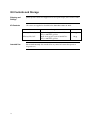
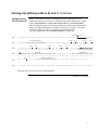
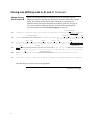
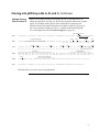
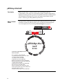
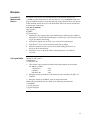
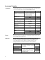
pEF4/myc-His A, B, and C Catalog no. V942-20 Rev. date: 30 December 2010 Manual part no. 25-0239 MAN0000081 Corporate Headquarters Invitrogen Corporation 1600 Faraday Avenue Carlsbad, CA 92008 T: 1 760 603 7200 F: 1 760 602 6500 E: [email protected] For country-specific contact information visit our web site at www.invitrogen.com User Manual ii Table of Contents Kit Contents and Storage........................................................................................................................... iv Introduction .............................................................................................................. 1 Product Overview ........................................................................................................................................1 Methods .................................................................................................................... 2 Cloning into pEF4/myc-His A, B, and C ...................................................................................................2 Transfection and Analysis...........................................................................................................................7 Creating Stable Cell Lines ...........................................................................................................................9 Appendix ................................................................................................................ 11 Human EF-1 Promoter ............................................................................................................................11 pEF4/myc-His Vector.................................................................................................................................12 pEF4/myc-His/lacZ....................................................................................................................................14 Zeocin™ ........................................................................................................................................................15 Recipes .........................................................................................................................................................17 Accessory Products ....................................................................................................................................18 Technical Support.......................................................................................................................................19 Purchaser Notification ...............................................................................................................................20 References....................................................................................................................................................21 iii Kit Contents and Storage Shipping and Storage pEF4/myc-His vectors are shipped on wet ice. Upon receipt, store vectors at -20°C. Kit Contents All vectors are supplied as detailed below. Store the vectors at –20°C. Vector Intended Use iv Composition Amount pEF4/myc-His A, B, and C 40 μL of 0.5 μg/μL vector in 10 mM TrisHCl, 1 mM EDTA, pH 8.0 20 μg pEF4/myc-His/lacZ 20 μg 40 μL of 0.5 μg/μL vector in 10 mM TrisHCl, 1 mM EDTA, pH 8.0 For research use only. Not intended for any animal or human therapeutic or diagnostic use. Introduction Product Overview Description of the System pEF4/myc-His A, B, and C are 5.9 kb vectors designed for overproduction of recombinant proteins in mammalian cell lines. Features of the vectors allow purification and detection of expressed proteins (see pages12–13 for more information). High-level stable and transient expression can be carried out in most mammalian cells. The vectors contain the following elements: Human elongation factor 1-subunit (hEF-1) promoter for high-level expression across a broad range of species and cell types (Goldman et al., 1996) (Mizushima and Nagata, 1990) (see page 11 for more information). Three reading frames to facilitate in-frame cloning with a C-terminal peptide encoding the myc epitope and a polyhistidine (6×His) metal-binding tag. Zeocin™ resistance gene for selection of stable cell lines* (Mulsant et al., 1988) (see page 15 for more information). Episomal replication in cell lines that are latently infected with SV40 or that express the SV40 large T antigen (e.g. COS7). The control plasmid, pEF4/myc-His/lacZ is included for use as a positive control for transfection, expression, and detection in the cell line of choice. Experimental Outline Use the following outline to clone and express your gene of interest in pEF4/mycHis. Consult the multiple cloning sites described on pages 3–5 to determine which vector (A, B, or C) should be used to clone your gene in frame with the Cterminal myc epitope and the polyhistidine tag. Ligate your insert into the appropriate vector and transform into E. coli. Select transformants on 50 to 100 g/mL ampicillin or 25 to 50 g/mL Zeocin™ in Low Salt LB. For more information, see page 17. Analyze your transformants for the presence of insert by restriction digestion. Select a transformant with the correct restriction pattern and use sequencing to confirm that your gene is in frame with the C-terminal peptide. Transfect your construct into the cell line of choice using your own method of transfection. Generate a stable cell line, if desired. Test for expression of your recombinant gene by western blot analysis or functional assay. For antibodies to the myc epitope or the C-terminal polyhistidine tag, see page 18. To purify your recombinant protein, you may use metal-chelating resin such as ProBond™. ProBond™ resin is available separately (see page 18 for ordering information). 1 Methods Cloning into pEF4/myc-His A, B, and C General Molecular Biology Techniques For help with DNA ligations, E. coli transformations, restriction enzyme analysis, purification of single-stranded DNA, DNA sequencing, and DNA biochemistry, refer to Molecular Cloning: A Laboratory Manual (Sambrook et al., 1989) or Current Protocols in Molecular Biology (Ausubel et al., 1994). E. coli Strain Many E. coli strains are suitable for the growth of this vector including TOP10F´, DH5F´, JM109, and INVF´. We recommend that you propagate vectors containing inserts in E. coli strains that are recombination deficient (recA) and endonuclease A deficient (endA). For your convenience, TOP10F´ is available as chemically competent or electrocompetent cells from Invitrogen. Transformation Method You may use any method of your choice for transformation. Chemical transformation is the most convenient for most researchers. Electroporation is the most efficient and the method of choice for large plasmids. Maintaining pEF4/myc-His To propagate and maintain the pEF4/myc-His vectors, use a small amount of the supplied 0.5 μg/μL stock solution in TE, pH 8.0 to transform a recA, endA E. coli strain like TOP10F´, DH5, JM109, or equivalent. Select transformants on LB plates containing 50–100 μg/mL ampicillin or 25 to 50 g/mL Zeocin™ in Low Salt LB. Be sure to prepare a glycerol stock of each plasmid for long term storage (see page 6). Cloning Considerations Your insert should contain a Kozak consensus sequence with an ATG initiation codon for proper initiation of translation (Kozak, 1987; Kozak 1990). An example of a Kozak consensus sequence is provided below. Other sequences are possible, but the G or A at position –3 and the G at position +4 (shown in bold) illustrates the most commonly occurring sequence with strong consensus. Replacing one of the two bases at these positions provides moderate consensus, while having neither results in weak consensus. The ATG initiation codon is shown underlined. (G/A)NNATGG If you wish to express your protein WITHOUT the C-terminal peptide, be sure to include a stop codon. Continued on next page 2 Cloning into pEF4/myc-His A, B, and C, Continued Multiple Cloning Site of Version A Below is the multiple cloning site for pEF4/myc-His A. Restriction sites are labeled to indicate the cleavage site. The boxed nucleotides indicate the variable region. Note that there is a stop codon between the Spe I site and the BstX I site. The multiple cloning site has been confirmed by sequencing and functional testing. For more information on the hEF-1 promoter, see page 11. The vector sequence of pEF4/myc-His A is available for downloading from www.invitrogen.com or from Technical Support (see page 19). 3´end of hEF-1a Intron 1 1581 GTTTGGATCT TGGTTCATTC TCAAGCCTCA GACAGTGGTT CAAAGTTTTT TTCTTCCATT TCAGGTGTCG TGAGGAATTA 1661 GCTTGGTACT AATACGACTC ACTATAGGGA GACCCAAGCT GGCTAGT TAA GCT TGG TAC CGA GCT CGG ATC CAC *** Ala Trp Tyr Arg Ala Arg Ile His 1735 TAG TCC AGT GTG GTG GAA TTC TGC AGA TAT CCA GCA CAG TGG CGG CCG CTC GAG TCT AGA GGG CCC *** Ser Ser Val Val Glu Phe Cys Arg Tyr Pro Ala Gln Trp Arg Pro Leu Glu Ser Arg Gly Pro 1801 TTC GAA CAA AAA CTC ATC TCA GAA GAG GAT CTG AAT ATG CAT ACC GGT CAT CAT CAC CAT CAC CAT Phe Glu Gln Lys Leu Ile Ser Glu Glu Asp Leu Asn Met His Thr Gly His His His His His His 1867 TGA G TTTAAACCCG CTGATCAGCC TCGACTGTGC CTTCTAGTTG CCAGCCATCT GTTGTTTGCC CCTCCCCCGT *** 1941 GCCTTCCTTG ACCCTGGAAG GTGCCACTCC CACTGTCCTT TCCTAATAAA ATGAGGAAAT TGCATCGCAT TGTCTGAGTA 5´ end of hEF-1a Exon 2 Acc65 I T7 promoter/priming site BstX I* BstB I EcoR V myc epitope Pme I *Note EcoR I BstX I* Not I Kpn I BamH I Spe I Xba I Polyhistidine tag BGH reverse priming site that there are two BstX I sites in the polylinker. Continued on next page 3 Cloning into pEF4/myc-His A, B, and C, Continued Multiple Cloning Site of Version B Below is the multiple cloning site for pEF4/myc-His B. Restriction sites are labeled to indicate the cleavage site. The boxed nucleotides indicate the variable region. The multiple cloning site has been confirmed by sequencing and functional testing. For more information on the hEF-1 promoter, see page 11. The vector sequence of pEF4/myc-His B is available for downloading from www.invitrogen.com or from Technical Support (see page 19). 3´end of hEF-1a Intron 1 1581 GTTTGGATCT TGGTTCATTC TCAAGCCTCA GACAGTGGTT CAAAGTTTTT TTCTTCCATT TCAGGTGTCG TGAGGAATTA 5´ end of hEF-1a Exon 2 Acc65 I T7 promoter/priming site Kpn I BamH I Spe I 1661 GCTTGGTACT AATACGACTC ACTATAGGGA GACCCAAGCT GGCTAGTT AAG CTT GGT ACC GAG CTC GGA TCC ACT Lys Leu Gly Thr Glu Leu Gly Ser Thr 1736 AGT CCA GTG TGG TGG AAT TCT GCA GAT ATC CAG CAC AGT GGC GGC CGC TCG AGT CTA GAG GGC CCG Ser Pro Val Trp Trp Asn Ser Ala Asp Ile Gln His Ser Gly Gly Arg Ser Ser Leu Glu Gly Pro 1802 CGG TTC GAA CAA AAA CTC ATC TCA GAA GAG GAT CTG AAT ATG CAT ACC GGT CAT CAT CAC CAT CAC Arg Phe Glu Gln Lys Leu Ile Ser Glu Glu Asp Leu Asn Met His Thr Gly His His His His His 1868 CAT TGA GTTTAAA CCCGCTGATC AGCCTCGACT GTGCCTTCTA GTTGCCAGCC ATCTGTTGTT TGCCCCTCCC His *** 1941 CCGTGCCTTC CTTGACCCT GAAGGTGCCA CTCCCACTGT CCTTTCCTAA TAAAATGAGG AAATTGCATC GCATTGTCTC BstX I* BstB I EcoR V myc epitope Pme I *Note EcoR I BstX I* Not I Xba I Polyhistidine tag BGH reverse priming site that there are two BstX I sites in the polylinker. Continued on next page 4 Cloning into pEF4/myc-His A, B, and C, Continued Multiple Cloning Site of Version C Below is the multiple cloning site for pEF4/myc-His C. Restriction sites are labeled to indicate the cleavage site. The boxed nucleotides indicate the variable region. The multiple cloning site has been confirmed by sequencing and functional testing. For more information on the hEF-1 promoter, see page 11. The vector sequence of pEF4/myc-His C is available for downloading from www.invitrogen.com or from Technical Support (see page 19). 3´end of hEF-1a Intron 1 1581 GTTTGGATCT TGGTTCATTC TCAAGCCTCA GACAGTGGTT CAAAGTTTTT TTCTTCCATT TCAGGTGTCG TGAGGAATTA 1661 GCTTGGTACT AATACGACTC ACTATAGGGA GACCCAAGCT GGCTAG TTA AGC TTG GTA CCG AGC TCG GAT CCA Leu Ser Leu Val Pro Ser Ser Asp Pro 1734 CTA GTC CAG TGT GGT GGA ATT CTG CAG ATA TCC AGC ACA GTG GCG GCC GCT CGA GGT CAC CCA TTC Leu Val Gln Cys Gly Gly Ile Leu Gln Ile Ser Ser Thr Val Ala Ala Ala Arg Gly His Pro Phe 1800 GAA CAA AAA CTC ATC TCA GAA GAG GAT CTG AAT ATG CAT ACC GGT CAT CAT CAC CAT CAC CAT TGA Glu Gln Lys Leu Ile Ser Glu Glu Asp Leu Asn Met His Thr Gly His His His His His His *** 1866 GTTTA AACCCGCTGA TCAGCCTCGA CTGTGCCTTC TAGTTGCCAG CCATCTGTTG TTTGCCCCTC CCCCGTGCCT 1941 TCCTTGACCC TGGAAGGTGC CACTCCCACT GTCCTTTCCT AATAAAATGA GGAAATTGCA TCGCATTGTC 5´ end of hEF-1a Exon 2 Acc65 I T7 promoter/priming site BstX I* EcoR I EcoR V *Note Not I BamH I Spe I BstE II BstB I Polyhistidine tag myc epitope Pme I BstX I* Kpn I BGH reverse priming site that there are two BstX I sites in the polylinker. Continued on next page 5 Cloning into pEF4/myc-His A, B, and C, Continued E. coli Transformation MEND ION AT RECOM Important Preparing a Glycerol Stock Transform your ligation mixtures into a competent recA, endA E. coli strain (e.g., TOP10F´, DH5) and select on LB plates containing 50–100 g/mL ampicillin or 25–50 g/mL Zeocin™ in Low Salt LB (see below). Select 10–20 clones and analyze for the presence and orientation of your insert. Any E. coli strain that contains the complete Tn5 transposable element (i.e. DH5F´IQ) encodes the ble (bleomycin resistance gene). These strains will confer resistance to Zeocin™. For the most efficient selection, we highly recommend that you choose an E. coli strain that does not contain the Tn5 gene (i.e. TOP10, DH5, DH10, etc.). We recommend that you sequence your construct with the T7 Forward and BGH Reverse primers to confirm that your gene is fused in frame with the myc epitope and the C-terminal polyhistidine tag. Refer to the diagrams on pages 3-5 for sequences and location of primer binding sites. Once you have identified the correct clone, be sure to purify the colony and make a glycerol stock for long-term storage. It is also a good idea to keep a DNA stock of your plasmid at –20°C. 1. 2. 3. 4. 5. 6 Streak the original colony out on an LB plate containing 50 μg/mL ampicillin or 25 μg/mL Zeocin™ in Low Salt LB. Incubate the plate at 37°C overnight. Isolate a single colony and inoculate into 1–2 mL of LB containing 50 μg/mL ampicillin or 25 μg/mL Zeocin™. Grow the culture to mid-log phase (OD600 = 0.5–0.7). Mix 0.85 mL of culture with 0.15 mL of sterile glycerol and transfer to a cryovial. Store at –80°C. Transfection and Analysis Introduction Once you have confirmed that your construct is in the correct orientation and fused in frame to the C-terminal peptide, you are ready to transfect your cell line of choice. We recommend that you include the positive control vector and a mock transfection to evaluate your results. Plasmid Preparation Plasmid DNA for transfection into eukaryotic cells must be very clean and free from phenol and sodium chloride. Contaminants will kill the cells and salt will interfere with lipid complexing, decreasing transfection efficiency. We recommend isolating plasmid DNA using the PureLink™ HiPure Miniprep Kit or the PureLink™ HiPure Midiprep Kit (see page 18). Methods of Transfection For established cell lines (e.g. HeLa), consult original references or the supplier of your cell line for the optimal method of transfection. We recommend that you follow exactly the protocol for your cell line. Pay particular attention to medium requirements, when to pass the cells, and at what dilution to split the cells. Further information is provided in Current Protocols in Molecular Biology. Methods for transfection include calcium phosphate (Chen and Okayama, 1987; Wigler et al., 1977), lipid-mediated (Felgner et al., 1989; Felgner and Ringold, 1989) and electroporation (Chu et al., 1987; Shigekawa and Dower, 1988). Invitrogen offers the Lipofectamine™ 2000 Reagent for mammalian transfection (see page 18 for ordering information). Positive Control pEF4/myc-His/lacZ is provided as a positive control vector for mammalian transfection and expression (see page 14), and may be used to optimize transfection conditions for your cell line. The gene encoding -galactosidase is expressed in mammalian cells under the control of the hEF-1 promoter. A successful transfection will result in -galactosidase expression that can be easily assayed (see below). Assay for -galactosidase Activity You may assay for -galactosidase expression by activity assay using cell-free lysates (Miller, 1972) or by staining the cells for activity. Invitrogen offers the -Gal Assay Kit and the -Gal Staining Kit for fast and easy detection of -galactosidase expression (see page 18 for ordering information). Continued on next page 7 Transfection and Analysis, Continued Detecting Fusion Proteins Several antibodies are available from Invitrogen to detect expression of your fusion protein from pEF4/myc-His (see page 18). To detect fusion protein by western blot, prepare a cell lysate from transfected cells. We recommend that you perform a time course to optimize expression of the fusion protein (e.g., 24, 48, 72 hours, etc. after transfection). To lyse cells: 1. Wash cell monolayers (~106 cells) once with phosphate-buffered saline (PBS). 2. Scrape cells into 1 mL PBS and pellet the cells at 1,500 × g for 5 minutes. 3. Resuspend in 50 μL Cell Lysis Buffer (see recipe below). Other lysis buffers may be suitable. 4. Incubate cell suspension at 37°C for 10 minutes to lyse the cells. 5. Centrifuge the cell lysate at 10,000 × g for 10 minutes to pellet nuclei and transfer the supernatant to a fresh tube. Assay the lysate for protein concentration. Note: Do not use protein assays utilizing Coomassie Blue or other dyes. NP-40 interferes with the binding of the dye with the protein. 6. Add SDS-PAGE sample buffer to a final concentration of 1X and boil the sample for 5 minutes. 7. Load 20 g of lysate onto an SDS-PAGE gel and electrophorese. Use the appropriate percentage of acrylamide to resolve your fusion protein. The C-terminal peptide containing the myc epitope and the polyhistidine tag will add approximately 3 kDa to the size of your protein. Purification 8 You will need lysate from 5 × 10⁶ to 1 × 10⁷ transfected cells for purification of your protein on a 2 mL ProBond™ column (or other metal-chelating column). Refer to the manufacturer's instructions before attempting to purify your fusion protein. To prepare cells for lysis, refer to the protocol on page 10. Creating Stable Cell Lines Selection in Mammalian Cell Lines Possible Sites for Linearization Enzyme To generate a stable cell line expressing your protein, you need to determine the minimum concentration of Zeocin™ required for killing your untransfected host cell line. Typically, concentrations between 50 and 1000 g/mL Zeocin™ are sufficient to kill the untransfected host cell line. Test a range of concentrations (see below) to ensure that you determine the minimum concentration necessary for your cell line. 1. Seed cells (2 × 10⁵ cells/60 mm plate) for each time point and allow cells to adhere overnight. 2. The next day, substitute culture medium with medium containing varying concentrations of Zeocin™ (e.g., 0, 50, 125, 250, 500, 750, and 1,000 g/mL). 3. Replenish the selective medium every 3–4 days, and observe the percentage of surviving cells. 4. Count the number of viable cells at regular intervals to determine the appropriate concentration of Zeocin™ that prevents growth. To obtain stable transfectants, you may choose to linearize your vector before transfection. While linearizing your vector may not improve the efficiency of transfection, it increases the chances that the vector does not integrate in a way that disrupts the gene of interest. The table below lists unique sites that may be used to linearize your construct prior to transformation. Other restriction sites are possible. Note that for the enzymes listed below, the cleavage site is indicated for versions A, B, and C of pEF4/myc-His. Be sure that your insert does not contain the restriction enzyme site you wish to use to linearize your vector. Restriction Site (bp) (A,B,C) Location Supplier Nru I 331 Upstream of EF-1 promoter Many Mlu I 351 Upstream of EF-1 promoter Many Bst1107 I 3688 (A), 3692 (B), 3684 (C) End of SV40 poly A AGS*, Fermentas, Takara, BoehringerMannhiem Eam1105 I 4960 (A), 4964 (B), 4956 (C) Ampicillin gene AGS*, Fermentas, Takara Fsp I 5182 (A), 5186 (B), 5178 (C) Ampicillin gene Many Pvu I 5330 (A), 5334 (B), 5326 (C) Ampicillin gene Many Sca I 5440 (A), 5444 (B), 5436 (C) Ampicillin gene Many *Angewandte Gentechnologie Systeme Continued on next page 9 Creating Stable Cell Lines, Continued Selecting Stable Integrants Once the appropriate Zeocin™ concentration is determined, you can generate a stable cell line with your construct. 1. Transfect your cells using the appropriate protocol for your cell line. Include a sample of untransfected cells as a negative control. 2. After transfection, wash the cells once with 1X PBS and add fresh medium to the cells. 3. 48 hours after transfection, split the cells into fresh medium containing Zeocin™ at the appropriate concentration for your cell line. Split the cells such that they are no more than 25% confluent. 4. Change selective medium every 3–4 days until Zeocin™-resistant colonies are detected. 5. Pick and expand colonies. Preparing Cells for Use the procedure below to prepare cells for lysis prior to purification of your protein on ProBond™. You will need 5 × 10⁶ to 1 × 10⁷ cells for purification of Lysis your protein on a 2 mL ProBond™ column (see ProBond™ Protein Purification manual). Lysis of Cells 1. Seed cells in either five T-75 flasks or 2 to 3 T-175 flasks. 2. Grow the cells in selective medium until they are 80–90% confluent. 3. Harvest the cells by treating with trypsin-EDTA for 2 to 5 minutes or by scraping the cells in PBS. 4. Inactivate the trypsin by diluting with fresh medium (if necessary) and transfer the cells to a sterile microcentrifuge tube. 5. Centrifuge the cells at 240 g for 5 minutes. Resuspend the cells in PBS. 6. Centrifuge the cells at 240 g for 5 minutes. You may lyse the cells immediately or freeze in liquid nitrogen and store at –80°C until needed. If you are using ProBond™ resin, refer to the ProBond™ Protein Purification manual for details about sample preparation for chromatography. If you are using other metal-chelating resin, refer to the manufacturer's instruction for recommendations on sample preparation. 10 Appendix Human EF-1 Promoter Description The diagram below shows all the features of the EF-1 promoter used in the pEF4/myc-His vectors (Mizushima and Nagata, 1990). Features are marked as per Uetsuki et al., 1989. 5´ end of human EF-1a promoter 339 GGAGTGCCTC GTGAGGCTCC GGTGCCCGTC AGTGGGCAGA GCGCACATCG CCCACAGTCC 399 CCGAGAAGTT GGGGGGAGGG GTCGGCAATT GAACCGGTGC CTAGAGAAGG TGGCGCGGGG 459 TAAACTGGGA AAGTGATGTC GTGTACTGGC TCCGCCTTTT TCCCGAGGGT GGGGGAGAAC Start of Transcription TATA box 519 CGTATATAAG TGCAGTAGTC GCCGTGAACG TTCTTTTTCG CAACGGGTTT GCCGCCAGAA Exon I 5´ end of Intron 1 579 CACAGGTAAG TGCCGTGTGT GGTTCCCGCG GGCCTGGCCT CTTTACGGGT TATGGCCCTT 639 GCGTGCCTTG AATTACTTCC ACCTGGCTGC AGTACGTGAT TCTTGATCCC GAGCTTCGGG 699 TTGGAAGTGG GTGGGAGAGT TCGAGGCCTT GCGCTTAAGG AGCCCCTTCG CCTCGTGCTT 759 GAGTTGAGGC CTGGCCTGGG CGCTGGGGCC GCCGCGTGCG AATCTGGTGG CACCTTCGCG 819 CCTGTCTCGC TGCTTTCGAT AAGTCTCTAG CCATTTAAAA TTTTTGATGA CCTGCTGCGA 879 CGCTTTTTTT CTGGCAAGAT AGTCTTGTAA ATGCGGGCCA AGATCTGCAC ACTGGTATTT 939 CGGTTTTTGG GGCCGCGGGC GGCGACGGGG CCCGTGCGTC CCAGCGCACA TGTTCGGCGA 999 GGCGGGGCCT GCGAGCGCGG CCACCGAGAA TCGGACGGGG GTAGTCTCAA GCTGGCCGGC 1059 CTGCTCTGGT GCCTGGCCTC GCGCCGCCGT GTATCGCCCC GCCCTGGGCG GCAAGGCTGG 1119 CCCGGTCGGC ACCAGTTGCG TGAGCGGAAA GATGGCCGCT TCCCGGCCCT GCTGCAGGGA 1179 GCTCAAAATG GAGGACGCGG CGCTCGGGAG AGCGGGCGGG TGAGTCACCC ACACAAAGGA 1239 AAAGGGCCTT TCCGTCCTCA GCCGTCGCTT CATGTGACTC CACGGAGTAC CGGGCGCCGT 1299 CCAGGCACCT CGATTAGTTC TCGAGCTTTT GGAGTACGTC GTCTTTAGGT TGGGGGGAGG 1359 GGTTTTATGC GATGGAGTTT CCCCACACTG AGTGGGTGGA GACTGAAGTT AGGCCAGCTT 1419 GGCACTTGAT GTAATTCTCC TTGGAATTTG CCCTTTTTGA GTTTGGATCT TGGTTCATTC 1479 TCAAGCCTCA GACAGTGGTT CAAAGTTTTT TTCTTCCATT TCAGGTGTCG TGA... Sp 1 Sp 1 Sp 1 Sp 1 Sp 1 Ap 1 3´ end of Intron 1 5´ end of Exon 2 11 pEF4/myc-His Vector Map of pEF4/myc-His The figure below summarizes the features of the pEF4/myc-His vectors. The sequences for pEF4/myc-His A, B, and C are available for downloading from www.invitrogen.com or from Technical Support (see page 19). 6xHis P -1 EF a BGH pA f1 Term Pme I T7 Acc65 I Kpn I BamH I Spe I BstX I EcoR I EcoR V BstX I Not I Xba I* BstE II* BstB I myc epitope or i ri 40 o SV C EF-1a promoter: bases 468-1653 T7 promoter/priming site: bases 1670-1689 Multiple cloning site: bases 1715-1806 myc epitope: bases 1804-1833 Polyhistidine tag: bases 1849-1866 BGH reverse priming site: bases 1889-1906 BGH polyadenylation signal: bases 1892-2119 f1 origin: bases 2165-2593 SV40 promoter and origin: bases 2621-2929 EM-7 promoter: bases 2977-3032 Zeocin resistance gene: bases 3051-3425 SV40 polyadenylation signal: bases 3555-3684 pUC origin: bases 4068-4741 Ampicillin resistance gene: bases 4886-5746 12 SV40 pA pU in Comments for pEF4/Myc-His A 5882 nucleotides EM-7 n 5.9 kb Ze oc A m p i c i l li pEF4/Myc-His A, B, C *There is a unique BstE II site, but no Xba I site in version C pEF4/myc-His Vector, Continued Features of pEF4/myc-His pEF4/myc-His A (5882 bp), pEF4/myc-His B (5886 bp), and pEF4/ myc-His C (5878 bp) contain the following elements. All features have been functionally tested. Feature Benefit Human elongation factor 1 (hEF-1) promoter Allows overexpression of your recombinant protein in a broad range of mammalian cell types (Goldman et al., 1996; Mizushima and Nagata, 1990). T7 promoter/priming site Allows for in vitro transcription in the sense orientation and sequencing through the insert. Multiple cloning site in three reading frames Allows insertion of your gene and facilitates cloning in frame with the myc epitope and C-terminal polyhistidine tag. myc epitope (Glu-Gln-Lys-Leu-Ile-SerGlu-Glu-Asp-Leu) Allows detection of your recombinant protein with the Anti-myc Antibody or Anti-myc-HRP Antibody (Evans et al., 1985). C-terminal polyhistidine (6xHis) tag Allows purification of your recombinant protein on metalchelating resin such as ProBond™. In addition, the C-terminal polyhistidine tag is the epitope for the Anti-His(C-term) Antibody (Lindner et al., 1997) and the Anti-His (C-term)-HRP Antibody. BGH reverse priming site Allows sequencing through the insert. Bovine growth hormone (BGH) polyadenylation signal Efficient transcription termination and polyadenylation of mRNA (Goodwin and Rottman, 1992). f1 origin Allows rescue of single-stranded DNA. SV40 early promoter and origin Allows efficient, high-level expression of the Zeocin™ resistance gene and episomal replication in cells expressing the SV40 large T antigen. EM-7 promoter Synthetic promoter based on the bacteriophage T7 promoter for expression of the Zeocin™ resistance gene in E. coli. Zeocin™ resistance gene Selection of transformants in E. coli and stable transfectants in mammalian cells (Drocourt et al., 1990; Mulsant et al., 1988). SV40 polyadenylation signal Efficient transcription termination and polyadenylation of mRNA. pUC-derived High-copy number replication and growth in E. coli. Ampicillin resistance gene Selection of transformants in E. coli. (-lactamase) 13 pEF4/myc-His/lacZ Description pEF4/myc-His/lacZ is a 8,932 bp control vector containing the gene for galactosidase. This vector was constructed by ligating a 3,976 bp BamH I-Bsm I fragment containing the EF-1 promoter from pEF4/myc-His B to a 4956 bp BamH I-Bsm I fragment containing the lacZ gene, myc epitope, polyhistidine tag and Zeocin™ resistance gene from pcDNA4/myc-His/lacZ. Map of Control Vector The figure below summarizes the features of the pEF4/myc-His/lacZ vector. The nucleotide sequence for pEF4/myc-His/lacZ is available for downloading from www.invitrogen.com or by contacting Technical Support (see page 19). P -1 EF a lacZ BGH pA Not I BstE II BstB I BamH I Spe I T7 f1 Pme I 6xHis myc epitope or i ri 40 o SV EF-1a promoter: bases 468-1653 T7 promoter/priming site: bases 1670-1689 LacZ ORF: bases 1770-4826 myc epitope: bases 4851-4880 Polyhistidine tag: bases 4896-4913 BGH reverse priming site: bases 4936-4953 BGH polyadenylation signal: bases 4939-5166 f1 origin: bases 5212-5640 SV40 promoter and origin: bases 5668-5968 EM-7 promoter: bases 6027-6082 Zeocin resistance gene: bases 6101-6475 SV40 polyadenylation signal: bases 6605-6735 pUC origin: bases 7118-7791 Ampicillin resistance gene: bases 7936-8796 14 C SV40 pA pU in Comments for pEF4/Myc-His/lacZ 8932 nucleotides EM-7 n 8.9 kb Ze oc A m p i c i l li pEF4/Myc-His/ lacZ Term Zeocin™ Introduction The pEF4/myc-His vectors contain the Zeocin™ resistance gene for selection of stable cell lines using Zeocin™. We recommend that you test the sensitivity of your mammalian host cell to Zeocin™ as natural resistance varies among cell lines. General information and guidelines are provided in this section for your convenience. Zeocin™ Zeocin™ is a member of the bleomycin/phleomycin family of antibiotics isolated from Streptomyces. Antibiotics in this family are broad spectrum antibiotics that act as strong anti-bacterial and anti-tumor drugs. They show strong toxicity against bacteria, fungi (including yeast), plants, and mammalian cells (Baron et al., 1992; Drocourt et al., 1990; Mulsant et al., 1988; Perez et al., 1989). The Zeocin™ resistance protein has been isolated and characterized (Calmels et al., 1991; Drocourt et al., 1990). This protein, the product of the Sh ble gene (Streptoalloteichus hindustanus bleomycin gene), is a 13.7 kDa protein that binds Zeocin™ and inhibits its DNA strand cleavage activity. Expression of this protein in eukaryotic and prokaryotic hosts confers resistance to Zeocin™. Molecular Weight, Formula and Structure The formula for Zeocin™ is C55H86 O21N20S2Cu.HCl and the molecular weight is 1,527.5 Da. Zeocin is an HCl salt. The diagram below shows the structure of Zeocin™. CONH2 H H2 N N H O H N CH3 HO N Cu NH O N H N N H N O O N O O ++ H2N H N CH3 HO R S N S CH3 H OH O O CH3 R = HN NH2 N NH NH2 OH H2N O O HO O HO OH OH O Continued on next page 15 Zeocin™, Continued Applications of Zeocin™ Zeocin™ is used for selection in mammalian cells (Mulsant et al., 1988); plants (Perez et al., 1989); yeast (Baron et al., 1992); and prokaryotes (Drocourt et al., 1990). Suggested concentrations of Zeocin™ for selection in mammalian cell lines and E. coli are listed below: Organism Zeocin™ Concentration and Selective Medium E. coli 25–50 g/mL in low salt LB medium* (see page 17 for recipe) Mammalian Cells 50–1000 μg/mL (varies with cell line) *Efficient selection requires that the concentration of NaCl be no more than 5 g/liter (<90 mM). Handling Zeocin™ 16 High salt and acidity or basicity inactivates Zeocin™. Therefore, we recommend that you reduce the salt in bacterial medium and adjust the pH to 7.5 to keep the drug active (see page 17). Store Zeocin™ at –20°C and thaw on ice before use. Zeocin™ is light sensitive. Store drug, plates, and medium containing drug in the dark. Wear gloves, a laboratory coat, and safety glasses or goggles when handling solutions containing Zeocin™. Zeocin™ is toxic. Do not ingest or inhale solutions containing the drug. Recipes Low Salt LB Medium with Zeocin™ For Zeocin™ to be active, the salt concentration of the medium must be low (<90 mM) and the pH must be 7.5. For selection in E. coli, it is imperative that you prepare LB broth and plates using the following recipe. Note the lower salt content of this medium. Failure to use low salt LB medium will result in non-selection due to inactivation of the drug. Low Salt LB Medium: 10 g Tryptone 5 g NaCl 5 g Yeast Extract Cell Lysis Buffer 1. Combine the dry reagents above and add deionized, distilled water to 950 mL. Adjust pH to 7.5 with 5 M NaOH. Bring the volume up to 1 liter. For plates, add 15 g/L agar before autoclaving. 2. Autoclave on liquid cycle at 15 lbs/sq. in. and 121°C for 20 minutes. 3. Thaw Zeocin™ on ice and vortex before removing an aliquot. 4. Allow the medium to cool to at least 55°C before adding the Zeocin™ to 25 g/mL final concentration. 5. Store plates at 4°C in the dark. Plates containing Zeocin™ are stable for 1-2 weeks. 50 mM Tris-HCl, pH 7.8 150 mM NaCl 1% Nonidet P-40 1. This solution can be prepared from the following common stock solutions. For 100 mL, combine: 1 M Tris base 5 M NaCl Nonidet P-40 5 mL 3 mL 1 mL 2. Bring the volume up to 90 mL with deionized water and adjust the pH to 7.8 with HCl. 3. Bring the volume up to 100 mL. Store at room temperature. Note: Protease inhibitors may be added at the following concentrations: 1 mM PMSF 1 g/mL pepstatin 1 g/mL leupeptin 17 Accessory Products Introduction The following products may be used with the pEF4/myc-His vectors. For details, visit www.invitrogen.com or contact Technical Support (page 19). Item Amount Catalog no. K850-01 ProBond™ Purification System 6 × 2 mL precharged, prepacked ProBond™ resin columns and buffers for native and denaturing purification 50 mL R801-01 150 mL R801-15 ProBond™ Resin ™ Anti-Xpress Antibody R910-25 Electrocomp™ TOP10F´ 5 × 80 μL One Shot® TOP10F´ Chemically Competent E. coli 20 × 50 μL PureLink™ HiPure Plasmid Miniprep Kit 100 preps K2100-03 PureLink™ HiPure Plasmid Midiprep Kit 25 preps K2100-04 80 mL K1455-01 1 kit K1465-01 1 gram R250-01 5 gram R250-05 0.75 mL 11668-027 -Gal Assay Kit -Gal Staining Kit Zeocin™ Lipofectamine™ 2000 Reagent C665-55 C3030-03 Primers For your convenience, Invitrogen offers a custom primer synthesis service. Visit www.invitrogen.com for more details. Antibodies If you do not have an antibody specific to your protein, Invitrogen offers the Anti-myc, or Anti-His(C-term) antibodies to detect your recombinant fusion protein. Horseradish peroxidase (HRP)- and alkaline phosphatase (AP)– conjugated antibodies are available for convenient one-step detection. Antibody Anti-myc Anti-myc-HRP Anti-myc-AP Anti-His(C-term) Anti-His(C-term)-HRP Anti-His(C-term)-AP 18 Epitope Catalog no. Detects a 10 amino acid epitope derived from c-myc (Evan et al., 1985): EQKLISEEDL R950-25 Detects the C-terminal polyhistidine tag (requires the free carboxyl group for detection) (Lindner et al., 1997): HHHHHH-COOH R951-25 R952-25 R930-25 R931-25 R932-25 Technical Support Web Resources Contact Us Visit the Invitrogen website at www.invitrogen.com for: Technical resources, including manuals, vector maps and sequences, application notes, SDSs, FAQs, formulations, citations, handbooks, etc. Complete technical support contact information Access to the Invitrogen Online Catalog Additional product information and special offers For more information or technical assistance, call, write, fax, or email. Additional international offices are listed on our website (www.invitrogen.com). Corporate Headquarters: 5791 Van Allen Way Carlsbad, CA 92008 USA Tel: 1 760 603 7200 Tel (Toll Free): 1 800 955 6288 Fax: 1 760 602 6500 E-mail: [email protected] Japanese Headquarters: LOOP-X Bldg. 6F 3-9-15, Kaigan Minato-ku, Tokyo 108-0022 Tel: 81 3 5730 6509 Fax: 81 3 5730 6519 E-mail: [email protected] European Headquarters: Inchinnan Business Park 3 Fountain Drive Paisley PA4 9RF, UK Tel: +44 (0) 141 814 6100 Tech Fax: +44 (0) 141 814 6117 E-mail: [email protected] SDS Safety Data Sheets (SDSs) are available on our website at www.invitrogen.com/sds. Certificate of Analysis The Certificate of Analysis provides detailed quality control and product qualification information for each product. Certificates of Analysis are available on our website. Go to www.invitrogen.com/support and search for the Certificate of Analysis by product lot number, which is printed on the box. Limited Warranty Invitrogen (a part of Life Technologies Corporation) is committed to providing our customers with high-quality goods and services. Our goal is to ensure that every customer is 100% satisfied with our products and our service. If you should have any questions or concerns about an Invitrogen product or service, contact our Technical Support Representatives. All Invitrogen products are warranted to perform according to specifications stated on the certificate of analysis. The Company will replace, free of charge, any product that does not meet those specifications. This warranty limits the Company’s liability to only the price of the product. No warranty is granted for products beyond their listed expiration date. No warranty is applicable unless all product components are stored in accordance with instructions. The Company reserves the right to select the method(s) used to analyze a product unless the Company agrees to a specified method in writing prior to acceptance of the order. Invitrogen makes every effort to ensure the accuracy of its publications, but realizes that the occasional typographical or other error is inevitable. Therefore the Company makes no warranty of any kind regarding the contents of any publications or documentation. If you discover an error in any of our publications, please report it to our Technical Support Representatives. Life Technologies Corporation shall have no responsibility or liability for any special, incidental, indirect or consequential loss or damage whatsoever. The above limited warranty is sole and exclusive. No other warranty is made, whether expressed or implied, including any warranty of merchantability or fitness for a particular purpose. 19 Purchaser Notification Limited Use Label License No. 22: Vectors and Clones Encoding Histidine Hexamer This product is licensed under U.S. and/or foreign patents from Hoffmann-LaRoche, Inc., Nutley, NJ and/or Hoffmann-LaRoche Ltd., Basel, Switzerland and is provided only for use in research. Information about licenses for commercial use is available from QIAGEN GmbH, MaxVolmer-Str. 4, D-40724 Hilden, Germany. Limited Use Label License No. 60: EF-1 Promoter EF-1alpha promoter products are the subject of U.S. and/or foreign patents and sold under license for research purposes only. The use of this product for any commercial purpose, including but not limited to, use in any study for the purpose of a filing of a new drug application, requires a license from: Mochida Pharmaceutical Co., Ltd., 7, Yotsuya 1-Chome, Shinjuku-Ku, Tokyo 160, Japan. Tel: 81-3-3225-5451; Fax: 81-3-3225-6091. 20 References Ausubel, F. M., Brent, R., Kingston, R. E., Moore, D. D., Seidman, J. G., Smith, J. A., and Struhl, K. (1994). Current Protocols in Molecular Biology (New York: Greene Publishing Associates and WileyInterscience). Baron, M., Reynes, J. P., Stassi, D., and Tiraby, G. (1992). A Selectable Bifunctional -Galactosidase: Phleomycin-resistance Fusion Protein as a Potential Marker for Eukaryotic Cells. Gene 114, 239-243. Calmels, T., Parriche, M., Burand, H., and Tiraby, G. (1991). High Efficiency Transformation of Tolypocladium geodes Conidiospores to Phleomycin Resistance. Curr. Genet. 20, 309-314. Chen, C., and Okayama, H. (1987). High-Efficiency Transformation of Mammalian Cells by Plasmid DNA. Mol. Cell. Biol. 7, 2745-2752. Chu, G., Hayakawa, H., and Berg, P. (1987). Electroporation for the Efficient Transfection of Mammalian Cells with DNA. Nuc. Acids Res. 15, 1311-1326. Drocourt, D., Calmels, T. P. G., Reynes, J. P., Baron, M., and Tiraby, G. (1990). Cassettes of the Streptoalloteichus hindustanus ble Gene for Transformation of Lower and Higher Eukaryotes to Phleomycin Resistance. Nuc. Acids Res. 18, 4009. Evans, G. I., Lewis, G. K., Ramsay, G., and Bishop, V. M. (1985). Isolation of Monoclonal Antibodies Specific for c-myc Proto-oncogene Product. Mol. Cell. Biol. 5, 3610-3616. Felgner, P. L., Holm, M., and Chan, H. (1989). Cationic Liposome Mediated Transfection. Proc. West. Pharmacol. Soc. 32, 115-121. Felgner, P. L., and Ringold, G. M. (1989). Cationic Liposome-Mediated Transfection. Nature 337, 387-388. Goldman, L. A., Cutrone, E. C., Kotenko, S. V., Krause, C. D., and Langer, J. A. (1996). Modifications of Vectors pEF-BOS, pcDNA1, and pcDNA3 Result in Improved Convenience and Expression. BioTechniques 21, 1013-1015. Goodwin, E. C., and Rottman, F. M. (1992). The 3´-Flanking Sequence of the Bovine Growth Hormone Gene Contains Novel Elements Required for Efficient and Accurate Polyadenylation. J. Biol. Chem. 267, 16330-16334. Kozak, M. (1987). An Analysis of 5´-Noncoding Sequences from 699 Vertebrate Messenger RNAs. Nuc. Acids Res. 15, 8125-8148. Kozak, M. (1991). An Analysis of Vertebrate mRNA Sequences: Intimations of Translational Control. J. Cell Biol. 115, 887-903. Kozak, M. (1990). Downstream Secondary Structure Facilitates Recognition of Initiator Codons by Eukaryotic Ribosomes. Proc. Natl. Acad. Sci. USA 87, 8301-8305. Lindner, P., Bauer, K., Krebber, A., Nieba, L., Kremmer, E., Krebber, C., Honegger, A., Klinger, B., Mocikat, R., and Pluckthun, A. (1997). Specific Detection of His-tagged Proteins With Recombinant Anti-His Tag scFv-Phosphatase or scFv-Phage Fusions. BioTechniques 22, 140-149. Miller, J. H. (1972). Experiments in Molecular Genetics (Cold Spring Harbor, New York: Cold Spring Harbor Laboratory). Mizushima, S., and Nagata, S. (1990). pEF-BOS, a Powerful Mammalian Expression Vector. Nuc. Acids Res. 18, 5322. Mulsant, P., Tiraby, G., Kallerhoff, J., and Perret, J. (1988). Phleomycin Resistance as a Dominant Selectable Marker in CHO Cells. Somat. Cell Mol. Genet. 14, 243-252. Continued on next page 21 References, Continued Perez, P., Tiraby, G., Kallerhoff, J., and Perret, J. (1989). Phleomycin Resistance as a Dominant Selectable Marker for Plant Cell Transformation. Plant Mol. Biol. 13, 365-373. Sambrook, J., Fritsch, E. F., and Maniatis, T. (1989). Molecular Cloning: A Laboratory Manual, Second Edition (Plainview, New York: Cold Spring Harbor Laboratory Press). Shigekawa, K., and Dower, W. J. (1988). Electroporation of Eukaryotes and Prokaryotes: A General Approach to the Introduction of Macromolecules into Cells. BioTechniques 6, 742-751. Uetsuki, T., Naito, A., Nagata, S., and Kaziro, Y. (1989). Isolation and Characterization of the Human Chromosomal Gene for Polypeptide Chain Elongation Factor-1. J. Biol. Chem. 264, 5791-5798. Wigler, M., Silverstein, S., Lee, L.-S., Pellicer, A., Cheng, Y.-C., and Axel, R. (1977). Transfer of Purified Herpes Virus Thymidine Kinase Gene to Cultured Mouse Cells. Cell 11, 223-232. ©2009, 2010 Life Technologies Corporation. All rights reserved. 22 Corporate Headquarters 5791 Van Allen Way Carlsbad, CA 92008 T: 1 760 603 7200 F: 1 760 602 6500 E: [email protected] For country-specific contact information, visit our web site at www.invitrogen.com User Manual