1
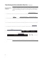
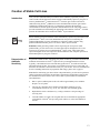
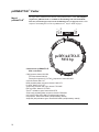
pcDNA4/TO-E™ Echo™-Adapted
Expression Vector
For cloning a gene of interest using the Echo™
Cloning System and inducible expression in
mammalian cells using the T-REx™ System.
Catalog nos. ET460-XX
Version F
28 October 2010
25-0327
Corporate Headquarters
Invitrogen Corporation
1600 Faraday Avenue
Carlsbad, CA 92008
T: 1 760 603 7200
F: 1 760 602 6500
E: [email protected]
For country-specific contact information visit our web site at www.invitrogen.com
User Manual
ii
Table of Contents
Table of Contents.................................................................................................................................................. iii
Kit Contents and Storage ........................................................................................................................................v
Introduction ..................................................................................................................1
Overview ................................................................................................................................................................1
Methods ........................................................................................................................5
Recombining Your Gene into pcDNA4/TO-E™ .....................................................................................................5
Transforming the Recombination Reaction ............................................................................................................6
Transfection and Analysis ....................................................................................................................................10
Creation of Stable Cell Lines................................................................................................................................13
Appendix.....................................................................................................................16
Recipes..................................................................................................................................................................16
pcDNA4/TO-E™ Vector .......................................................................................................................................18
pcDNA4/TO-E/Uni-lacZ™....................................................................................................................................20
Zeocin™.................................................................................................................................................................21
Product Qualification............................................................................................................................................23
Related Products ...................................................................................................................................................24
Purchaser Notification ..........................................................................................................................................26
Technical Service .................................................................................................................................................29
References ............................................................................................................................................................30
iii
iv
Kit Contents and Storage
Several pcDNA4/TO-E™ Echo™ Cloning System Kits are available. The table below lists
the kits that include the pcDNA4/TO-E™ Echo™-Adapted Expression Vector.
Types of Kits
Kit
pcDNA4/TO-E™ Echo™-Adapted
Expression Vector Kit
Reagents Supplied
pcDNA4/TO-E™ vector
Catalog nos.
ET460-01
Expression control vector
Cre Recombinase and 10X buffer
CMV Forward Primer
™
™
pcDNA4/TO-E Echo -Adapted
Expression Vector Kit with a choice
of Donor Vector Kit and One Shot®
TOP10 Chemically Competent
E. coli (see page 25 for more
information on donor vectors)
pUni/V5-His TOPO® TA Cloning Kit
®
ET460-10C
pUniBlunt/V5-His TOPO Cloning Kit
ET460-20C
pUni/V5-His A, B, and C
ET460-30C
®
pUniD/V5-His TOPO Cloning Kit
ET460-40C
Shipping/Storage
The pcDNA4/TO-E™ Echo™-Adapted Vector Kit is shipped on dry ice. Upon receipt,
store the pcDNA4/TO-E™ reagents at -20°C. Store the One Shot® Competent E. coli at
-80°C.
pcDNA4/TO-E™
Reagents
The pcDNA4/TO-E™reagents are listed below. Store at -20°C.
Item
pcDNA4/TO-E
Concentration
™
Cre Recombinase
Amount
Supercoiled, lyophilized in TE, pH 8.0
20 µg
Please check the label on the tube for exact
concentration of the enzyme
12 µl
Enzyme supplied in:
50 mM Tris-HCl, pH 8.0
5 mM EDTA
1 mM EGTA
10 mM β-mercaptoethanol
20% Glycerol
10X Recombinase Buffer
25 µl
500 mM Tris-HCl, pH 7.5
100 mM MgCl2
300 mM NaCl
1.0 mg/ml BSA
CMV Forward Primer
(21 mer)
Expression Control
Lyophilized in TE Buffer, pH 8.0
2 µg
(5´-CGCAAATGGGCGGTAGGCGTG-3´)
(306 pmoles)
Supercoiled, lyophilized in TE, pH 8.0
20 µg
™
(pcDNA4/TO-E/Uni-lacZ )
Continued on next page
v
Kit Contents and Storage, Continued
One Shot®
Reagents
(Optional)
The table below describes the items included in the One Shot® Competent E. coli kit.
Store at -80°C.
Item
Concentration
SOC Medium
2% Tryptone
(may be stored at room
temperature or at
+4°C)
0.5% Yeast Extract
Amount
6 ml
10 mM NaCl
2.5 mM KCl
10 mM MgCl2
10 mM MgSO4
20 mM glucose
Genotype of
TOP10
TOP10 E. coli
--
11 x 50 µl
pUC19 Control DNA
10 pg/µl in 5 mM Tris-HCl, 0.5 mM
EDTA, pH 8
50 µl
TOP10: Use this strain for general cloning of your gene of interest. Note: This strain
cannot be used for growth and transformation of donor vectors.
F- mcrA ∆(mrr-hsdRMS-mcrBC) φ80lacZ∆M15 ∆lacX74 recA1 araD139 ∆(araleu)7697 galU galK rpsL (StrR) endA1 nupG
vi
Introduction
Overview
Introduction
The Echo™ Cloning System allows direct recombination of your gene of interest
downstream of an appropriate promoter for expression in the host system of choice.
pcDNA4/TO-E™ is a member of the Echo™ Cloning System family of expression vectors
and is specifically designed for inducible expression in mammalian cells. The vector
allows tetracycline-regulated expression of the gene of interest in mammalian host cells
cotransfected with the pcDNA6/TR vector (Catalog no. V1025-20).
For more information about pcDNA6/TR and the T-REx™ System, please refer to the
T-REx™ System manual, our World Wide Web site (www.invitrogen.com), or call
Technical Service (see page 29).
Echo™ Cloning
System
The Echo™ Cloning System is based on the univector plasmid-fusion system (UPS) described by Elledge and coworkers to quickly and easily recombine a gene of interest into a
series of recipient (acceptor) vectors (Liu et al., 1998; Liu et al., 1999). The system consists of the univector (donor) vector containing the gene of interest and recipient (acceptor) vectors containing various regulatory sequences for expression in the host of choice.
The Echo™ System utilizes the cre-lox site-specific recombination system of bacteriophage P1 (Abremski et al., 1983; Sternberg et al., 1981a). The product of the cre gene is a
site-specific recombinase that catalyzes conservative recombination between two 34 bp
loxP or loxH sequences to resolve P1 dimers generated by replication of circular lysogens.
Plasmid Fusion
The donor vector and the acceptor vector (i.e. pcDNA4/TO-E™) each contains a lox site.
The donor vector (pUni) contains a loxP site, while the acceptor vector contains either a
loxP or a loxH site (see the next page for more information about loxH). You have already
constructed the donor vector containing the PCR product of interest via the TOPO®
Cloning method. pcDNA4/TO-E™ allows you to regulate expression of your PCR product
using the T-REx™ System. The unique loxH site is located downstream of the regulatory
sequences. By mixing the donor vector containing the PCR product of interest with
pcDNA4/TO-E™ in the presence of Cre recombinase, a plasmid fusion is created that
expresses the PCR product in mammalian cells. A generic diagram is shown below.
KanR
r
ote
om
Pr
Kg
X
lox*
e
recombinase
or
i
C
i
C
pU
pAcceptor
(2.5 to 5.8 kb)
or
Recombinant
Plasmid
(4.8 kb + gene to
8.1 kb + gene)
Cre
pU
r
ote
om
Pr
gen
R
loxP
gene
Kan
or
i
lox*
R6
Kg
R6
pUni
(2.3 kb + gene)
AmpR
AmpR
P
lox
lox* = loxP or loxH depending on acceptor vector
Continued on next page
1
Overview, Continued
loxP or loxH Sites
The sequence of the loxP site is shown below. The loxP site consists of a 34 bp sequence
containing two 13 bp inverted repeats (see underlined bases) separated by an 8 bp spacer
(Hoess et al., 1982). The inverted repeats may form a stem and loop structure that may
reduce expression of the gene of interest in some cases. A variation of the loxP site (loxH,
see below) was created to eliminate the formation of a stem and loop structure and improve
expression. Mutated bases are shown in boldface. Please note that some acceptor vectors
including pcDNA4/TO-E™ contain a loxH site. Cre-mediated recombination can still occur
between a loxP and a loxH site although the efficiency may be slightly reduced.
•
loxP: ATA ACT TCG TAT AGC ATA CAT TAT ACG AAG TTA T
•
loxH: ATT ACC TCA TAT AGC ATA CAT TAT ACG AAG TTA T
Cre Recombinase
Cre recombinase (MW = 35 kDa) is a site-specific recombinase that binds to specific
sequences (loxP and loxH sites), brings together the target sites, cleaves, and covalently
attaches to the DNA. Recombination occurs following two pairs of strand exchanges and
ligation of the DNAs in a novel (recombinant) form. A nucleophilic hydroxylated tyrosine
initiates the DNA cleavage event by attack on a specific phosphodiester bond followed by
the covalent attachment of the recombinase to the target sequence through a phosphoamino
acid bond (Abremski and Hoess, 1992; Argos et al., 1986). The reaction does not require
any host factors or ATP, but does require Mg2+ or spermidine for activity (Abremski et al.,
1983). Recombination between two supercoiled substrates, each containing a loxP or loxH
site, results in a supercoiled dimer. The extent of the reaction is 10-20% and appears to be
stoichiometric (Abremski and Hoess, 1984; Abremski et al., 1983).
Selection of
Recombinants
By fusing the two plasmids, kanamycin resistance is now linked to the pUC origin of
replication. The recombination reaction is transformed into TOP10 E. coli and
recombinants selected by plating the transformation reaction onto plates containing
kanamycin. Because the donor plasmid carries the R6Kγ origin of replication, it will not
propagate in E. coli such as TOP10 which do not carry the pir gene. In addition, the
acceptor vector, which carries the ampicillin resistance gene will not be selected. Therefore
every colony that is selected on kanamycin will represent a recombined fusion plasmid.
pcDNA4/TO-E™
pcDNA4/TO-E™ is a 5.0 kb vector derived from pcDNA4/TO and designed for high-level,
inducible expression in most mammalian hosts. The vector contains the following elements:
•
•
•
Cytomegalovirus (CMV) immediate-early promoter containing two tetracycline
operator 2 (TetO2) sites for tetracycline-regulated expression of your gene of interest in
mammalian cells (Yao et al., 1998).
A loxH site for univector plasmid fusion
Zeocin™ resistance gene for selection of stable cell lines (Mulsant et al., 1988)
• The pUC origin for high-copy replication and maintenance in most E. coli strains
• The ampicillin (bla) resistance gene for selection in E. coli
For a map and a description of pcDNA4/TO-E™, please refer to page 18.
Other Echo™-adapted acceptor vectors are available separately and are provided with their
own manuals. For more information on other available acceptor vectors, please visit our
Web site (www.invitrogen.com) or call Technical Service (see page 29).
Continued on next page
2
Overview, Continued
Repression and
Derepression
The TetO2 sequences in the pcDNA4/TO-E™ vector serve as binding sites for four Tet
repressor molecules (comprising two Tet repressor homodimers) and confer tetracyclineresponsiveness to your gene of interest. The Tet repressor is expressed from the
pcDNA6/TR plasmid. For more information about the TetO2 sequences, please see
below. For more information about the pcDNA6/TR plasmid and the Tet repressor,
please refer to the T-REx™ System manual. The T-REx™ System manual is available for
downloading from our Web site (www.invitrogen.com) or from Technical Service (see
page 29).
In the absence of tetracycline, expression of your gene of interest is repressed by the
binding of Tet repressor homodimers to the TetO2 sequences. Addition of tetracycline to
the cells derepresses the hybrid CMV/TetO2 promoter in pcDNA4/TO and allows
expression of your gene of interest
Tet Operator
Sequences
The promoters of bacterial tet genes contain two types of operator sequences, O1 and O2,
that serve as high-affinity binding sites for the Tet repressor (Hillen and Berens, 1994;
Hillen et al., 1983). Each O1 and O2 site binds to one Tet repressor homodimer. While
Tet repressor homodimers bind to both tet operators with high affinity, studies have
shown that the affinity of the Tet repressor homodimer for O2 is three- to five-fold
higher than it is for O1 (Hillen and Berens, 1994).
Tet operators have been incorporated into heterologous eukaryotic promoters to allow
tetracycline-regulated gene expression in mammalian cells (Gossen and Bujard, 1992;
Yao et al., 1998). In the T-REx™ System, two copies of the O2 operator sequence
(TetO2) were inserted into the strong CMV promoter of pcDNA4/TO-E™ to allow
regulated expression of your gene of interest by tetracycline. We use the TetO2 operator
sequence in pcDNA4/TO-E™ to maximize repression of basal gene expression. For more
detailed information about tet operators, please refer to Hillen and Berens (1994).
Yao et al. (1998) have recently demonstrated that the location of tet operator sequences
in relation to the TATA box of a heterologous promoter is critical to the function of the
tet operator. Regulation by tetracycline is only conferred upon a heterologous promoter
by proper spacing of the TetO2 sequences from the TATA box (Yao et al., 1998). For
this reason, the first nucleotide of the TetO2 operator sequence has been placed 10
nucleotides after the last nucleotide of the TATA element in the CMV promoter in
pcDNA4/TO-E™. Please refer to the diagram on page 8 for the sequence and placement
of the TetO2 sequences in relation to the TATA box.
In other tetracycline-regulated systems, the TetO2 sequences are located upstream of the
TATA element in the promoter of the inducible expression vector (Gossen and Bujard,
1992). These systems differ substantially from the T-REx™ System in that they use
regulatory molecules composed of the Tet repressor fused to a viral transactivation
domain. The presence of viral transactivation domains appears to overcome the
requirement for specific positioning of the TetO2 sequences in relation to the TATA box
of the heterologous promoter. However, the presence of viral transactivation domains
has been found to have deleterious effects in some mammalian cell lines.
Continued on next page
3
Overview, Continued
Experimental
Outline
The table below describes the general steps needed to recombine, transform, and express
your protein of interest.
Step
4
Action
Page
1
Perform the recombination reaction using your donor vector and
pcDNA4/TO-E™.
5
2
Transform the recombination reaction into competent TOP10 E. coli
cells.
6
3
Select transformants on LB plates containing 50 µg/ml kanamycin.
7
4
Analyze transformants by restriction digestion.
7
5
Select the correct clone and cotransfect your construct and pcDNA6/TR 10
into the mammalian cell line of interest using your method of choice.
6
Induce expression of your recombinant protein with tetracycline and
analyze by western blot or functional assay.
10
7
Generate a double stable cell line, if desired.
13
8
Purify your protein, if desired.
15
Methods
Recombining Your Gene into pcDNA4/TO-E™
Introduction
At this point, you should have a plasmid preparation of your donor vector construct in
addition to pcDNA4/TO-E™. Please review the information below and on the next page
before performing the recombination reaction.
Preparation and
Maintenance of
pcDNA4/TO-E™
To prepare pcDNA4/TO-E™ for use, add 20 µl sterile water to create a 1 µg/µl stock
solution. You can further dilute a small aliquot of plasmid or use the stock solution as is.
Store the stock solution at -20°C when you are finished.
If you wish to propagate the pcDNA4/TO-E™ plasmid or prepare plasmid DNA, you
may transform the plasmid into TOP10 E. coli as described on page 6. Use 10-100 ng of
plasmid DNA for transformation and select transformants on LB plates containing 50 to
100 µg/ml ampicillin. Be sure to prepare a glycerol stock of your plasmid-containing
TOP10 strain for long-term storage (see page 9).
Before Starting
Recombination
Reaction
You will need the following reagents and equipment.
•
100 ng of your donor vector construct
•
100 ng of pcDNA4/TO-E™ (included in kit)
•
Microcentrifuge tubes
•
Heat blocks set at 37°C and 65°C
•
Ice bucket with ice
•
Cre recombinase (included in the kit)
•
10X Recombinase Buffer (included in the kit)
1.
Set up each 20 µl recombination reaction on ice as follows:
x µl
Donor vector (100 ng)
pcDNA4/TO-E (100 ng)
y µl
10X Recombinase Buffer
2 µl
™
Deionized water
add to a total volume of 17 µl
Cre Recombinase
Final Volume
1 µl
20 µl
2.
Incubate at 37°C for 20 minutes.
3.
Incubate at 65°C for 5 minutes to inactivate the recombinase.
4.
Place the tube on ice and proceed to Transformation, next page. If you run out of
time, you may store the recombination reaction at +4°C or -20°C overnight. Longer
storage times have not been tested.
5
Transforming the Recombination Reaction
Introduction
Once you have performed the recombination reaction, you are ready to transform your
E. coli host. We recommend using TOP10 E. coli (available with the kit) for
transformation, but other strains are suitable. E. coli strains should be endonuclease A
deficient (endA) and recombination deficient (recA) to ensure quality plasmid
preparations and reduce the chances of recombination, respectively.
Materials Supplied In addition to general microbiological supplies (i.e. plates, spreaders), you will need the
following reagents and equipment.
by the User
Important
Preparation for
Transformation
•
42°C water bath
•
LB plates containing 50 µg/ml kanamycin (see Important, below)
•
37°C shaking and non-shaking incubator
•
SOC (supplied in the One Shot® kit)
It is important to select for the fusion plasmid using kanamycin. Remember that the
donor vector contains the R6Kγ origin. This origin can only be maintained in E. coli
strains containing the pir gene. After the donor vector and pcDNA4/TO-E™ acceptor
vectors have recombined to form the fusion plasmid, the kanamycin resistance gene
(from the donor vector) is linked to the pUC origin (from pcDNA4/TO-E™). The fusion
plasmid can be maintained in E. coli strains that do not contain the pir gene (i.e. TOP10).
By selecting for kanamycin resistance, you ensure that only colonies containing the
fusion vector are selected.
The following transformation protocol is for use with the TOP10 One Shot® competent
cells available with the kit. If you are using other competent cells, please follow the
manufacturer’s protocol.
For each transformation, you will need one vial of One Shot® TOP10 competent cells and
two selective plates. Perform the following steps before beginning.
•
Equilibrate a water bath to 42°C.
•
Thaw the vial of SOC medium from the One Shot® box and bring to room temperature.
•
Warm LB plates containing 50 µg/ml kanamycin at 37°C for 30 minutes.
•
Thaw on ice 1 vial of One Shot® cells for each transformation.
Continued on next page
6
Transforming the Recombination Reaction, Continued
One Shot®
Transformation
Reaction
Analysis of
Positive Clones
1.
Add 5 µl of the recombination reaction to a vial of One Shot® TOP10 E. coli and mix
gently. Do not mix by pipetting up and down.
2.
Heat-shock the cells for 30 seconds at 42°C without shaking.
3.
Immediately transfer the tubes to ice.
4.
Add 500 µl of room temperature SOC medium.
5.
Cap the tube tightly and shake the tube horizontally at 37°C for 45 minutes.
6.
Spread 50 µl from each transformation onto a prewarmed LB plate containing
50 µg/ml kanamycin. Pellet the remaining cells, resuspend the cell pellet in 50 µl
SOC and plate. Incubate overnight at 37°C.
7.
An efficient recombination reaction will produce hundreds of colonies. Pick
~5 colonies for analysis.
1.
Culture the 5 colonies (see previous page) overnight in 2-5 ml LB or SOB medium
containing 50 µg/ml kanamycin. See pages 16-17 for recipes for LB and SOB
medium.
2.
Isolate plasmid DNA using your method of choice. If you need ultra-pure plasmid
DNA for automated or manual sequencing, we recommend the S.N.A.P. ™ MiniPrep
Kit (10-15 µg DNA, Catalog no. K1900-01) or the S.N.A.P. ™ MidiPrep Kit (10200 µg DNA, Catalog no. K1910-01).
3.
Analyze the plasmids by restriction analysis. Use an enzyme or enzymes that cut once
in the donor vector and once in the acceptor vector to yield two fragments that are
distinguishable from one another. Please note that other strategies are possible.
4.
(Optional) To sequence the fusion plasmid to confirm the fusion junctions, use the
CMV Forward and Uni1 Forward sequencing primers. Please refer to the diagram on
the following page for the sequence around the pcDNA4/TO-E™ loxH site. Refer to
the donor vector manual for the sequence around the donor vector loxP site.
If you need help with setting up restriction enzyme digests or DNA sequencing, please
refer to general molecular biology texts (Ausubel et al., 1994; Sambrook et al., 1989).
7
Transforming the Recombination Reaction, Continued
Sequencing Your
Construct
The sequence surrounding your insert is shown below. Unique restriction sites are labeled
to indicate the cleavage site. Please note that the complete sequence of pcDNA4/TO-E™ is
available for downloading from our Web site (www.invitrogen.com) or from Technical
Service.
CMV Forward priming site
721 AAAATCAACG GGACTTTCCA AAATGTCGTA ACAACTCCGC CCCATTGACG CAAATGGGCG GTAGGCGTGT
Tetracycline operator (TetO2)
TATA box
Tetracycline operator (TetO2)
791 ACGGTGGGAG GTCTATATAA GCAGAGCTCT CCCTATCAGT GATAGAGATC TCCCTATCAG TGATAGAGAT
861 CGTCGACGAG CTCGTTTAGT GAACCGTCAG ATCGCCTGGA GACGCCATCC ACGCTGTTTT GACCTCCATA
931 GAAGACACCG GGACCGATCC AGCCTCCGGA CTCTAGCGTT TAAACTTAAG CTT ATT ACC TCA
loxH site
1001 TAT AGC ATA CAT TAT ACG AAG TTA T
Gene of
interest
C-terminal tag
(optional)
Uni1 Forward priming site
donor vector
Apa I
loxP site
donor vector
Pme I
GGG CCCGTTTAAA CCCGCTGATC
AGCCTCGACT GTGCCTTCTA GTTGCCAGCC ATCTGTTGTT TGCCCCTCCC CCGTGCCTTC
Continued on next page
8
Transforming the Recombination Reaction, Continued
Fusion Vector
Analysis
It should be clear from restriction analysis that you have a dimer plasmid consisting of the
donor vector and pcDNA4/TO-E™. Occasionally, trimers will result. Trimers usually
consist of two donor vector molecules and one acceptor molecule. In theory, trimers may
result from two sequential fusion events or a single fusion event between a pre-existing
monomeric substrate and a dimeric substrate. The production of trimers can be eliminated if
gel-purified monomeric supercoiled DNA is used in the recombination reaction. Please
note that trimers usually express as well as the dimer product.
Preparing a
Glycerol Stock for
Long-Term
Storage
Once you have identified the correct clone, prepare a glycerol stock for long term storage.
1. Streak out the original colony on LB plates containing 50 µg/ml kanamycin to isolate
single colonies.
2. Select a single colony and inoculate into 1-2 ml of LB containing 50 µg/ml kanamycin.
3. Grow overnight until culture is saturated.
4. Mix 0.85 ml of culture with 0.15 ml of sterile glycerol and transfer to a cryovial.
5.
Store at -80°C. (You may also want to store a stock of plasmid DNA at -20°C.)
9
Transfection and Analysis
Introduction
Once you have created the pcDNA4/TO-E™ fusion plasmid, have verified its integrity,
and have prepared clean plasmid preparations of both the fusion plasmid and
pcDNA6/TR, you are ready to cotransfect the plasmids into the mammalian cell line of
choice. Please refer to the T-REx™ System manual for complete information on
pcDNA6/TR, transfection, and induction of expression. We recommend that you include
the positive expression control vector and a mock transfection (negative control) to
evaluate your results.
Plasmid
Preparation
Plasmid DNA for transfection into eukaryotic cells must be very clean and free from
phenol and sodium chloride. Contaminants will kill the cells, and salt will interfere with
lipids decreasing the transfection efficiency. We recommend isolating plasmid DNA
using the S.N.A.P. ™ MidiPrep Kit (Catalog no. K1910-01) or other resin-based method.
Important
Because tetracycline-regulated expression in the T-REx™ System is based on a repression/
derepression mechanism, the amount of Tet repressor that is expressed in the host cell line
from pcDNA6/TR will determine the level of transcriptional repression of the Tet operator
sequences in your pcDNA4/TO-E™ fusion vector. Tet repressor levels should be
sufficiently high to suitably repress basal level transcription. We recommend that you
cotransfect your mammalian host cell line with a ratio of at least 6:1 (w/w)
pcDNA6/TR:pcDNA4/TO-E™ fusion vector DNA. You may want to try varying ratios of
pcDNA6/TR:pcDNA4/TO-E™ fusion vector to optimize repression and expression for
your particular cell line and your gene of interest.
General guidelines are provided below to cotransfect your pcDNA4/TO-E™ fusion vector
Cotransfection
and Induction with (or the control plasmid) and pcDNA6/TR into your cell line of interest and to induce
expression of your protein of interest with tetracycline. Please refer to the T-REx™
Tetracycline
System manual for more information on transfection and the preparation and handling of
tetracycline.
•
Use cells that are approximately 60% confluent for transfection.
•
Cotransfect your pcDNA4/TO-E™ fusion vector and pcDNA6/TR at a ratio of 6:1
(w:w) into the cell line of choice using your preferred method. Absolute amounts of
plasmid used for transfection will vary depending on the method of transfection and
the cell line used.
•
After transfection, add fresh medium and allow the cells to recover for 24 hours
before induction.
•
Remove medium and add fresh medium containing the appropriate concentration of
tetracycline to the cells. In general, we recommend that you add tetracycline to a
final concentration of 1 µg/ml (5 µl of a 1 mg/ml stock solution per 5 ml of medium)
to the cells and incubate the cells for 24 hours at 37°C.
•
Harvest the cells and assay for expression of your gene of interest.
Continued on next page
10
Transfection and Analysis, Continued
Positive
Expression
Control
pcDNA4/TO-E/Uni-lacZ™ is provided as a positive control vector for mammalian cell
transfection and expression and may be used to optimize transfection conditions for your
cell line. Cotransfection of the positive control vector and pcDNA6/TR results in the
expression of β-galactosidase following the addition of tetracycline. A successful
cotransfection will result in positive β-galactosidase expression and can be easily assayed
by staining with X-gal (see below).
Assay for
β-galactosidase
Activity
You may assay for β-galactosidase expression by activity assay using cell-free lysates
(Miller, 1972) or by staining the cells for activity. Invitrogen offers the β-Gal Assay Kit
(Catalog no. K1455-01) and the β-Gal Staining Kit (Catalog no. K1465-01) for fast and
easy detection of β-galactosidase expression.
Detection of
β-Galactosidase
If you wish to detect expression of β-galactosidase by western blot, you may use an
antibody to β-galactosidase.
Detection of
Recombinant
Fusion Proteins
If you have expressed your protein as a fusion to the C-terminal V5 epitope and
polyhistidine (6xHis) tag, you can detect expression using the Anti-V5 or Anti-His(C-term)
antibodies (see page 25 for ordering information). You may also use an antibody to your
protein of interest.
To detect your fusion protein by western blot, you will need to prepare a cell lysate from
transfected cells. We recommend that you perform a time course to optimize expression of
your fusion protein (e.g. 12, 24, 48, 72 hours, etc. after transfection). Use the protocol
below to lyse cells. Other protocols and lysis buffers are also suitable.
1.
Wash cell monolayers (~5 x 10⁵ to 1 x 10⁶ cells) once with phosphate-buffered saline
(PBS, see recipe on the next page).
2.
Scrape cells into 1 ml PBS and pellet the cells at 1500 x g for 5 minutes.
3.
Resuspend pellet in 50 µl Cell Lysis Buffer (see recipe on the next page) and vortex.
4.
Incubate cell suspension at 37°C for 10 minutes to lyse the cells. Note: You may
prefer to lyse the cells at room temperature or on ice if degradation of your protein is a
potential problem.
5.
Centrifuge the cell lysate at 10,000 x g for 10 minutes to pellet nuclei and transfer the
supernatant to a fresh tube. Assay the lysate for protein concentration. Note: Do not
use protein assays utilizing Coomassie Blue or other dyes. NP-40 interferes with the
binding of the dye with the protein.
6.
Add SDS-PAGE sample buffer to a final concentration of 1X and boil the sample for 5
minutes.
7.
Load 20 µg of lysate onto an SDS-PAGE gel and electrophorese. Use the appropriate
percentage of acrylamide to resolve your fusion protein.
Continued on next page
11
Transfection and Analysis, Continued
The C-terminal peptide containing the V5 epitope and the polyhistidine (6xHis) tag will
add approximately 5 kDa to the size of your protein.
Cell Lysis Buffer
50 mM Tris, pH 7.8
150 mM NaCl
1% Nonidet P-40
1.
This solution can be prepared from the following common stock solutions. For
100 ml, combine:
1 M Tris base
5 ml
5 M NaCl
3 ml
Nonidet P-40
1 ml
2.
Bring the volume up to 90 ml with deionized water and adjust the pH to 7.8 with HCl.
3.
Bring the volume up to 100 ml. Store at room temperature.
Note: Just before use, add protease inhibitors to a small amount of buffer at the following
final concentrations:
1 mM PMSF
1 µg/ml pepstatin
1 µg/ml leupeptin
PhosphateBuffered Saline
(PBS)
137 mM NaCl
2.7 mM KCl
10 mM Na2HPO4
1.8 mM KH2PO4
1.
Dissolve the following in 800 ml of deionized water:
8 g NaCl
0.2 g KCl
1.44 g Na2HPO4
0.24 g KH2PO4
12
2.
Adjust pH to 7.4 with concentrated HCl.
3.
Bring the volume up to 1 liter and autoclave for 20 minutes on liquid cycle.
4.
Store at +4°C or room temperature.
Creation of Stable Cell Lines
Introduction
Once you have established that your construct can be inducibly expressed, you may
create a stable cell line that carries the tet repressor and inducibly expresses your gene of
interest. pcDNA4/TO-E™ contains the Zeocin™ resistance gene to allow selection of
stable lines using Zeocin™, pcDNA6/TO carries the blasticidin resistance gene. Before
establishing a double-stable cell line, it is important to determine the minimum
concentration of selection agents required to kill untransfected cells. The protocol below
provides information for determining the appropriate concentration of Zeocin™. A similar
protocol for blasticidin can be found in the T-REx™ System manual.
Please note that your gene of interest will be constitutively expressed if you transfect your
pcDNA4/TO-E™ fusion vector into mammalian host cells prior to transfecting the
pcDNA6/TR plasmid. For more information on selection of stable cell lines using
pcDNA6/TR and blasticidin, please refer to the T-REx™ System manual.
Reminder: When generating a stable cell line expressing the Tet repressor (from
pcDNA6/TR), you will want to select for clones that express the highest levels of Tet
repressor to use as hosts for your pcDNA4/TO-E™ fusion vector. Those clones that
express the highest levels of Tet repressor should exhibit the most complete repression of
basal transcription of your gene of interest.
Determination of
Antibiotic
Sensitivity
To generate a stable cell line expressing your protein of interest, you need to determine the
minimum concentration of Zeocin™ required to kill your untransfected host cell line.
Typically, concentrations between 50 and 1000 µg/ml Zeocin™ are sufficient to kill the
untransfected host cell line. Test a range of concentrations (see below) to ensure that you
determine the minimum concentration necessary for your cell line. For more information on
Zeocin™ including instructions on preparation and storage, please refer to page 21.
Note: Before transfecting your host cell line with pcDNA6/TR, you will need to perform a
similar experiment to determine the minimum concentration of blasticidin required to kill
the untransfected cell line. Please refer to the T-REx™ System manual for information
about blasticidin.
•
Plate or split a confluent plate so the cells will be approximately 25% confluent.
Prepare a set of 7 plates.
•
The next day, substitute culture medium with medium containing varying
concentrations of Zeocin™ (e.g. 0, 50, 125, 250, 500, 750, and 1000 µg/ml).
•
Replenish the selective medium every 3-4 days, and observe the percentage of
surviving cells.
•
Count the number of viable cells at regular intervals to determine the appropriate
concentration of Zeocin™ that prevents growth within 1-2 weeks after addition of
Zeocin™.
13
Creation of Stable Cell Lines, Continued
Effect of Zeocin™
on Sensitive and
Resistant Cells
The method of killing of Zeocin™ is quite different from blasticidin, neomycin, and
hygromycin. Cells do not round up and detach from the plate. Sensitive cells may exhibit
the following morphological changes upon exposure to Zeocin™:
•
Vast increase in size (similar to the effects of cytomegalovirus infecting permissive cells)
•
Abnormal cell shape
•
Presence of large empty vesicles in the cytoplasm (breakdown of the endoplasmic
reticulum and golgi apparatus or scaffolding proteins)
•
Breakdown of plasma and nuclear membrane (appearance of many holes in these
membranes)
Eventually, these "cells" will completely break down and only "strings" of protein will remain.
Zeocin™-resistant cells should continue to divide at regular intervals to form distinct colonies.
There should not be any distinct morphological changes in Zeocin™-resistant cells when
compared to cells not under selection with Zeocin™. For more information about Zeocin™,
please see page 21.
Plasmid
Linearization
We have found that it is not necessary to linearize the pcDNA4/TO-E™ fusion vector prior
to transfection.
Selection of Stable Once you have determined the appropriate Zeocin™ concentration to use for selection, you
can generate a stable cell line expressing pcDNA6/TR and your pcDNA4/TO-E™ fusion
Integrants
vector. We recommend that you first generate a stable cell line expressing pcDNA6/TR and
then use this cell line as the host for your pcDNA4/TO-E™ fusion vector.
1.
Once you have obtained a stable cell line expressing the Tet repressor, follow the steps
below to transfect your stable cell line with the pcDNA4/TO-E™ fusion vector. Use
Zeocin™ to select for double stable clones. Remember to maintain your cells in
medium containing blasticidin as well.
2.
Transfect your cell line of choice with your pcDNA4/TO-E™ fusion vector using the
desired protocol. Include a sample of untransfected cells as a negative control.
3.
24 hours after transfection, wash the cells and add fresh medium to the cells.
4.
48 hours after transfection, split the cells into fresh medium containing Zeocin™ at the
appropriate concentration for your cell line. Split the cells such that they are no more
than 25% confluent. If the cells are too dense, the antibiotic will not kill the
untransfected cells.
5.
Replenish selective medium every 3-4 days until Zeocin™-resistant colonies are
detected.
6.
Pick at least 20 foci and expand them to test for tetracycline-inducible gene expression.
Continued on next page
14
Creation of Stable Cell Lines, Continued
Dual Selection of
Stable Integrants
If you wish to select for stable cell lines by dual selection, you may cotransfect your
pcDNA4/TO-E™ fusion vector and pcDNA6/TR into your cell line of choice and select
with Zeocin™ and blasticidin. Pick and expand at least 40 foci to screen for tetracyclineregulated expression of your gene of interest.
Purification
If you have expressed your protein as a fusion to the C-terminal polyhistidine (6xHis) tag,
you can purify it using ProBond™ Resin (or other metal-chelating resin). Please refer to
the manufacturer’s instructions before attempting to purify your fusion protein.
Preparation of
Cells for Lysis
Use the procedure below to prepare cells for lysis if you will be purifying your protein on
ProBond™ Resin. You will need 5 x 10⁶ to 1 x 10⁷ stably transfected cells for purification
of your protein on a 2 ml ProBond™ column (see ProBond™ Protein Purification manual).
1.
Lysis of Cells
Seed cells in either five T-75 flasks or 2 to 3 T-175 flasks.
2.
Grow the cells in selective medium until they are approximately 80% confluent.
3.
Harvest the cells by treating with trypsin-EDTA for 2 to 5 minutes or by scraping the
cells in PBS.
4.
Inactivate the trypsin by diluting with fresh medium (if necessary) and transfer the
cells to a microcentrifuge tube.
5.
Centrifuge the cells at 1500 rpm for 5 minutes. Resuspend the cell pellet in PBS.
6.
Centrifuge the cells at 1500 rpm for 5 minutes. You may lyse the cells immediately or
freeze in liquid nitrogen and store at -70°C until needed.
If you are using ProBond™ resin, refer to the ProBond™ Protein Purification manual for
details about sample preparation for chromatography.
If you are using other metal-chelating resin, please refer to the manufacturer’s instructions
for recommendations on sample preparation.
15
Appendix
Recipes
LB (Luria-Bertani)
Medium and
Plates
Composition:
1.0% Tryptone
0.5% Yeast Extract
1.0% NaCl
pH 7.0
1.
For 1 liter, dissolve 10 g tryptone, 5 g yeast extract, and 10 g NaCl in 950 ml
deionized water.
2.
Adjust the pH of the solution to 7.0 with NaOH and bring the volume up to 1 liter.
3.
Autoclave on liquid cycle for 20 minutes at 15 psi. Allow solution to cool to 55°C
and add antibiotic if needed.
4.
Store at room temperature or at +4°C.
LB agar plates
Low Salt LB
Medium with
Zeocin™
1.
Prepare LB medium as above, but add 15 g/L agar before autoclaving.
2.
Autoclave on liquid cycle for 20 minutes at 15 psi.
3.
After autoclaving, cool to ~55°C, add antibiotic (50 µg/ml of kanamycin), and pour
into 10 cm plates.
4.
Let harden, then invert and store at +4°C, in the dark.
™
For Zeocin to be active, the salt concentration of the medium must be low (< 90 mM) and
the pH must be 7.5. Use the medium below to prepare plates and liquid medium for selection
in E. coli. Failure to use low salt LB medium will result in non-selection due to
inactivation of the drug.
10 g Tryptone
5 g NaCl
5 g Yeast Extract
1.
2.
3.
4.
5.
Combine the dry reagents above and add deionized, distilled water to 950 ml. Adjust
pH to 7.5 with 5 M NaOH. Bring the volume up to 1 liter. For plates, add 15 g/L agar
before autoclaving.
Autoclave on liquid cycle for 20 minutes.
™
Thaw Zeocin on ice and vortex before removing an aliquot.
Allow the medium to cool to at least 55°C before adding the Zeocin to 25-50 µg/ml
final concentration.
™
Store plates at +4°C in the dark. Plates containing Zeocin are stable for 1-2 weeks.
™
Continued on next page
16
Recipes, Continued
SOB Medium (with SOB (per liter)
Kanamycin)
2% Tryptone
0.5% Yeast Extract
0.05% NaCl
2.5 mM KCl
10 mM MgCl2
1.
Dissolve 20 g tryptone, 5 g yeast extract, and 0.5 g NaCl in 950 ml deionized water.
2.
Make a 250 mM KCl solution by dissolving 1.86 g of KCl in 100 ml of deionized
water. Add 10 ml of this stock KCl solution to the solution in Step 1.
3.
Adjust pH to 7.5 with 5 M NaOH and add deionized water to 1 liter.
4.
Autoclave this solution, cool to ~55°C, and add 10 ml of sterile 1 M MgCl2. You may
also add kanamycin to 50 µg/ml.
5.
Store at +4°C. Medium is stable for only ~1 month.
17
pcDNA4/TO-E™ Vector
Apa I
Pme I
The figure below summarizes the features of the pcDNA4/TO-E™ vector. The complete
sequence for pcDNA4/TO-E™ is available for downloading from our World Wide
Web site (www.invitrogen.com) or from Technical Service (see page 29) Details of the
sequences surrounding the loxH site in pcDNA4/TO-E™ may be found on page 8.
Pme I
Afl II
Hind III
Map of
pcDNA4/TO-E™
loxH
BGH pA
f1
or
i
P
ri
40 o
SV
CM
V
O2
Tet
2X
5032 bp
EM7
A m p i cil li
pcDNA4/TO-E
ci
n
n
UC
Ze
p
Comments for pcDNA4/TO-E:
5032 nucleotides
o ri
S
o
A
V40 p
CMV promoter: bases 232-958
TATA box: bases 804-810
Tetracycline operator 2 (2X TetO2) sequences: bases 820-859
loxH site: bases 984-1017
BGH polyadenylation sequence: bases 1049-1273
f1 origin: bases 1319-1747
SV40 early promoter and origin: bases 1752-2095
EM7 promoter: bases 2137-2203
Zeocin resistance gene: bases 2204-2578
SV40 early polyadenylation sequence: bases 2708-2838
pUC origin: bases 3221-3891 (complementary strand)
bla promoter: bases 4897-4995 (complementary strand)
Ampicillin (bla) resistance gene: bases 4036-4896 (complementary strand)
TM
Continued on next page
18
pcDNA4/TO-E™ Vector, Continued
Features of
pcDNA4/TO-E™
pcDNA4/TO-E™ (5032 bp) contains the following elements. All features have been
functionally tested.
Feature
Benefit
Human cytomegalovirus (CMV)
immediate early promoter
Permits high-level expression of your gene
of interest (Andersson et al., 1989; Boshart
et al., 1985; Nelson et al., 1987).
CMV Forward priming site
Allows sequencing in the sense orientation
Tetracycline operator (O2) sequences
Two tandem 19 nucleotide repeats which
serve as binding sites for Tet repressor
homodimers (Hillen and Berens, 1994)
loxH site
Allows recombination between the donor
vector and pcDNA4/TO-E™ (Hoess et al.,
1982)
BGH reverse priming site
Permits sequencing through the insert
Bovine growth hormone (BGH)
polyadenylation sequence
Permits efficient transcription termination
and polyadenylation of mRNA (Goodwin
and Rottman, 1992).
f1 origin
Allows rescue of single-strand DNA
SV40 early promoter and origin
Allows efficient, high-level expression of
the neomycin resistance gene and episomal
replication in cells expressing SV40 large
T antigen
EM-7 promoter
Synthetic prokaryotic promoter for
expression of the Zeocin™ resistance gene
in E. coli
Permits selection of stable transfectants in
Zeocin™ resistance (Sh ble ) gene
(expressed from the SV40 early promoter mammalian cells and transformants in
E. coli (Drocourt et al., 1990; Mulsant
or the EM-7 promoter)
et al., 1988)
SV40 early polyadenylation signal
Allows efficient transcription termination
and polyadenylation of mRNA
pUC origin
Permits high-copy number replication and
growth in E. coli
bla promoter
Allows expression of the ampicillin (bla)
resistance gene
Ampicillin resistance gene (β-lactamase)
Allows selection of transformants in E. coli
19
pcDNA4/TO-E/Uni-lacZ™
Description
pcDNA4/TO-E/Uni-lacZ™ is a 10405 bp expression control vector that contains the lacZ
gene (3056 bp). The lacZ gene was amplified and TOPO® Cloned into pUni/V5His/Gene-TOPO®. The resulting vector was recombined with pcDNA4/TO-E™ using cre
recombinase to create pcDNA4/TO-E/Uni-lacZ™. Note: pUni/V5-His/Gene-TOPO® is
similar to pUni/V5-His-TOPO® except that it contains additional DNA between the
TOPO® Cloning site and the V5 epitope.
™
Map of Expression The figure below summarizes the features of the pcDNA4/TO-E/Uni-lacZ vector. The
™
complete nucleotide sequence for pcDNA4/TO-E/Uni-lacZ is available for
Control Vector
downloading from our World Wide Web site (www.invitrogen.com) or by
contacting Technical Service (see page 29).
MV
PC
2X
TetO 2
loxH
Lac
Z
H
BG
Am
pi
c
pA
ori
R6
Kg
40
ox
P
SV
pA
40
SV
ri
oc
in
Ori
p UC o
Ze
EM 7
SV40 ori
CMV promoter: bases 232-958
TATA box: bases 804-810
Tetracycline operator 2 (2X TetO2) sequences: bases 820-859
loxH site: bases 984-1017
LacZ ORF: bases 1041-4097
BGH polyadenylation sequence: bases 4260-4468
Kanamycin promoter: bases 5585-5722 (complementary strand)
Kanamycin resistance gene: bases 4790-5584 (complementary strand)
R6Kg origin: bases 5940-6342
loxP site: bases 6357-6390
BGH polyadenylation sequence: bases 6422-6646
f1 origin: bases 6692-7120
SV40 early promoter and origin: bases 7125-7468
EM7 promoter: bases 7510-7576
Zeocin resistance gene: bases 7577-7951
SV40 early polyadenylation sequence: bases 8081-8211
pUC origin: bases 8594-9264 (complementary strand)
bla promoter: bases 10270-10368 (complementary strand)
Ampicillin (bla) resistance gene: bases 9409-10269 (complementary strand)
TM
20
Kanamycin
pA
i l li
n
Comments for pcDNA4/TO-E/Uni-lacZ:
10405 nucleotides
pcDNA4/TO-E/Uni-lacZ
10405 bp
ri
f1 o
BG
A
Hp
l
Zeocin™
Zeocin™
Zeocin™ belongs to a family of structurally related bleomycin/phleomycin-type
antibiotics isolated from Streptomyces. Antibiotics in this family are broad spectrum
antibiotics that act as strong antibacterial and antitumor drugs. They show strong toxicity
against bacteria, fungi (including yeast), plants, and mammalian cells (Baron et al., 1992;
Drocourt et al., 1990; Mulsant et al., 1988; Perez et al., 1989).
The Zeocin™ resistance protein has been isolated and characterized (Calmels et al., 1991;
Drocourt et al., 1990). This protein, the product of the Sh ble gene (Streptoalloteichus
hindustanus bleomycin gene), is a 13.7 kDa protein that binds Zeocin™ and inhibits its
DNA strand cleavage activity. Expression of this protein in eukaryotic and prokaryotic
hosts confers resistance to Zeocin™.
Molecular Weight,
Formula, and
Structure
The formula for Zeocin™ is C60H89N21O21S3 and the molecular weight is 1,535. The
diagram below shows the structure of Zeocin™.
H
CONH2
H2
N
N
H
O
H
N
CH3
HO
N
O
++
Cu
N
H
N
H
N
O
O
N
O
NH
O
N
H2N
H
N
CH3
HO
R
S
N
S
CH3
H
OH
O
O
CH3
R =
NH2
N
HN
NH
NH2
OH
H2N
O
O
HO
O
MW = 1,535
O
HO
Applications of
Zeocin™
OH
OH
Zeocin™ is used for selection in mammalian cells (Mulsant et al., 1988), plants (Perez
et al., 1989), yeast (Baron et al., 1992), and prokaryotes (Drocourt et al., 1990).
Suggested concentrations of Zeocin™ for selection in mammalian cell lines and E. coli
are listed below:
Organism
Zeocin™ Concentration and Selective Medium
E. coli
25-50 µg/ml in low salt LB medium*
Mammalian Cells
50-1000 µg/ml (varies with cell line)
*Efficient selection requires that the concentration of NaCl be no more than 5 g/liter (< 90 mM).
Continued on next page
21
Zeocin™, Continued
Handling Zeocin™
Preparing and
Storing Zeocin™
22
•
High salt and acidity or basicity inactivate Zeocin™. Therefore, we recommend
that you reduce the salt in bacterial medium and adjust the pH to 7.5 to keep the drug
active (see Low Salt LB Medium, page 16). Please note that the pH and salt
concentration do not need to be adjusted when preparing tissue culture medium
containing Zeocin™.
•
Store Zeocin™ at -20°C and thaw on ice before use.
•
Zeocin™ is light sensitive. Store the drug, and plates or medium containing drug, in
the dark at +4°C. Culture medium containing Zeocin™ may be stored at +4°C
protected from exposure to light for up to 1 month.
•
Wear gloves, a laboratory coat, and safety glasses or goggles when handling
Zeocin™-containing solutions.
•
Zeocin™ is toxic. Do not ingest or inhale solutions containing the drug.
Zeocin™ is available from Invitrogen (see page 24 for ordering information). For your
convenience, Zeocin™ is prepared in autoclaved, deionized water in 1.25 ml aliquots at
a concentration of 100 mg/ml. The stability of Zeocin™ is guaranteed for six months, if
stored at -20°C protected from exposure to light.
Product Qualification
Vectors
pcDNA4/TO-E™ and pcDNA4/TO-E/Uni-lacZ™ are qualified by restriction digest. The
table below lists the restriction enzymes and the expected fragments.
pcDNA4/TO-E™
Restriction Enzyme
pcDNA4/TO-E/Uni-lacZ™
Avr II (linearizes)
5032 bp
Not tested
Bgl II
4208 bp, 824 bp
Not tested
Hind III
5032 bp
5327 bp, 5078 bp
BamH I (linearizes)
Not tested
10405 bp
Pme I
Not tested
5429 bp, 4976 bp
Primers
The CMV Forward Sequencing Primer has been lot-qualified by DNA sequencing
experiments using the dideoxy chain termination technique.
Cre Recombinase
Purity: >95% homogeneity
Endonuclease activity: Negative
Exonuclease activity: Negative
Functional Assay: Cre recombinase is qualified using the assay on page 23 of this manual.
The donor vector is pUni/lacZ and the acceptor vector is pcDNA3.1-E. Five microliters of
the recombination reaction is transformed into 50 µl TOP10 One Shot® competent E. coli
using the protocol on page 7. Twenty-five µl of the transformation reaction is plated on
LB plates containing 50 µg/ml kanamycin (performed in duplicate). One microliter of Cre
recombinase should yield >500 blue, kanamycin-resistant transformants.
One Shot®
Competent E. coli
All competent cells are qualified as follows:
•
Cells are tested for transformation efficiency using the control plasmid included in
the kit. Transformed cultures are plated on LB plates containing 100 µg/ml
ampicillin and the transformation efficiency is calculated. Test transformations are
performed in duplicate. Transformation efficiency should be ~1 x 10⁹ cfu/µg DNA
for chemically competent cells and >1 x 10⁹ for electrocompetent cells.
•
To verify the absence of phage contamination, 0.5-1 ml of competent cells are
added to LB top agar and poured onto LB plates. After overnight incubation, no
plaques should be detected.
•
Untransformed cells are plated on LB plates 100 µg/ml ampicillin, 25 µg/ml
streptomycin, 50 µg/ml kanamycin, or 15 µg/ml chloramphenicol to verify the
absence of antibiotic-resistant contamination.
23
Related Products
Additional
Products
The T-REx™ Echo™ Expression Support Kit and individual reagents for inducible
expression of your gene of interest from the pcDNA4/TO-E™ fusion vector are available
from Invitrogen. Ordering information is provided below.
T-REx™ Echo™
Expression
System Core Kit
The T-REx™ Echo™ Expression System Support Kit contains the pcDNA6/TR vector which
expresses the Tet repressor, Zeocin™, blasticidin, tetracycline, and a comprehensive
instruction manual detailing the procedure for inducible expression in mammalian cell lines.
Item
Catalog no.
™
™
T-REx Echo Expression System Support Kit
T-REx™ Cell Lines
For your convenience, Invitrogen offers four mammalian cell lines that stably express the
Tet repressor. Expression of your gene of interest from pcDNA4/TO may be assayed by
transfection of your pcDNA4/TO construct into any of the T-REx™ cell lines and
induction with tetracycline. Ordering information is provided below.
Cell Line
Source
Catalog no.
™
Human embryonic kidney
R710-07
™
Human cervical adenocarcinoma
R714-07
™
Chinese hamster ovary cells
R718-07
™
Human lymphocyte
R722-07
T-REx -293
T-REx -HeLa
T-REx -CHO
T-REx -Jurkat
T-REx™ System
Components
K1020-03
Many of the reagents used in the T-REx™ System are available separately from Invitrogen.
See the table below for ordering information.
Item
Amount
Catalog no.
pcDNA6/TR
20 µg, lyophilized
V1025-20
Tetracycline
5 g, powder
Q100-19
1g
R250-01
5g
R250-05
50 mg, powder
R210-01
Zeocin
™
Blasticidin
Continued on next page
24
Related Products, Continued
Many of the reagents supplied with the pcDNA4/TO-E™ Echo™-Adapted
Expression Vector, as well as additional reagents that may be used with the
pcDNA4/TO-E™, are available separately from Invitrogen. Ordering information is
provided below.
Additional
Products
Product
Catalog no.
11 x 50 µl
C1010-10
Cre Recombinase
10 reactions
R100-10
Anti-His(C-term) Antibody
50 µl
R930-25
Anti-His(C-term)-HRP Antibody
*
50 µl
R931-25
Anti-V5 Antibody
50 µl*
R960-25
Anti-V5-HRP Antibody
50 µl*
R961-25
*
™
6 purifications K850-01
™
ProBond Purification System with AntiHis(C-term)-HRP Antibody
1 kit
K853-01
ProBond™ Purification System with Anti-V5-HRP
Antibody
1 kit
K854-01
ProBond™ Metal-Binding Resin
50 ml
R801-01
150 ml
R801-15
ProBond Purification System
Purification Columns
50
R640-50
One Shot® TOP10 chemically competent E. coli
21 x 50 µl
C4040-03
*
Donor Vectors
Amount
PIR1 One Shot E. coli (chemically competent)
®
Quantity supplied is sufficient for 25 western blots.
The table below lists a variety of donor vectors currently available from Invitrogen to
facilitate cloning of your gene of interest for use with Echo™ Cloning System.
Product
pUni/V5-His-TOPO® TA
Cloning Kit
Application
Cloning A-tailed PCR products
Quantity
10 reactions
Catalog no.
ET001-10
pUniBlunt/V5-His-TOPO®
Cloning Kit
Cloning blunt PCR products
10 reactions
ET002-10
pUniD/V5-His-TOPO®
Cloning Kit
Directional cloning of blunt PCR
products
10 reactions
ET004-10
pUni/V5-His A, B, and C
Cloning DNA fragments using
restriction enzymes
10 reactions
ET003-10
25
Purchaser Notification
Limited Use Label
License
No: 5 Invitrogen
Technology
The purchase of this product conveys to the buyer the non-transferable right to
use the purchased amount of the product and components of the product in
research conducted by the buyer (whether the buyer is an academic or for-profit
entity). The buyer cannot sell or otherwise transfer (a) this product (b) its components or (c) materials made using this product or its components to a third
party or otherwise use this product or its components or materials made using
this product or its components for Commercial Purposes. The buyer may transfer
information or materials made through the use of this product to a scientific
collaborator, provided that such transfer is not for any Commercial Purpose, and
that such collaborator agrees in writing (a) not to transfer such materials to any
third party, and (b) to use such transferred materials and/or information solely
for research and not for Commercial Purposes. Commercial Purposes means any
activity by a party for consideration and may include, but is not limited to: (1)
use of the product or its components in manufacturing; (2) use of the product or
its components to provide a service, information, or data; (3) use of the product
or its components for therapeutic, diagnostic or prophylactic purposes; or (4)
resale of the product or its components, whether or not such product or its
components are resold for use in research. For products that are subject to
multiple limited use label licenses, the terms of the most restrictive limited use
label license shall control. Life Technologies Corporation will not assert a claim
against the buyer of infringement of patents owned or controlled by Life
Technologies Corporation which cover this product based upon the
manufacture, use or sale of a therapeutic, clinical diagnostic, vaccine or
prophylactic product developed in research by the buyer in which this product
or its components was employed, provided that neither this product nor any of
its components was used in the manufacture of such product. If the purchaser is
not willing to accept the limitations of this limited use statement, Life
Technologies is willing to accept return of the product with a full refund. For
information about purchasing a license to use this product or the technology
embedded in it for any use other than for research use please contact Out
Licensing, Life Technologies, 5791 Van Allen Way, Carlsbad, California 92008;
Phone (760) 603-7200 or e-mail: [email protected].
Limited Use Label
License
No: 22 Vectors
and Clones
Encoding
Histidine Hexamer
This product is licensed under U.S. Patent Nos. 5,284,933 and 5,310,663 and
foreign equivalents from Hoffmann-LaRoche, Inc., Nutley, NJ and/or
Hoffmann-LaRoche Ltd., Basel, Switzerland and is provided only for use in
research. Information about licenses for commercial use is available from
QIAGEN GmbH, Max-Volmer-Str. 4, D-40724 Hilden, Germany.
Continued on next page
26
Purchaser Notification, Continued
Limited Use Label
License
No: 119 Echo™
Cloning Products
No license is conveyed to use this product with any recombination sites other
than those purchased from Life Technologies Corporation or its authorized
distributor. The buyer cannot modify the recombination sequence(s) contained
in this product for any purpose.
27
Technical Service
Web Resources
Visit the Invitrogen Web site at www.invitrogen.com for:
• Technical resources, including manuals, vector maps and sequences,
application notes, MSDSs, FAQs, formulations, citations, handbooks, etc.
• Complete technical service contact information
• Access to the Invitrogen Online Catalog
• Additional product information and special offers
Contact Us
For more information or technical assistance, call, write, fax, or email. Additional
international offices are listed on our Web page (www.invitrogen.com).
Corporate Headquarters:
Invitrogen Corporation
1600 Faraday Avenue
Carlsbad, CA 92008 USA
Tel: 1 760 603 7200
Tel (Toll Free): 1 800 955 6288
Fax: 1 760 602 6500
E-mail:
[email protected]
Japanese Headquarters:
Invitrogen Japan
LOOP-X Bldg. 6F
3-9-15, Kaigan
Minato-ku, Tokyo 108-0022
Tel: 81 3 5730 6509
Fax: 81 3 5730 6519
E-mail:
[email protected]
European Headquarters:
Invitrogen Ltd
Inchinnan Business Park
3 Fountain Drive
Paisley PA4 9RF, UK
Tel: +44 (0) 141 814 6100
Tech Fax: +44 (0) 141 814 6117
E-mail:
[email protected]
Material Data
Safety Sheets
(MSDSs)
MSDSs are available on our Web site at www.invitrogen.com. On the home
page, click on Technical Resources and follow instructions on the page to
download the MSDS for your product.
Limited Warranty
Invitrogen is committed to providing our customers with high-quality goods and
services. Our goal is to ensure that every customer is 100% satisfied with our
products and our service. If you should have any questions or concerns about an
Invitrogen product or service, contact our Technical Service Representatives.
Invitrogen warrants that all of its products will perform according to
specifications stated on the certificate of analysis. The company will replace, free
of charge, any product that does not meet those specifications. This warranty
limits Invitrogen Corporation’s liability only to the cost of the product. No
warranty is granted for products beyond their listed expiration date. No
warranty is applicable unless all product components are stored in accordance
with instructions. Invitrogen reserves the right to select the method(s) used to
analyze a product unless Invitrogen agrees to a specified method in writing prior
to acceptance of the order.
Invitrogen makes every effort to ensure the accuracy of its publications, but
realizes that the occasional typographical or other error is inevitable. Therefore
Invitrogen makes no warranty of any kind regarding the contents of any
publications or documentation. If you discover an error in any of our
publications, please report it to our Technical Service Representatives.
Invitrogen assumes no responsibility or liability for any special, incidental,
indirect or consequential loss or damage whatsoever. The above limited
warranty is sole and exclusive. No other warranty is made, whether expressed
or implied, including any warranty of merchantability or fitness for a
particular purpose.
28
References
Abremski, K., and Hoess, R. (1984). Bacteriophage P1 Site-Specific Recombination. Purification and Properties of
the Cre Recombinase Protein. J. Biol. Chem. 259, 1509-1514.
Abremski, K., Hoess, R., and Sternberg, N. (1983). Studies on the Properties of P1 Site-Specific Recombination:
Evidence for Topologically Unlinked Products Following Recombination. Cell 32, 1301-1311.
Abremski, K. E., and Hoess, R. H. (1992). Evidence for a Second Conserved Arginine Residue in the Integrase
Family of Recombination Proteins. Protein Eng. 5, 87-91.
Andersson, S., Davis, D. L., Dahlbäck, H., Jörnvall, H., and Russell, D. W. (1989). Cloning, Structure, and
Expression of the Mitochondrial Cytochrome P-450 Sterol 26-Hydroxylase, a Bile Acid Biosynthetic Enzyme. J.
Biol. Chem. 264, 8222-8229.
Argos, P., Landy, A., Abremski, K., Egan, J. B., Haggard-Ljungquist, E., Hoess, R. H., Kahn, M. L., Kalionis, B.,
Narayana, S. V. L., Pierson III, L. S., Sternberg, N., and Leong, J. M. (1986). The Integrase Family of Site-Specific
Recombinases: Regional Similarities and Global Diversity. EMBO J. 5, 433-440.
Ausubel, F. M., Brent, R., Kingston, R. E., Moore, D. D., Seidman, J. G., Smith, J. A., and Struhl, K. (1994).
Current Protocols in Molecular Biology (New York: Greene Publishing Associates and Wiley-Interscience).
Baron, M., Reynes, J. P., Stassi, D., and Tiraby, G. (1992). A Selectable Bifunctional b-Galactosidase::Phleomycinresistance Fusion Protein as a Potential Marker for Eukaryotic Cells. Gene 114, 239-243.
Boshart, M., Weber, F., Jahn, G., Dorsch-Häsler, K., Fleckenstein, B., and Schaffner, W. (1985). A Very Strong
Enhancer is Located Upstream of an Immediate Early Gene of Human Cytomegalovirus. Cell 41, 521-530.
Calmels, T., Parriche, M., Burand, H., and Tiraby, G. (1991). High Efficiency Transformation of Tolypocladium
geodes Conidiospores to Phleomycin Resistance. Curr. Genet. 20, 309-314.
Drocourt, D., Calmels, T. P. G., Reynes, J. P., Baron, M., and Tiraby, G. (1990). Cassettes of the Streptoalloteichus
hindustanus ble Gene for Transformation of Lower and Higher Eukaryotes to Phleomycin Resistance. Nucleic
Acids Res. 18, 4009.
Goodwin, E. C., and Rottman, F. M. (1992). The 3´-Flanking Sequence of the Bovine Growth Hormone Gene
Contains Novel Elements Required for Efficient and Accurate Polyadenylation. J. Biol. Chem. 267, 16330-16334.
Gossen, M., and Bujard, H. (1992). Tight Control of Gene Expression in Mammalian Cells by TetracyclineResponsive Promoters. Proc. Natl. Acad. Sci. USA 89, 5547-5551.
Hillen, W., and Berens, C. (1994). Mechanisms Underlying Expression of Tn10 Encoded Tetracycline Resistance.
Annu. Rev. Microbiol. 48, 345-369.
Hillen, W., C., G., Altschmied, L., Schollmeier, K., and Meier, I. (1983). Control of Expression of the Tn10Encoded Tetracycline Resistance Genes: Equilibrium and Kinetic Investigations of the Regulatory Reactions. J.
Mol. Biol. 169, 707-721.
Hoess, R. H., Ziese, M., and Sternberg, N. (1982). P1 Site-Specific Recombination: Nucleotide Sequence of the
Recombining Sites. Proc. Natl. Acad. Sci USA 79, 3398-3402.
Liu, Q., Li, M. Z., Leibham, D., Cortez, D., and Elledge, S. (1998). The Univector Plasmid-Fusion System, a
Method for Rapid construction of Recombinant DNA Without Restriction Enzymes. Current Biology 8, 1300-1309.
Continued on next page
29
References, Continued
Liu, Q., Li, M. Z., Liu, D., and Elledge, S. J. (1999). Rapid Construction of Recombinant DNA by the Univector
Plasmid-Fusion System. Methods in Enzymology, in press.
Miller, J. H. (1972). Experiments in Molecular Genetics (Cold Spring Harbor, New York: Cold Spring Harbor
Laboratory).
Mulsant, P., Tiraby, G., Kallerhoff, J., and Perret, J. (1988). Phleomycin Resistance as a Dominant Selectable
Marker in CHO Cells. Somat. Cell Mol. Genet. 14, 243-252.
Nelson, J. A., Reynolds-Kohler, C., and Smith, B. A. (1987). Negative and Positive Regulation by a Short Segment
in the 5´-Flanking Region of the Human Cytomegalovirus Major Immediate-Early Gene. Molec. Cell. Biol. 7, 41254129.
Perez, P., Tiraby, G., Kallerhoff, J., and Perret, J. (1989). Phleomycin Resistance as a Dominant Selectable Marker
for Plant Cell Transformation. Plant Mol. Biol. 13, 365-373.
Sambrook, J., Fritsch, E. F., and Maniatis, T. (1989). Molecular Cloning: A Laboratory Manual, Second Edition
(Plainview, New York: Cold Spring Harbor Laboratory Press).
Sternberg, N., Hamilton, D., Austin, S., Yarmolinsky, M., and Hoess, R. (1981a). Site-Specific Recombination and
its Role in the Life Cycle of P1. CSH Symp. Quant. Biol. 45, 297-309.
Yao, F., Svensjo, T., Winkler, T., Lu, M., Eriksson, C., and Eriksson, E. (1998). Tetracycline Repressor, tetR,
Rather than the tetR-Mammalian Cell Transcription Factor Fusion Derivatives, Regulates Inducible Gene
Expression in Mammalian Cells. Hum. Gene Ther. 9, 1939-1950.
©1999-2006, 2010 Invitrogen Corporation. All rights reserved.
For research use only. Not intended for any animal or human therapeutic or diagnostic use.
30
Notes
31
32
Corporate Headquarters
Invitrogen Corporation
1600 Faraday Avenue
Carlsbad, CA 92008
T: 1 760 603 7200
F: 1 760 602 6500
E: [email protected]
For country-specific contact information visit our web site at www.invitrogen.com
User Manual