1
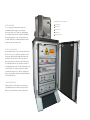
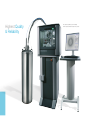
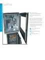
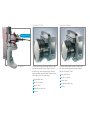
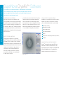
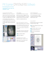
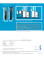
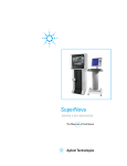
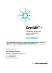
PX Scanner + SuperNova
driving X-ray innovation
PX Scanner™+ SuperNova™
Unique In-situ X-ray
and Optical Imager
Highest Quality
& Reliability Protein System
PX Scanner + SuperNova
PX Scanner
The PX Scanner is Oxford Diffraction’s unique
instrument for the X-ray screening of protein crystals,
in-situ in the multi-well crystallisation plate.
A combined optical imager and X-ray system, the PX
Scanner allows the visualisation, identification and X-ray
evaluation of protein crystals undisturbed within the
growth media.
Benefits of the PX Scanner
Early stage identification of crystalline material
Discrimination of salt from protein crystal
Identification of optimal crystal growth conditions
An unlimited number of crystals can be queued for X-ray
analysis, either within one well or over an entire plate.
The PX Scanner then automatically processes the queue,
leaving the user free to continue with other crystallisation
work before reviewing their diffraction results.
Comparison of crystal diffraction quality
Automated crystal screening leading to
accelerated results
Study of crystals too small and fragile to extract
By removing the manual handling of potential crystals
during screening, the PX Scanner can accelerate the
study of crystals at all stages of the crystallisation
work flow, including:
Phi Axis
Having identified a suitable crystal using in-situ
diffraction, the crystal can then be extracted from
the well and mounted atop the Phi Axis. Accepting
protein crystals in either loops or capillaries the Phi Axis
attachment is inserted into the front of the PX Scanner
and allows full X-ray data collection. The Phi Axis has a
rotational range of up to 360 degrees and a resolution
limit of up to 2.0Å
High throughput crystallisation condition screening and
Crystallisation scale-up for diffraction crystals
Multi-well crystallisation plate and Phi Axis
Applications
Optical and in-situ screening
Comparison of crystal
diffraction quality
Small, fragile crystals
Structure
determination
Screen
sensitive,
membrane
proteins
The complete PX Scanner system including
monitor / keyboard table
PX Scanner + SuperNova
PX Scanner
The System
The PX Scanner is unique as the world’s only
commercially available system for in-situ X-ray screening
of protein crystals. Specially designed for this purpose,
the PX Scanner is a compact system occupying a
footprint of approximately 0.5m2 (not including separate
monitor table). Contained within a fully interlocked
radiation proof cabinet, the PX Scanner consists of an
x-y-omega goniometer table and crystallisation plate
carriage, a high intensity X-ray source and a 165 mm
diameter, large active area CCD detector.
Entirely self contained, the PX Scanner requires only a
low power mains electricity and water supply, having
a built in control computer, system interface, X-ray
generator and compact internal chiller units.
For ease of integration into high throughput research
laboratories, the PX Scanner comes complete with a
dedicated barcode reader allowing the quick and easy
identification of the plate prior to loading.
PX Scanner - Loading a crystallisation plate
View inside the PX Scanner showing the:
Optical imaging
The x-y-omega goniometer table positions the
crystallisation plate beneath a series of digital
microscopes and cool LED lights for optical imaging
of the droplets within the well. Using Oxford Diffraction
proprietary algorithms, a series of optical images are
recorded of differing focal depth and lighting. A single
optimised composite image results.
In-situ X-ray imaging
Housed within the base of the system the high intensity
Nova™ X-ray source is orientated to provide a vertical
X-ray beam. Having optically imaged the plate and a
potential crystal selected for in-situ X-ray study, the
plate and crystal are accurately positioned directly over
the X-ray beam which shoots vertically up through the
horizontal plate and crystal. The plate and thereby crystal
can be tilted up to 6° using the goniometer, allowing
scanned X-ray images to be collected. The diffraction
data is collected above the plate by an Oxford Diffraction
165 mm CCD detector.
Plate compatibility
Highly versatile, the PX Scanner accepts virtually all
crystallisation plates, micro-fluidic chips and counterdiffusion set-ups which fully conform to SBS format.
X-ray safety panel
System interface
System computer
X-ray generator
X-ray and CCD chiller
PX Scanner + SuperNova
SuperNova
As the latest protein diffraction system from Oxford Diffraction, the
SuperNova is compact and easy to use, providing the highest quality
crystallographic data from a system of the highest reliability.
SuperNova is the ideal companion to the PX Scanner.
Together the two systems provide all you need for the
complete protein crystallography laboratory.
Kappa platform
With its 4-circle kappa goniometer, the SuperNova is
ideally suited for fast, strategy based data collections as
well as high resolution, in-house phasing experiments.
The kappa goniometer incorporates a motorised sample
to detector distance for the study of large cell proteins
and a rotatable beam stop for ease of crystal mounting
using cryo-tools.
The SuperNova combines Oxford Diffraction’s high
intensity Nova X-ray micro-source with the 165 mm fast,
high performance Titan™ CCD detector. Mounted on
a 4-circle goniometer, the copper radiation Nova X-ray
source provides up to 3x the intensity of a 5kW rotating
anode generator with optics and the data quality of a
micro-focus rotating anode.
Occupying a 0.5m2 footprint, the SuperNova’s stylish
design of safety interlocked and radiation proof cabinet
is entirely self contained and incorporates the X-ray
generator, system interface, system computer and
two compact water chiller units. For installation, the
SuperNova requires only a low power mains electricity
and water supply.
Ease of sample mounting on the SuperNova. Configuration shown
includes the optional second wavelength X-ray source
Optional Cryo-devices
The SuperNova goniometer has been designed
to accept all major open flow cryogenic sample
cooling devices, including Oxford Diffraction’s liquid
nitrogen device (90 - 300K).
Applications
Molecular
Replacement
(MR)
Screening
Ligand binding
SAD Phasing
or Heavy
Atom Phasing
Small
Molecules
Highest Quality
& Reliability
The complete SuperNova system including
monitor / keyboard table and optional cryo-device
PX Scanner + SuperNova
SuperNova
Nova X-ray Micro-source
The SuperNova has been designed around the low
power consumption yet high intensity Nova X-ray
micro-source.
Up to 3x more intense than a 5 kW rotating anode and
with more than 30 systems installed, the Nova is the best
selling, most reliable micro-source from any manufacturer
in single crystal X-ray diffraction. The Nova combines a
state-of-the-art multi-layer optic with a micro-focus X-ray
tube running at 50W. Power to the Nova is provided by a
software controlled X-ray generator mounted in the base
of the SuperNova system.
Copper wavelength, 50W X-ray micro-source
Multi-layer X-ray optic
Up to 3x more intense than a 5kW rotating
anode with optic
Approximately 300 micron beam size
View of the SuperNova goniometer
Titan CCD Detector
Atlas CCD detector
The SuperNova incorporates Oxford Diffraction’s high
performance, fast CCD detector, Titan. At 165 mm
in diameter, the Titan is the largest single chip CCD
detector available today and is ideal for high resolution
protein data collection using fine slicing.
For studies requiring higher sensitivity combined with
large active area, the SuperNova can be configured
with the 135 mm Atlas™ CCD.
Multi-layer optic inside
Nova X-ray source
Active area Ø165 mm
Gain, 24 e-/X-ray (Cu)
Precision, 18 bit
Speed, 0.28 sec (duty cycle)
Low noise
Active area Ø135 mm
Gain, 65 e-/X-ray (Cu)
Precision, 18 bit
Speed, 0.28 sec (duty cycle)
Low noise
PX Scanner + SuperNova
SuperNova CrysAlisPro Software
The SuperNova comes complete with CrysAlisPro™, Oxford Diffraction’s intelligent data
collection and data processing software for protein crystallography. Designed around an
easy to use graphical user interface, CrysAlisPro can be operated under fully automatic,
semi-automatic or completely manual control.
AUTOMATIC CRYSTAL SCREENING
At the heart of CrysAlisPro are the automatic crystal
screening and data collection strategy software modules.
For a typical crystal, a pre-experiment lasting less than 5
minutes is recorded to evaluate the crystal quality. From
the first frame of data, CrysAlisPro automatically evaluates
the crystal quality and provides the user with information
regarding the unit cell, angular resolution and suggested
frame exposure time.
Automatic and concurrent data reduction
Data reduction and processing initialise automatically
with the start of data collection and intelligent routines
tune the parameters for the best data quality. Since data
reduction is concurrent with data collection, processed
data are always available and accompanied by real time
on-screen feedback of data quality and completeness.
Specialist crystallographic tools
In addition to the automatic routines, CrysAlisPro provides
hundreds of specialist tools and functions for dealing
with non-standard and problematic crystals, including:
Single image screening
Advanced unit cell finding
Reciprocal space viewer
Space group determination
Twinning
Remote device control (X-ray generators, Cryo-devices)
Fastest Strategy software
Following the pre-experiment, the sophisticated
CrysAlisPro strategy software automatically calculates the
optimal data collection conditions for fast, high quality,
complete data. All strategies are calculated based on the
specific crystal orientation and unit cell and the user has
complete control to optimise the data collection for
a wide variety of targets including multiplicity, time
and resolution. Strategy calculations are extremely
fast and efficient allowing the user to quickly adapt
the data collection conditions.
Axial photos
Compatibility and software licences
CrysAlisPro automatically outputs data in MTZ format
and interfaces seamlessly with the CCP4 suite and
third-party data reduction packages including MOSFLM,
XDS and HKL2000. CrysAlisPro is provided with the
SuperNova under a multi-site, multi-user licence.
Main CrysAlisPro GUI
PX Scanner CrystalEyes Software
Once the multi-well plate has been loaded, user
interaction with the PX Scanner is entirely via the
CRYSTALEYES™ software.
Easy to use, graphical interface
CRYSTALEYES can be used online to control the PX
Scanner or offline for the review of previously recorded
data. The easy to use graphical user interface (GUI) uses
a schematic of the crystallisation plate for user selection
of wells. Dependent upon the type of plate, one or
more drops are displayed, together with a list of all data
recorded, be it optical or X-ray. The user can then select
any part of this data for display as a large image within
the GUI.
Optical imaging
Using CRYSTALEYES the PX Scanner can optically
image either the entire plate or individual user selected
wells / drops. All tasks are added to an on-screen queue
allowing the PX Scanner to be used either in real time
manual mode or unattended automatic mode.
Mouse selection of objects for X-ray
Whilst reviewing optical images, objects for in-situ X-ray
are easily added by simply clicking on the object using
the mouse cursor. The software automatically queues
the task, centres the crystal on the X-ray beam and
collects data.
Once recorded, X-ray images can be analysed within
CRYSTALEYES to provide the following information:
Angular resolution limit of diffraction
Ease of export of results
All results from CRYSTALEYES can be exported
as a concise html report with more in depth
crystallographic analysis available using Oxford
Diffraction’s CrysAlisPro software.
Phi Axis software
CRYSTALEYES supplies a simple step by step guide to
Phi Axis data collections, including:
Crystal mounting and alignment using the PX Scanner’s
video microscope
Crystal screening for evaluation of the unit cell
Automatic strategy based data collection
Automatic and concurrent data reduction
and finalisation
Possible unit cell parameters
Main CRYSTALEYES GUI in plate mode
Secure login and data storage
CRYSTALEYES operates a sophisticated, custom
database which uses secure user login to control
access to personal and group data.
CRYSTALEYES GUI in Phi Axis mode
1500 mm
1650 mm
LT
Option
110-240 v
drain
water supply
110-240 v
Vacuum
pump
drain
water supply
Vacuum
pump
110-240 v
Suggested system layout (in mm)
110-240 v
System dimensions
LT Option
470
Monitor
Electronics
Ø400
790
Monitor
SuperNova
790
PX Scanner
710
130
710
Keyboard
Keyboard
Table
1000
570
door
100
510
Table
500
570
door
100
510
500
790 mm
670 mm
810 mm
Environmental requirements for each system
Power
240 /110 V (switchable) ± 10% / 16 A fuse protected
Water
2 l/min flow required / 3 -5 bar gauge / 10 – 20 °C range / drain required
Relative Humidity
<80 % non-condensing
Weight loading
Able to bear system weight of 300 kg
Temperature
18 – 23 °C
Please refer to the system user manual for the most up to date and complete requirements.
For more information on Oxford Diffraction or its products please contact us at:
[email protected]
Please refer to the system user manual for the most up to date
Europe UK: +44 (0)1235 443630
North America: +1 540 443 9272
Latin America: +1 540 443 9272
and complete requirements
Asia Pacific Hong Kong: +852 2571 9188
For other sales offices and dealers throughout the world - please visit our website at:
www.oxford-diffraction.com or www.varianinc.com
Chromatography • Spectroscopy • Mass Spectrometry • Magnetic Resonance Spectroscopy and Imaging • X-Ray Crystallography • Dissolution • Consumables • Data Systems • Vacuum
Oxford Diffraction reserves the right to change product specifications without notice.
© Oxford Diffraction Ltd, 2008. All trademarks, copyrights and registrations acknowledged. All rights reserved. Printed by Real Affinity Agency in England. SI-1617/A/07/08