1
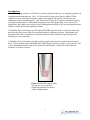
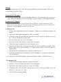
Product Manual Rac2 Activation Assay Kit Catalog Number STA-401-2 20 assays FOR RESEARCH USE ONLY Not for use in diagnostic procedures Introduction Small GTP-binding proteins (or GTPases) are a family of proteins that serve as molecular regulators in signaling transduction pathways. Rac2, a 21 kDa protein, belongs to the family of Rho GTPases regulates a variety of biological response pathways that include cell motility, cell division, gene transcription, and cell transformation. Like other small GTPases, Rac2 regulates molecular events by cycling between an inactive GDP-bound form and an active GTP-bound form. In its active (GTPbound) state, Rac2 binds specifically to the p21-binding domain (PBD) of p21-activated protein kinase (PAK) to control downstream signaling cascades. Cell Biolabs’ Rac2 Activation Assay Kit utilizes PAK PBD Agarose beads to selectively isolate and pull-down the active form of Rac from purified samples or endogenous lysates. Subsequently, the precipitated GTP-Rac is detected by western blot analysis using an anti-Rac2 specific polyclonal antibody (see Assay Principle). Cell Biolabs’ Rac2 Activation Assay Kit provides a simple and fast tool to monitor the activation of Rac2. The kit includes easily identifiable PAK1 PBD Agarose beads (see Figure 1), pink in color, and a Rac2 Immunoblot Positive Control for quick Rac2 identification. Each kit provides sufficient quantities to perform 20 assays. Figure 1:PAK RBD Agarose beads, in color, are easy to visualize, minimizing potential loss during washes and aspirations. 2 Assay Principle Related Products 1. STA-400: Pan-Ras Activation Assay Kit 2. STA-400-H: H-Ras Activation Assay Kit 3. STA-400-K: K-Ras Activation Assay Kit 4. STA-400-N: N-Ras Activation Assay Kit 5. STA-401-1: Rac1 Activation Assay 6. STA-402: Cdc42 Activation Assay Kit 7. STA-403-A: RhoA Activation Assay Kit 8. STA-405: RhoA/Rac1/Cdc42 Activation Assay Combo Kit 9. STA-410: Raf1 RBD Agarose Beads 3 10. STA-457: Ras Expression Vector Set 11. STA-459: Active Ras Expression Vector Set Kit Components 1. PAK1 PBD Agarose (Part No. STA-411): One vial – 800 μL of 50% slurry, 400 μg PBD in PBS containing 50% glycerol. Note: Agarose bead appears pink in color for easy identification, washing, and aspiration. 2. 100X GTPγS (Part No. 240103): One vial – 50 μL of 10 mM GTPγS dissolved in sterile water. 3. 100X GDP (Part No. 240104): One vial – 50 μL of 100 mM GDP dissolved in sterile water. 4. 5X Assay/Lysis Buffer (Part No. 240102): One bottle – 30 mL of 125 mM HEPES, pH 7.5, 750 mM NaCl, 5% NP-40, 50 mM MgCl2, 5 mM EDTA, 10% Glycerol. 5. Anti-Rac2, Rabbit Polyclonal (Part No. 240112): One vial – 40 μL in PBS, pH 7.4, 0.05% NaN3, 0.1% BSA. Note: This polyclonal antibody specifically reacts with human and mouse Rac2; Additional unknown higher MW proteins may be detected in some preparations. 6. Rac2 Immunoblot Positive Control (Part No. 240111): One vial – 100 μL of partially purified, recombinant Rac2 from E. coli (provided ready-to-use in 1X reducing SDS-PAGE Sample Buffer, pre-boiled). Materials Not Supplied 1. 2. 3. 4. 5. 6. 7. 8. 9. 10. 11. 12. 13. 14. Stimulated and non-stimulated cell lysates Rac2 activators Protease inhibitors 0.5 M EDTA in water 1 M MgCl2 30°C incubator or water bath 4°C tube rocker or shaker 2X reducing SDS-PAGE sample buffer Electrophoresis and immunoblotting systems Immunoblotting wash buffer such as TBST (10 mM Tris-HCl, pH 7.4, 0.15 M NaCl, 0.05% Tween-20) Immunoblotting blocking buffer (TBST containing 5% Non-fat Dry Milk) PVDF or nitrocellulose membrane Secondary Antibody ECL Detection Reagents 4 Storage Store all kit components at -20ºC. The 5X Assay/Lysis Buffer may be stored at either -20ºC or 4ºC. Avoid multiple freeze/thaw cycles. Preparation of Reagents • 1X Assay/Lysis Buffer: Mix the 5X Stock briefly and dilute to 1X in deionized water. Just prior to usage, add protease inhibitors such as 1 mM PMSF, 10 μg/mL leupeptin, and 10 μg/mL aprotinin. Preparation of Samples Note: It is advisable to use fresh cell lysates because GTP-Rac2 is quickly hydrolyzed to GDPRac2; frozen lysates stored at -70 °C may be used. Performing steps at 4 °C or on ice may reduce hydrolysis. Avoid multiple freeze/thaw cycles of lysates. I. Adherent Cells 1. Culture cells to approximately 80-90% confluence. Stimulate cells with Rac2 activator(s) as desired. 2. Aspirate the culture media and wash twice with ice-cold PBS. 3. Completely remove the final PBS wash and add ice-cold 1X Assay/Lysis Buffer to the cells (0.5 - 1 mL per 100 mm tissue culture plate). 4. Place the culture plates on ice for 10-20 minutes. 5. Detach the cells from the plates by scraping with a cell scraper. 6. Transfer the lysates to appropriate size tubes and place on ice. 7. If nuclear lysis occurs, the cell lysates may become very viscous and difficult to pipette. If this occurs, lysates can be passed through a 27½-gauge syringe needle 3-4 times to shear the genomic DNA. 8. Clear the lysates by centrifugation for 10 minutes (14,000 x g at 4°C). 9. Collect the supernatant and store samples on ice for immediate use, or snap freeze and store at 70°C for future use. 10. Proceed to GTPγS/GDP Loading for positive and negative controls, or Pull-Down Assay. II. Suspension Cells 1. Culture cells and stimulate with Rac2 activator(s) as desired. 2. Perform a cell count, and then pellet the cells by centrifugation. 3. Aspirate the culture media and wash twice with ice-cold PBS. 4. Completely remove the final PBS wash and add ice-cold 1X Assay/Lysis Buffer to the cell pellet (0.5 – 1 mL per 1 x 10⁷ cells). 5. Lyse the cells by repeated pipetting. 5 6. Transfer the lysates to appropriate size tubes and place on ice. 7. If nuclear lysis occurs, the cell lysates may become very viscous and difficult to pipette. If this occurs, lysates can be passed through a 27½-gauge syringe needle 3-4 times to shear the genomic DNA. 8. Clear the lysates by centrifugation for 10 minutes (14,000 x g at 4°C). 9. Collect the supernatant and store samples on ice for immediate use, or snap freeze and store at 70°C for future use. 10. Proceed to GTPγS/GDP Loading for positive and negative controls, or Pull-Down Assay. Assay Protocol Important Note: Before running any Small GTPase pulldown assay, it is always a good practice to run a Western Blot directly on the cell lysate using the antibody provided in this kit. For example: load 5 µg, 10 µg and 20 µg of lysate onto an SDS-PAGE gel, transfer and blot. When proceeding with the pulldown assay, use 100-times the amount of lysate that gave you a clear band of your desired small GTPase in the direct Western blot. For example: if the 5 µg band was faint but the 10 µg band was clear and strong, use 100 x 10 µg = 1 mg of lysate in the assay. Using sufficient lysate in the pulldown assay is critical to success. I. GTPγS/GDP Loading (Positive and Negative Controls) Note: Samples that will not be GTPγS/GDP loaded may be kept on ice during the loading of controls. 1. Aliquot 0.5 – 1 mL of each cell lysate to two microcentrifuge tubes. Note: Typical protein content/sample is > 0.5 mg. 2. Adjust the volume of each sample to 1 mL with 1X Assay Lysis Buffer. 3. Add 20 µL of 0.5 M EDTA to each sample. 4. Add 10 µL of 100X GTPγS to one tube (positive control) and 10 µL of 100X GDP to the other tube (negative control). Mix and label each tube appropriately. 5. Incubate the tubes for 30 minutes at 30°C with agitation. 6. Stop the loading by adding 65 µL of 1 M MgCl2 to each tube. Mix and place tubes on ice. 7. Continue with Pull-Down assay. II. Rac2 Pull-Down Assay 1. Aliquot 0.5 – 1 mL of cell lysate (treated with Rac2 activators or untreated) to a microcentrifuge tube. 2. Adjust the volume of each sample to 1 mL with 1X Assay Lysis Buffer. 3. Thoroughly resuspend the PAK PBD Agarose bead slurry by vortexing or titurating. 4. Quickly add 40 µL of resuspended bead slurry to each tube (including GTPγS/GDP controls). 5. Incubate the tubes at 4°C for 1 hour with gentle agitation. 6. Pellet the beads by centrifugation for 10 seconds at 14,000 x g. 6 7. Aspirate and discard the supernatant, making sure not to disturb/remove the bead pellet. 8. Wash the bead 3 times with 0.5 mL of 1X Assay Buffer, centrifuging and aspirating each time. 9. After the last wash, pellet the beads and carefully remove all the supernatant. 10. Resuspend the bead pellet in 40 µL of 2X reducing SDS-PAGE sample buffer. 11. Boil each sample for 5 minutes. 12. Centrifuge each sample for 10 seconds at 14,000 x g. III. Electrophoresis and Transfer 1. Load 20 µL/well of pull-down supernatant to a polyacrylamide gel. Also, it’s recommended to include a pre-stained MW standard (as an indicator of a successful transfer in step 3). Note: If desired, 10 µL/well of Rac2 Immunoblot Positive Control (provided ready-to-use, preboiled) can be added as an immunoblot positive control. 2. Perform SDS-PAGE as per the manufacturer’s instructions. 3. Transfer the gel proteins to a PVDF or nitrocellulose membrane as per the manufacturer’s instructions. IV. Immunoblotting and Detection (all steps are at room temperature, with agitation) 1. Following the electroblotting step, immerse the PVDF membrane in 100% Methanol for 15 seconds, and then allow it to dry at room temperature for 5 minutes. Note: If Nitrocellulose is used instead of PVDF, this step should be skipped. 2. Block the membrane with 5% non-fat dry milk in TBST for 1 hr at room temperature with constant agitation. Incubate the membrane with Anti-Rac2 Antibody, freshly diluted 1:1000 in 5% non-fat dry milk/TBST, for 1-2 hr at room temperature with constant agitation. Note: To conserve antibody, incubations should be performed in a plastic bag. 3. Wash the blotted membrane three times with TBST, 5 minutes each time. 4. Incubate the membrane with a secondary antibody (e.g. Goat Anti-Rabbit IgG, HRPconjugate), freshly diluted in 5% non-fat dry milk/TBST, for 1 hr at room temperature with constant agitation. 5. Wash the blotted membrane three times with TBST, 5 minutes each time. 6. Use the detection method of your choice. reagents from Pierce. 7 We recommend enhanced chemiluminescence Example of Results The following figure demonstrates typical results seen with Cell Biolabs Rac2 Activation Assay Kit. One should use the data below for reference only. Figure 2: Rac2 Activation Assay. Left Image: Rac2 Immunoblot Positive Control. Right Image: Demonstrates Anti-Rac2 polyclonal antibody specificity by dot blot. References 1. Raftopoulou M., and Hall A. (2004) Dev Biol. 265: 23-32. 2. Bar-Sagi D., and Hall A. (2000) Cell 103: 227-38. 3. Benard, V., Bohl, B. P., and Bokoch, G. M. (1999) J. Biol. Chem. 274, 13198-13204. Recent Product Citation Baetta, R. et al. (2015). Atorvastatin reduces long pentraxin 3 expression in vascular cells by inhibiting protein geranylgeranylation. Vascul Pharmacol. doi: 10.1016/j.vph.2014.11.008. Warranty These products are warranted to perform as described in their labeling and in Cell Biolabs literature when used in accordance with their instructions. THERE ARE NO WARRANTIES THAT EXTEND BEYOND THIS EXPRESSED WARRANTY AND CELL BIOLABS DISCLAIMS ANY IMPLIED WARRANTY OF MERCHANTABILITY OR WARRANTY OF FITNESS FOR PARTICULAR PURPOSE. CELL BIOLABS’ sole obligation and purchaser’s exclusive remedy for breach of this warranty shall be, at the option of CELL BIOLABS, to repair or replace the products. In no event shall CELL BIOLABS be liable for any proximate, incidental or consequential damages in connection with the products. 8 Contact Information Cell Biolabs, Inc. 7758 Arjons Drive San Diego, CA 92126 Worldwide: +1 858-271-6500 USA Toll-Free: 1-888-CBL-0505 E-mail: [email protected] www.cellbiolabs.com 2010-2015: Cell Biolabs, Inc. - All rights reserved. No part of these works may be reproduced in any form without permissions in writing. 9