1
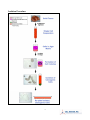
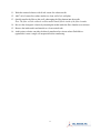
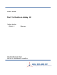
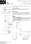
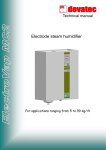
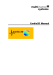
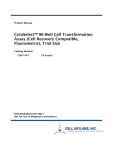
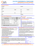
Product Manual CytoSelect™ Clonogenic Tumor Cell Isolation Kit Catalog Number CBA-155 5 preps CBA-155-5 5 x 5 preps FOR RESEARCH USE ONLY Not for use in diagnostic procedures Introduction Neoplastic transformation occurs via a series of genetic and epigenetic alterations that yield a cell population that is capable of proliferating independently of both external and internal signals that normally restrain growth. Anchorage independence is an important hallmark of the transformation that correlates with tumorgenicity. It has been shown that cells from most types of human solid tumors can form colonies in soft agar culture with plating efficiency from 0.01 to 1%; furthermore, cells recovered from individual colonies can form tumors when transplanted into mice. Stem cells are defined as clonogenic cells capable of both self-renewal and multilineage differentiation. Recent data from both hematologic malignancies and solid tumors have suggested that there are only minor populations of cells in each malignancy that are capable of tumor initiation. These tumor initiating cells have the functional properties of a tumor stem cell. They appear to be capable of asymmetric division and self renewal, and are only a minor faction among the bulk of more differentiated cells in the tumor. These observations have profound implications for tumor biology research as well as successful tumor therapy. Cell Biolabs CytoSelect™ Clonogenic Tumor Cell Isolation Kit is designed for the isolation of a small population of colony forming cells from solid tumor samples. After digestion of a solid tumor biopsy sample, cells are incubated 6-8 days in a proprietary semisolid agar media. Once these cells form colonies, they are isolated from single cells by a size filter (see Isolation Procedure below). The viable cells in these colonies are then easily recovered for further culturing and testing (FACS, protein/DNA array analysis, cancer vaccine development). NOD/SCID mouse engraftment is needed to further establish the true stem nature of these isolated cells. Each kit provides sufficient quantities to perform five isolations in a 6-well plate or 35 mm dish. On average when 500,000 cells from solid tumor are plated, after one week culture, 50-1000 colonies are expected. 2 Isolation Procedure 3 Related Products 1. CBA-100: CytoSelect™ 24-Well Cell Migration Assay (8μm, Colorimetric) 2. CBA-106: CytoSelect™ 96-Well Cell Migration Assay (8µm, Fluorometric) 3. CBA-106-C: CytoSelect™ 96-Well Cell Migration and Invasion Assay (8µm, Fluorometric) 4. CBA-112: CytoSelect™ 96-Well Cell Invasion Assay (Basement Membrane, Fluorometric) 5. CBA-130: CytoSelect™ 96-Well Cell Transformation Assay (Soft Agar Colony Formation) 6. CBA-135: CytoSelect™ 96-Well Cell Transformation Assay (Cell Recovery, Colorimetric) 7. CBA-140: CytoSelect™ 96-Well Cell Transformation Assay (Cell Recovery, Fluorometric) 8. CBA-145: CytoSelect™ 384-Well Cell Transformation Assay 9. CBA-150: CytoSelect™ 96-Well In Vitro Tumor Sensitivity Assay (Soft Agar Colony Formation) 10. CBA-320: CytoSelect™ 96-Well Hematopoietic Colony Forming Cell Assay Kit Components 1. 10X CytoSelect™ Agar Matrix Solution (Part No. 114001): One sterile bottle – 10.0 mL 2. CytoSelect™ Matrix Diluent (Part No. 114002): One sterile bottle – 4.0 mL 3. 5X DMEM Medium (Part No. 20103): One sterile bottle – 5.0 mL 4. 100X Osmotic Lysis Buffer (Part No. 115501): One sterile tube – 1.0 mL 5. 10X Assay Buffer (Part No. 115502): One sterile bottle – 15.0 mL 6. Filters (Part No. 115503): Five sterile filters Materials Not Supplied 1. Solid Tumor 2. Tumor Digestion Enzyme Mixture (containing Collagenase/DNase) 3. Razor blade or scalpel 4. 70-100 µm sterile mesh filter or Cell Strainer 5. 37ºC Incubator, 5% CO2 Atmosphere 6. Light Microscope 7. 37ºC and boiling water baths Storage Store all components at 4ºC until their expiration dates. 4 Preparation of Reagents • 2X DMEM/20% FBS Medium: In a sterile tube, dilute the provided 5X DMEM in sterile cell culture grade water to 2X containing 20% FBS. For example, to prepare a 5 mL solution, add 2 mL of 5X DMEM, 1 mL of FBS and 2 mL of sterile cell culture grade water. Sterile filter the 2X media to 0.2 µm. • 1X Osmotic Lysis Buffer: Prepare 1X Osmotic Lysis Buffer by diluting the provided 100X stock 1:100 in sterile cell culture grade water. Sterile filter the 1X solution to 0.2 µm. • 10X CytoSelect™ Agar Matrix Solution: Heat the Agar Matrix Solution bottle to 90-95ºC in a water bath for 30 minutes, or until agarose liquefies (microwaving is optional). Transfer the bottle to a 37ºC water bath for 20 minutes and maintain until needed. • 1X Assay Buffer: Prepare 1X Assay Buffer by diluting the provided 10X stock 1:10 in sterile cell culture grade water. Sterile filter the 1X solution to 0.2 µm. Note: Only prepare 1X Assay Buffer for colony recovery and re-plating, since the 10X stock is required for tumor preparation. Cell Isolation Protocol (must be under sterile conditions) The following assay protocol is written for a 6-well plate or 35-mm dish. Culture Dish Base Agar Matrix Layer (μL/well) Cell Suspension/Agar Matrix Layer (μL/well) Culture Media (μL/well) 6-well or 35-mm dish 1000 1500 1000 Table 1: Dispensing Volumes I. Solid Tumor Preparation 1. Dissect tumor and place into a 10 cm petri dish with 5-10 mL of desired media (e.g. HBSS, RPMI). 2. Quickly mince the tumor into small fragments with a scalpel/razor blade (pieces should be approximately 1-3 mm3). 3. To further dissociate single cells from the tumor fragments, add tumor digestion enzyme mixture to the media and mix well. 4. Incubate the pieces at 37ºC for 1-3 hours, periodically mixing/shaking the solution. 5. Titurate the fragment mixture 3-5 times with a 10-mL pipette. 6. Pass the solution through a 70-100 µm sterile mesh filter (not provided) to remove any large, undigested tumor pieces. 7. Collect the filtered solution. 5 8. Pellet the cells by centrifugation at 1200 rpm for 5 minutes. 9. Aspirate the supernatant. 10. Resuspend the pellet in 9 mL of 1X Osmotic Lysis Buffer for 30 seconds, mixing well. Note: This step removes most of the contaminating RBC. 11. Immediately add 1 mL of 10X Assay Buffer, mixing well. 12. Pellet the remaining cells at 1200 rpm for 5 minutes. 13. Aspirate the supernatant and resuspend the cells in 2 mL of complete culture medium. 14. Determine the cell concentration and adjust to 2 - 5 x 10⁶ cells/mL. II. Preparation of Base Agar Matrix Layer 1. Heat the 10X CytoSelect™ Agar Matrix Solution to 90-95ºC in a water bath for 30 minutes, or until agarose liquefies (microwaving is optional). Transfer the bottle to a 37ºC water bath for 20 minutes and maintain until needed. 2. Warm the 2X DMEM/20% FBS medium (see Preparation of Reagents section) to 37ºC in a water bath. Allow at least 30 minutes for the temperature to equilibrate. 3. According to Table 2 (below), prepare the desired volume of Base Agar Matrix Layer in the following sequence: a. In a sterile tube, add the appropriate volume of 2X DMEM/20% FBS medium. b. Next, add the corresponding volume of sterile water. Mix well. c. Finally, add the corresponding volume of 10X CytoSelect™ Agar Matrix Solution. Mix well. Note: The 10X CytoSelect™ Agar Matrix Solution is slightly viscous; care should be taken in accurately pipetting the appropriate volume. 2X DMEM/20% FBS Medium (mL) 2.5 1.5 0.5 Sterile Water (mL) 10X Total Volume of CytoSelect™ Base Agar Matrix Agar Matrix Layer (mL) Solution (mL) 2 0.5 5 1.2 0.3 3 0.4 0.1 1 Table 2: Preparation of Base Agar Matrix Layer # of Tests in 6-well Plate (1 mL/test) 5 3 1 4. After mixing, maintain the Base Agar Matrix Layer at 37 ºC to avoid gelation. 5. Dispense 1 mL of Base Agar Matrix Layer into each well of a 6-well, sterile plate. Gently tap the plate a few times to ensure the Base Agar Matrix Layer evenly covers the wells. Notes: • Work quickly with the layer to avoid gelation. Also, try to avoid adding air bubbles to the well. 6 • To avoid fast and uneven evaporation that leads to aberrant results, we suggest not using the wells on the plate edge, or filling the edge wells with medium to reduce evaporation. 6. Transfer the plate to 4ºC for 30 minutes to allow the Base Agar Matrix Layer to solidify. 7. Prior to adding the Cell Suspension/Agar Matrix Layer (Section III), allow the plate to warm to room temperature for 30 minutes. III. Addition of Cell Suspension/Agar Matrix Layer 1. Heat the 10X CytoSelect™ Agar Matrix Solution to 90-95ºC in a water bath for 30 minutes, or until agarose liquefies (microwaving is optional). Transfer the bottle to a 37ºC water bath for 20 minutes and maintain until needed. 2. Warm the 2X DMEM/20% FBS medium (see Preparation of Reagents section) and CytoSelect™ Matrix Diluent to 37ºC in a water bath. Allow at least 30 minutes for the temperature to equilibrate. 3. Warm the recovered cell suspension solution (Section I, step 14) in a 37ºC water bath. 4. According to Table 3 (below), prepare the desired volume of Cell Suspension/Agar Matrix Layer in the following sequence: a. In a sterile tube, add the appropriate volume of 2X DMEM/20% FBS medium. b. Next, add the corresponding volume of CytoSelect™ Matrix Diluent. Mix well. c. Next, add the corresponding volume of 10X CytoSelect™ Agar Matrix Solution. Mix well. d. Finally, add the corresponding volume of cell suspension. Mix well. Note: The CytoSelect™ Matrix Diluent and 10X CytoSelect™ Agar Matrix Solution are slightly viscous; care should be taken in accurately pipetting the appropriate volumes. 2X DMEM/20% FBS Medium (mL) 3.5 2.1 0.7 CytoSelect™ Matrix Diluent (mL) 10X Cell Total Volume of CytoSelect™ Suspension Cell Suspension/ Agar Matrix (mL) Agar Matrix Solution (mL) Layer (mL) 2.75 0.75 0.5 7.5 1.65 0.45 0.3 4.5 0.55 0.15 0.1 1.5 Table 3: Preparation of Cell Suspension/Agar Matrix Layer # of Tests in 6-well Plate (1.5 mL/test) 5 3 1 5. After mixing, incubate the Cell Suspension/Agar Matrix Layer at room temperature for 5 minutes. 7 6. Immediately dispense 1.5 mL of Cell Suspension/Agar Matrix Layer into each well of the 6well plate, already containing the Base Agar Matrix Layer (Section II). Notes: • Work quickly with the layer to avoid gelation, but gently pipette as not to disrupt the base layer integrity. Also, try to avoid adding air bubbles to the well. • Always include negative control wells that contain no cells in the Cell Suspension/Agar Matrix Layer. 7. Transfer the plate to 4ºC for 20 minutes to allow the Cell Suspension/Agar Matrix Layer to solidify. 8. Allow the plate to warm to room temperature for 30 minutes. 9. Carefully add 1 mL of culture medium to each well, in a dropwise fashion, as to not disturb the layers. 10. Incubate the cells for 6-8 days at 37ºC and 5% CO2. Examine the colony formation under a light microscope. IV. Recovery of Clonogenic Colonies and Re-plating: 1. Add 3.5 mL of 1X Assay Buffer (see Preparation of Reagents section) to each well. 2. Pipette each well 10-12 times to mix thoroughly. 3. Transfer the colony suspension mixture (~ 7 mL) to a 50-mL conical tube. 4. Add 40 mL of serum-free culture media to the colony mixture, mixing well. 5. Pellet the colonies by centrifugation at 1200 rpm for 5 minutes. 6. Carefully aspirate the 40 mL of the supernatant. 7. Add another 40 mL of serum-free culture media to the remaining colony suspension, mixing well. 8. Place a provided recovery filter into a clean 50-mL conical tube (see picture below). 9. Carefully pipet the colony suspension (step 7) through center of the filter with a steady, semiforceful pipet stream. Note: Clonogenic colonies will be retained on the filter top, while single cells will pass through. 10. Transfer the filter to another clean 50 mL conical tube. 8 11. Wash the retained colonies with 40 mL serum-free culture media. 12. Add 7 mL of serum-free culture media to a clean well of a 6-well plate. 13. Quickly transfer the filter to this well, submerging the filter bottom into the media. Note: The filter will tilt within the well but media should fill the inside of the filter chamber. 14. Recover the clonogenic colonies by titurating the media inside the filter chamber sereval times. 15. Remove the inside media and transfer to a clean conical tube. 16. At this point, colonies can either be directly transferred to a tissue culture flask/dish or typsinized to create a single cell suspension before transferring. 9 Example of Results The following figures demonstrate typical results with the CytoSelect™ Clonogenic Tumor Cell Isolation Kit. One should use the data below for reference only. This data should not be used to interpret actual results. Figure 1. Clonogenic Colony Formation, Isolation, and Re-plating. Mouse lung tumor was excised, minced and digested before seeding 500,000 cells/well in agar matrix suspension, according to the assay protocol. Panel A demonstrates clonogenic colony formation after 7 days (red arrows show colonies, black arrows show single cells). Panel B demonstrates recovered clonogenic colonies (single cells have been removed). Panel C demonstrates re-plated clonogenic colonies after 3 days (without prior typsinization). Panel D demonstrates re-plated clonogenic colonies after 1 day (colonies were typsinized/titurated to create a single-celled suspension before plating). References 1. Pavelic ZP, Slocum HK, Rustum YM, Creaven PJ, Nowak NJ, Karakousis C, Takita H, Mittelman A. Cancer Res. 1980; 40(11):4151-8. 2. Heppner G H. Cancer Res. 1984; 44:2259–2265. 10 3. Hamburger A W, Salmon S E. Science 1977; 197:461–463. 4. Al-Hajj M, Wicha MS, Benito-Hernandez A, Morrison SJ, Clarke MF. Proc Natl Acad Sci 2003; 100(7):3983-8. 5. Reya T, Morrison SJ, Clarke MF, Weissman IL. Nature 2001; 414:105–111. Warranty These products are warranted to perform as described in their labeling and in Cell Biolabs literature when used in accordance with their instructions. THERE ARE NO WARRANTIES THAT EXTEND BEYOND THIS EXPRESSED WARRANTY AND CELL BIOLABS DISCLAIMS ANY IMPLIED WARRANTY OF MERCHANTABILITY OR WARRANTY OF FITNESS FOR PARTICULAR PURPOSE. CELL BIOLABS’ sole obligation and purchaser’s exclusive remedy for breach of this warranty shall be, at the option of CELL BIOLABS, to repair or replace the products. In no event shall CELL BIOLABS be liable for any proximate, incidental or consequential damages in connection with the products. Contact Information Cell Biolabs, Inc. 7758 Arjons Drive San Diego, CA 92126 Worldwide: +1 858-271-6500 USA Toll-Free: 1-888-CBL-0505 E-mail: [email protected] www.cellbiolabs.com 2007-2011: Cell Biolabs, Inc. - All rights reserved. No part of these works may be reproduced in any form without permissions in writing. 11