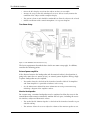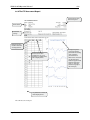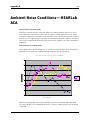
1
HEARLab System
Operator’s Manual
January 08, 2010
Copyright © 2010 Frye Electronics, Inc.
All Rights Reserved January 08, 2010
Table of Contents
iii
Contents
Chapter 1: INTRODUCING HEARLAB . . . . . . . . . . . . . . . . . . . . . . . . . . . . . . . . . . . . . . . . . . . . . . . . . . . . . . 1
1.1 System Overview . . . . . . . . . . . . . . . . . . . . . . . . . . . . . . . . . . . . . . . . . . . . . . . . . . . . . . . . . . . . . . . . . . . . . . . . . . . . . . . . . . . . . . . 1
1.2 Intended Use . . . . . . . . . . . . . . . . . . . . . . . . . . . . . . . . . . . . . . . . . . . . . . . . . . . . . . . . . . . . . . . . . . . . . . . . . . . . . . . . . . . . . . . . . . . 1
1.3 Safety and Precautions . . . . . . . . . . . . . . . . . . . . . . . . . . . . . . . . . . . . . . . . . . . . . . . . . . . . . . . . . . . . . . . . . . . . . . . . . . . . . . . . . 2
1.3.1 Meaning of Symbols . . . . . . . . . . . . . . . . . . . . . . . . . . . . . . . . . . . . . . . . . . . . . . . . . . . . . . . . . . . . . . . . . . . . . . . . . . . . . . 2
1.3.2 Reporting/Factory Location . . . . . . . . . . . . . . . . . . . . . . . . . . . . . . . . . . . . . . . . . . . . . . . . . . . . . . . . . . . . . . . . . . . . . . . 3
1.3.3 Safety Classification . . . . . . . . . . . . . . . . . . . . . . . . . . . . . . . . . . . . . . . . . . . . . . . . . . . . . . . . . . . . . . . . . . . . . . . . . . . . . . 4
1.3.4 Electromagnetic Compatiblity . . . . . . . . . . . . . . . . . . . . . . . . . . . . . . . . . . . . . . . . . . . . . . . . . . . . . . . . . . . . . . . . . . . . 4
1.3.5 Cautions and Warnings . . . . . . . . . . . . . . . . . . . . . . . . . . . . . . . . . . . . . . . . . . . . . . . . . . . . . . . . . . . . . . . . . . . . . . . . . . . 5
1.3.6 Interpretation of Results . . . . . . . . . . . . . . . . . . . . . . . . . . . . . . . . . . . . . . . . . . . . . . . . . . . . . . . . . . . . . . . . . . . . . . . . . . 7
1.4 Maintenance . . . . . . . . . . . . . . . . . . . . . . . . . . . . . . . . . . . . . . . . . . . . . . . . . . . . . . . . . . . . . . . . . . . . . . . . . . . . . . . . . . . . . . . . . . . 7
1.4.1 Service . . . . . . . . . . . . . . . . . . . . . . . . . . . . . . . . . . . . . . . . . . . . . . . . . . . . . . . . . . . . . . . . . . . . . . . . . . . . . . . . . . . . . . . . . . . . 7
1.4.2 Cleaning . . . . . . . . . . . . . . . . . . . . . . . . . . . . . . . . . . . . . . . . . . . . . . . . . . . . . . . . . . . . . . . . . . . . . . . . . . . . . . . . . . . . . . . . . . 7
1.4.3 Disposal of Equipment . . . . . . . . . . . . . . . . . . . . . . . . . . . . . . . . . . . . . . . . . . . . . . . . . . . . . . . . . . . . . . . . . . . . . . . . . . . . 8
1.5 Warranty . . . . . . . . . . . . . . . . . . . . . . . . . . . . . . . . . . . . . . . . . . . . . . . . . . . . . . . . . . . . . . . . . . . . . . . . . . . . . . . . . . . . . . . . . . . . . . . 8
Chapter 2: THE HEARLAB HARDWARE . . . . . . . . . . . . . . . . . . . . . . . . . . . . . . . . . . . . . . . . . . . . . . . . . . . . 9
2.1 Main Hardware Components . . . . . . . . . . . . . . . . . . . . . . . . . . . . . . . . . . . . . . . . . . . . . . . . . . . . . . . . . . . . . . . . . . . . . . . . . . . 9
2.2 Optional Accessories . . . . . . . . . . . . . . . . . . . . . . . . . . . . . . . . . . . . . . . . . . . . . . . . . . . . . . . . . . . . . . . . . . . . . . . . . . . . . . . . . . 11
2.3 Stimulus Controller Connections . . . . . . . . . . . . . . . . . . . . . . . . . . . . . . . . . . . . . . . . . . . . . . . . . . . . . . . . . . . . . . . . . . . . . . 12
2.4 Installation . . . . . . . . . . . . . . . . . . . . . . . . . . . . . . . . . . . . . . . . . . . . . . . . . . . . . . . . . . . . . . . . . . . . . . . . . . . . . . . . . . . . . . . . . . . . 13
2.4.1 Site considerations . . . . . . . . . . . . . . . . . . . . . . . . . . . . . . . . . . . . . . . . . . . . . . . . . . . . . . . . . . . . . . . . . . . . . . . . . . . . . . 13
2.4.2 Physical layout of system components . . . . . . . . . . . . . . . . . . . . . . . . . . . . . . . . . . . . . . . . . . . . . . . . . . . . . . . . . . . 13
2.4.3 Connecting the system components . . . . . . . . . . . . . . . . . . . . . . . . . . . . . . . . . . . . . . . . . . . . . . . . . . . . . . . . . . . . . 16
2.5 Start-up . . . . . . . . . . . . . . . . . . . . . . . . . . . . . . . . . . . . . . . . . . . . . . . . . . . . . . . . . . . . . . . . . . . . . . . . . . . . . . . . . . . . . . . . . . . . . . . 16
2.6 Calibration . . . . . . . . . . . . . . . . . . . . . . . . . . . . . . . . . . . . . . . . . . . . . . . . . . . . . . . . . . . . . . . . . . . . . . . . . . . . . . . . . . . . . . . . . . . . 16
2.7 Summary Specifications . . . . . . . . . . . . . . . . . . . . . . . . . . . . . . . . . . . . . . . . . . . . . . . . . . . . . . . . . . . . . . . . . . . . . . . . . . . . . . . 17
Chapter 3: HEARLAB ACA Measurements . . . . . . . . . . . . . . . . . . . . . . . . . . . . . . . . . . . . . . . . . . . . . . . 21
3.1 Starting-up . . . . . . . . . . . . . . . . . . . . . . . . . . . . . . . . . . . . . . . . . . . . . . . . . . . . . . . . . . . . . . . . . . . . . . . . . . . . . . . . . . . . . . . . . . . . 21
3.2 Working with Clients . . . . . . . . . . . . . . . . . . . . . . . . . . . . . . . . . . . . . . . . . . . . . . . . . . . . . . . . . . . . . . . . . . . . . . . . . . . . . . . . . . 22
3.2.1 Adding a new client . . . . . . . . . . . . . . . . . . . . . . . . . . . . . . . . . . . . . . . . . . . . . . . . . . . . . . . . . . . . . . . . . . . . . . . . . . . . . 23
3.2.2 Working with an existing client record . . . . . . . . . . . . . . . . . . . . . . . . . . . . . . . . . . . . . . . . . . . . . . . . . . . . . . . . . . . 24
3.2.2.1 Searching for a client record . . . . . . . . . . . . . . . . . . . . . . . . . . . . . . . . . . . . . . . . . . . . . . . . . . . . . . . . . . . . . 24
3.2.2.2 Listing all existing clients . . . . . . . . . . . . . . . . . . . . . . . . . . . . . . . . . . . . . . . . . . . . . . . . . . . . . . . . . . . . . . . . . 25
3.2.2.3 Selecting a client record . . . . . . . . . . . . . . . . . . . . . . . . . . . . . . . . . . . . . . . . . . . . . . . . . . . . . . . . . . . . . . . . . . 25
3.2.3 Modifying details or deleting an existing client . . . . . . . . . . . . . . . . . . . . . . . . . . . . . . . . . . . . . . . . . . . . . . . . . . 26
3.3 Performing an Assessment . . . . . . . . . . . . . . . . . . . . . . . . . . . . . . . . . . . . . . . . . . . . . . . . . . . . . . . . . . . . . . . . . . . . . . . . . . . . 27
3.3.1 Preparing a client for testing . . . . . . . . . . . . . . . . . . . . . . . . . . . . . . . . . . . . . . . . . . . . . . . . . . . . . . . . . . . . . . . . . . . . . 27
3.3.2 Specifying assessment settings . . . . . . . . . . . . . . . . . . . . . . . . . . . . . . . . . . . . . . . . . . . . . . . . . . . . . . . . . . . . . . . . . . 27
3.3.3 Specifying run settings . . . . . . . . . . . . . . . . . . . . . . . . . . . . . . . . . . . . . . . . . . . . . . . . . . . . . . . . . . . . . . . . . . . . . . . . . . 30
3.3.4 Impedance check . . . . . . . . . . . . . . . . . . . . . . . . . . . . . . . . . . . . . . . . . . . . . . . . . . . . . . . . . . . . . . . . . . . . . . . . . . . . . . . . 31
3.3.5 Performing a test (acquisition) . . . . . . . . . . . . . . . . . . . . . . . . . . . . . . . . . . . . . . . . . . . . . . . . . . . . . . . . . . . . . . . . . . . 32
iv
HEARLab ACA Operators’ Manual
3.4 CAEP Wave Morphology . . . . . . . . . . . . . . . . . . . . . . . . . . . . . . . . . . . . . . . . . . . . . . . . . . . . . . . . . . . . . . . . . . . . . . . . . . . . . . . 37
3.5 The Results Window . . . . . . . . . . . . . . . . . . . . . . . . . . . . . . . . . . . . . . . . . . . . . . . . . . . . . . . . . . . . . . . . . . . . . . . . . . . . . . . . . . 39
3.5.1 Accessing the Results window . . . . . . . . . . . . . . . . . . . . . . . . . . . . . . . . . . . . . . . . . . . . . . . . . . . . . . . . . . . . . . . . . . 40
3.5.2 Adding notes . . . . . . . . . . . . . . . . . . . . . . . . . . . . . . . . . . . . . . . . . . . . . . . . . . . . . . . . . . . . . . . . . . . . . . . . . . . . . . . . . . . 40
3.5.3 Viewing the Statistical Analysis . . . . . . . . . . . . . . . . . . . . . . . . . . . . . . . . . . . . . . . . . . . . . . . . . . . . . . . . . . . . . . . . . 40
3.5.4 Viewing Averaged Responses . . . . . . . . . . . . . . . . . . . . . . . . . . . . . . . . . . . . . . . . . . . . . . . . . . . . . . . . . . . . . . . . . . . 41
3.5.5 Viewing the Epoch Count . . . . . . . . . . . . . . . . . . . . . . . . . . . . . . . . . . . . . . . . . . . . . . . . . . . . . . . . . . . . . . . . . . . . . . . . 43
3.5.6 Changing the Run Selection . . . . . . . . . . . . . . . . . . . . . . . . . . . . . . . . . . . . . . . . . . . . . . . . . . . . . . . . . . . . . . . . . . . . . 43
3.5.7 Viewing Residual Noise . . . . . . . . . . . . . . . . . . . . . . . . . . . . . . . . . . . . . . . . . . . . . . . . . . . . . . . . . . . . . . . . . . . . . . . . . 44
3.5.8 Exporting Run Results . . . . . . . . . . . . . . . . . . . . . . . . . . . . . . . . . . . . . . . . . . . . . . . . . . . . . . . . . . . . . . . . . . . . . . . . . . . 45
3.5.9 Deleting a Run . . . . . . . . . . . . . . . . . . . . . . . . . . . . . . . . . . . . . . . . . . . . . . . . . . . . . . . . . . . . . . . . . . . . . . . . . . . . . . . . . . . 45
3.6 Printing Assessment Reports . . . . . . . . . . . . . . . . . . . . . . . . . . . . . . . . . . . . . . . . . . . . . . . . . . . . . . . . . . . . . . . . . . . . . . . . . 46
3.6.1 Previewing the Print Report . . . . . . . . . . . . . . . . . . . . . . . . . . . . . . . . . . . . . . . . . . . . . . . . . . . . . . . . . . . . . . . . . . . . 46
3.6.2 Printing the Report without Preview . . . . . . . . . . . . . . . . . . . . . . . . . . . . . . . . . . . . . . . . . . . . . . . . . . . . . . . . . . . . 46
3.6.3 The ACA Assessment Report . . . . . . . . . . . . . . . . . . . . . . . . . . . . . . . . . . . . . . . . . . . . . . . . . . . . . . . . . . . . . . . . . . . . 47
3.6.4 The CTE Assessment Report . . . . . . . . . . . . . . . . . . . . . . . . . . . . . . . . . . . . . . . . . . . . . . . . . . . . . . . . . . . . . . . . . . . . 48
3.7 Saving Assessment to Database . . . . . . . . . . . . . . . . . . . . . . . . . . . . . . . . . . . . . . . . . . . . . . . . . . . . . . . . . . . . . . . . . . . . . . . 49
3.8 Deleting Assessments . . . . . . . . . . . . . . . . . . . . . . . . . . . . . . . . . . . . . . . . . . . . . . . . . . . . . . . . . . . . . . . . . . . . . . . . . . . . . . . . 49
Chapter 4: Sound Field Calibration . . . . . . . . . . . . . . . . . . . . . . . . . . . . . . . . . . . . . . . . . . . . . . . . . . . . 51
4.1 Calibration Setup . . . . . . . . . . . . . . . . . . . . . . . . . . . . . . . . . . . . . . . . . . . . . . . . . . . . . . . . . . . . . . . . . . . . . . . . . . . . . . . . . . . . . . 51
4.2 Free Field Calibration Wizard . . . . . . . . . . . . . . . . . . . . . . . . . . . . . . . . . . . . . . . . . . . . . . . . . . . . . . . . . . . . . . . . . . . . . . . . . 52
4.3 Calibration Results . . . . . . . . . . . . . . . . . . . . . . . . . . . . . . . . . . . . . . . . . . . . . . . . . . . . . . . . . . . . . . . . . . . . . . . . . . . . . . . . . . . 56
Chapter 5: Tools & Utilities . . . . . . . . . . . . . . . . . . . . . . . . . . . . . . . . . . . . . . . . . . . . . . . . . . . . . . . . . . . . 57
5.1 Administrative Tools . . . . . . . . . . . . . . . . . . . . . . . . . . . . . . . . . . . . . . . . . . . . . . . . . . . . . . . . . . . . . . . . . . . . . . . . . . . . . . . . . . . 57
5.1.1 User Administration . . . . . . . . . . . . . . . . . . . . . . . . . . . . . . . . . . . . . . . . . . . . . . . . . . . . . . . . . . . . . . . . . . . . . . . . . . . . . 57
5.1.2 Changing Your Password . . . . . . . . . . . . . . . . . . . . . . . . . . . . . . . . . . . . . . . . . . . . . . . . . . . . . . . . . . . . . . . . . . . . . . . . 59
5.1.3 Changing the Setup Location . . . . . . . . . . . . . . . . . . . . . . . . . . . . . . . . . . . . . . . . . . . . . . . . . . . . . . . . . . . . . . . . . . . . 59
5.2 Database Backup and Restore . . . . . . . . . . . . . . . . . . . . . . . . . . . . . . . . . . . . . . . . . . . . . . . . . . . . . . . . . . . . . . . . . . . . . . . . . 59
5.2.1 Backing up the Database . . . . . . . . . . . . . . . . . . . . . . . . . . . . . . . . . . . . . . . . . . . . . . . . . . . . . . . . . . . . . . . . . . . . . . . 60
5.2.2 Restoring the Database to a Previous Version . . . . . . . . . . . . . . . . . . . . . . . . . . . . . . . . . . . . . . . . . . . . . . . . . . 60
5.3 Utilities . . . . . . . . . . . . . . . . . . . . . . . . . . . . . . . . . . . . . . . . . . . . . . . . . . . . . . . . . . . . . . . . . . . . . . . . . . . . . . . . . . . . . . . . . . . . . . . . 61
5.3.1 Checking the Impedance . . . . . . . . . . . . . . . . . . . . . . . . . . . . . . . . . . . . . . . . . . . . . . . . . . . . . . . . . . . . . . . . . . . . . . . . 61
5.3.2 Checking the Stimuli Presentation Levels . . . . . . . . . . . . . . . . . . . . . . . . . . . . . . . . . . . . . . . . . . . . . . . . . . . . . . . . 61
5.3.3 Ambient Noise Measurement . . . . . . . . . . . . . . . . . . . . . . . . . . . . . . . . . . . . . . . . . . . . . . . . . . . . . . . . . . . . . . . . . . . 62
Chapter 6: Reference . . . . . . . . . . . . . . . . . . . . . . . . . . . . . . . . . . . . . . . . . . . . . . . . . . . . . . . . . . . . . . . . . . 65
6.1 Toolbars . . . . . . . . . . . . . . . . . . . . . . . . . . . . . . . . . . . . . . . . . . . . . . . . . . . . . . . . . . . . . . . . . . . . . . . . . . . . . . . . . . . . . . . . . . . . . . 65
6.1.1 Main Toolbar . . . . . . . . . . . . . . . . . . . . . . . . . . . . . . . . . . . . . . . . . . . . . . . . . . . . . . . . . . . . . . . . . . . . . . . . . . . . . . . . . . . 65
6.1.2 Client Toolbar . . . . . . . . . . . . . . . . . . . . . . . . . . . . . . . . . . . . . . . . . . . . . . . . . . . . . . . . . . . . . . . . . . . . . . . . . . . . . . . . . . 65
6.1.3 Assessment Toolbar . . . . . . . . . . . . . . . . . . . . . . . . . . . . . . . . . . . . . . . . . . . . . . . . . . . . . . . . . . . . . . . . . . . . . . . . . . . . 65
6.2 Menus . . . . . . . . . . . . . . . . . . . . . . . . . . . . . . . . . . . . . . . . . . . . . . . . . . . . . . . . . . . . . . . . . . . . . . . . . . . . . . . . . . . . . . . . . . . . . . . . 66
6.2.1 Main Menu Bar . . . . . . . . . . . . . . . . . . . . . . . . . . . . . . . . . . . . . . . . . . . . . . . . . . . . . . . . . . . . . . . . . . . . . . . . . . . . . . . . . 66
6.2.2 Client Menu Bar . . . . . . . . . . . . . . . . . . . . . . . . . . . . . . . . . . . . . . . . . . . . . . . . . . . . . . . . . . . . . . . . . . . . . . . . . . . . . . . . 68
6.2.3 Asssement Menu Bar . . . . . . . . . . . . . . . . . . . . . . . . . . . . . . . . . . . . . . . . . . . . . . . . . . . . . . . . . . . . . . . . . . . . . . . . . . . 68
Table of Contents
Chapter 7: Overview of the ACA and CTE Modes . . . . . . . . . . . . . . . . . . . . . . . . . . . . . . . . . . . . . . . . 69
7.1 ACA Mode . . . . . . . . . . . . . . . . . . . . . . . . . . . . . . . . . . . . . . . . . . . . . . . . . . . . . . . . . . . . . . . . . . . . . . . . . . . . . . . . . . . . . . . . . . . . . 69
7.2 CTE Mode . . . . . . . . . . . . . . . . . . . . . . . . . . . . . . . . . . . . . . . . . . . . . . . . . . . . . . . . . . . . . . . . . . . . . . . . . . . . . . . . . . . . . . . . . . . . . . 70
7.3 Notes on the Use of HEARLab ACA . . . . . . . . . . . . . . . . . . . . . . . . . . . . . . . . . . . . . . . . . . . . . . . . . . . . . . . . . . . . . . . . . . . . 71
7.3.1 Monitoring of test participant state . . . . . . . . . . . . . . . . . . . . . . . . . . . . . . . . . . . . . . . . . . . . . . . . . . . . . . . . . . . . . . 71
7.3.2 Interpretation of results . . . . . . . . . . . . . . . . . . . . . . . . . . . . . . . . . . . . . . . . . . . . . . . . . . . . . . . . . . . . . . . . . . . . . . . . . 72
7.3.3 Noise Indicator . . . . . . . . . . . . . . . . . . . . . . . . . . . . . . . . . . . . . . . . . . . . . . . . . . . . . . . . . . . . . . . . . . . . . . . . . . . . . . . . . . 72
7.3.4 Detection Statistics . . . . . . . . . . . . . . . . . . . . . . . . . . . . . . . . . . . . . . . . . . . . . . . . . . . . . . . . . . . . . . . . . . . . . . . . . . . . . . . 72
7.3.5 Cortical Latencies . . . . . . . . . . . . . . . . . . . . . . . . . . . . . . . . . . . . . . . . . . . . . . . . . . . . . . . . . . . . . . . . . . . . . . . . . . . . . . . . 74
Chapter 8: Overview of the ACA and CTE Modes . . . . . . . . . . . . . . . . . . . . . . . . . . . . . . . . . . . . . . . . 75
8.1 Physiological Mechanisms of the CAEP . . . . . . . . . . . . . . . . . . . . . . . . . . . . . . . . . . . . . . . . . . . . . . . . . . . . . . . . . . . . . . . . 75
8.2 Characteristics of the CAEP . . . . . . . . . . . . . . . . . . . . . . . . . . . . . . . . . . . . . . . . . . . . . . . . . . . . . . . . . . . . . . . . . . . . . . . . . . . . 76
8.3 Clinical Application of the CAEP . . . . . . . . . . . . . . . . . . . . . . . . . . . . . . . . . . . . . . . . . . . . . . . . . . . . . . . . . . . . . . . . . . . . . . . 77
8.4 Relationship of CAEP to audibility . . . . . . . . . . . . . . . . . . . . . . . . . . . . . . . . . . . . . . . . . . . . . . . . . . . . . . . . . . . . . . . . . . . . . 78
8.5 CAEP hardware and measurement parameters . . . . . . . . . . . . . . . . . . . . . . . . . . . . . . . . . . . . . . . . . . . . . . . . . . . . . . . . 79
8.5.1 Transducers . . . . . . . . . . . . . . . . . . . . . . . . . . . . . . . . . . . . . . . . . . . . . . . . . . . . . . . . . . . . . . . . . . . . . . . . . . . . . . . . . . . . . 79
8.5.2 Speech stimuli . . . . . . . . . . . . . . . . . . . . . . . . . . . . . . . . . . . . . . . . . . . . . . . . . . . . . . . . . . . . . . . . . . . . . . . . . . . . . . . . . . 80
8.5.3 Polarity . . . . . . . . . . . . . . . . . . . . . . . . . . . . . . . . . . . . . . . . . . . . . . . . . . . . . . . . . . . . . . . . . . . . . . . . . . . . . . . . . . . . . . . . . 80
8.5.4 Repetition rates . . . . . . . . . . . . . . . . . . . . . . . . . . . . . . . . . . . . . . . . . . . . . . . . . . . . . . . . . . . . . . . . . . . . . . . . . . . . . . . . 80
8.5.5 Tone burst characteristics . . . . . . . . . . . . . . . . . . . . . . . . . . . . . . . . . . . . . . . . . . . . . . . . . . . . . . . . . . . . . . . . . . . . . . . 81
8.5.6 Masking . . . . . . . . . . . . . . . . . . . . . . . . . . . . . . . . . . . . . . . . . . . . . . . . . . . . . . . . . . . . . . . . . . . . . . . . . . . . . . . . . . . . . . . . . 81
8.5.7 Number of signal channels and electrodes . . . . . . . . . . . . . . . . . . . . . . . . . . . . . . . . . . . . . . . . . . . . . . . . . . . . . . 81
8.5.8 Electrode placement . . . . . . . . . . . . . . . . . . . . . . . . . . . . . . . . . . . . . . . . . . . . . . . . . . . . . . . . . . . . . . . . . . . . . . . . . . . . 82
8.5.9 Display . . . . . . . . . . . . . . . . . . . . . . . . . . . . . . . . . . . . . . . . . . . . . . . . . . . . . . . . . . . . . . . . . . . . . . . . . . . . . . . . . . . . . . . . . 82
8.6 Statistical processing . . . . . . . . . . . . . . . . . . . . . . . . . . . . . . . . . . . . . . . . . . . . . . . . . . . . . . . . . . . . . . . . . . . . . . . . . . . . . . . . . 83
8.7 Reference List . . . . . . . . . . . . . . . . . . . . . . . . . . . . . . . . . . . . . . . . . . . . . . . . . . . . . . . . . . . . . . . . . . . . . . . . . . . . . . . . . . . . . . . . 84
APPENDIX A: Practical Aspects of CAEP Testing with Infants . . . . . . . . . . . . . . . . . . . . . . . . . . . . 89
APPENDIX B: Ambient Noise Conditions—HEARLab ACA . . . . . . . . . . . . . . . . . . . . . . . . . . . . . . . 95
APPENDIX C: Sources of Interference—HEARLab . . . . . . . . . . . . . . . . . . . . . . . . . . . . . . . . . . . . . . . 97
APPENDIX D: Distraction Techniques for Testing Adults . . . . . . . . . . . . . . . . . . . . . . . . . . . . . . . . . 99
APPENDIX E: Quick Start Guide to HEARLab ACA Software . . . . . . . . . . . . . . . . . . . . . . . . . . . . . 101
APPENDIX F: Proper Insertion of Insert Earphones . . . . . . . . . . . . . . . . . . . . . . . . . . . . . . . . . . . . 103
APPENDIX G: Troubleshooting . . . . . . . . . . . . . . . . . . . . . . . . . . . . . . . . . . . . . . . . . . . . . . . . . . . . . . . . 105
GLOSSARY . . . . . . . . . . . . . . . . . . . . . . . . . . . . . . . . . . . . . . . . . . . . . . . . . . . . . . . . . . . . . . . . . . . . . . . . . . . 107
v
Chapter 1
v1.0
Introducing HEARLab
1.1 System Overview
Welcome to the HEARLab™ System, manufactured by Frye Electronics, Inc. under license
by the National Acoustics Laboratory (NAL) of Australia.
This manual describes the Aided Cortical Assessment (ACA) software module, which
is intended to help determine if a person who has been fitted with a hearing assistance
device (such as a hearing aid) is actually hearing sounds in the low, mid and high frequency bands associated with normal hearing.
The HEARLab ACA module also contains the Cortical Tone Evaluation (CTE) sub-module. In the CTE mode a number of frequency specific tone bursts can be used to evaluate
the audibility of specific frequencies at different levels.
The HEARLab system consists of a personal computer running the HEARLab ACA software module in the Microsoft Windows® environment, and a set of specialized but
versatile hardware components. The hardware contains electronic circuits that can be
configured and controlled by the software to deliver the correct acoustic stimulus and to
acquire the evoked cortical response. The software performs the required signal processing, manages record keeping and interacts with the tester.
ELECTRODES
MONITOR
SPEAKER
(or Insert earphones
or Bone conductor)
SUBJECT
HEARLab
COMPUTER
HEARLab
By
STIMULUS
CONTROLLER
By
ELECTRODE
PROCESSOR
1.2 Intended Use
The HEARLab ACA device is intended for use in the recording and analysis of human
electro-physiological data, in the form of the Cortical Auditory Evoked Potential (CAEP)
as an aid in the assessment of auditory function.
HEARLab ACA presents stimuli to the test participant via free-field loudspeakers, insert
earphones or bone conduction. It allows the audibility of speech sounds and highly frequency-specific tone bursts to be assessed by recording, analyzing and displaying the cortical response waveforms evoked by these stimuli. It is designed to be used as a tool for
audiologists or other hearing health care professionals, who have training in the recording of evoked potentials.
Page 1
HEARLab ACA Operators’ Manual v1.0
HEARLab ACA can be used for assessment of all age groups, from newborn infants to the
very elderly, given that the test participant is able to remain awake, alert and reasonably
still and quiet during the recording time. It is important to note that if the test participant is too active cortical responses may be masked by noise. Because CAEPs to acoustic
stimuli are involuntary, HEARLab is especially useful in assessing individuals when
behavioral audiometric results cannot be obtained, or when behavioral test results are
deemed unreliable. For example, babies who are developmentally too young for behavioural testing (such as Visual Reinforcement Orientation Audiometry, or VROA), adults
who have physical or cognitive difficulties, or children and adults who will not cooperate
in standard behavioural tests (i.e., suspected non-organic hearing loss cases).
In these “difficult to test” populations, HEARLab ACA can be used as an adjunct to hearing aid evaluation, allowing the audiologist to assess the audibility of speech sounds
when amplified by a particular hearing aid gain-frequency response. It has been purposefully developed for testing infants who have been referred for hearing aid fitting following neonatal hearing screening, and who are too young for VROA testing.
HEARLab ACA offers two test modes (modules) referred to as ACA mode and CTE mode.
Each of these modules has a specific application.
See Chapter 7 for more information on how and when to use the ACA and CTE modules. Chapter 8 has information on the scientific background of Cortical Auditory Evoked
Potential (CAEP) measurements.
1.3 Safety and Precautions
1.3.1 Meaning of Symbols
These symbols are found on the Stimulus Controller.
Patient applied parts of HEARLab are type BF.
Read the accompanying documents. Please read this manual before operating
HEARLab.
Replace fuses only with same type and rating (1A, 250V, Time LAG).
CE signifies compliance with the European Union’s Medical Devices
Directive 93/42/EEC.
Page 2
Introducing HEARLab v1.0
Shipping
The following symbols are included on the electronic module shipping box:
Keep dry. The HEARLab System should not be exposed to water or
other fluids.
65ºC
The shipping/storage temperature of the HEARLab System is -40 to
65 degrees Celsius (-40 to 149 degrees Fahrenheit)
-40ºC
%
95%
The shipping/storage Humidity of the HEARLab System is 10 to 95
percent relative humidity (non-condensing)
10%
1.3.2 Reporting/Factory Location
If you are located in the European Union, please report all safety-related concerns to
our authorized representative:
Siemens Hearing Instruments, Ltd
Alexandra House
Newton Road
Manor Royal
Crawley
West Sussex RH109TT
ENGLAND
Otherwise, please report all safety-related concerns to the Frye factory:
Frye Electronics, Inc.
9826 SW Tigard St
Tigard OR 97223
Phone: 503-620-2722, 800-547-8209
Fax: 503-639-0128
Email: [email protected] (operational help), [email protected] (sales/price questions), [email protected] (malfunctioning equipment)
Frye Electronics, Inc. is a Registered Firm of British Standards
Institution, and we conform to the ISO 13485 standard.
Page 3
HEARLab ACA Operators’ Manual v1.0
1.3.3 Safety Classification
The HEARLab system complies with:
• IEC 60601-1 and AS/NZS3200.1.0 Medical electrical equipment - Part 1.0: General
requirements for basic safety and essential performance.
• IEC 60601-1-2, collateral standard for Electromagnetic compatibility requirements
(See Section 1.3.4 for more details.)
• Australian Therapeutic Goods Administration, Class 1 (Rule 4.1)
• European Medical Device Directive, Class 11a (Rule 10)
• U.S. FDA, Class 2
• ISO 13485: 2003
Safety Classification for IEC 60601-1
Type of protection against electrical shock: Class 1
Degree of protection against electrical shock: Type BF
Mode of operation: Continuous
Degree of protection against the ingress of water: Ordinary
The HEARLab System does not require sterilization.
Warning: The HEARLab System is not suitable for use in the presence of a flammable
anesthetic mixture with air or with oxygen or nitrous oxide.
The HEARLab ACA software is indicated for use in the recording and analysis of
human electro-physiological data as an aid in the assessment of hearing and hearingrelated functions. The data is obtained with electrodes attached to the scalp of the
subject to detect the presence or absence of electro-physiological signals that may be
evoked in response to auditory stimulus. These electro-physiological signals being
monitored are not intended to indicate signals vital to life or health or state of well
being of the patient.
Note that for conformance to IEC 60601-1 Type BF patient isolation requirements the
HEARLab System must be setup in accordance with instructions in Section 2.2.
It should be noted that it is the user’s responsibility to ensure that conformance to IEC
60601-1 Type BF patient isolation requirements are maintained when patient-connected
equipment or accessories not supplied by Frye Electronics are used with or in conjunction
with the HEARLab System.
1.3.4 Electromagnetic Compatiblity
The HEARLab System acquires and analyses signals of low amplitudes with circuits that
that are sensitive to electrical interference. Some forms of electrical interference may cause
interruption of the cortical measurement tests. For further information, see Appendix C for
notes that may be helpful in identifying and resolving sources of interference.
HEARLab complies with IEC 60601-1-2 with the following deviations:
Page 4
Introducing HEARLab v1.0
36.202.2 Electrostatic discharge (ESD): The HEARLab Cortical Analyzer is sensitive to
high voltage discharges. However, the analyzer software monitors measurements for
electrical noise that exceed the dynamic range of the system, including that which may
result from this type of discharge, and discards it from final test results. Furthermore,
no adverse harm comes to the patient from the application of these discharges.
36.202.3 Radiated RF electromagnetic fields: While testing at 3 V/m, between 80 MHz
and 700 MHz, the EEG and most recent recent epoch graph will show invalid measurement data. If this electrical noise exceeds the dynamic range of the system, it will be
automatically blocked from final test results. If this noise is within the dynamic range
of the system, it will be included in test results, but should be averaged out if the noise
is short in duration.
36.202.4 Electrical fast transients and bursts: Power fluctuations may cause the Cortical
Analyzer software to close. This will lead to the loss of data for any running test, but
will not affect saved test results or the safety of the user or patient. To prevent this
occurence, it is recommended to use a surge protector or uninterruptible power supply
(UPS power backup).
36.202.6 Conducted disturbances, induced by RF fields: While testing at 3 Vrms, the
EEG and most recent epoch graphs will include noise and may result in a moderate to
loud tone from the speaker. In most cases, this noise will exceed the dynamic range of
the system and will be automatically discarded and excluded from final test results.
Noise that does not exceed the dynamic range of the system may be included in test
results, but will be averaged out of long term results as long as the signal is short in
duration. Any tone(s) issuing from the loudspeaker that is not synchronous with the
computer driven stimulus is ignored by the averaging system and will not affect test
results.
1.3.5 Cautions and Warnings
This section contains important safety precautions. Please read carefully before operating
the HEARLab ACA software.
The HEARLab System should only be used for purposes described in this operator’s manual.
Acoustical Levels and Patient Safety:
The software and hardware are designed to limit the acoustical signal to levels that are not
harmful to a patient, even if a malfunction should occur. Malfunction of the device, including the software, may result in uncomfortably loud levels but is unlikely to cause even
minor injury to the patient.
Acoustically evoked electro-physiological signals are picked up by passive electrodes placed
on the scalp of the subject. Malfunction of the device, including the software, is unlikely to
cause minor injury or discomfort to the patient.
Setup and Connections:
To avoid electric shock hazard, care must be taken to connect cables and accessories in
exact accordance with the user instructions. Only appropriately trained and skilled personnel should attempt to install the HEARLab System.
Page 5
HEARLab ACA Operators’ Manual v1.0
The HEARLab System should be used in a clinical test environment with low ambient
noise and electromagnetic field levels. Unsuitable test environments could lead to bad
test results.
Safe use of the HEARLab System may be compromised if the user connects unsuitable
equipment to the Stimulus Controller Unit.
Use only the electrode cables supplied with the HEARLab System. Do not attempt to
substitute any other cables. The use of alternate electrode cables could lead to bad test
results.
Avoid the use of extension cables, or placing power cables where they present a tripping
hazard. Keep hardware and cables out of children’s reach.
Keep the HEARLab System hardware isolated in a well ventilated area. It should not be
operated in a tightly enclosed area or stacked with other electrical equipment. Failure to
do so could overheat the equipment and lead to failure.
All electrical equipment within the patient test area should have approved medical safety
status. Failure to follow these instructions could lead to interference with nearby medical
equipment.
Electrode application and removal:
Electrodes must be applied by suitably qualified and experienced personnel. Universal
precautions should be applied to reduce the risk of patient infection.
Avoid contact with broken skin, or patients with existing skin conditions. Electrode contact in these circumstances could further damage the skin.
Substances used to prepare electrode sites (e.g., abrasive liquids, gel, cream or paste),
or to adhere electrodes to the skin (e.g., self adhesive electrodes, medical tapes), or the
electrode itself, may produce allergic or other dermatologic reaction in some individuals. Check for history of allergic reactions before preparing electrode sites/applying electrodes.
Be aware that some patient populations may be at particular risk (e.g., burns patients,
premature infants). If in any doubt, seek medical advice.
Substances used for electrode preparation and placement should be selected with regard
to the manufacturer’s recommendations for use with patients. Always read and follow the
manufacturer’s written instructions. Avoid contact with the patient’s eyes. Alcohol preparation swabs are not recommended for use on infants.
Clinicians who have skin sensitivities should also avoid prolonged contact with preparation substances or consider wearing gloves during electrode preparation application and
removal.
Dispense preparation substances in a hygienic manner using single-use, disposable applicators (e.g., cotton swabs). To reduce the risk of discomfort or infection, avoid aggressive
abrading of the skin when preparing electrode sites. Avoid the use of dry abrasive pads.
Page 6
Introducing HEARLab v1.0
Do not use undue physical force when preparing skin or applying electrodes. Take particular care when applying electrodes to infants, particularly the vertex electrode, where
the fontanel area is susceptible to injury.
Thoroughly clean the skin immediately after electrode removal using clean water. Harsh
agents (e.g., alcohol, acetone) are not recommended.
1.3.6 Interpretation of Results
The HEARLab ACA software is intended to be user-friendly and simple to use. However,
the system should only be used for electrophysiological assessment and only by personnel with appropriate training and experience.
Results may be invalid if:
• The calibration procedures are not correctly followed prior to testing (Section 3.6)
• Transducers (e.g., insert earphones) are changed and the system is not recalibrated
prior to use (Section 3.9)
• Electrode contact is poor (> 5 kΩ) (Section 3.4.4)
• Excessive electromagnetic interference is present in the test environment
• The patient is drowsy or asleep.
• The patient is restless and/or overheated.
• Insufficient quality (noise-free) cortical recordings are obtained.
1.4 Maintenance
This section describing servicing, cleaning and disposal of the HEARLab Cortical
Analyzer.
1.4.1 Service
The HEARLab System hardware must only be repaired or serviced by a qualified
technician. There are no user serviceable parts inside the Stimulus Controller or the
Electrode Processor. Do not attempt to open the external casings of these components.
Calibration should be performed at least once a year in accordance with the procedures
described in Section 3.9.
1.4.2 Cleaning
The Stimulus Controller and the Electrode Processor may be wiped with a dry cloth.
Do not allow liquids to come into contact with the HEARLab System hardware.
Single-use, disposable sensor pads are used with the HEARLab electrode connector system. Refer to the manufacturer’s information to ensure appropriate use. To avoid the possibility of cross-infection, do not attempt to clean or re-use disposable sensor pads.
When insert earphones are used as stimulus generators, do not reuse the insert earphone
eartips. Sterilization of these eartips is not possible. When performing these measurements, make sure to use a new tip for each patient.
Page 7
HEARLab ACA Operators’ Manual v1.0
1.4.3 Disposal of Equipment
The HEARLab System and some of its accessories contain lead. When the user has decided that the HEARLab equipment is not longer serving its intended purpose and is to be
discarded, the HEARLab devices should be disposed of in accordance with local waste
disposal regulations. Consult the local waste disposal authority on the relevant regulations applicable.
1.5 Warranty
The HEARLab System and all accessories except the electrodes are guaranteed to be
free of manufacturing defects that would prevent the product from meeting its specifications (given in Appendix D of this manual) for a period of one year from the date
of purchase when the following conditions are met:
• The cases of the Stimulus Controller and the Electrode Processor have only been
opened for modification or repair by persons authorized by Frye Electronics or one
of its official distributors.
• The installation of the HEARLab System complies with relevant requirements.
• No additional software has been installed on the personal computer supplied with
the device.
• All components connected to the HEARLab System have been provided by Frye
Electronics.
The electrodes have a limited three month warranty covering manufacturing defects
only. This warranty does not extend to cover customer damage.
Page 8
Chapter 2 v1.0
The HEARLab Hardware
This chapter describes the HEARLab hardware components and how to set them up for
testing.
2.1 Main Hardware Components
Personal Computer
The HEARLab System is built around a standard PC operating under Windows XP
Professional. The HEARLab ACA softare is installed at the factory. The computer includes
the following components:
The PC is connected to the Stimulus Controller with a USB cable.
Stimulus Controller
The Stimulus Controller (SC) is the HEARLab
System’s main hardware unit. It connects directly to
the PC with a USB cable and functions as an Input/
Output device that connects to various transducers.
A power cable for the SC module is also included.
Electrode Processor
The Electrode Processor (EP) is a small box that is
used for connecting the electrodes. It is connected
to the jack on the Stimulus Controller labeled “To
Electrode Processor”.
BLUE
ELECTRODES
YELLOW
BLACK
ELECTRODE
2
ELECTRODE
1
ELECTRODE
3
HEARLab
By
REFERENCE
ELECTRODE
GROUND
ELECTRODE
ELECTRODE PROCESSOR (top panel)
To Stimulus
Controller
ELECTRODE PROCESSOR (side panel)
Page 9
HEARLab ACA Operators’ Manual v1.0
Electrodes
Consist of custom set of cables that incorporate
amplification at the electrode connector snaps. The
recommended sensor pads (Blue Sensor) are single
use and commercially available.
Three electrodes are included with HEARLab ACA.
A ground electrode with a black connector (p/n:
GNDELEC), a reference electrode with a blue connector
(p/n: REFELEC), and an active electrode with a yellow
connector (p/n: ACTELEC). The electrodes connect to
the Electrode Processor.
Electrode Sensor Pads
A package of 50 Ambu Blue Sensors (p/n: 026-002000) are included with the HEARLab System.
Control Microphone
This microphone is used for the calibration of the
sound field during ACA measurements. (p/n: REFMIC)
It is connected to the jack labeled “Control Microphone”
on the Stimulus Controller.
Free Field Loudspeaker
This free-standing speaker is used for performing ACA
measurements. (p/n: 034-1261-00) It is meant to be
placed in front of the patient on a table or other flat surface. A cable (p/n: 072-0262-00) is included to connect
it to the jack labeled “Speaker Left” on the Stimulus
Controller. An adapter is provided (p/n: 044-0047-00)
for connecting to the speaker.
Insert Earphones
A pair of ER3A earphones for performing CTE measurements. (p/n: 015-0416-00) An assortment of
disposable eartips is also included. The insert earphones use the “Insert Right” and “Insert Left” jacks
on the back of the Stimulus Controller.
Page 10
The HEARLab Hardware v1.0
Bone Conductor
A Radioear B-71 bone conductor for perfoming
CTE measurements. (p/n: 015-1224-00) It is connected to the “Bone” connector on the back of the
Stimulus Controller.
SC/EP Cable
This “firewire” cable is used to connect the Stimulus
Controller and the Electrode Processor.
(p/n: 072-0312-00)
USB Cable
This cable is used to connect the Stimulus
Controller to the Personal Computer. The square
connector connects to the back of the Stimulus
Contoller. The rectangular connector connects to
the computer. (p/n: 072-0313-00)
2.2 Optional Accessories
Monitor Speaker
This is a second loudspeaker for use in a tworoom setup. (p/n: 034-1261-00) It is placed in the
control room and outputs sounds picked up by
the control microphone. It requires a cable (p/n:
072-0262-00) and an adapter (p/n: 044-0047-00).
Speaker Floor Stand
This is a speaker plus the necessary mounting hardware
and a floor stand. (p/n ACC/STAND/HEARLAB)
Note: The speaker that is normally included with HEARLab
does not have the mounting hardware necessary to connect
it to this floor stand.
Page 11
HEARLab ACA Operators’ Manual v1.0
Speaker Swing Arm
This is a speaker plus the necessary hardware to
mount it to the included swing arm. The swing arm
can be attached to a table top or a wall. (p/n ACC/
SWING/HEARLAB)
Note: The speaker that is normally included with
HEARLab does not have the mounting hardware
necessary to connect it to the swing arm.)
Kid Pack
The Kid Pack (p/n KIDPACK PKG) is a selection
of silent toys that are used to entertain babies and
young children during the cortical test. Colors and
shapes may vary, but the Kid Pack includes:
1 drawstring bag (p/n MESH BAG), 1 Oball Jelly (p/n
OBALL), 1 Plushy (p/n PLUSHY), 1 Hand Puppet
(p/n PUPPET), 3 headbands (P/N HEADBAND PKG),
1 container of bubbles (p/n BUBBLES).
2.3 Stimulus Controller Connections
The following connectors with function labels are available on the back of the Stimulus
Controller. For the most part, connectors located in the upper row are outputs. Connectors
in the bottom row are inputs.
Figure 2.3: Stimulus Controller connectors
Upper Row
Page 12
Distracter Headphones
(Not used in HEARLab ACA software)
Line Out
(Not used in HEARLab ACA software)
Insert Right
Right ER3A insert earphone
Headphone Right
(Not used in HEARLab ACA software)
Speaker Right
(Not used in HEARLab ACA software)
The HEARLab Hardware v1.0
To Amplifier
Connects left and right stimulus signals to the input of
an external amplifier
Caution: Do not use any of the other jacks to connect the
input of an external amplifier to the HEARLab System!
From Amplifier
Left and right speaker drive power from an external amplifier
Speaker Left
Left sound field speaker. This is the speaker drive jack
used for ACA stimulus presentations.
Headphone Left
(Not used in HEARLab ACA software)
Bone
B71 Bone vibrator headset
Insert Left
Left ER3A insert earphone
Monitor Speaker
Speaker that monitors the stimulus and sounds in the
test area. Input is from the control microphone.
Lower Row
Power connector/ fuse
Use a type 3AG size fuse, 1/2 amp, slow blow.
Connect the HEARLab System to power line, 100 to 240
VAC, 50 to 60 Hz.
To Electrode Processor
Connection for Electrode Processor box
USB from Computer
USB connection to personal computer
External Control
(Not used in HEARLab ACA software)
Auxiliary In
(Not used in HEARLab ACA software)
Response In
(Not used in HEARLab ACA software)
Control Microphone
Control (reference) microphone used for calibration
Line In
(Not used in HEARLab ACA software)
Speech Microphone
(Not used in HEARLab ACA software)
2.4 Installation
2.4.1Site considerations
When choosing a site for the HEARLab System setup, take into account the site’s
ambient noise levels and potential sources of electro-magnetic interference. See
Section 5.3.3 and Appendix B for more information on Ambient Noise Levels and
how to measure them. When performing CTE measurements, a sound treated room is
required. When performing ACA measurements, a quiet room may be sufficient.
HEARLab ACA and CTE measurements are designed to operate within the following
ranges:
Page 13
HEARLab ACA Operators’ Manual v1.0
Temperature: 10-35º C
Relative humidity: 20-85% (non-condensing)
An air conditioned environment is recommended. On initial installation, allow 7
hours for the system to stabilize in the operating environment for reliable operation.
Cortical measurements are susceptible to electro-magnetic interference. See
Appendix C for details.
2.4.2Physical layout of system components
The device generally may operate in one or the other of the following physical configurations:
1. One-Room Setup: Subject and clinician are in the same room during measurements (Figure 2.3.2A).
2. Two-Room Setup: Clinician and Subject are located in separate environments such
as a sound treated test booth and adjacent control room (Figure 2.3.2B).
One-Room Setup
Figure 2.3.2A: HEARLab ACA One Room Setup
• Ideally an area is provided that maintains a distance of 1.5 meters between the
patient and any electrically unknown object. Adhering to this distance will make it
impossible or remote that the subject may be in contact with any object or equipment that is not electrically isolated to a medical level.
• Allow space for an assistant, often the infant’s parent, who may present objects to
the patient that attract attention. The objects should be presented in the general
direction where the loudspeaker is located, so as not turn the subject’s head away
from the loudspeaker.
• A TV showing a silent movie can be used as a distracter to attract the patient’s
attention.
Page 14
The HEARLab Hardware v1.0
• Orient the PC’s display away from the subject so that it is not visible.
• Arrange the setup so that the audiologist or tester is able to visually monitor the
condition of the subject and the computer display
• The patient’s chair or sofa should be comfortable to allow the subject to be relaxed.
• Include installation of the control microphone. See separate diagram.
Two-Room Setup
Figure 2.4.2B: HEARLab ACA Two-Room Setup
The basic requirements described above for the one room setup apply. In addition,
consider the following points:
External power amplifier
If the distance between the loudspeaker and the nominal subject’s head position is
going to be more than 1.2 meters (4 feet), an external power amplifier driving a higher power loudspeaker may be required.
• Two sockets located at the back of the Stimulus Controller labeled “To Amplifier”
and “From Amplifier” is provided to facilitate such a setup.
• See the Maintenance Manual for more information on using a two-room setup,
including a diagram of the amplifier sockets.
Monitor loudspeaker
For 2 room setup, a monitor loudspeaker may be employed to allow the tester in the
control/observation room to acoustically monitor the test space, including the acoustic stimulus, subject and distracter’s voices.
• The socket labeled “Monitor Speaker” at the back of the Stimulus Controller is provided for cabling.
• The software allows the tester to adjust the volume of the monitor speaker to suit.
Page 15
HEARLab ACA Operators’ Manual v1.0
2.4.3Connecting the system components
2.5 Start-up
To turn on HEARLab, locate the POWER on/off switch at the front of the SC. Once
the power is on, the LED on both the SC and EP lights green, and there will be several audible ‘clicks’ as the hardware is initialised. After approximately 10 seconds,
the system will be ready for use in testing. It is recommended that that the electrode
cables are connected to the EP correctly and the electrode connectors are attached to
the resistor pads before the hardware system is powered up.
If you attempt to start ACA before powering on HEARLab, you will be advised with
an on-screen message to indicate that only results viewing will be allowed. If you
want to conduct an assessment, you will need to exit and restart the software application.
Do not install or run any other software programs on the computer that is used for
HEARLab.
2.6 Calibration
The insert earphones and bone conductor are calibrated at the factory prior of shipment. The calibration procedure is described in the HEARLab Maintenance Manual
which can be referenced for more information.
The user is responsible for calibrating the free field loudspeaker used during ACA
testing. The procedure is described in Chapter 4.
Page 16
The HEARLab Hardware v1.0
2.7 Summary Specifications
Tests Available
ACA
Aided Cortical Assessment (ACA)
Cortical Tone Evaluation (CTE)
Input Channels
Stimulus Controller
2 Microphone channels
Electrode Processor
2-Channel recording (the cortical response signal and
the stimulus signal)
Output Connections
Stimulus Controller
Free field loudspeaker (test loudspeaker)
Free field loudspeaker (monitor loudspeaker)
Insert earphone, left and right
Bone conductor
Stimuli and Levels
ACA Stimuli Speech sounds /m/, /t/, /g/
ACA Output and Levels Free field output only @ 55, 65 and 75 dB SPL*
CTE Stimuli
Pure tones 500, 1k, 1k5, 2k, 3k, 4k Hz
CTE Output and Level
Insert phones: -10 — 110 dB HL, 5 dB steps
Bone conductor: -10 — 70 dB HL, 5 dB steps (60 dB HL
for 500 Hz)
Masking Signals
CTE:
Narrow-band noise
See the HEARLab Maintenance Manual for more detailed technical specifications.
Page 17
Warning Page
v1.0
WARNING
Electrode application and removal:
Electrodes must be applied by suitably qualified and experienced
personnel. Universal precautions should be applied to reduce the risk of
patient infection.
Avoid contact with broken skin, or patients with existing skin conditions.
Contact with electrodes could further damage the skin.
Substances used to prepare electrode sites (e.g., abrasive liquids, gel,
cream or paste), or to adhere electrodes to the skin (e.g., self adhesive
electrodes, medical tapes), or the electrode itself, may produce allergic
or other dermatologic reaction in some individuals. Check for history of
allergic reactions before preparing electrode sites/applying electrodes.
Be aware that some patient populations may be at particular risk (e.g.,
burns patients, premature infants). If in any doubt, seek medical advice.
Substances used for electrode preparation and placement should be
selected with regard to the manufacturer’s recommendations for use with
patients. Always read and follow the manufacturer’s written instructions.
Avoid contact with the patient’s eyes. Alcohol preparation swabs are not
recommended for use on infants.
Clinicians who have skin sensitivities should also avoid prolonged
contact with preparation substances or consider wearing gloves during
electrode preparation application and removal.
Dispense preparation substances in a hygienic manner using single
use, disposable applicators (e.g., cotton swabs). To reduce the risk of
discomfort or infection, avoid aggressive abrading of the skin when
preparing electrode sites. Avoid the use of dry abrasive pads.
Do not use undue physical force when preparing skin or applying
electrodes. Take particular care when applying electrodes to infants,
particularly the vertex electrode, where the fontanel area is susceptible
to injury.
Thoroughly clean the skin immediately after electrode removal using
clean water. Harsh agents (e.g., alcohol, acetone) are not recommended.
Page 19
Chapter 3
v1.0
HEARLab ACA Measurements
ACA is the first ERA test module in the HEARLab test suite. Two test protocols are provided in ACA: Aided Cortical Assessment (ACA) and Cortical Tone Evaluation (CTE).
Only trained clinicians should use HEARLab to acquire information to be used in diagnosing a hearing loss or in verifying the effectiveness of a hearing aid.
Aided Cortical Assessment
ACA involves presenting speech sounds at conversational levels to an aided or unaided
client via free field, and measures the evoked cortical responses to obtain information
about the client’s speech perception. For an aided client, the responses give an indication
of the audibility of speech sounds amplified by the hearing aid. This is particularly useful for testing infants or young children who have been fitted with hearing aids and are
too young to have behavioral threshold testing performed.
Three speech sounds (/m/, /t/ and /g/), representing speech in the low, mid, and high frequency respectively, are available for choice, and they can be presented at 55, 65 or 75
dB SPL.
Cortical Tone Evaluation
CTE involves presenting tonal stimuli to an unaided client via insert phones or bone
conductors, and measures the evoked cortical responses to determine the audibility of
highly-frequency specific tones.
Six tonal stimuli (500 Hz, 1 kHz, 1.5 kHz, 2 kHz, 3 kHz and 4 kHz) are available for testing. Levels from 0 dB HL – 110 dB HL can be presented through insert phones, and levels
from 0 dB HL – 70 dB HL can be presented through bone conductors. Masking signal
(narrow-band noise) can also be applied where appropriate at levels -30 dB – 10 dB relative to stimulus level.
3.1 Starting up
To power up the HEARLab System and turn on the HEARLab ACA software:
1. Turn on HEARLab using the POWER switch on the Stimulus Controller (SC).
2. Double-click the ACA icon.
3. A log-in window will appear.
4. Type your assigned username and
password, then click OK or hit
ENTER to proceed.
Page 21
HEARLab ACA Operators’ Manual v1.0
The Login window
3.2 Working with Clients
The Client window is the first window displayed when the HEARLab ACA program is
opened. See Figure 3.2.
T he C lient Scr een
Figure
3.2: The Client window
When you are in the Client window, you can:
• Add a new client record or open an existing client record
• Modify client’s details or delete a client record
• Open a client’s assessment results
• Delete client assessments
To access the Client window at other times:
• Click the Client button on the main toolbar, or
Page 22
The HEARLab ACA Measurements v1.0
• Open the Main menu and select Client.
Note: It is necessary to select a client before you can proceed to a new assessment
3.2.1Adding a new client
1. Open the Add New Client window (see Figure 3.2.1).
• Click the Add New Client button on the toolbar, or
• Open the Client menu and select Add New...
T he Add New Cl ient Dia log
Figure 3.2.1: The Add New Client window
2. Input the following client details:
• ClientID – this is the unique reference number for each client. You can enter a
maximum of 10 digits
• Surname
• First name
• Date of birth (dd/mm/yyyy)
• Gender
• Notes – any additional comments you may wish to make about the client. You
may type up to 500 characters.
3. Click OK to save the new client record to the database. The newly added client
record will be automatically selected.
Page 23
HEARLab ACA Operators’ Manual v1.0
3.2.2Working with an existing client record
To open an existing client record, you can:
• Search for the client using client details, OR
• Find the client after listing all the clients in the database
3.2.2.1 Searching for a client record
1. Open the Client Search window (Figure 3.2.2A).
• Click the Client Search button on the toolbar, or
• Open the Client menu and select Search for Client.
Figure 3.2.2A: The Client Search window
2. Type the details you want to use for your search. This can be:
• Exact ClientID
• Exact surname or partial surname with the wildcard character % (for example,
use ‘Smi%’ to search for all clients with a surname starting with S-m-i)
• Exact first name or partial name with the wildcard character ‘%’
• Exact date of birth (DOB) or date range:
Page 24
The HEARLab ACA Measurements ––
Before a certain date
––
After a certain date
––
Between two certain dates
v1.0
• Gender
• Combination of client details. (Note: ClientID takes precedence over all other
fields. If you put in a ClientID as well as other details, the search will be performed on the ClientID only.)
3. Click Search or press ENTER. A list of matching records or a “No client found”
message will appear.
3.2.2.2 Listing all existing clients
1. Open the Client Search window (Figure 3.2.2A):
• Click the Client Search button on the toolbar, or
• Open the Client menu and select Search for Client.
2. Click the List All Clients button in the Client Search window. This will display all
existing client records in the database.
3.2.2.3 Selecting a client record
Once you have located the client record (by either searching for the client by using
search criteria, or finding the client in the list of all existing clients), open it by either
double-clicking it, or single-clicking on it and clicking Select Client.
The selected client details, along with a list of the client’s previous assessment records,
will be displayed in the Client window. See Figure 3.2.2B.
Details of the
selected client
Click these tabs to sort the assessments by,
• Assessment Date,
• Assessment Time,
• Assessment Type,
• Ear Tested,
• Aided/Unaided, and
• Assessment By,
List details of all
previous assessments
belonging to the client
Figure 3.2.2B: Client window with assessment records
Page 25
HEARLab ACA Operators’ Manual v1.0
3.2.3Modifying details or deleting an existing client
Cli ck to mod ify clien t
de tail s
Cli ck to de lete clien t
Modifying details of an existing client
1. Follow the steps listed in the previous section to select a client.
2. Click Modify in the Client window.
dow will appear. (Figure 3.2.3)
The Modify Client Details win-
Figure 3.2.3: The Modify Client Details Dialog box
3. Make any desired modifications. (You cannot modify the ClientID.)
4. Click OK to save the modifications or Cancel to discard them.
Deleting a client
1. Follow the steps listed in the previous section to select a client.
2. Click Delete in the Client window. This will open a confirmation window.
3. Click Yes to delete the client or No to cancel the deletion. All assessment records
belonging to the selected client will also be deleted.
Page 26
The HEARLab ACA Measurements v1.0
3.3 Performing an Assessment
An assessment is defined as a series of contiguous tests (referred to as “runs”) of different
stimuli and presentation levels performed aided/unaided on the same ear, using the same
test protocol and presentation method. Each run is defined by the presentation level and
the test stimuli. An assessment can have an unlimited number of runs. (It is not possible
to repeat the same stimulus at the same level within the same assessment.)
Performing an assessment generally involves the following:
1. Preparing the client for testing.
2. Specifying assessment settings
3. Specifying presentation stimuli and level for each run
4. Checking electrode impedance before testing begins
5. Acquiring evoked cortical responses to presented stimuli
Note: It must be ensured that calibration of the transducers and free field environment
are correct and up-to-date before performing an assessment. If no calibration has been
performed, all testing will be disallowed and you will be advised by an on-screen warning.
3.3.1Preparing a client for testing
Seat the client comfortably at the test position. Prepare the client’s scalp, then place the
electrodes on the client’s head as per the diagram below:
• The Gnd (ground) electrode is to be attached on the forehead
• The Ref (reference) electrode is to be attached to the left
or right mastoid
• The Cz (active) electrode is to be attached to the vertex
position
For details on how to prepare a pediatric client for testing, please refer to Appendix A.
3.3.2Specifying assessment settings
1. Open the New Assessment window. See Figure 3.3.2.
• Click the New Assessment button on the toolbar, or
• Open the Main menu and select New Assessment.
Page 27
HEARLab ACA Operators’ Manual Note: If no calibration has yet been performed for free field environment or for
other output transducers, a warning message will be displayed on the screen to
indicate testing is disallowed for either ACA or CTE, or both.
T he New As sessment D ia log
Figure 3.3.2: The New Assessment window
2. Specify the test type:
• Click the ACA for an ACA assessment, OR
• Click the CTE for a CTE assessment
3. Specify whether the test will be Aided or Unaided.
4. Select the ear to be tested. To test binaurally, select both ears.
5. Select the stimuli presentation from the Stim. Presentation list:
• Free Field (only available for ACA)
• Insert Earphone (only available for CTE)
• Bone Conductor (only available for CTE)
Page 28
v1.0
The HEARLab ACA Measurements v1.0
6. For CTE assessments, you must also specify the stimulus transducer used to present the stimuli. Do this by selecting the transducer from the Stim. Transducer
list (all the calibrated transducers of the transducer type selected from Stim.
Presentation will be shown)
7. For ACA assessments, you may need to specify the loudspeaker position (if there
is more than one loudspeaker set up). Do this by selecting the position from the
Loudspeaker Position list.
8. Select the masking type from the Masking to use list:
• None
• Narrow Band Noise (only available for CTE)
9. Where applicable, specify the masking presentation from the Masking
Presentation list:
• Insert Earphone
• Bone Conductor
10. Where applicable, specify the calibrated masking transducer that is to be used to
present the masking from the Masking Transducer list.
11. Where applicable, select the masking presentation level relative to the stimulus
level from the Masking Level list: -40 dB, -30 dB, -20 dB, -10 dB, 0 dB, +10 dB.
12. Acquisition of cortical responses will stop automatically when the stop criterion is
reached. This stop criterion is based on the minimum number of accepted epochs
for responses to each stimulus. Specify this number as shown below:
13. Click OK to proceed with assessment or Cancel if you do not wish to proceed. The New Run window (Figure 3.3.3) will be displayed after OK is clicked.
Page 29
HEARLab ACA Operators’ Manual v1.0
3.3.3Specifying run settings
1. To open the New Run window (Figure 3.3.3):
• Click the New Run button on the Assessment toolbar, or
• Open the Assessment menu and select New Run…
T he New Ru n D ia log
Figure 3.3.3: The New Run window
Note: The New Run window is automatically displayed after a new assessment has
been specified.
2. Select the presentation level first using the Presentation Level list:
• 55, 65, or 75 dB SPL (for ACA only)
• -10 dB HL — 110 dB HL in 5 dB steps (for CTE only, for insert earphones)
• -10 dB HL — 70 dB HL in 5 dB steps (for CTE only, for bone conductors)
3. Select the stimuli to be presented from the Stimuli list:
• /m/, /t/, /g/ (for ACA only)
• 500 Hz, 1 kHz, 1.5 kHz, 2 kHz, 3 kHz, 4 kHz (for CTE only)
Page 30
The HEARLab ACA Measurements v1.0
Note: At least one stimulus needs to to be selected. For CTE assessments, up to
three stimuli can be selected.
4. If the selected stimulus has been tested at the selected level, a message will be displayed:
If you would like to repeat the stimulus at the selected level, click Yes.
• All other stimuli presented together with the stimulus previously tested will be
repeated too, and will be automatically selected.
• Repeated runs cannot be displayed simultaneously. You may select the specific
run result to view from the Results Screen (Section 3.5).
5. Click OK to proceed with the assessment.
3.3.4Impedance check
It is important to ensure the electrode connections are good before proceeding to acquire
cortical responses. This can be done by accessing the Impedance window (Figure 3.3.4)
and checking that both Ref and Cz electrodes have an impedance of < 5 kΩ.
The colors shown on each of the bar for each electrode represents how good the electrode
connection is:
• Green: [0 - 5 kΩ], good impedance
• Yellow: [5 – 10 kΩ], satisfactory impedance
• Marginal: [10 – 15 kΩ], marginal impedance
• Red: > 20 kΩ, poor impedance
Monitor the Reference and Cz electrode impedances, and perform any necessary adjustments until both readings give a Good impedance status. While the Impedance window
is open, impedance measurements will be performed continuously at 1 second intervals.
Page 31
HEARLab ACA Operators’ Manual v1.0
The coloured bars show
the impedance condition
of the reference and Cz
electrodes
Good
Satisfactory
Marginal
Poor
Figure 3.3.4: The Impedance window
To access the Impedance window:
• Click the Impedance Check button on the Assessment toolbar, or
• Open the Assessment menu and select Impedance Check.
3.3.4Performing a test
Acquisition for a run can be performed by accessing the Acquisition window.
To access the Acquisition window (Figure 3.3.4):
• Click the Acquisition button on the Assessment toolbar, or
Page 32
The HEARLab ACA Measurements •
v1.0
Open the Assessment menu and select Acquisition.
Statistical results of
differentiation of stimuli
Controls to
start, pause or
stop acquisition
Averaged cortical
responses
Most recent obtained
cortical response
Control for
adjusting/muting
the monitoring
volume
Displays status of current run:
• # of responses accepted/rejected
• current stimulus being presented
• latest p value obtained from
statistical analysis
• time elapsed from start of current
run
Indication of signal to noise
relationship for responses
to each stimulus
Ongoing brain
activity (EEG)
Figure 3.3.4: The Acquisition window
Once in the Acquisition screen, acquisition of cortical responses can be started, paused,
resumed, or terminated at any time during a run.
Starting the Acquisition
Click the Start button
to start / resume acquisition or click the Pause button
to resume acquisition after pausing the current run. During acquisition, statistical analysis for determining the detection and differentiation of the responses will be performed
online.
• While acquisition is progressing, both the menu and toolbars will be disabled.
• Monitor the EEG and the incoming responses. If the EEG is very noisy and many
epochs are not being accepted, or if there is no signal, terminate the acquisition
immediately and double check all the connections. In the latter case, you may
need to restart the both hardware and the application.
Pausing the Acquisition
Click the Pause button
to pause acquisition for the current run.
Once acquisition has been paused, you may access the Impedance window and the
Results window. You may also choose to save the test.
Page 33
HEARLab ACA Operators’ Manual v1.0
Completing the Acquisition
Click the Stop button
to stop the test and finish the acquisition for the current run.
Once acquisition has been completed, all toolbar and menu items will be enabled. You
may then wish to view results, save results, specify a new run or assessment, or return to
the Client winow.
• Acquisition is automatically completed when the specified stop criterion has been
met.
• When the Client window is closed, the current assessment is considered complete,
and it is not possible to specify a new run for that assessment.
Test Results in the Acquisition window
During the acquisition, the test window will be updated with the acquired data and other
results, which include:
• ongoing brain activity
• the most recent response to the presented stimulus
• the averaged responses to the presented stimuli
• statistical analysis results for response detection
• the number of responses accepted and rejected for each stimulus
• the time elapsed for the current run
• the Q factor (a signal to noise indicator) for each stimulus
Ongoing brain activity
The ongoing gross brain activity is displayed on the Ongoing EEG plot:
Most recent response
The most recent response to the presented stimulus is displayed in the Most Recent
Epoch plot:
Page 34
The HEARLab ACA Measurements v1.0
Averaged responses
The averaged responses to all the presented stimuli are displayed in the Cumulative
Averages plot. This data is displayed for informational purposes only. You should normally use the statistical analysis results to determine audibility of the signal.
The shaded region shown in the averaged response graph indicates the typical latency range for the patient’s age. This is for informational purposes only. See Section
7.3.5 for more information.
Statistical analysis results
The ongoing statistical analysis results are displayed in the Detection p plots:
The detection p-value indicates the probability that the response is significantly different
than noise. A p-value < 0.05 indicates a significant result.
The actual p-value of the latest statistical analyses can be viewed on the left hand panel
of the screen:
This information should be used together with the residual noise measurement to determine the likelihood that the signal is audible to the patient. In some cases, the statistical
analysis will correctly predict the audibility of a signal even when a visual analysis of the
CAEP waveform is not immediately apparent to the clinician. See the section on residual
noise below for information on how it can impact the statistical analysis of the signal.
Page 35
HEARLab ACA Operators’ Manual v1.0
Number of accepted / rejected responses
The number of accepted and rejected responses for each presented stimulus can be
viewed on the left hand panel of the screen:
Time elapsed for current run
The time elapsed since the start of the current run is displayed on the left hand side
panel:
Residual Noise
Residual noise is an indicator of the quality of the cortical response recording in relation
to the noise level in the signal:
• A red colour indicates a high level of noise in the signal, thus the quality of the
responses are not good and the statistical results may not be reliable
• A yellow/orange colour indicates a moderate level of noise in the signal, thus the
quality of the responses is satisfactory. However, there may still be a relatively high
number of false positives / negatives in the statistical results.
• A green colour indicates a low level of noise in the signal, thus the quality of the
response is good and the statistical results are more reliable.
If the test changes from a high noise state (as evidenced by the patient’s behavior, a
large amplitude on the continuous EEG record, and a high residual noise in the traffic light window) to a lower noise state (as evidenced by the patient’s behaivor and a
smaller amplitude on the continuous EEG record) as a test progresses, it is possible that
a low residual noise in the averaged response will be obtained more quickly if the test is
restarted than if the test is continued.
Page 36
The HEARLab ACA Measurements v1.0
3.4 CAEP Wave Morphology
CAEP waveforms are highly individual and vary greatly from person to person. This
section contain a few examples. Note the typical appearance of a single positive peak
in the infant ACA example of figure 3.4B, and the marked negativity followed by a
clear positive peak that is typical in adult cases shown in figure 3.4A and 3.4C.
Figure 3.4A: Example of a hearing impaired adult ACA
Figure 3.4B: Example of a normal hearing infant ACA
Page 37
HEARLab ACA Operators’ Manual Page 38
v1.0
Figure 3.4C: Example of a hearing impaired
adult CTE at 500 Hz
Figure 3.4D: Example of a hearing impaired
adult CTE at 2000 Hz
Figure 3.4E: Example of a normal hearing
adult CTE at 500 Hz
Figure 3.4F: Example of a normal hearing
adult CTE at 2000 Hz
The HEARLab ACA Measurements v1.0
3.5 The Results Window
The Results window can be accessed to view results for the current assessment or for previously saved assessments. See Figure 3.5.
Assessment results consist of:
• a summary of assessment settings (e.g. whether it was aided/unaided, test ear etc)
• the statistical analysis results
• the averaged responses to the presented stimuli at different levels
• any additional comments about the assessment
Assessment and run settings can be viewed at any time during the assessment, including
when acquisition is progressing. A summary of the assessment settings is located on the
located on the Results window.
• Detailed assessment settings can be viewed by opening the Assessment menu,
pointing at View Settings, and selecting Assessment.
• The current run’s settings can be viewed by opening the Assessment menu, pointing at View Settings and selecting Run.
Statistical results
Click to view
epoch count
Summary of
test conditions
Click to select the
runs to display
Click to view statistical analysis history
Click to view p values instead of ticks/dashes
For entering additional
comments on the
assessment
Averaged cortical
responses to presented
stimuli are grouped by
presentation levels
Figure 3.5: The Results window
Page 39
HEARLab ACA Operators’ Manual v1.0
3.5.1Accessing the Results window
To access the Results window during the time of an assessment:
• Click the Results button on the Assessment toolbar, or
• Opening the Assessment menu and selecting View Results
To access a client’s previous assessment results:
1. Open the Client window. The selected client’s previous assessments are displayed
in a list.
2. Double-click on the assessment you wish to view, or right click on it and select
View Assessment.
3.5.2Adding notes
To add notes to the assessment, type them into the Additional Notes box. A maximum of
500 characters can be used.
3.5.3Viewing the Statistical Analysis
Statistical analysis results for detection / differentiation of responses are displayed in a
tabular format for each stimulus at each presentation level (See Figure 3.5.3A). By default,
checkmarks and dashes are used to represent a significant or an insignificant result
respectively.
Use this button to view the actual p value from the last analysis performed.
Page 40
The HEARLab ACA Measurements v1.0
Figure 3.5.3A: Statistical Analysis on the Assessment
Using the Statistical Analysis History Window
Use this button to view the results of the ongoing statistical analysis
during the acquisition.
To view the numerical values of a particular curve in the Statistical Analysis History
window, click on the curve. The mouse cursor will become a +, the selected trace will be
bolded, and a rectangular box will appear next to the cursor with the p value obtained at
the nth analysis. See Figure 3.5.3B. Double-click to return to normal mouse operations.
Figure 3.5.3B: The Statistical Analysis History window
3.5.4Viewing Averaged Responses
For ACA assessments, averaged responses to each stimulus are grouped by stimulus presentation level. Thus, each graph displays the averaged responses to the stimuli that have
been presented at the indicated presentation level.
Page 41
HEARLab ACA Operators’ Manual v1.0
For CTE assessments, averaged responses are grouped by stimuli, Thus, each graph displays the averaged responses to the indicated stimulus at various presentation levels.
Selecting an Averaged Response
To select an averaged response trace, move the mouse over the trace of the response of
interest. Left-click once on the trace to select it. The mouse cursor will become a +, the
selected trace will be bolded, and a rectangular box will appear next to the cursor with
latency and amplitude information displayed. You can then move the cursor along the
trace to view the values for a particular point on the trace. Double-click to return to normal mouse operation.
Marking Latencies on an Averaged Response
To mark latencies on an averaged response:
1. Select the trace.
2. Move the cursor to the appropriate peak or trough and right-click on it. A menu
will appear.
3. Select the appropriate marker (P1, N1 or P2). It will appear on the graph where
you clicked on the graph.
Page 42
The HEARLab ACA Measurements v1.0
To remove a latency marker, right-click on the trace and select the marker to remove.
Note: The latency markers will be displayed with latency and amplitude values only
when the trace is selected.
3.5.5Viewing the Epoch Count
Use this button to access information about the number of accepted/
rejected responses for each stimulus at the levels it was presented, This
will open the Epoch Count window. See Figure 3.5.5.
Click to select the level at which
you wish to view the number of
accepted/rejected responses
The number of accepted/rejected
responses for each stimulus
presented at the selected level
Figure 3.5.5: Epoch Count window
Use the list to select the presentation level at which you wish to view the epoch counts.
3.5.6Changing the Run Selection
Even if multiple runs have been performed with a particular stimulus and ampitude, only
one can be displayed at a time. To select which run is displayed, use the Run Selection
button. See Figure 3.5.6.
Page 43
HEARLab ACA Operators’ Manual v1.0
List of all runs in the assessment
Averaged responses grouped
by presentation level
Click on the level to view
average responses for the
selected presentation level
Figure 3.5.6: Run Selection window
Select the runs you wish you use. The plot window will be updated accordingly as you
make your selections. Click OK when you have finished your selections.
3.5.7Viewing Residual Noise
To view the residual noise level values for the run:
1. Open the Run Selection window.
2. Right-click on the run in the run list and select View Residual Noise Values... The
Residual Noise for Run window will be displayed. See Figure 3.5.7. The displayed
value is the noise calculated for the last response to the specified stimuli.
Residu3.5.7:
al NoiResidual
se for RuNoise
n Dia log
Figure
for Run window
The meaning of the residual noise values:
Page 44
Value
Indicator Color
Description
< 3.2 μV
Green
Low level of noise in recorded signal
3.2 – 3.6 μV
Yellow
Moderate level of noise in recorded signal
> 3.6 μV
Red
High level of noise in recorded signal
The HEARLab ACA Measurements v1.0
3.5.8Exporting Run Results
To export run results:
1. Open the Run Selection window.
2. Right-click on the run in the run list and select Export Run Data… A Save As window will appear.
3. Specify the location and name of the file to export the results.
4. Click Save to save exported run data. The data will be saved as a text file.
The exported file will be in the following format:
[Client]
ClientID={value}
Client Name= {value}
[Run Settings]
Level Tested={value}
Level Unit={dB SPL / dB HL}
Test date={dd mmm yyyy}
Test time={value}
[Averaged Responses Data]
Stimulus={value}
# of samples={value}
# of accepted ={value}
Data={values}
[Statistical Analysis History for Detection p]
Stimulus={value}
p values={values}
[Residual Noise History]
Stimulus={value}
Residual noise values={values}
3.5.9Deleting a Run
To remove a run from an assessment:
1. Open the Run Selection window.
2. Right-click on the run in the run list and select Delete Run…
3. Confirm that you want to delete the run.
Page 45
HEARLab ACA Operators’ Manual v1.0
3.6 Printing Assessment Reports
The assessment reports for ACA and CTE assessments are slightly different from each
other in terms of layout, but they both present the same information. You may choose to
look at the print preview of the assessment report before printing it out, or you may print
it directly without a preview.
3.6.1Previewing the Print Report
To access print preview before printing, you can either click the Print Preview button on
the Assessment toolbar or open the Assessment menu, point at Report, and select Print
Preview. See Figure 3.6.1.
Click to print report
Click to close preview
Click to zoom
This is what the printed
report will look like
Figure 3.6.1: Print Preview
To print results, click Print on the top of the Print Preview window. This will close
the Print Preview window, display the Results window, and open a Print window.
Choose the desired printer and click OK to print.
3.6.2Printing the Report without Preview
To print the test report without previewing, click the Print Report button on the
Assessment toolbar, or open the Assessment menu, point at Report, and select Print.
This will open a Print window. Choose the printer you want to use and click OK.
Page 46
The HEARLab ACA Measurements v1.0
3.6.3 The ACA Assessment Report
Assessment date, time,
clinician and place
Client details
Assessment
settings
Legend for
graphs
Averaged responses grouped by
stimulus presentation level.
When there is more than one
stimulus presentation level,
additional graphs will be added on
and the number of pages of the
report will increase accordingly
Statistical analysis results
for detection of stimuli
Any additional comments
will be displayed here
If latencies have been
marked for P1, N1, or P2,
the latency (in milliseconds)
will be displayed here.
The ACA Assessment Report
Page 47
HEARLab ACA Operators’ Manual v1.0
3.6.4The CTE Assessment Report
Assessment date, time,
clinician and place
Client details
Assessment
settings
Statistical analysis
results for detection
are displayed here.
If latencies have been
marked for P1, N1, or P2,
the latency (in milliseconds)
will be displayed here.
Any additional comments
will be displayed here
The CTE Assessment Report
Page 48
Averaged responses
grouped by stimulus,
presented in descending
order of stimulus
presentation level.
When there is more than
one stimulus tested,
additional graphs will be
added on subsequent
pages (i.e. each page
contains one graph in
the CTE report)
The HEARLab ACA Measurements v1.0
3.7 Saving Assessment to Database
To save the assessment runs and modifications (e.g. latencies, comments):
• Click Save on the Assessment toolbar, or
• Open the Assessment menu and select Save.
3.8 Deleting Assessments
To delete a client’s previous assessment:
1. Open the Client window.
2. Right-click on the assessment you want to delete.
3. Select Delete Assessment…
4. Confirm the deletion.
Page 49
Chapter 4
v1.0
Sound Field Calibration
This chapter describes how to calibrate the sound field for ACA measurements. We
recommend that this simple procedure be performed on a daily basis for best accuracy of test results.
These instructions assume that control microphone has been calibrated. The control
microphone calibration needs to be performed by an experienced technician using
proper equipment. See the Hearlab System Maintenance Manual for instructions on
performing the control microphone calibration as well as calibration of the insert earphones and bone vibrator for CTE measurements.
The sound field calibration process involves obtaining the frequency response of a
free field environment (room acoustics + sound field frequency response) and equalising the complex signals that are to be presented in that environment.
4.1 Calibration Setup
To set up the room for free field calibration, have everything set up as you would for an
ACA assessment. Place the control microphone at the approximate location where a client’s head is likely to be situated.
> 0.6 m
Calibrated free field
microphone at
reference point
Free field
loudspeaker
> 1 m apart
> 1.2 m
> 0.6 m
> 0.6 m
Typical room setup for free field calibration
Page 51
HEARLab ACA Operators’ Manual v1.0
4.2 Free Field Calibration Wizard
The Free Field Calibration Wizard will guide you step-by-step through the calibration process.
Warning: When the sound field is calibrated, the old calibration results will be replaced
with the new results. See Section 4.3 for instructions on how to keep a permanent record
of old calibration results.
1. Open the Calibration window by pressing CTRL+SHIFT+ALT+K.
2. Click the Free Field button. This will open the Free Field Calibration Wizard.
Free field button
T he F ree F ield Calib r ati on Wi za rd – S tep 1 : Speaker Po si tion
Selection
The Free Field Calibration Wizard
3. Specify the loudspeaker position in the room by using the Loudspeaker Position
list. The last calibrated position is selected by default.
Loudspeaker position selection box
Page 52
Sound Field Calibration v1.0
4. Click Next to proceed to the next step in the wizard.
5. Specify the loudspeaker to be calibrated by using the Loudspeaker ID list. If the
loudspeaker you want to calibrate is not listed, see the Maintenance Manual for
instructions on adding a new loudspeaker to the list.
Loudspeaker selection box
6. Specify the distance between the loudspeaker and the test position (where the control microphone has been placed).
Distance beteween microphone and speaker
7. Click Next to proceed to the next step in the calibration wizard.
8. Select Automatic (using a calibrated microphone). For instructions on how to
calibrate using a sound level meter instead of the control microphone, see the
HEARLab System Maintenance manual.
9. Use the list to specify the microphone that is to be used in the calibration process.
This field will normally not be changed.
10. Click Next to proceed to the next step in the calibration wizard. This step contains
a diagram on how to set up the equipment for the calibration process.
T he F ree F ield Calib r ati on Wi za r d—Step 4: Roo m Setup
The Free Field Calibration Wizard: Room Setup
11. Click Next to procede to the next step in the calibration wizard. This step contains
a list of all the connections to check before performing the calibration. • Make sure the control microphone (used in the calibration process) is plugged
into the Control Mic socket of the Stimulus Controller (SC) and properly
placed at the test position.
Page 53
HEARLab ACA Operators’ Manual v1.0
• Make sure that loudspeaker is plugged into the Left Speaker socket of the SC.
T he F ree F ield Calib rati on Wi za r d – S tep 5 Eq uipment setup
( instruc tions) for automatic f ree field cal ibration
Equipment setup instructions
8. Click Next to proceed with calibration. A series of warble tones (from 125 Hz to 8
kHz in one-third octaves) will be presented through the loudspeaker. The signals
will be recorded and measured by the microphone in order to obtain the frequency
response of the free field environment.
T he F ree F ield Calib r ati on Wi za rd – S tep 6 : A utomatic ca li bration
Automatic calibration in progress
in pro gress
• You may pause or abort the calibration process at any time.
• During the calibration process, the Back and Next buttons on the wizard will
be disabled.
• If the system could not obtain a signal, or finds that the obtained frequency
response exceeds ± 8 dB of the reference frequency but is still within ± 11 dB
of it, a warning message will be displayed on-screen and you will be prompted
to restart the automatic calibration process or to continue.
• After the first frequency sweep, the system will make automatic level adjustments as the second frequency sweep is presented in order to produce a (near)
flat frequency response. The signals will again be recorded and measured by
the microphone.
Page 54
Sound Field Calibration v1.0
• Upon the completion of the second frequency sweep, the filtered (filter generated from obtained frequency response) ACA stimuli will be presented and
recorded.
• The Back and Next buttons on the wizard will be enabled again after the automatic calibration process has completed.
T he F ree F ield Calib r ati on Wi za rd – S tep 5 : A utomatic ca li bration
Automatic calibration completed
completed
9. If the measured frequency response of the field exceeds ±10 dB, or if the adjusted
frequency response still exceeds IEC 645-2 tolerances, you will be notified and
asked whether or not you wish to retry the calibration.
You may wish to view the preliminary calibration results before making the decision. If you choose to retry, the calibration process will be repeated, and all previous adjustments will be discarded.
Before trying the calibration process again, You may wish to adjust the room setup
(e.g. moving furniture around, moving the loudspeaker and test position further
away from the walls) to improve the frequency response of the sound field.
10. Click Next to proceed to the next step in the calibration wizard.
11. Type any additional notes regarding the calibration (e.g. sound level meter used,
name of the operators performing the calibration).
12. Click Finish to save the results and exit, or Cancel to discard the results and exit.
Unsuccessful calibration results cannot be saved.
Page 55
HEARLab ACA Operators’ Manual v1.0
T he F ree F ield Calib r ati on Wi za rd – S tep 7 : F inishing of f
Comments in the Calibration Wizard
4.3Calibration Results
You can view current calibration results, save them to a text file, or print a hardcopy.
1. Open the Calibration window by pressing CTRL+SHIFT+ALT+K.
2. Click the Free Field button. This will open the Free Field Calibration Wizard.
3. Click View Results... This will open the Last Calibration Record for My Room window.
Room’s last calibration
results for selected
loudspeaker position
Click to save last
calibration results
as an ASCII text file
Click to print last
calibration results
Click to return to free
field calibration
• Click the Save As... button to save the calibration results into a text file.
• Click the Print... button to print the calibration results.
Page 56
Chapter 5
v1.0
Tools & Utilities
In ACA, various administrative options and utility functions are provided for software
housekeeping and purposes. These functions include:
• Administrative tools—user administration, user passwords, and setup location
• Database tools—database backup and restoration
• Utilities—quick impedance, ambient noise, and stimuli presentation level checks
5.1 Administrative Tools
The tools described in this section include how to add a new user or motify an existing user, how to change the user password, and how to change the setup location for
HEARLab.
5.1.1 User Administration
You must have HEARLab administrator privileges to perform the actions described in this
section.
To Open the User Administration window
Open the Tools menu and select User Administration…
Click once to select
(highlight) the user
List of all users with
access to ACA
Click to add a new
user access
Click to modify
selected user’s details
(e.g. password)
Click to remove selected user’s access
To Add a New User
Click the Add User… button in the User Administration window. This will open the Add
User window.
Page 57
HEARLab ACA Operators’ Manual v1.0
Add User window
• To create a new user, select New User. Enter the user’s ID details, username and
password, access privilege, and login options.
• To modify an existing user from this window, select Existing User and use the list
to select the user.
• Click OK to save the changes.
To Modify an Existing User
Open the User Administration window, select the existing user, and click Modify. The
Modify User Details window will appear.
Modify User Details window
Modify the user’s details as desired, then click OK to save the changes.
Delete a User
Open the User Administration window, select the existing user, and click Remove. The
user will no longer have access to HEARLab ACA.
Page 58
Tools & Utilities
v1.0
5.1.2 Changing Your Password
The current user may choose to change his/her password at anytime, and does not
require administrator privileges to do so.
1. Open the Tools menu and select Change My Password... This will open the
Password Change window.
Password change window
2. Type in the current password, and then type in the new password twice in the
fields indicated.
3. Click OK to save the new password.
5.1.3 Changing the Setup Location
The setup location of the HEARLab system is saved in the database in order to identify
calibration results. Therefore if the system has been moved to another location, the setup
location should be updated accordingly.
1. Open the Tools menu and select Update Local Settings... The Update Room
Location window will be displayed.
2. Type the new setup location into the box.
3. Click OK to save the changes.
5.2 Database Backup and Restore
It is important to regularly backup the database to prevent the loss of clinical records
in the case of a mishap. The database restore function allows the clinical records to be
restored to a previous backed up version. Currently, the software does not provide a function for the merging of clinical records from different HEARLab setups.
Page 59
HEARLab ACA Operators’ Manual v1.0
5.2.1Backing up the Database
1. Open the Tools menu, point at Database Administration, and select Backup
Database. A Browse For Folder dialog will be displayed.
Select folder for database backup
2. Select the folder you want to use to store the database backup. A database backup
directory containing the backed up data will be created in that folder. If you want
to create a new folder, click Make New Folder.
3. Click OK to start the backup. A message box will be displayed to indicate the success / failure of the database backup.
5.2.2Restoring the Database to a Previous Version
1. Open the Tools menu, point at Database Administration, and select Restore
Database. A Browse For Folder dialog will be displayed.
Restore Database browser
2. Select the folder that contains the version of the database that you want to restore.
3. Click OK to start the restore. A message box will be displayed to indicate the success / failure of the database restore.
Page 60
Tools & Utilities
v1.0
5.3 Utilities
Several utility functions are provided in ACA to allow the user to perform quick checks on:
• Impedance of electrodes
• Output presentation levels of test stimuli
• Ambient noise measurement
5.3.1 Checking the Impedance
You may check the impedance at any time with out needing to go through the client selection process.
• Open the Tools menu, point at Utilities, and select Impedance Check... This will
display the Impedance Check window.
Impedance Check window
• The impedance of the reference and active electrodes will be automatically measured in 1 second intervals.
• Close this window when finished.
5.3.2 Checking the Stimuli Presentation Levels
This function is used to check the output presentation levels of the ACA and CTE stimuli,
thus ensuring the HEARLab calibration is still valid.
1. Open the Tools menu, point at Utilities, and select Stimulus Presentation Level
Check…The Stimulus Presentation Check window will be displayed.
2. Specify whether the ACA or CTE stimuli are to be checked.
3. Specify the transducer you want to check by using the Output Pathway list.
• Free Field (ACA)
• Insert Earphone (CTE)
• Bone conductor (CTE)
Page 61
HEARLab ACA Operators’ Manual v1.0
Stimulus Presentation Check window
4. If you are checking the free field calibration:
• Place the control microphone at the reference test position such that the output
can be measured.
• Specify the loudspeaker position from which the stimuli are to be presented.
5. If you are checking the insert earphone or bone conductor calibrations:
• Specify the Output Channel to test.
• Specify the output transducer you are checking in the Stim. Transducer list.
6. Select the output level for the stimulus presentation.
7. Select the stimulus you wish to check.
8. Click Check stimulus output. The stimulus will be presented through the selected
transducer at the specified presentation level. For ACA, the dB SPL output of the
stimulus will be measured by the microphone and displayed in a message box.
9. Click Done when you have finished checking the stimulus presentation levels.
5.3.3Ambient Noise Measurement
Before performing an assessment or calibration, it is good practice to first measure the
ambient noise of the room and check that the noise level is below the recommended levels. You can measure the ambient noise with the Ambient Noise Measurement window.
To perform this test, make sure the control microphone is connected to the Stimulus
Controller. Position it at the test position (where the patient will be sitting).
1. Open the Tools menu, point at Utilities, and select Ambient Noise Check… The
Ambient Noise Measurement window will be displayed.
Page 62
Tools & Utilities
v1.0
Ambient Noise Measurement window
2. Click Start to begin the ambient noise measurement. The microphone will record
the ambient sounds, and the recording is filtered at 1/3 octave bands to produce
the noise spectrum.
3. Once the analysis has been completed, the noise spectrum and the dB A level is
updated.
Ambient Noise Measurement (completed) window
4. Click View ambient noise requirements to check the ISO 8253-1 requirements for
for testing. A dialog with a table of the maximum permissible ambient sound pressure levels at each frequency for the selected transducer and test level is displayed.
Select the transducer (insert earphone or bone conductor) and the test level (in
dB HL) in order to view the updated figures. In general, the ambient noise level
should not exceed 35 dB A.
Page 63
HEARLab ACA Operators’ Manual ISO 8253-1 Ambient Noise Requirements
5. Click Finish to exit the test.
Page 64
v1.0
Chapter 6 v1.0
Reference
This chapter contains a list of all the toolbars and menu items in the HEARLab ACA
software.
6.1 Toolbars
There are three toolbars in the ACA module:
• Main toolbar – Displayed in all windows
• Client toolbar – Displayed in the Client window
• Assessment toolbar – Displayed in the Impedance, Acquisition, and Results windows.
6.1.1Main Toolbar
Click this button to access the Client window.
Click this button to specify a new assessment for the selected client.
6.1.2 Client Toolbar
Click this button to add a new client to the database
Click this button to search for an existing client OR to list all existing clients
6.1.3 Assessment Toolbar
Click this button to access the Impedance window.
Page 65
HEARLab ACA Operators’ Manual v1.0
Click this button to access the Acquisition window.
Click this button to specify a new run for the current assessment.
Click this button to view results for the current assessment.
Click this button to save assessment results to the database.
Click this button to access the assessment report’s print preview.
Click this button to print the assessment report directly without previewing.
6.2 Menus
There are three menus bars available in HEARLab ACA.
• Main Menu Bar — available in all windows
• Client Menu Bar — available in the Client window
• Assessment Menu Bar — available in the Impedance, Acquisition, and Results windows
Note: The menu bar displayed at any one time is a combination of the main menu bar
and one of the other two menus bars.
6.2.1Main Menu Bar
File menu
• Switch User: Log-in as a different user
• Exit: Exit ACA
Main menu
• Client: Access the Client window
• New Assessment: Specify a new assessment for the selected
client (this option is unavailable if no client has been selected)
Page 66
Reference v1.0
Tools menu
• Utilities: View the submenu
• User Administration… View/edit registered users
• Database Administration… View the submenu
• Update Local Settings… Update the physical location
of HEARLab (this information is used for calibration
purposes).
• Change My Password… Modify your login password
• View Log Activity… View the submenu
Utilities submenu
• Impedance Check… Check electrode impedance without going through the client selection process
• Stimuli Presentation Level Check… Check the output
level of the various ACA and CTE stimuli to ensure the
calibration results are still valid
• Ambient Noise Check… Measure the ambient noise of
the room
Database Administration submenu
• Backup Database: Perform database backup
• Restore Database: Perform database restore
View Log Activity submenu
• Click Module Log… (or press M on the keyboard) to
view the log activities for the module
• Click System Log… (or press S on the keyboard) to
view the system log activities
Page 67
HEARLab ACA Operators’ Manual v1.0
Help menu
• ACA 1.0 Help: Access online help
• About ACA… View HEARLab ACA version information
6.2.2 Client Menu Bar
• Add New… Add a new client to the database
• Search for Client: Search for an existing client or to list all
existing clients
6.2.3 Asssement Menu Bar
• Impedance Check: Access the Impedance window
• Acquisition: Access the Acquisition window
• New Run… Specify a new run for the current assessment
• View Results: View the current assessment’s results
• Save: Save the results of the assessment to the database
• Report: View the submenu
• View Settings: View the submenu
• View Client Info: View current client information
Report submenu
• Print Preview: Access print preview of the assessment report
• Print: Print the assessment report directly without preview
View Settings submenu
• Assessment: View the detailed assessment settings of the loaded assessment
• Run: View the current run settings (applicable only for the
active assessment)
Page 68
Chapter 7 v1.0
Overview of the ACA and CTE Modes
This chapter contains an overview of the Aided Cortical Assessment (ACA) and
Cortical Tone Evaluation (CTE) modes.
7.1 ACA Mode
What is the ACA mode intended for?
The ACA mode allows speech stimuli to be presented in the free-field, and is an
additional tool to assist in the evaluation of hearing aid fittings, particularly for
young infants or older people unable to give feedback to the audiologist about
the suitability of their hearing aid fitting. It is most applicable in cases where the
individual’s pure tone hearing threshold levels are unknown or uncertain, making
accurate hearing aid prescription and real ear evaluation difficult. The ACA module
allows the audiologist to determine whether the hearing aid wearer is able to detect
speech sounds of different frequency spectra at average conversational levels, with or
without a hearing aid fitted. The most common scenarios for considering using the
ACA mode are:
• The hearing aid wearer is not developmentally ready for behavioural aided (or
unaided) hearing assessment (e.g., normally developing children under the age
of 10 months)
• The hearing aid wearer has disabilities in addition to hearing loss which make
behavioural hearing testing unreliable, or impossible (e.g., children or adults
with significant cognitive or motor impairments)
• The pattern of audiological results for the hearing aid wearer is complex, and
there is great uncertainty about the pure tone audiogram on which the hearing
aid prescription is based (e.g. infants who have evident otoacoustic emissions
but an abnormal ABR, and may be affected by auditory neuropathy spectrum
disorder).
What acoustic stimuli are available for presentation in ACA mode?
Three “natural” speech stimuli, /m/, /t/ and /g/ are available in the ACA mode. They
are speech tokens extracted from recordings of running speech. They were chosen
for their dominant energy spectrum in different frequency bands; /m/ for the low
frequencies, /g/ for the mid frequencies and /t/ for the high frequencies. The aim is
to gives some indication to the tester as to whether speech information of low-, midand high-frequency bands are independently detectable by the subject at the cortical
level.
The presentation levels available in the ACA mode are closely indicative of the longterm rms level of continuous speech in which these sounds occur. The spectral energy of the speech stimuli is shown in Appendix B.
Page 69
HEARLab ACA Operators’ Manual v1.0
What presentation levels available for ACA mode?
Three presentation levels are available for each of the speech stimuli. These levels
are:
• 55 dB SPL, representing speech at soft levels
• 65 dB SPL, representing normal (average conversational) speech levels and
• 75 dB SPL, representing speech at louder levels
The test parameters (stimulus type and presentation level) can be selected by the
tester prior to each new test run. Typically, the tester may commence an audibility
assessment with hearing aids for the three speech sounds at 65 dBSPL. If the cortical
response is present, testing at 55 dBSPL may be desirable. If the cortical response is
not present, the tester may choose to go on and test at 75 dBSPL.
NOTE: The ACA mode is not intended for frequency-specific threshold testing. The
CTE mode should be used for evaluating the audibility of highly-frequency specific
tones.
Can ACA testing be performed monaurally?
Masking is not available for testing in ACA mode. However, the non-test ear may
be blocked with an earplug (e.g. foam EAR plug). For aided evaluation, if monaural
results are desired the hearing aid in the non-test ear can be left in-situ but switched
off.
7.2 CTE Mode
What is CTE mode intended for?
The CTE mode is intended for evaluating the audibility of frequency-specific tone-bursts.
Common clinical indications include:
• Testing adult participants who are uncooperative for behavioral frequency specific
audibility assessment (e.g., claimants in medico-legal investigations)
• Testing older children or adults who have multiple impairments such that behavioral hearing assessments are unreliable or impossible (e.g., stroke victims or those
with severe cognitive impairments),
• CTE can, in principle, be used with younger children; however due to the time
taken to perform multiple test runs, and the need for the child to be in a quiet
alert state, the clinician may consider alternate frequency-specific test techniques
that can be performed while the child is asleep (Auditory Brainstem Response, or
Auditory Steady State Response) for this age group.
What acoustic stimuli are available for presentation in CTE mode?
There is a choice of six tone-burst frequencies in the CTE mode. The envelope of these
tone-bursts comprises a rise time and a fall time of 10 ms with a plateau of 30 ms. The
duration of these tone-bursts is equivalent to 40 ms. The tone-burst frequencies available are 500, 1000, 1500, 2000, 3000 and 4000 Hz. The calibration procedure employs
a continuous tone that has the same peak level as the tone-burst stimuli used during the
cortical testing. To compensate for the short duration of the tone bursts, the CTE mode
Page 70
Reference v1.0
automatically applies a 6 dB correction to the levels determined in the continuous tone
calibration.
Choice of transducer and test levels in CTE mode
In the CTE mode the acoustic stimulus can be presented by air conduction (via an insert
earphone) or by bone conduction. The choice of test levels available is:
Insert phone: -10 dB HL to 110 dB HL (5 dB step size)
Bone conductor: -10dB HL to the maximum levels specified below (5 dB step size):
Center
Max dB
Frequency HL
(Hz)
500
1000
1500
2000
3000
4000
60
70
70
70
70
60
Masking for CTE testing
When testing with insert phones, masking is available for the non-test ear. The masking signal is a narrow band noise centered at the test frequency. Narrow band noise,
centred at the test frequency, is routinely and effectively applied in conventional audiometry (Hall, 2007; Hyde, 1997) and is appropriate for cortical testing with tone burst
stimuli delivered via air or bone conduction (Goldstein et al, 1999). The level of masking applied should follow normal audiometric rules (Reid & Thornton, 1983). Given the
increased inter-aural attenuation offered by insert earphones, masking to the NTE may
not be needed for stimulus levels if the TE level ≤70 dB HL. Inter-aural attenuation for
insert earphones may be as great as 60 dB (Hall, ibid). Where masking is indicated, the
CTE mode offers a masking range of +10 to -30 dB (relative to the stimulus presentation
level). Testers should be aware that with extreme stimulus presentation levels (e.g. 110
dB in the test ear) the contralateral masking may be 80 dB or more which may present
the possibility of over-masking.
7.3 Notes on the Use of HEARLab ACA
7.3.1 Monitoring of test participant state
The state of alertness of the test participant is critical in cortical testing. This applies
equally when using the HEARLab device in either the ACA or CTE mode. Sleep is a
known to affect the amplitude of cortical responses (Hall, 2007). There is evidence that
attention affects response amplitude and waveform shape, particularly near the lower
limits of audibility.
It is also important to maintain the most favourable signal to noise ratio (SNR) conditions
possible, and the test participant themselves can be a source of noise. Vocalisation can
Page 71
HEARLab ACA Operators’ Manual v1.0
be a significant noise source (usually when testing young children). Physical movements
(voluntary or involuntary) and neck muscle tension can also introduce unwanted electrophysiological noise.
It is recommended that cortical testing be carried out in a quiet, comfortable test environment. Ideally, the test participant should be awake and alert, yet physically as relaxed as
possible. The tester needs to monitor the test participant constantly to be aware of any
changes in their level of alertness and/or physical state. To ensure that adults and older
children remain in an ideal state, allowing them to watch a silent DVD of interest to
them is a good technique. When testing younger children/infants, a trained test assistant
can be an advantage in both monitoring the child’s state and keeping them as quiet and
content as possible. A parent or caregiver may also be able to assist in this respect. Refer
Appendix A for more detailed pediatric distraction techniques.
As well as observing the test participant, the tester may be alerted to problems with the
participant’s state by noting changes in the EEG and/or a high epoch reject rate (refer also
to the following section regarding the “noise indicator”).
If test conditions become unfavourable for any reason, the tester has the option of pausing the recording and resuming the test as soon as favourable conditions are restored, or
truncating the test and restarting it.
7.3.2Interpretation of results
Is the acoustic stimulus audible to the test participant?
HEARLab calculates and displays the averaged response waveforms. The tester can
review these waveforms and mark key features (i.e., peak amplitudes and latencies)
before saving and printing reports. HEARLab also provides a detection statistic and a
noise indicator to assist the tester in determining whether the stimulus is audible to the
participant. These indicators are aimed at improving test reliability, and enabling meaningful interpretation of results to be achieved, even by testers with limited experienced in
interpreting cortical response waveforms.
7.3.3Noise Indicator
Noise in the recorded cortical responses can affect the reliability of the statistical analysis
results, thus the higher the residual noise level, the less reliable the resulting statistical
analysis. The morphology of the average waveform may also be adversely affected by
noise.
HEARLab incorporates a display which indicates the quality of the cortical response
recording in relation to the noise level of the signal. This is shown prominently on the
test screen in the “residual noise traffic light” window. A residual noise level value ≤ 3.2
µV (represented by a green color) indicates a good quality recording; a value between 3.2
and 3.6 µV (represented by a yellow color) indicates a slightly compromised recording;
and a value > 3.6 µV (represented by a red color) indicates a poor quality recording.
7.3.4 Detection Statistics
HEARLab ACA employs statistical techniques to determine whether a CAEP is actually
present. The analysis result is expressed as a p-value. This value represents the prob-
Page 72
Reference v1.0
ability that the response to a stimulus is significantly different from random noise. A
detection p-value < 0.05 is often taken to represent a significant response. The lower the
p-value that is obtained, the greater is the certainty that a CAEP is present.
Guide on use of the noise indicator and the detection statistics
HEARLab ACA calculates and displays the average waveforms and probability values,
without any need for off-line processing by the tester. The tester can decide whether a
stimulus is audible to the test participant, based on the p-value combined with a favourable noise residual indication. Research conducted by NAL has shown that this technique is as reliable as (and sometimes more reliable than) visual interpretation of the
waveforms by an experienced tester. Note, however, that a high residual noise (red or yellow) should caution against making a decision. This is particularly the case if the p-value
is not significant. That is, if the residual noise level is high, it can NOT be concluded that
the test sound is inaudible, just because the p-value is greater than 0.05. The noise may
in fact be masking the presence of an actual cortical response.
If the test participant starts the test run in a restless state (resulting in a large amplitude
on the continuous EEG record, and a high residual noise in the traffic light indicator) but
subsequently settles to a more desirable state, it is possible that a good quality averaged
response will be obtained more quickly if the test is restarted than if the test is continued. This is because the large proportion of rejected responses during the “noisy” part of
the test will contribute unduly to the overall response.
If the Noise Indicator is green and the p-value is greater than 0.05, the tester should
ensure that the test participant is not falling asleep.
Deciding on the number of epochs to collect for each test run
The greater the number of accepted epochs in a test run, the higher the quality of the
resulting average waveform is likely to be. However, the number of epochs impacts on
the test time and so is a consideration, particularly if the tester wishes to conduct a large
number of runs.
During a test run, acquisition of cortical responses stops automatically when the number
of accepted epochs, preset by tester, is reached for all stimuli selected for that test run. If
the test participant is in an ideal test state, the tester may choose to preset a lower number of minimum accepted epochs (e.g., 50). If the subject’s state is less than ideal, or likely to be variable (e.g., in the case of an active young infant) the tester may prefer to preset
a higher number of accepted epochs (e.g., 200 or more), then manually stop the testing
when enough information has been obtained and the noise indicator is favourable. This
is because more epochs are needed to reduce the overall background noise level.
In any case, the tester can save test time by stopping a run early when the p-value is less
than 0.05 and the noise indicator is green. There is no minimum required number of
epochs if these two conditions have been achieved. If the p-value is only just less than
0.05, it is wise to allow the test to continue further to ensure that a significant p-value
continues to be obtained. Remember that adopting a p-value criterion of 0.05 is equivalent to accepting that 1 time in 20, a “significant” response will appear to be present even
when there is in fact no response present.
Please note that in the ACA mode it is possible to have two or more stimuli selected in
a single test run. If the preset number of accepted epochs is reached first for one of the
Page 73
HEARLab ACA Operators’ Manual v1.0
stimuli, further presentation of this stimulus automatically ceases. The remaining stimuli
continue to be presented until the preset number is reached in each case.
It should also be noted that as stimuli are presented in blocks of 25 the stop criteria can
occasionally be exceeded, if the required number is achieved part-way through a presentation block.
7.3.5Cortical Latencies
The averaged CAEP waveforms of adults typically exhibit 2 positive and a negative peak
that are labeled as P1, N1 and P2. In infants, only one positive peak is present. The negative peak, and second positive peak, develop with increasing age. HEARLab indicates
the typical range of latencies for the single positive peak, when the age is less than 15
years (calculated from the date of birth entered in the client details screen). The range is
a function of age and is calculated from a regression equation based on previous research
(see scientific background to the CAEP section).
HEARLab indicates the typical range of latencies for subjects older than 15 years according to the following ranges
Peak
P1
N1
P2
Latency range (msec)
40-100
75-150
150-300
The age-dependent typical ranges for latency should be considered as guidelines only.
The significance of an individual having latencies falling outside the normal range has
not been extensively investigated. There is, however, evidence that:
• Auditory deprivation during the first months or years of life is likely to cause
delayed latency when sound is first experienced, although latency is likely to progress towards and eventually reach normal latencies if auditory stimulation (via
cochlear implantation) occurs before the age of 3.5 years and may reach the normal latency range if stimulation occurs before the age of 7 years (Sharma, Dorman
& Spahr, 2002).
• Longer latencies are associated with poorer speech perception for wearers of
cochlear implants (Sharma, 2008).
Page 74
Chapter 8
v1.0
Scientific Background of CAEP
This chapter contains information on the scientific principals of Cortical Auditory
Evoked Potentials (CAEPs).
CAEPs have been recorded both in clinical and research settings for decades. As early as
the 1960s, studies have reported the application of the CAEP technique (Coles & Mason,
1984; Cone-Wesson & Wunderlich, 2003; Davis, 1965; Hyde et al., 1986; Rickards et al.,
1996).
8.1 Physiological Mechanisms of the CAEP
The CAEPs of interest for HEARLab are the P1, N1 and P2 responses. These responses
are assumed to be generated at different levels of the auditory cortex, which is a complex
network of neural cells and fibres. The physiological mechanisms of the CAEP are complex and have been the subject of much study and debate (Hall, 2007). Much of what is
understood regarding the mechanisms of cortical processes in humans has been inferred
from animal studies. Several comprehensive texts can provide the reader with an overview of this debate (e.g. Burkard et al., 2007, Hall, ibid). Eggermont (in Burkard et al, ibid)
describes the auditory cortex as a hierarchical structure, with three broad subdivisions,
referred to as the primary, secondary and tertiary cortex. The cortex as a whole is organized in terms of six laminae, or layers, parallel to the folded cortex surface (Katz, 1985).
The origins of the voltages observed on the scalp can be understood as follows (Burkard et
al., ibid). The auditory cortex receives excitatory input from specific auditory areas in the
thalamus. Axons from thalamic neurons communicate synaptically with the long fibre-like
dendrites of cortical neurons. These dendrites run perpendicular to the cortical laminae
and cortical outer surface. The inflow of positive ions that occurs following excitation at a
synapse creates a positive-going post-synaptic potential within the dendrite (i.e. depolarization). As a result of the inflow, the extracellular environment local to the inflow position becomes more negative. Extra-cellular current then flows throughout the cortex from
positively to negatively charged regions to equalize the extra-cellular potential throughout
the cortex. If the synaptic excitation (and hence depolarization) happens relatively deep
below the cortical surface, for instance at layer IV, the extra-cellular currents predominantly flow from superficial layers to the deeper layers. Positions on the scalp closest to
the superficial layer therefore acquire a positive voltage relative to positions on the scalp
closer to the deeper layers. Because the surface of the auditory cortex lies mostly within
the Sylvian fissure, most dendritic fibers run tangential to the scalp. Consequently the
upper half of the scalp, including the vertex, becomes positive relative to the lower half of
the scalp, including the mastoid. This is thought to be the origin of the P1 peak, and to be
one of the origins of the P2 peak. Conversely, if the original synaptic excitation and consequent depolarization occur in a more superficial layer (e.g. layer II), then the extra-cellular
current flows from deeper to more superficial layers, and the vertex will become negative
with respect to the mastoid. This is thought to be the origin of the N1 response. As dendrites and synaptic connections do not form in the superficial layers until after about five
years of age, this is consistent with the absence of an N1 response in infants (Kraus, 1993).
Page 75
HEARLab ACA Operators’ Manual v1.0
Amplitudes of auditory evoked potentials, including the CAEP, are thought to be affected
by the summation of contributions from multiple sources that may have opposite polarities and hence have partially cancelling effects (Burkard et al., ibid).
Multiple mechanisms contribute to the latency of the cortical responses, including the
cochlear travelling wave (which is more relevant to the low frequency components)
and the time taken for the neural activity to travel through the brainstem to the cortex.
However, the latencies of the P1 peak (60 ms in adults), N1 peak (100 ms in adults) and
the P2 peak (180 ms in adults) are far too long to be fully accounted for by these factors,
particularly the latter two peaks. The additional delay may be accounted for by neural
activity forming synchronised circuits between different layers and areas of cortex and
between the cortex and thalamus (Burkard et al., ibid). EEG activity in the cortex can
be detected because the dendrites predominantly run in “vertical” columns (i.e. at right
angles to the cortical surface). Because they are parallel, the extra-cellular currents they
induce sum coherently, and hence can be detected at a distance, on the scalp. Conversely,
dendrites in the thalamus are not arranged in a parallel structure, so the activity cannot
be detected at the head surface, perhaps accounting for the periods between the peaks of
the CAEP waveform.
8.2 Characteristics of the CAEP
The major components of the CAEP change substantially over the first 14-16 years of
life (Hyde, 1997; Pasman et al, 1999; Rotteveel et al, 1986). These changes are not fully
understood (Kushnerenko et al, 2002; Wunderlich and Cone-Wesson, 2006), however, it is
believed they reflect underlying developmental changes in the response generators, such
as increased speed of propagation arising from increased axon myelination and from maturation of intra- and inter-hemispheric connections throughout the cortex (Cunningham
et al, 2000; Eggermont and Ponton, 2003). The newborn infant cortical response is
dominated by a prominent positive peak at 200 to 300 ms when recorded at the midline
(Kurtzberg, 1989; Sharma, Dorman, & Spahr, 2002; Stapells & Kurtzberg, 1991). By adult
years (i.e., over 20 years), the dominant component is a negative peak (N1 at 80 – 120 ms)
that is preceded and followed by positive components (i.e., P1 at 50 to 70 ms, and P2 at
150 – 200 ms) (Davis, 1965; Ponton et al., 2000).
Response amplitude, latency and wave morphology also vary substantially between and
within subjects due to a number of other factors, such as:
a. varying levels of alertness (Hyde, 1997; Wunderlich & Cone-Wesson, 2006),
b. inadequate signal to noise ratio, due to an inadequate numbers of epochs within
the averaged response (Molfese, 1978)
c. excessive levels of background noise from a participant’s restless state (Hyde,
1994).
CAEPs in young children can show even more variability than those of adults because
of increased electrophysiological noise brought about by movement of the electrode-skin
interface when physical movement occurs, or sudden alterations of psychological state
(Hyde, ibid).
Page 76
Reference v1.0
8.3 Clinical Application of the CAEP
In the past, the main application of the CAEP technique has been in estimating auditory
threshold to frequency specific stimuli in adults who are unable, or unwilling, to participate in normal behavioral testing (Coles & Mason, 1984; Cone-Wesson & Wunderlich,
2003; Davis, 1965; Hyde et al, 1986; Rickards et al, 1996). In particular thresholds
inferred from CAEP responses are valuable in evaluating claims for hearing loss in worker’s compensation cases.
CAEPs have not been routinely used as a threshold estimation technique for infants,
given the availability and popularity of alternative techniques such as Auditory
Brainstem Response (ABR) and Auditory Steady State Response (ASSR) (Purdy et al,
2005). It is well established that Cortical Auditory Evoked Potentials (CAEPs) can be
reliably recorded in infants, and as early as 1967, CAEPs were explored as a technique
in evaluating hearing aid fittings for children (Rapin & Graziani, 1967). Carter et al. (in
review) cite several more contemporary references to the recording of CAEPs in infants
(Cone-Wesson et al, 2003; Kurtzberg, 1989; Steinschneider, 1992). Purdy et al. (ibid) also
reports that the developmental time course of the CAEP in infants has been investigated
extensively (e.g. Kurtzberg et al., 1984; Novak et al., 1989; Ponton et al., 1996). There
has been a renewed interest in the clinical applicability of CAEPs to the infant population during the early years of the 21st century, stimulated by the need to verify hearing
aid fittings for infants referred early in life by newborn hearing screening programs.
CAEP is particularly appealing in this context, as in contrast to other electrophysiological
techniques (ABR and ASSR) The recording of CAEPs verifies the detection of the speech
stimuli at the highest level of the auditory system.
While the relationship between CAEPs and speech perception is yet to be fully documented, there is clear evidence indicating that CAEPs relate well to behavioural measures
of auditory perception (Purdy et al., ibid). For example:
i. Studies have investigated the effects of changes in speech stimulus parameters,
e.g. voice onset time and place of articulation (e.g., Tremblay et al., 2003).
ii. Changes in CAEP as a result of listening training (that has resulted in improved
speech discrimination, confirmed behaviourally) have been documented (Tremblay
& Kraus, 2002).
iii. A clear relationship between speech perception and the presence of CAEP has
been demonstrated in children diagnosed with auditory neuropathy/dys-synchrony
(Rance et al., 2002).
iv. Several authors report on the recording of CAEPs to verify the audibility of stimuli
for infants fitted with hearing aids (Cone-Wesson et al., 2003; Gravel et al., 1989).
v. A statistically significant relationship has been shown to exist between the observations of parents regarding their child’s auditory function in everyday situations
(as recorded using the Parent’s Evaluation of Aural/Oral Performance in Children)
and the presence of CAEPs (Golding et al., 2007).
vi. The latency of CAEPs in children who have received cochlear implants has been
shown to be closely associated with the speech perception ability of those children
(Sharma, 2008).
Page 77
HEARLab ACA Operators’ Manual v1.0
8.4 Relationship of CAEP to audibility
HearLab was used to assess the audibility of speech for 10 adults with mild–severe hearing loss wearing hearing aids. The sounds /m/, /t/ and /g/, filtered in various ways (highfrequency emphasized, no filtering, and low-frequency emphasized) were presented
through a loudspeaker. The behavioral thresholds for each of the nine sounds were measured for each participant. The sounds were then presented by HearLab at sound pressure levels of 40, 50, and 60 dB SPL, the cortical responses were registered on HearLab,
and their statistical significance was calculated by HearLab. (HearLab normally presents
levels of 55, 65 and 75 dB SPL, chosen to represent soft, average and loud speech respectively. Levels 15 dB lower than the levels normally produced by HearLab were used
in this experiment to increase the proportion of sounds below behavioral threshold, so
that the relationship between audibility and cortical responses could more critically be
examined.) This attenuation and the filtering were achieved with equipment interposed
between the HearLab speaker output socket and the speaker itself.
Figure 8.4: z-scores versus behavioral sensational level for HearLab data obtained on adults wearing hearing aids, for the sounds /g/, /t/ and /m/ under 3 filtering conditions (flat, high boost, and low
boost)
Figure 8.4 shows the p-value displayed on the HearLab screen, transformed to z-scores
to linearize the scale, versus the sensation level (relative to behavioral threshold) of each
stimulus. A p-value of 0.05 transforms to a z-score of -1.64, as indicated by the dotted
horizontal line. For sounds below threshold (i.e. SL<0), there was only 1 case out of 45
for which HearLab indicated a significant response. (At the p=0.05 significance level,
2 such cases would be expected by chance alone.) For sounds more than 10 dB above
threshold, there were only 2 cases out of 147 for which HearLab did not indicate a significant response, one of which was for a sensation level of 10.5 dB. As sensation levels
Page 78
Reference v1.0
increase from 0 to 10 dB, there is a progression towards significance, with a total of 43
significant responses out of 69 cases within this range. The many results with a z-score
of -4 correspond to the cases where the certainty of a significant response was so high,
HearLab indicated a p-value of 0.0000.
For adults with mild to severe sensorineural hearing loss, listening to speech via their
hearing aids, HearLab very reliably distinguishes cases where the sound is below behavioral threshold from cases where the sound is more than 10 dB above behavioral threshold. The results for infants would be similar, though less precise, because of the greater
difficulty of accurately estimating the infant’s behavioral thesholds. Also, cortical noise
levels are larger in infants, making detection more difficult, though this is largely offset
by the CAEP responses also being larger, which makes detection easier. For adults, CAEP
responses to sounds just above behavioural threshold are much greater for people with
sensorineural hearing loss than for people with normal hearing. This is to be expected
when there is loss of outer hair cell function, and consequent recruitment. The same
phenomenon should therefore occur in infants with sensorineural hearing loss, though
this has not yet been directly tested.
8.5 CAEP hardware and measurement parameters
8.5.1Transducers
HEARLab presents speech sound test signals in the free-field only. This is appropriate for the evaluation of hearing both pre- and post-fitting of hearing aids. Distances
between the participant’s head and the loudspeaker in the free-field have varied across
studies from 7.6 cm (Brown, Klein, & Snydee, 1999), 20 cm (Kushnerenko et al., 2002),
to 1 meter (Simos & Molfese, 1997). The angle between the head and the speaker is not
always reported in studies, but when it is noted, it varies significantly (Kurtzberg, 1989;
Kushnerenko et al., ibid). The use of a loudspeaker positioned at 90º azimuth relative to
the test ear (TE) has been reported (Brown et al., ibid) as has the use of two loudspeakers
positioned at 45º facing the right and left ears (Purdy et al., ibid). One study has reported
the use of a loudspeaker positioned directly above the participant and equidistant to both
ears (Simos et al., ibid). It can be concluded that while it is important to measure the
stimulus intensity at the TE, the issues of angle to the speaker and distance from the participant are not critical to the recording of the cortical responses.
The HearLab CTE module delivers stimuli by insert earphones and bone conduction (BC)
only. Insert earphones offer a number of advantages over headphones that apply equally
to evoked potential testing and standard audiometry. First, they offer increased accuracy
in the estimation of threshold in the low frequencies because of reduced variability arising from air leaks around standard headphones (Gordon, Phillips, Helt, Konrad-Martin,
& Fausti, 2005; Zwislocki et al., 1988). Second, the insertion depth that is required for
positioning the device attenuates physiological noise in the ear and external ambient
noise which makes the use of sound-treated test facilities less critical (Clemis, Ballad, &
Killion, 1986; Gordon et al., ibid; Zwislocki et al., ibid)). Third, the potential for ear canal
collapse in older adults, and hence false air conduction (AC) thresholds, is eliminated
(Clemis et al., ibid; Gordon et al., ibid). Fourth, insert earphones maximize inter-aural
attenuation and reduce the need for masking in the non test ear (NTE) which is particularly useful when significant asymmetric or conductive hearing loss exists (Clemis et
al., ibid; Gordon et al., ibid; Killion & Villchur, 1989; Zwislocki et al., ibid). Finally, they
Page 79
HEARLab ACA Operators’ Manual v1.0
provide increased comfort when worn for extended periods (Clemis et al., ibid; Gordon et
al., ibid).
The recording of cortical responses to bone conducted tone-burst stimuli is not performed routinely but its application should not be considered any more problematic than
in behavioural testing (Durrant & Hyre, 1993). For unmasked bone conducted responses,
placement of the device on the electrode-free mastoid (see 3.1.2.2) should be satisfactory
given that the head offers very little inter-aural attenuation to BC stimulation in adult
subjects (Hall, 1992).
8.5.2Speech stimuli
Three speech stimuli (/m/, /t/, /g/) are available through the ACA unit. They have been
extensively used in cortical response projects at NAL (Dillon, Golding, Purdy, & Katsch,
2006; Golding, Dillon, Seymour, Purdy, & Katsch, 2006; Golding, Pearce, Seymour, Cooper
& Ching, ibid) for the assessment of infants with normal hearing and those fitted with
hearing aids. The duration of these stimuli have been determined based on experimental
evidence gathered at NAL (Golding et al. 2006).
8.5.3Polarity
Speech stimuli are presented with alternating polarity in the ACA unit. The selection of
signal polarity is not critical to recording CAEPs (Hall, 2007; Hyde, 1997), but as the use
of stimuli that alternate in polarity is known to be effective in reducing the contaminating effects of stimulus artifact upon the waveform (Hall, ibid; Hyde, ibid; Goldstein &
Aldrich, 1999), it has been incorporated in this module. As the statistical analysis of the
response waveform is based on that portion of the waveform occurring after the stimulus
has finished, there is no chance of stimulus artifact affecting the statistical detection process, even were polarity alternation not to be used.
8.5.4Repetition rates
Speech sound test signals are presented in the ACA unit with an inter stimulus interval (ISI) (i.e., the period between stimulus offset and the following stimulus onset) of
1125ms. In studies where adult-generated cortical responses are recorded, an ISI of 1
or 2 seconds has been reported as clinically satisfactory (Hyde, 1997; Stapells, 2002).
Variations in the ISI with the speech stimuli /uh/, have been recently shown to impact
on the components of the cortical response in children 3-12 years in a highly complex
manner (Gilley, Sharma, Dorman, & Martin, 2005) but the impact of ISI change in infants
has not been widely reported. In a NAL study using the original /m/ and /t/ stimuli, the
stimulus duration was fixed at 79 ms but the ISI was varied from 750 ms to 1125 ms and
1500 ms. In infants, the amplitude of the cortical response increased significantly as the
ISI increased from 750 to 1125 ms but not from 1125 ms to 1500 ms (Golding et al., 2006,
ibid). As there was no clear advantage in using the 1500 ms rate and test time would be
increased, the shorter 1125 ms ISI appears to be a satisfactory and appropriate interval.
The ISI for tone-burst stimuli within the CTE module is also 1125 ms. This is well within the recommended range of one to two per second (Abramovic, 1990) that optimizes the
amplitude of the cortical response (Goldstein et al., 1999).
Page 80
Reference v1.0
8.5.5Tone burst characteristics
The tone-burst envelope comprises a rise and fall time of 10 ms each and a plateau of 30
ms. The rise and fall shape follows the cosine curve over 90° with an equivalent overall
period of 40 ms. Variations to the rise/fall and the duration of tonal stimuli have complex
implications for the amplitude and latency of adult cortical responses. When the rise/fall
is brief (e.g., 3 ms), an increase in response amplitude and a decrease in response latency
might be expected as the duration of the stimulus plateau varies from 0 – 30 ms and
up to 70 ms (Alain, Woods, & Covarrubias, 1997; Onishi & Davis, 1968). When the rise/
fall is 5 ms, the amplitude of the cortical response is nearly constant as the duration is
varied from 2 – 320 ms (Davis & Zerlin, 1965). Optimal responses are, however, recorded
with rise/fall times and plateau times of greater than 10 ms (Onishi et al., ibid; Rothman,
Davis, & Hay, 1970). The stimulus envelope offered in the CTE module is therefore selected to meet this requirement, but it is not long enough to overlap with the response.
8.5.6Masking
Narrow-band noise is offered as the masking signal in the CTE module.
Narrow bands of noise centered at pure-tone test frequencies are routinely and effectively
applied to the NTE in audiometry (Hall, 2007; Hyde, 1997) and are appropriate for cortical testing when tone burst stimuli are delivered via AC or BC (Goldstein et al., 1999).
The level of masking applied to the NTE may follow normal audiometric rules (Reid &
Thornton, 1983). This would involve unmasked evaluation of audibility for all frequencies of interest in both ears before re-evaluation (with masking applied to the NTE) for
those frequencies where a cross-heard stimulus is possible. Given the increased interaural attenuation offered by insert earphones, masking to the NTE may not be needed for
stimulus levels in the TE ≤ 70 dB HL as inter-aural attenuation for insert earphones may
be as much as 60 dB (Hall, 2007). For stimulus presentation levels above this (or when
stimulation is by BC), the CTE module offers a masking level range of +10 to -30 dB
(relative to the stimulus presentation level). With an extreme stimulus presentation in the
TE of 110 dB, the contralateral masking will be 80 dB or more, which may lead to overmasking in some cases.
8.5.7Number of signal channels and electrodes
The ACA and CTE modules offer a single channel for signal detection.
For cortical response detection a single channel is quite satisfactory (Roger & Thornton,
2007). And given the practical challenges of attaching electrodes and maintaining placement during testing, HEARLab’s single channel recording system is appropriate. In a
recent NAL study where the validity of a statistical technique in the detection of cortical
responses was evaluated, cortical responses from ten adult subjects were recorded to two
speech stimuli presented at five sensation levels (relative to their auditory threshold).
While recordings were made at three electrode sites; C3, Cz, C4, no discernable difference in response morphology could be observed across the three sites (Golding et al., in
review). As a result reporting was restricted to responses recorded using the single channel of Cz.
Page 81
HEARLab ACA Operators’ Manual v1.0
8.5.8Electrode placement
For ACA and CTE modules, the examiner is advised to position the active electrode on
the vertex (Cz*), the reference electrode on the earlobe (A1 or A2*) or on the mastoid
(M1 or M2*), and the ground electrode on the forehead (FPz*).
For cortical response detection, placement of the active electrode at Cz or within two
to three cm of the site ensures optimal amplitude of the cortical response in most cases
(Hall, 2007). Based on measurements of the distribution of potentials across the scalp
(Sussman et al., 2008) and on theoretical considerations arising from the angle of inclination of the surface of the auditory cortex, positions forward of the vertex (such as FCz*)
may offer higher cortical response amplitudes. Even a forehead position is a feasible
alternative to placement at the vertex but an undesirable increase in eye movement artifact will be evident in the recording (Hyde, 1994). In recent NAL studies, where the effect
of lateral variations in electrode position on infant and adult cortical responses was specifically examined, the dominant adult N1 response was largest at Cz (compared with C3
and C4) (Purdy et al., 2006) while the dominant infant P1 was largest at C3 by 1 to 2 µV
(Golding et al., 2006).
The use of mastoid or earlobe placement for the reference electrode is common
(Goldstein et al., 1999; McPherson, 1996) but given the small surface area of an infant’s
earlobe, mastoid placement may be more practical in these cases. The detection of the
cortical response is independent of the ear under stimulation and so it is not necessary
to move the reference electrode from one mastoid to the other during testing (Stapells,
2002). Although many positions for the ground electrode are plausible, a forehead position is one of the easiest to achieve (Goldstein et al., ibid).
8.5.9Display
The cumulative averaged cortical response is displayed in both the ACA and CTE modules. A shaded age-typical latency band for the first positive peak (mean P1 latency± 2
SD) is displayed for children, and shaded typical latency bands for P1, N1 and P2 is displayed for adults.
The cumulative average response is shown in the time domain with latency (measured
in ms) displayed on the X-axis as a function of amplitude (measured in µV) on the Y-axis.
The latency scale is fixed at - 200 to + 600 ms (although waveforms cease at 550 ms)
while the amplitude scale defaults to -15 to +15 µV but it can be modified by the examiner (unlimited scale).
As cortical responses are particularly susceptible to changes in client state (Goldstein et
al., 1999; Hall, 1992; Hyde, 1994; Stapells, 2002), the addition of a pre-stimulus baseline
(-200 to 0 ms) is advantageous (Hall, 1992). The baseline period serves two purposes.
First, if the waveforms are being evaluated visually (which is not necessary in the case
of HearLab because of its internal statistical calculator) the baseline portion enables the
size of fluctuations where there should not be any response to be compared to those in
the region where a response might be expected. Second, the voltage averaged across
the baseline period should be zero. Any non-zero value is therefore the effect of lowfrequency noise from the head or the measurement system that has not been removed by
averaging. This value is subtracted from the entire waveform, a process known as baseline correction.
Page 82
* According to Jasper (1958). The ten-twenty electrode system of the international federation.
Reference v1.0
As stated, the morphology of the major components of the cortical response change substantially with respect to the shape and latency over the first 14-16 years of life. The
duration of the X-axis is therefore sufficient to accommodate these age-related changes to
latency.
The age-appropriate latency band was derived from a NAL study (Golding et al., 2006).
The latency results for P1 in 54 infants, aged 0.2 to 0.75 years were plotted together with
adult latency values and data published by Sharma and colleagues in which the P1 latency for adult normal hearers and those with cochlear implants is reported (Sharma et al.,
2002). The shaded band that is superimposed on the averaged display represents these
findings.
The ACA and CTE modules also display an indicator of residual noise, which is based on
the inter-epoch variability and the number of accepted epochs in each averaged stimulusgenerated response. This indicator provides the examiner with desirable information on
the likelihood of a response, if present, being detectable given the number of accepted
epochs and electrical activity within the accumulated epochs.
The rms value of the residual noise that is superimposed on the averaged accepted
epochs is calculated concurrent with the detection p-value (see 3.1.4.1) and the magnitude is shown as the HearLab “traffic light”. The color “red” is used to denote a calculated rms residual noise voltage in the average waveform > 3.6 µV; “yellow” denotes 3.2
µV < residual noise rms value < 3.6 µV; and “green” denotes residual noise rms value <
3.2 µV.
The residual noise rms values were previously calculated for data collected from 14
babies with normal hearing listening to sounds at 10, 20 and 30 dB sensation level
(Carter et al., in review). Detectability of the cortical response was high whenever the
residual noise was less than 3.4 µV, which provided the basis for the criterion adopted.
8.6 Statistical processing
ACA and CTE modules apply Hotelling’s T2 (Flury & Riedwyl, 1988; Harris, 2001) for
detection of cortical responses.
To achieve this, the sampling points for this averaged response are reduced to nine variables to form a “response” condition for further analysis. As the latency of key response
components varies with age, the point of commencement and duration of the analysis
period also varies. For infants 0 – 2 years, the analysis period commences at 101 ms poststimulus onset with an end point at 550 ms; for 2 – 10 year olds, analysis commences
at 76 ms (end point at 450 ms); for ages > 10 years, analysis commences at 51 ms (end
point at 350 ms). With ten averaged responses per stimulus, Hotelling’s T2 is applied to
calculate the probability (p) that the mean value of any linear combination of the nine
variables is significantly different from zero. The resulting p-value is presented numerically and also graphically using a color-code that is consistent with the cumulative average display. This value is continually refreshed with every increment of ten in averaged
responses, until the stop-criteria for the test is reached.
Automated and machine scoring methods for detection of evoked responses is not new,
but it has not been routinely applied in the detection of cortical responses. The application of Hotelling’s T2 in cortical response detection has been recently reported using both
adult- and infant-generated cortical responses (Golding et al., in review; Carter et al., in
Page 83
HEARLab ACA Operators’ Manual v1.0
review). Its accuracy in detecting the presence of a cortical response when a stimulus
was present and its accuracy in reporting the absence of a response when a non-stimulus
trial was presented, was compared with that of human examiners. Results suggested that
Hotelling’s T2 was at least equal to, if not better at detecting cortical responses than the
average human expert.
8.7 Reference List
Abramovic, S. (1990). Electric response audiometry in clinical practice. Churchill
Livingstone.
Agung, K., Purdy, S. C., McMahon, C., & Newall, P. (2006). The use of cortical auditory
evoked potentials to evaluate encoding of speech sounds in adults. Journal American
Academy Audiology, 17, 559-572.
Alain, C., Woods, D. L., & Covarrubias, D. (1997). Activation of duration-sensitive auditory cortical fields in humans. Electroencephalography and Clinical Neurophysiology,
104, 531-539.
Beagley, H. A. & Knight, J. J. (1967). Changes in auditory evoked response with intensity.
Journal of Laryngology and Otology, 81, 861-873.
Brown, E., Klein, A. J., & Snydee, K. A. (1999). Hearing aid processed tone pips:
Electroacoustic and ABR characteristics. Journal American Academy Audiology, 10, 190197.
Burkard RF., Don M., Eggermont, JJ (2007). Auditory Evoked Potentials Basic Principles
and Clinical Application. Lippincott Williams & Wilkins, Philadelphia.
Carter, L., Golding, M., Dillon, H., Seymour, J. (Manuscript in review). The detection of
infant Cortical Auditory Evoked Potentials (CAEPs) using statistical and visual detection
techniques.
Clemis, J. D., Ballad, W. J., & Killion, M. C. (1986). Clinical use of an insert earphone.
Annals of Otology, Rhinology and Laryngology, 95, 520-524.
Coles, R. A. & Mason, S. M. 1984. The results of cortical electric response audiometry in
medico-legal investigations. Br J Audiol, 18, 71-78.
Cone-Wesson, B. & Wunderlich, J. (2003). Auditory evoked potentials from the cortex:
audiology applications. Current Opinion in Otolaryngology and Head and Neck Surgery,
11, 372-377.
Cunningham, J., Nicol, T., Zecker, S., Kraus, N. (2000). Speech-evoked neurophysiological
responses in children with learning problems: development and behavioural correlates of
perception. Ear & Hearing 21: 554-568.
Davis, H. (1965). Slow cortical responses evoked by acoustic stimuli. Acta
Otolaryngologica, 59, 179-185.
Davis, H. & Zerlin, S. (1965). Acoustic relations of the human vertex potential. Journal
Acoustical Society of America, 39, 109-116.
Page 84
Reference v1.0
Dillon, H. (2005). Page Ten: So, baby, how does it sound? Cortical assessment of infants
with hearing aids. The Hearing Journal, 58, 10-17.
Dillon, H., Golding, M., Purdy, S. C., & Katsch, R. (2006). Automated detection of cortical auditory evoked potentials.[Abstract] The Australian and New Zealand Journal of
Audiology, 28 (Suppl.), 20.
Durrant, J.D. & Hyre, R. (1993). Relative effective frequency response in bone versus air
conduction stimulation examined via masking. Audiology, 32: 175 - 184.
Eggermont, J.J., Ponton, C.W. (2003). Auditory-evoked potential studies of cortical maturation in normal hearing and implanted children: correlations with changes in structure
and speech perception. Acta Otolaryngologica 123:249-252.
Flury, B. & Riedwyl, H. (1988). Multivariate Statistics: A practical approach. London:
Chapman and Hall.
Gilley, P., Sharma, A., Dorman, M. F., & Martin, K. (2005). Developmental changes in
refractoriness of the cortical auditory evoked potential. Clinical Neurophysiology, 116,
648-657.
Golding, M., Dillon, H., Seymour, J., & Carter, L. (Manuscript in review). The detection of
adult Cortical Auditory Evoked Potentials (CAEPs) using an automated statistic and visual
detection.
Golding, M., Dillon, H., Seymour, J., Purdy, S. C., & Katsch, R. (2006). Obligatory Cortical
Auditory Evoked Potentials (CAEPs) in infants — a five year review. National Acoustic
Laboratories Research & Development Annual Report 2005/2006. Chatswood, NSW,
Australia: Australian Hearing, 15 - 19.
Golding, M., Pearce, W., Seymour, J., Cooper, A., Ching, T. Y. C., & Dillon, H. (2007). The
relationship between obligatory cortical auditory evoked potentials (CAEPs) and functional measures in young infants. Journal American Academy Audiology, 18, 117-125.
Golding, M., Purdy, S. C., Sharma, M., & Dillon, H. (2006). The effect of stimulus duration
and interstimulus interval on cortical responses in infants. Australian and New Zealand
Journal of Audiology, 28, 122-136.
Goldstein, R. & Aldrich, A. M. (1999). Evoked potential audiometry: Fundamentals and
Applications. Allyn and Bacon.
Gordon, J. S., Phillips, D. S., Helt, W. J., Konrad-Martin, D., & Fausti, S. A. (2005).
Evaluation of insert earphones for high-frequency bedside ototoxicity monitoring. Journal
of Rehabilitation, Research & Development, 42, 353-362.
Gravel, J., Kurtzberg, D., Stapells, D.R., Vaughan, H.G., Wallace, I.F. (1989). Case studies.
Seminars in Hearing 10:272-287.
Hall, J. W. (1992). Handbook of auditory evoked responses. Allyn and Bacon.
Hall, J. W. (2007). New handbook of auditory evoked responses. Boston: Pearson, Allyn
and Bacon.
Page 85
HEARLab ACA Operators’ Manual v1.0
Harris, R. J. (2001). A primer of multivariate statistics. (3rd ed.) Mahwah, NJ: Lawrence
Erlbaum Associates.
Hyde, M. (1994). Signal processing and analysis. In J.T.Jacobson (Ed.), Principles and
applications in auditory evoked potentials (1st ed., pp. 47-84). Needham Heights: Allyn
and Bacon.
Hyde, M. (1994). The slow vertex potential: properties and clinical applications. In: J.
T. Jacobson (ed.) Principles and Applications in auditory evoked potentials. Needham
Heights MA: Allyn & Bacon, pp. 179-218.
Hyde, M. (1997). The N1 Response and Its Applications. Audiology and Neuro-otology, 2,
281-307.
Hyde, M., Alberti, P. W., Matsumoto, N. & Yao-Li Li (1986). Auditory evoked potentials in
audiometric assessment of compensation and medicolegal patients. Annals of Otology,
Rhinology and Laryngology, 95, 514-519.
Jacobson, G. P., Lombardi, M. A., Gibbens, N. D., Ahmad, B. K., & Newman, C. W. (1992).
The effects of stimulus frequency and recording site on the amplitude and latency of
multichannel cortical auditory evoked potential (CAEP) component N1. Ear and Hearing,
13, 300-306.
Jasper, H.H. (1958). The ten twenty electrode system of the international federation.
Electroencephalography and clinical neurophysiology, 10, 371 -375.
Katz, J. Handbook of Clinical Audiology; Third Edition. 1985. Williams & Wilkins,
Baltimore.
Killion, M. C. & Villchur, E. (1989). Comments on “Earphones in audiometry”. Journal
Acoustical Society of America, 85, 1775-1778.
Kraus, N., McGee, T., Carrell, T., Sharma, A., & Micco, A. (1993) Speech-evoked cortical
potentials in children. Journal of the American Academy of Audiology, 4(4): 238-248.
Kurtzberg, D. (1989). Cortical event-related potential assessment of auditory system function. Seminars in Hearing, 10, 252-262.
Kurtzberg, D., Hilpert, P.L., Kreuzer, J.A., Vaughan, H.G., Jr. (1984) Differential maturation
of cortical auditory evoked potentials to speech sounds in full-term and very low-birthweight infants. Developmental Medicine and Child Neurology. 26(4):466-475.
Kurtzberg, D. (1989) Cortical Event-Related Potential assessment of auditory system function. Seminars in Hearing, 10 (3):252-261.
Kushnerenko, E., Ceponiene, R., Balan, P., Fellman, V., Huotilainen, M., & Naatanen, R.
(2002). Maturation of the auditory event-related potentials during the first year of life.
Cognitive Neuroscience and Neuropsychology, 13, 47-51.
McPherson, D. L. (1996). Late Potentials of the Auditory System.
Molfese, D. L. 1978. Neuroelectrical correlates of categorical speech perception in adults.
Brain Lang, 5 (1), 25-35.
Page 86
Reference v1.0
Novak, G.P., Kurtzberg, D., Kreuzer, J.A., Vaughan, H.G. Jr. (1989) Cortical responses to
speech sounds and their formants in normal infants: Maturational sequence and spatiotemporal analysis. Electroencephalography and Clinical Neurophysiology 73(4): 295305.
Onishi, S. & Davis, H. (1968). Effects of duration and rise time of tone bursts on evoked V
potentials. Journal of the acoustical society of America, 44, 582-591.
Pasman, J.W.. Rotteveel, J.J., de Graaf, R., Maassen, B., and Notermans, S.L.H (1991).
Detectability of auditory evoked response components in preterm infants. Early Human
Development 26 (2): 129-141.
Ponton, C.W, Don, M., Eggermont, J.J., Waring, M.D., Masuda, A. (1996). Maturation of
human cortical auditory function: Differences between normal hearing children and children with cochlear implants. Ear & Hearing 17(5):430-437.
Ponton, C.W, Eggermont, J.J., Kwong, B., & Don, M. (2000). Maturation of human central auditory system activity: evidence from multi-channel evoked potentials. Clinical
Neurophysiology 111(2):220-236.
Purdy, S. C., Katsch, R., Dillon, H., Storey, L., Sharma, M., & Agung, K. (2005). Aided
cortical auditory evoked potentials for hearing instrument evaluation in infants. In
R.Seewald (Ed.), A sound foundation through early amplification ( Warrenville, IL:
Phonak.
Purdy, S. C., Katsch, R., Storey, L., Dillon, H., Agung, K., & Sharma, M. (2006). Obligatory
cortical auditory evoked potentials to speech and tonal stimuli in infants and adults with
normal hearing. Manuscript submitted for publication.
Rance, G., Cone-Wesson, B., Wunderlich, J., Dowell, R. (2002). Speech perception and
cortical event related potentials in children with auditory neuropathy. Ear and Hearing
23(3):239-253.
Rapin, I., Granziani, L.J. (1967) Auditory-evoked responses in normal, brain-damaged,
and deaf infants. Neurology 17:881-894.
Reid, A., & Thornton, A.R.D. (1983). The effects of contralateral masking upon brainstem
electric responses. British Journal of Audiology, 17, 155-162.
Rickards, F. W., DeVidi, S. & McMahon, D. S. 1996. Cortical evoked response audiometry
in noise induced hearing loss claims. Australian Journal of Otolaryngology, 2 (3), 237241.
Roger, A. & Thornton, A.R.D. (2007). Instrumentation and recording parameters. In
Burkard, R.F., Don., M., & Eggermont, J.J. Auditory evoked potentials: Basic principles and
clinical application. Baltimore: Lippincott Williams & Wilkins,
Rothman, H. H., Davis, H., & Hay, I. S. (1970). Slow evoked cortical potentials and temporal features of stimulation. Electroencephalography and Clinical Neurophysiology, 29,
225-232.
Page 87
HEARLab ACA Operators’ Manual v1.0
Rotteveel, J.J., Colon, E.J., Notermans, L.H., Stoelinga, G.B.A., de Graaf, R., Visco, Y.M.
(1986). The central auditory conduction at term date and three months after birth. IV.
Auditory cortical responses. Scandinavian Audiology 15:85-85.
Sharma, A. (2008).
Sharma, A., Dorman, M. F., & Spahr, A. J. (2002). A sensitive period for the development
of the central auditory system in children with cochlear implants: implications for age of
implantation. Ear and Hearing, 23, 532-539.
Simos, P. G. & Molfese, D. L. (1997). Electrophsiological responses from a temporal order
continuum in the newborn infant. Neuropsychologia, 35, 89-98.
Stapells, D. R. (2002). Cortical event-related potentials to auditory stimuli. In J.Katz (Ed.),
Handbook of Clinical Audiology (pp. 378-406). Philadelphia: Lippincott Williams and
Wilkins.
Stapells, D. R. & Kurtsberg, D. (1991). Evoked potential assessment of auditory system
integrity in infants. Clinics in Perinatology, 18, 497-518.
Steinschneider, M., Kurtzberg, D., Vaughn, H.G. (1992) Event-related potentials in developmental neuropsychology. In Rapin, I., Segalowitz, S.J. (eds) Handbook of developmental neuropsychology. Amsterdam: Elsevier Science Publishers, 239-299.
Sussman, E.; Steinschneider, M.; Gumenyuk, V.; Grushko, J., and Lawson, K. (2008). The
maturation of human evoked brain potentials to sounds presented at different stimulus
rates. Hear Res. 236(1-2):61-79.
Tremblay, K.L., Friesen, L., Martin, B.A., Wright, R. (2003). Test-retest reliability of cortical evoked potentials using naturally produced speech sounds. Ear & Hearing 24(3): 225232.
Tremblay, K.L., Kraus, N., (2002) Auditory training induces asymmetrical changes in cortical neural activity. Journal of Speech, Language and Hearing Research 45(3):564-572.
Wunderlich, J., Cone-Wesson, B. (2006). Maturation of CAEP in infants and children: A
review. Hearing Research 212:212-223.
Zerlin, S. & Naunton, R. F. (1974). Early and late averaged electroencephalic responses at
low sensation levels. Audiology, 13, 366-378.
Zwislocki, J., Kruger, B., Miller, J. D., Niemoeller, A. F., Shaw, E. A., & Studebaker, G.
(1988). Earphones in audiometry. Journal Acoustical Society of America, 83, 1688-1689.
Page 88
Appendix A v1.0
Practical Aspects of CAEP Testing with
Infants
The HEARLab system has been designed to make objective audiological assessment as easy
and efficient as possible. However, testing young children, regardless of the hardware and
software employed, presents practical challenges.
The application of CAEPs to a pediatric population may be relatively new to mainstream
clinical practice; however, the general techniques and strategies used in other areas of pediatric audiology are still highly relevant. Experienced clinicians will be well aware of how
to best manage the test environment, and will have developed many of their own solutions
to overcoming the issues that inevitably arise when working with infants.
The clinical validation trials of the new HEARLab system involved repeated CAEP measurements, conducted in a controlled and systematic way. This provided a valuable opportunity to investigate different approaches and test techniques, and to consider their relative merits, without the usual restrictions of a routine clinical assessment appointment, in
which a diagnostic outcome must be achieved within a limited time frame.
Some general suggestions, based on observations made during the study, are summarized
in this Appendix. It is hoped that this information may provide useful guidance, particularly for clinicians with less experience in pediatric audiology, or those who are new to using
electrophysiological assessment techniques with young children.
Before the appointment
• Prior to the appointment, provide parents/care givers with information (written and
verbal) about the procedure. This will reduce the time spent in explanation at the
assessment.
• When arranging the appointment, ask about the child’s routines. Try to book the test
at a time of day when the child is likely to be in a “good” mood, and less likely to be
overtired and irritable. Allow plenty of time so that the appointment is not rushed,
and it is possible to take breaks if needed.
• Check whether the parent/care giver intends to bring sibling/s to the appointment. If
it is necessary for siblings to attend, make sure there will be suitable activities/supervision away from the test room. The parent should be free to focus their attention on
the child having the test.
• Ask the parent/care giver to bring food, drinks or dummies (“pacifiers”) for the child
to the appointment. Some favorite toys, that are suitable as quiet distracters, can also
be useful in making the child feel more secure in the test environment. DVDs that
the child enjoys can also provide familiarity and useful distraction.
• Suggest that the child be dressed for the assessment in layers of clothing that can be
easily removed. Electrode contact can be compromised if the child becomes overheated and “sweaty” and it may be necessary to remove clothing to cool them down.
It is better not to have to pull clothes over the child’s head once they are “wired up”.
Page 89
HEARLab ACA Operators’ Manual v1.0
• Ask parents/care givers to call and postpone the appointment if their child is
unwell, particularly if the child has a temperature. A restless and irritable state is
not conducive to quality recordings.
• Call the parent/care giver to confirm the appointment the day before, and take the
opportunity to check whether they have any questions or concerns they would like
to discuss.
Test environment
• Make the test environment “child friendly.” For example, decorate the test booth
and surrounding areas using items such as mobiles, displays of soft toys (out of
the child’s reach), and fabric motifs. Avoid hard reflective objects that will cause
sound reflections. Minimize technical “clutter.” Keep wires out of view and laboratory supplies in drawers. Children, particularly if they have undergone medical
treatment or hospitalization in the past, may associate such items with unpleasant
procedures.
• Keep the test area clean and tidy. A plastic backed ‘Draped’ sheet (available from
medical suppliers) is a useful surface for arranging preparation materials, and for
wrapping used electrodes, cotton tips, etc. afterwards for disposal.
• Provide a chair that is as large enough for the child to sit comfortably, either on
their parent’s lap or beside them, during testing. Some children become irritable if
they feel overly restrained or restricted in their movements. Where possible, try to
let them settle into a position which they prefer. A recliner chair works well as it
can be used for adult testing as well.
• Use washable covers on the chair (e.g., bath toweling) and change between assessments to maintain hygiene. This makes “dribbles” and food spills easier to contain
and reduces parents inclination to perform immediate “clean ups,” which can disrupt the testing. Have tissues or baby wipes at hand if needed.
• Have a container on hand to collect items that require cleaning, according to infection control guidelines (e.g., toys that have been in the child’s mouth).
• Some younger infants may be comfortable in a rocker “Fraser chair,” but don’t rock
the baby during recordings. “Bounces” can be evident in recordings and rocking
can make a child sleepy.
• Fluorescent lighting can cause problems of electrical interference. Incandescent
lighting should be used in preference. As well as providing a technical advantage,
incandescent lamps can create a pleasant, relaxed ambience for children and parents. Novelty lamps (e.g., artificial fish tanks, “lava” lamps) can provide the child
with visual distraction, as well as providing illumination.
• It is important that the tester is able to monitor the test environment and the
baby’s state throughout the test. A strategically placed video camera can be
extremely helpful if the arrangement of the test booth makes it difficult to maintain a clear view. A video camera that works well in low light is recommended.
• Where the tester is in a separate observation room, an audio monitor is essential
in monitoring the ambient noise level in the test environment to ensure consistent
stimulus delivery, and is also useful in communicating with the distracter.
Preparation for testing
• Children generally have a short attention span, and their mood and state can
change quickly. Have all test equipment switched on, checked and calibrated
before the child arrives. Have the recording system software open and ready on the
impedance check screen to avoid unnecessary delays.
Page 90
Practical Aspects of CAEP Testing with Infants v1.0
• If testing is with hearing aids on, change the batteries and check the devices on
arrival. Having another staff member do this while you are interviewing the parent
will minimize the delay in commencing testing.
• Ensure that the parent understands what is involved in the test. The child is more
likely to be relaxed and cooperative during the assessment if their parent is confident and relaxed about what is happening.
• Make the parent comfortable. Providing a hot drink or glass of water can help put
parents at ease. It is a good idea to have a safe place to put a drink beside a parent
so that it cannot be knocked onto the baby or electrical equipment.
• Ensure the parent feels in control of the situation. Seek their advice about the
child’s preferences and the best strategies for preparing them for testing. Respect
their opinion and follow their suggestions.
• Try to build some rapport with the child. A little physical interaction with the
child while you are interviewing the parent (e.g., patting or stroking their arm or
head) can be useful in gauging how they will react to the preparation for electrode
sites, and may possibly help the child accept it more readily.
• Make sure the child’s physical needs (e.g., nappy/diaper changing) are attended to
before starting preparation for the electrode placement.
• Have any items that might be needed (e.g., bottles, food, toys) close at hand in
order to minimize noise and disruption during testing. Check with parents to see if
bottles need warming or food needs preparation before you start.
• Attempt otoscopy and tympanometry first if indicated, but don’t persevere if it
causes the child to become too active or distressed.
• Ask parents to switch off mobile phones or pagers as these may cause distraction
to the child if they ring during testing, and they may also be a potential source of
electrical interference.
Preparation of electrode sites
• Attaching the electrodes is potentially the most challenging part of the test procedure. Approach the preparation confidently but not too forcefully. Smile, and talk
to the child reassuringly.
• Start the preparation in a position where the child is comfortable and not overly
restrained. For example, try starting while an older infant is playing on the floor, or
at a child’s table and chair.
• Try not to physically “stand over” the child while doing skin preparation. Working
from behind the child may be a good option. For your own health and safety, try
to maintain a posture that is ergonomic and doesn’t place strain on your back or
neck. Sitting on the floor beside baby while chatting with Mom or Dad and gently
rubbing the skin can be a very non-threatening way to get the skin prepared.
• Electrode sites are generally prepared by abrading with a cotton applicator ‘bud’
and a medical gel intended for the purpose. Rub firmly and vigorously enough to
cause a slight redness on the skin surface, but not so hard that the child becomes
obviously distressed by the sensation. Rubbing gently, but firmly, back and forth
works better than “dabbing” at the skin. Rub on the back of Mom or Dad’s hand
first so they know what it feels like.
• Work as quickly as possible and minimize the number of physical contacts with
the child. Don’t fuss or “overdo” it, but be mindful that it is better to prepare skin
thoroughly than to have to repeat the whole process.
• If the child needs reassurance about the preparation, modeling the procedure (e.g.,
by rubbing the forehead of the parent or a doll with a cotton bud, and sticking on
Page 91
HEARLab ACA Operators’ Manual •
•
•
•
v1.0
an electrode) can be helpful. Try letting an older child have a “turn” at putting an
electrode on a toy or on their parent.
Some children will be reassured by watching the preparation in a mirror, but this
may make others more apprehensive.
Television can be a good distraction during electrode placement. Use a range of
children’s DVDs with lots of color and movement. If the child becomes interested,
the DVD can be left playing (with the sound muted) when testing starts. This is the
time to use your noisy, fun toys, before the real testing begins! Make sure you put
these out of sight before you start.
Wherever possible have a trained distracter (in addition to the parent) to interact
with the child during electrode placement, as well as during testing. Toys that
involve some fine motor manipulation (e.g., block stacking, button pressing) can
help keep hands away from the electrode sites, and at this stage toys that make a
noise are suitable.
Cleaning skin with an isopropyl alcohol prep swab after abrading is sometimes
recommended, and may improve contact, but it can make the electrode stick very
firmly and make it difficult to remove. It can also feel “stingy” – try it on yourself
if you don’t believe this! Therefore, alcohol preparation with an alcohol wipe is not
needed and is not recommended for the delicate skin of infants.
Optimizing and maintaining electrode contact
• Use a liberal amount of electrode paste under the vertex electrode even if a disposable electrode (that already contains conductive gel) is used.
• If using disposable electrodes, a spot of double sided tape (the type used for retaining hearing aids) on the underside of the plastic tab of the electrode stud can give
a firmer hold, particularly for mastoid or forehead sites (i.e., where the skin is free
of hair).
• A headband is very helpful in keeping the electrodes in place, particularly at the
vertex, but some children are less accepting of wearing a headband than others.
• To make wearing a headband more appealing to the child, choose colorful, soft and
stretchy materials. Give older children a choice of colors or designs (e.g., have a
selection of different motifs sewn on a selection of headbands). Having a choice of
“girls” or ‘”boys’” styles can be important to the child, and sometimes also to the
parent. Use fabrics that are easy to wash and dry after use.
• Dividing the top of the headband before use (by cutting a slit a few inches long
across its center) allows a section of fabric to be stretched forward to hold the forehead electrode in place.
• Passing the leads under the headband can help reduce pulling and strain on the
electrode site during the test.
• Elastic bandage, particularly of material that allows the ends to adhere without
pins or tape (e.g., “peg” bandage), can be a reasonable alternative, but tends to be
more “fiddly” to put on than a headband. If an inexpensive type is chosen, it can
also be disposed of after use, which can be an advantage.
• Micropore tape (either on its own or with a headband) is appropriate for keeping
re-usable type electrodes attached.
• Once the electrodes are attached, try to drape the leads behind the child, avoiding
contact with the child’s face or neck. If the child can feel them, they will be more
inclined to pull at them. Try to keep the leads away from clothing (e.g., don’t let
them become tangled in bibs or collars).
Page 92
Practical Aspects of CAEP Testing with Infants v1.0
• Loosely taping the leads to the back of the child’s clothing (using micropore tape)
may be helpful, but ensure they are not taped so tightly that the electrode leads are
pulled off the head if the child suddenly leans forward.
• Make sure if the parent is holding the child that the electrode leads are not
cramped or pulled under the parent’s arm. Try directing the leads up and over the
parent’s shoulder
• Avoid the child leaning back onto the electrode leads, or making sudden large
movements, such as lunging forward. Strategic use of distraction toys can help in
this respect.
• If the child starts to touch the leads or electrodes, don’t over-react (e.g., grab suddenly at the child’s hands). In preference, try to distract the child by offering them
an alternative item to play with.
• Once the electrodes are in place, avoid touching them unless really necessary (i.e.,
they are obviously slipping/becoming unstuck). Drawing the child’s attention to
them will often result in renewed efforts to remove them.
• Don’t let the child overheat. This can result in electrodes lifting and the reject rate
increasing. If the child gets very restless, it can be better to suspend testing than
let it proceed until they are “hot and bothered”.
Distraction techniques
• The distracter is best seated in a comfortable position at, or below, the child’s eye
level. Care must be taken to maintain an appropriate position in relation to the
speaker if the stimulus is presented free-field.
• Have a wide selection of age appropriate toys that are not too noisy. Keep toys and
other distraction aids in easy reach, to minimize noise and disruption as the test
proceeds. Choose toys that can be cleaned according to infection control procedures. Toys that can’t be cleaned (e.g., soft toys) should be kept out of the child’s
reach.
• For very young infants, the main aim of distraction is to keep them alert and awake
(they do not have the motor skills to pull at the leads or electrodes). Mobiles, hand
and finger puppets are all useful items. Visual novelties (e.g., toys with lights and
motion) can be excellent. Some mechanized toys are too noisy to use while the
testing is in progress but can be good to use during breaks in order to regain a
child’s interest and increase alertness.
• Toys that are used for VROA distraction are generally suitable for children in the
7- 24 month age group. Examples include; stacking plastic rings or cups, farm animals, large counting and threading beads, puzzles, colorful teething rings and so
on. Items that keep hands occupied are ideal for children old enough to manage
them.
• For younger children or those who are not developmentally ready to “play” with
items, toys with texture (e.g., spiky plastic balls, plastic animals, etc.) can be interesting for the child to touch or mouth (make sure you put them in the “washing”
container after use).
• Action toys (e.g., water-wheels, small spinning tops, “pecking” birds, ‘”wobbly”
animals, clear plastic balls that contain moving toys) can be useful as long as they
are not too noisy. If children are allowed to hold the items, they should not contain
small parts that may be a choking hazard, and they must be easy to clean. Watch
that water-filled toys don’t leak.
Page 93
HEARLab ACA Operators’ Manual v1.0
• For older infants (around 24 months and over) try coloring books with big colorful
crayons, play-dough (if past eating it, or well supervised), paper with stickers or
self-inking stamps.
• Books can be good for children of all ages. Heavy cardboard books of various
shapes, or with flaps/pop-up features, can provide “hands on” activity. Books made
of plastic (i.e., intended for bath time) can be ideal for very young children as they
are easy to clean if mouthed.
• Almost every child enjoys watching bubbles being blown. The small bottles
used for parties are inexpensive and easy to use. Avoid “sticky” bubbles that are
designed not to burst. They tend to leave messy residue in the test environment.
• Eating and drinking are excellent distracters. Good choices include baby bottles or
infant sipping cups, and soft foods such as banana, custard, fruit gel, or sultanas.
Avoid hard foods (e.g., crunchy crackers) or large pieces of food of that require a
lot of chewing, as the resulting noise and jaw movements can affect recordings.
• Breast-feeding is good for calming infants, but often can induce sleep. If the baby
must feed during the assessment, watch very carefully and rouse the child gently
if they begin to doze off, or their eyes start to appear “unfocussed.” Be prepared to
pause testing if the child’s state becomes inappropriate. Sometimes a short break to
have a feed can give the baby a boost to keep them going for a bit more testing.
• Avoid distraction activities that are too stimulating or that encourage increased
vocalization, for example, physically vigorous play, or games/gestures that encourage the child to answer questions or name objects. Parents may sometimes need
some guidance about activities that are inappropriate in this respect.
• If the child is content and quiet it can be best for the distracter to sit quietly or
withdraw and leave them to their own devices. If the child is unsettled, sometimes
it can help for the distracter to get right out of their view and let the parent try to
calm the child before proceeding.
• Try to end the test on a “happy note” rather than persisting until the child (and
potentially the parent) is distressed. This is especially important if the child will
have to attend on another occasion.
Page 94
Appendix B v1.0
Ambient Noise Conditions—HEARLab
ACA
Cortical Tone Evaluation (CTE)
To obtain a reliable measure of hearing ability, the ambient acoustic noise levels need
to be sufficiently low relative to the levels of acoustic stimuli employed in tests. While
electrophysiological test methods are not included in the relevant international standard,
ISO 8253-1, the requirements it lays down on maximum permissible ambient sound pressure levels is recommended for CTE when either insert earphones or a bone conductor is
employed.
Aided Cortical Assessment (ACA)
ACA employs three speech sounds /m/, /g/ and /t/ as acoustic stimuli. These stimuli have
dominant energy in the low, middle and high frequency bands respectively.
1/3 o c ta v e s p e c tr a o f s p e e c h s tim u li @ 65d B S P L
70
60
50
40
/m/
/g/
/t/
30
20
10
0
100
1000
10000
Centre frequency (Hz)
With the ACA procedure, the speech sounds are presented at default supra-threshold
level of 65 dB SPL. It is recommended that the 1/3 octave ambient noise levels be below
35 dB SPL.
Page 95
HEARLab ACA Operators’ Manual v1.0
HEARLab ACA tools
HEARLab ACA includes a tool for automated measurement of ambient noise. The results
are displayed in 1/3 octave band levels. For CTE applications, the software can also display the ambient noise limits for either a bone conductor or insert earphone against dB
HL. See Section 5.3.3 in this manual.
Page 96
Appendix C v1.0
Sources of Interference—HEARLab
HEARLab used in evoked potential recording involves detection and analysis of signals
of a few microvolts or lower in the frequency range of near DC to 5 kHz. The following
notes are intended to assist in reducing electrical interference that may affect recordings.
• Magnetic field related to power supply: Reduce pick-up from transformers and
inductor motors by minimizing areas enclosed by the connector cables. Loosely
“braiding” the electrode connector cables together is often practical.
• Electrostatic field related to power supply: Unshielded fluorescent tubes and
lamps and cables carrying power are sources of interference. This can be reduced
with distance and shielding
• Magnetic induction loops: Some facilities are equipped with induction loops to
transmit audio signals to hearing aids. They may need to be switched off if the
interference is serious.
• HF interference: Interference from radios, mobile phones, local communication
systems and TV broadcasting, if serious, may require shielding. Similarly, medical
equipment in proximity, such as short wave diathermy, can be the source of interference.
• Static and movement: Movement of electrode cables within a static field can
induce interference.
Page 97
Appendix D v1.0
Distraction Techniques for Testing
Adults
To keep adult subjects relaxed and alert during the assessment, playing a silent movie
is recommended. Some DVD players make too much noise, therefore it is important to
choose one that is quiet. Locate the display, such as an LCD TV, more or less is in line
with the loudspeaker so that the subject will be facing the test loudspeaker.
For some subjects, reading may also be a good alternative to watching a silent movie.
Page 99
Appendix E
v1.0
Quick Start Guide to HEARLab ACA
Software
Before opening the HEARLab ACA software application, ensure the following:
1. The device components are correctly connected
2. The device is turned on
3. Allow time after device switch-on before opening the application
After opening the application, take care to:
1. Not disconnect the Stimulus Controller from the Personal Computer
2. Not disconnect the Electrode Processor from the Stimulus Controller
3. Not switch off the device before closing the application
To ensure accuracy of stimuli presentation:
1. Ensure all transducer calibrations are up to date
2. Perform a quick stimuli presentation level check before the client arrives
Page 101
Appendix F
v1.0
Proper Insertion of Insert Earphones
The following instructions are from the instruction manual for E-A-RTONE 3A Insert
Earphones
Preliminary Procedures for Use of the E-A-RTONE® 3A
Initially examine the ear canal for obstruction(s) and evaluate the proper size E-A-RLINK®
disposable foam eartip needed to fit the particular ear canal. Although the standard E-ARLINK® 3A fits most ear canals, for smaller ear canals, the smaller diameter E-A-RLINK®
3B eartips are available. It is important to obtain a good seal and achieve the proper insertion depth. Insertion procedures are the same for the E-A-RLINK® 3A and E-A-RLINK®
3B, but deep insertion may be difficult to achieve in extremely small ear canals. In these
instances, a shallow placement will have to suffice.
• Secure the section of black tubing, which protrudes from the E-A-RLINK® eartips to
the sound tube nipple located at the end of the E-A-RTONE® sound tube.
• Slowly roll the E-A-RLINK® tip into the smallest diameter possible and insert the
E-A-RLINK® well into the ear canal (see illustrations). Since the eartips are 12 mm
long, the correct insertion depth into the ear canal is obtained when the rear edge of
the E-ARLINK ® is 2-3mm inside the entrance of the ear canal.
• Hold E-A-RLINK® in ear canal until expanded.
• After the test is completed, remove the E-A-RLINK® eartips and replace with a new
pair for the next subject.
E-A-RLINK®
Full Insertion Shallow Insertion
Page 103
HEARLab ACA Operators’ Manual v1.0
Proper insertion depth is necessary to achieve maximum ambient noise attenuation
and interaural attenuation. The graph below depicts interaural attenuation as a function of insertion depth. The described interaural attenuation is obtained for each ear
in which the E-A-RLINK® is fully inserted, regardless of whether it is the “sending”
ear or “receiving” ear.
Page 104
Appendix G
v1.0
Troubleshooting
Hearlab ACA/CTE Troubleshooting
If you are not getting good results with HEARLab ACA/CTE:
• Check the electrode contact using the Impedance button in the Assessment toolbar.
For best testing results, the Impedance should be below 5.
• Make sure the patient is not drowsy. When performing HEARLab ACA or CTE tests,
the patient should be alert but quiet.
• Make sure the patient is not restless or overheated. Patient movement will cause
noisy measurements, and sweat can affect the electrode contact.
• For ACA measurements, check that the sound field has been calibrated. It’s a good
idea to calibrate the sound field once a day. For best accuracy, the sound field
should be calibrated for each patient.
• Make sure the transducers (such as insert earphones, bone vibrators, or sound field
speaker) have not been changed without the HEARLab System being recalibrated.
Sometimes in a busy office, a clinician might “borrow” transducers from another
piece of equipment and later mistakenly replace them with different transducers.
But calibrations are transducer specific (even if it is the same model and brand) and
the calibration will be invalid if changed, and this will affect test results.
• Environmental noise. ACA and CTE measurements are audiometric measurements
that need to be performed at least 10 dB over the environmental noise of the room.
Make sure the patient is in a quiet environment for testing.
• Excessive electromagnetic noise in the test environment. Cortical test results can be
affected by electromagnetic noise, such as from an older-style CRT monitor, power
conduits, or even overhead lights. If you are getting consistently bad measurements
and have tried all of the previous steps, try moving HEARLab to a different location
and see if you get better results.
HEARLab Preparation Checklist
• Calibrate the sound field speaker. See Chapter 4 in the Operator’s Manual for
instructions. This should be performed at least daily.
• Prepare the scalp of the patient carefully with the electrode prep gel and cotton
swabs. This is necessary to get a good electrode contact.
• Check the impedance (this window will appear automatically when you start a new
test). For faster and better test results, it is worth taking a few extra minutes to get a
better initial electrode contact. Normally this should be 3-4 for the Ref and Cz.
• If testing adults, have a silent DVD ready for viewing. This will help keep the
patient alert during testing.
• If testing children, have silent toys ready and be prepared to entertain the patient
during testing. Frye has an optional “Kid pack” of silent toys that can be purchased
for this purpose.
Page 105
Glossary v1.0
Glossary
A
ACA
Acronym for Aided Cortical Assessment. See Aided Cortical
Assessment
Aided Cortical Assessment
One of the two hearing assessment protocols included in the
HEARLab ACA software. The protocol involves presenting
speech tokens /m/, /t/ and /g/ at conversational levels and
measuring the cortical auditory-evoked responses to such
stimuli. A present cortical response represents that the
stimulus can be detected by the listener at the cortical level.
Assessment
A test defined by the hearing assessment protocol, test ear,
and the stimuli presentation pathway. Each assessment is
comprised of one or more runs. See Run
C
CAEP
Acronym for cortical auditory-evoked potentials. See Cortical
Auditory-Evoked Potentials
Cortical Auditory Evoked Potentials Brain responses to auditory stimuli measured at the cortical
level (scalp).
Cortical Threshold Estimate
One of the two hearing assessment protocols included in the
HEARLab ACA software. The protocol involves presenting
pure tone burst stimuli at various hearing levels to assess the
audibility to highly-frequency specific tones.
CTE
Acronym for cortical threshold estimate. See Cortical
Threshold Estimate
Cumulative Average
Non-weighted average of all accepted cortical responses to a
stimulus up to the current accepted response.
D
Detection P
The p-value that represents the probability that the response
to a stimulus is significantly different to random noise. A
detection p-value < 0.05 represents a significant response,
i.e. the stimulus can be detected. See p-Value
Distracter
A person or device or object that is used to keep the patient
who is undergoing cortical tests quiet, engaged and awake.
Page 107
HEARLab ACA Operators’ Manual v1.0
E
EEG
Acronym for electroencephalogram. See Electroencephalogram.
EP
Acronym for electrode processor. See Electrode Processor.
Electrode Processor
The hardware unit to which electrodes are connected.
Epoch
A single presentation of a cortical test signal.
P
p-Value
A probability value used in statistical analysis for indication
of statistical significance. In HEARLab, a very low p-value
is an indication that the probability is high of there being a
relationship between the signals presented during the epochs
and the averaged data presented on the analysis screen.
R
Residual Noise Level
A calculated value that indicates the quality of the cortical
response recording in relation to the noise level of the signal. A
residual noise level value ≤ 0.32 (represented by a green color)
indicates a good quality recording; a value≤ 0.36 (represented
by a yellow color) indicates a mediocre quality recording; and
a value > 0.36 (represented by a red color) indicates a poor
quality recording. Noise in the recorded responses can affect
the reliability of the statistical analysis results, thus the higher
the residual noise level, the less reliable the resulting statistical
analysis.
Run
A single test within an assessment, defined by the stimuli
presented and the level at which the stimuli are presented.
Page 108