1
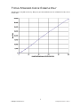
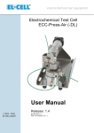
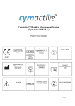
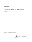
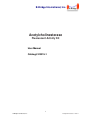
B-Bridge International, Inc.
Acetylcholinesterase
Fluorescent Activity Kit
User Manual
Catalog # K3015-1
1
B-Bridge International, Inc.
Acetylcholinesterase 110811
TABLE OF CONTENTS
Intended Use
3
Background
3
Assay Principle
3
Kit Components
4
Materials Required
4
Precautions
4
Reagent Preparation
5
Sample Preparation
6
Assay Protocol
6
Calculations
6
Typical Standard Curve: Example
7
Notice to Purchaser
This product is to be used for Research Purposes Only. It is not to be used for Drug or
Diagnostic Purposes, nor is it intended for Human Use. B-Bridge products may not be
resold, modified for resale, or used to manufacture commercial products without the
express written consent of B-Bridge International, Inc.
EXCEPT AS OTHERWISE EXPRESSLY SET FORTH IN THIS USER MANUAL, B-BRIDGE
DOES NOT MAKE ANY REPRESENTATION OR WARRANTIES OR CONDITIONS OF ANY
KIND, EITHER EXPRESSED OR IMPLIED, WITH RESPECT TO THE PRODUCTS, OR
INFORMATION DISCLOSED HEREUNDER, INCLUDING, BUT NOT LIMITED TO, THE IMPLIED
WARRANTIES OF MERCHANTABILITY, FIT FOR A PARTICULAR PURPOSE, OR
NONINFRINGEMENT OF THE INTELLECTUAL PROPERTY RIGHTS OF THIRD PARTIES.
B-Bridge International, Inc. All Rights Reserved.
2
B-Bridge International, Inc.
Acetylcholinesterase 110811
INTENDED USE
The B-Bridge Acetylcholinesterase Fluorescent Activity Kit (catalog # K3015-1) quantitatively measures
Acetylcholinesterase (AChE) activity in serum, EDTA and heparin plasmas, and RBC ghosts (Erythrocyte
Membranes) from a variety of species. This assay is species independent. It will also measure AChE in
extracted tissue samples and cell lysates.
BACKGROUND
Acetylcholinesterase appears critical to both development and function of the nervous system. The use of
AChE inhibitors as therapeutic agents and pesticides has spurred detailed investigations of cholinesterase.
Acetylcholine (ACh) is an essential neurotransmitter in the central and peripheral nervous systems. In the
brain multiple areas exist where cholinergic neurons are concentrated. Nicotinic and muscarinic ACh
receptors are recognized as binding and effector proteins to mediate chemical neurotransmission at neurons,
ganglia, heart and smooth muscle fibers and glands. This traditional view of AChE acting solely as
neurotransmitter has to be revised based on the findings published both early and late in the last century,
demonstrating the non-neuronal cholinergic system.
AChE
Acetylcholine
Acetylcholinesterase is encoded by the single AChE gene; and the structural diversity in the gene products
arises from alternative nRNA splicing and post translational associations of catalytic and structural subunits.
The major form of acetylcholinesterase found in brain, muscle, and other tissues is the hydrophilic species,
which forms disulfide-linked oligomers with collagenous, lipid-containing structural subunits. The other
alternatively-spliced form, expressed primarily in the erythroid tissues, is structurally different at the
C-terminal end and contains a cleavable peptide with a GPI anchor. It associates with membrane receptors
through phosphoinositide moieties added post-translational.
Impairment of cholinergic neurotransmission is well-established in Alzheimer’s disease, but there is
controversy about its relevance at the early stages of the disease as well as in mild cognitive impairment. In
vivo positron emission tomography imaging of cortical AChE activity as a marker of cholinergic function that is
expressed by cholinergic axons and neurons has demonstrated a reduction of this enzyme activity in
manifest Alzheimer’s patients. Other intentional or environmental methods of with anticholinergic properties
which are used as insecticides worldwide or as warfare agents. Thousands of cases of acute poisoning have
been reported. This acute toxicity inhibits AChE at nerve terminals where inhibition causes accumulation of
ACh. This, in turn, induces overstimulation of nicotinic and muscarinic receptors in the central and peripheral
nervous systems and the consequent signs and symptoms.
ASSAY PRINCIPLE
The Acetylcholinesterase Fluorescent Activity kit is designed to quantitatively measure acetylcholinesterase
(AChE) activity in a variety of samples. The kit utilizes a proprietary non-fluorescent molecule, Thio, that
covalently binds to the thiol product of the reaction between the AChE Substrate and AChE in the standards
or samples, yielding a fluorescent product read at 510 nm in a fluorescent plate reader with excitation at 390
nm.
Because the readout of AChE activity is purely chemical, there are few interferants that will affect the
readings obtained.
1. Sample or standard added to well.
2. The reaction is initiated with the addition of the Reaction Mix containing AChE, Substrate, and Thio
Detection Reagent
3. Incubate for 20 minutes and read fluorescent signal. Calculate AChE activity from standard curve.
4. Alternatively samples can be read kinetically. Follow steps 1 and 2 above.
Add Reaction Mix and read signal at 510 nm over time. Compare rates for samples and standards to
determine sample AChE activity.
3
B-Bridge International, Inc.
Acetylcholinesterase 110811
KIT COMPONENTS
Black 96-well plates
2 plates
Acetylcholinesterase Standard (1,000 mU/ml)
225 µl
Thio Detection Reagent
-Stored in desiccator
2 vials
AChE Substrate
2 vials
- Acetylthiocholine iodide freeze dried with stabilizers
Dimethyl Sulfoxide Solvent (DMSO)
14 ml
- DMSO will freeze at 2-8°C. May be stored at room temperature.
10X Assay Buffer
28 ml
Store all components at 2-8°C, except DMSO. Store DMSO tightly capped at room temperature.
MATERIALS REQUIRED BUT NOT SUPPLIED
- Deionized or distilled water
- Isotonic saline (0.9%)
- Triton X-100
- Centrifuge
- Fluorescence 96-well plate reader capable of reading fluorescent emission at 510 nm with excitation at 390 nm.
-Software for converting raw relative fluorescent unit (FLU) readings from the plate reader and
carrying out four parameter logistic curve (4PLC) fitting.
PRECAUTIONS
This kit should only be used by qualified personnel who have had laboratory safety instruction. The complete
User Manual should be read and understood before using this product.
Dimethyl sulfoxide (DMSO) is a powerful aprotic organic solvent that has been shown to enhance the
rate of skin absorption of skin-permeable substances. Wear protective gloves, safety glasses and
clothing when working with DMSO. Consult your institution’s safety procedures for working with
hazardous chemicals.
The Acetylcholinesterase Standard is derived from human erythrocytes. It has been extensively tested for
viral contamination, but all human blood products should be treated as potentially infectious and adequate
precautions taken. Protective clothing, gloves, and safety glasses should be worn. Consult your institution’s
safety procedures for working with human samples.
Thio Detection Reagent should be stored at 4°C in the desiccator. Allow the desiccator to warm to room
temperature prior to opening. Thio Detection Reagent will react with strong nucleophiles. Buffers containing
the preservatives sodium azide, Proclin™ and Kathon™ will react with the substrate.
4
B-Bridge International, Inc.
Acetylcholinesterase 110811
REAGENT PREPARATION
Allow the kit reagents to come to room temperature for 30 minutes.
We recommend that all standards and samples be run in duplicate to accurately determine AChE activity.
Ensure that all samples have reached room temperature and have been diluted as appropriate prior to
assaying.
1X Assay Buffer
Prepare a 1X Assay Buffer by diluting 1 part 10X Assay Buffer with 9 parts deionized water. 1X Assay Buffer
is stable for up to 3 months at 4°C.
10X Thio Detection Reagent
Make a 10X Thio Detection Reagent by adding 700 µl of DMSO to 1 vial of Thio Reagent, vortex thoroughly.
Store any unused reconstituted Detection Reagent at 4°C in the desiccator and use within 2 weeks.
10X Acetylcholinesterase Substrate
Make a 10X AChE Substrate Reagent by adding 700 µl of DMSO to 1 vial of AChE Substrate, vortex
thoroughly. Store any unused reconstituted AChE Substrate at room temperature and use within 2 weeks.
Reaction Mix
Follow the table below to make the appropriate volume of Reaction Mix. Use the Reaction Mix within 1 hour of
preparation. Protect from light.
10X AChE Substrate
10X Thio Detection Reagent
DMSO
Half Plate
300 µl
300 µl
2.4 ml
Whole Plate
550 µl
550 µl
4.4 ml
Standard Preparation
AChE Standards are prepared by labeling five test tubes as #1 through #5. Briefly spin vial of standard in a
microcentrifuge to ensure contents are at bottom of the vial.
1. Pipet 450 µl of 1X Assay Buffer into tube #1 and 250 µl into tubes #2 to #5.
2. Carefully add 50 µl of the AChE Standard to tube #1 and vortex completely.
3. Take 250 µl of the AChE solution in tube #1 and add it to tube #2 and vortex completely.
4. Repeat these serial dilutions for tubes #3 through #5. The activity of AChE in tubes 1 through 5 will
be 100, 50, 25, 12.5, and 6.25 mU/ml.
Use all standards within 2 hours of preparation
Reagent
Assay Buffer Volume
AChE Stock
Standard 1
Standard 2
Standard 3
Standard 4
Final Concentration (mU/mL)
Standard
1
450 µl
50 µl
Standard
2
250 µl
Standard
3
250 µl
Standard
4
250 µl
Standard
5
250 µl
250 µl
250 µl
250 µl
100
50
25
12.5
250 µl
6.25
5
B-Bridge International, Inc.
Acetylcholinesterase 110811
SAMPLE PREPARATION
Serum and Plasma
Store serum or plasma on ice until assaying or freeze in aliquots for later use. Samples containing visible
particulate should be centrifuged prior to using.
Samples must be diluted in 1X Assay Buffer prior to assaying. Serum and plasma typically have to be diluted
≥ 1:300 to read in the assay. Any samples with AChE activity outside the standard curve range should be
diluted further with 1X Assay Buffer to obtain readings within the standard curve.
Use all samples within 2 hours of dilution
Erythrocytes (RBCs)
Blood is collected in the presence of heparin or EDTA. The sample is then centrifuged and the plasma and
white cell layer are removed from the RBC layer. The RBCs are suspended and gently washed twice with
three volumes of isotonic saline (0.9%), separating the cells by centrifugation at 600 x g for 10 minutes and
discarding the saline after each step. To lyse the RBCs, four volumes of cold deionized water are added to
the RBCs. The cells are then vortexed and incubated for 10 minutes at 4˚C, or allowed to undergo a
freeze/thaw. Samples are centrifuged at 14,000 rpm for 10 minutes at 4˚C and the supernatant discarded.
Further wash the membrane pellet with two or three volumes of isotonic saline, with centrifugation
in between, until it is only slightly pink. The smaller dark red pellet on the bottom is non-lysed RBCs and
should be avoided. Solubilize the white membrane ghost pellet with Triton X-100.
Dilute solubilized RBC ghost with 1X Assay Buffer. Final assay dilution of the solubilized RBC ghost sample
must be sufficient that the assay sample contains ≤ 0.01% Triton X-100.
Use all samples within 2 hours of dilution
ASSAY PROTOCOL
Standards and samples should be run in duplicate
1. Pipet 100 µl of samples or standards into duplicate wells in the plate
2. Pipet 100 µl of 1X Assay Buffer into duplicate wells as a zero standard.
3. Add 50 µl of prepared Reaction Mix to each well using a repeater or multi-channel pipet
4. Gently tap the sides of the plate to ensure adequate mixing of reagents
5. Incubate at room temperature for 20 minutes
6. Read the fluorescent emission at 510 nm with excitation at 370-410 nm.
CALCULATIONS
Average the duplicate fluorescent unit (FLU) readings for each standard and sample. Create a standard
curve by reducing the data using the 4PLC (4 parameter logistic curve) fitting routine on the plate reader, after
subtracting the mean FLU for the zero standard. The sample activity obtained should be multiplied by the
dilution factor to obtain neat sample values.
6
B-Bridge International, Inc.
Acetylcholinesterase 110811
TYPICAL STANDARD CURVE: EXAMPLE ONLY
Standard curves vary with each assay. Always run your own standard curves for calculation of results; do not
use this data
7
B-Bridge International, Inc.
Acetylcholinesterase 110811