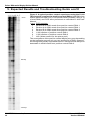
1
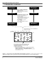
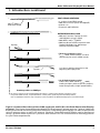
CLONTECH
Innovative
Tools to
Accelerate
Discovery
Delta® Differential
Display Kit
User Manual
PT1173-1 (PR19324)
Published 05 September 2001
Catalog #: K1810-1
Storage Conditions: Positive control RNAs
All other components
FOR RESEARCH USE ONLY
–70°C
–20°C
Delta® Differential Display Kit User Manual
Table of Contents
I.
Introduction
3
II.
List of Components
8
III.
Additional Materials Required
9
IV.
Protocol for Delta Differential Display
12
A. General Considerations
12
B. Preparation and Handling of Total RNA
12
C. (Optional) DNase I Treatment of Total RNA
13
D. First-Strand cDNA Synthesis
15
E. Design Considerations for Delta Experiments
17
F. Differential Display PCR
19
G. Electrophoresis and Autoradiography
22
V.
Expected Results and Troubleshooting Guide
23
VI.
Additional Procedures
26
A. Purification of DNA Fragments from Dried Polyacrylamide Gels
26
B. Reamplification of the Band of Interest
26
C. Northern Blot Analysis
27
D. Additional Experiments
28
References
30
VIII. Related Products
31
Appendix A: Design of the Delta Primers
32
Appendix B: Size of Delta Differential Display Experiments
33
VII.
Notice to Purchaser
This product is intended to be used for research purposes only. It is not to be used for drug or diagnostic
purposes nor is it intended for human use. CLONTECH products may not be resold, modified for resale,
or used to manufacture commercial products without written approval of CLONTECH.
This product is optimized for use in the Polymerase Chain Reaction (“PCR”) covered by patents
owned by Hoffmann-La Roche, Inc. and F. Hoffmann-La Roche, Ltd. (“Roche”). No license under
these patents to use the PCR process is conveyed expressly or by implication to the purchaser by
the purchase of this product. A license to use the PCR process for certain research and development
activities accompanies the purchase of certain reagents from licensed suppliers such as
CLONTECH Laboratories, Inc., when used in conjunction with an authorized thermal cycler, or is
available from Perkin-Elmer Corporation. Further information on purchasing licenses to practice the
PCR process may be obtained by contacting the Director of Licensing at the Perkin-Elmer
Corporation, 850 Lincoln Centre Drive, Foster City, CA 94404, or at Roche Molecular Systems, Inc.,
1145 Atlantic Avenue, Alameda, CA 94501.
CLONTECH Laboratories, Inc.
2
www.clontech.com
Protocol # PT1173-1
Version # PR19324
Delta ® Differential Display Kit User Manual
I. Introduction
The Delta® Differential Display Kit (formerly named the Delta RNA Fingerprinting
Kit) enables researchers to identify RNAs that are expressed in one RNA
population but missing in another (Diachenko et al., 1996). The identification of
such “differentially expressed” RNAs is a powerful tool for studying differentiation, the cell cycle, carcinogenesis, inductive events, and other biological
phenomena that involve changes in gene expression.
Delta Differential Display is a highly reproducible method for detecting differentially
expressed RNAs. In our hands, more than 95% of Delta results are reproducible,
and more than 85% of the differentially expressed bands can be confirmed by
Northern blot analysis. The method requires only 2 µg of total RNA per sample,
and the primers in the kit give you more than 150 different combinations. The
protocol requires only a single cDNA synthesis for each different RNA sample,
in contrast to the multiple cDNA synthesis reactions required for similar methods.
The longer primers in the Delta Kit allow higher stringency PCR, which, in turn,
leads to much greater reproducibility of the resulting displays. The use of enzyme
mixes suitable for long-distance PCR enables the Delta system to resolve
differentially expressed bands of up to 2 kb and results in a higher fidelity of PCR
products to the original RNA templates.
Protocol overview
The Delta Kit is based on improvements to the methods described by McClelland
et al. (1993). The protocol consists of two stages: cDNA synthesis and differential
display PCR (Figure 1). First-strand cDNA is synthesized using each of the RNAs
of interest as a template and oligo(dT) as a primer. In contrast to the 9–12
syntheses required for each different RNA sample in other differential display
protocols (Liang & Pardee, 1992; Liang et al., 1993), the Delta protocol requires
only a single cDNA synthesis reaction for each RNA sample. (The multiple cDNA
syntheses in other methods use different oligo(dT)11NM primers to subdivide the
pool of total RNA. In Delta Differential Display, this subdivision occurs during the
PCR.) The single cDNA synthesis saves considerable time, and, since reverse
transcriptase is an expensive reagent, it reduces costs as well. We also find the
subsequent PCR to be more reproducible.
Another time- and cost-saving feature of the Delta Kit is that each cDNA
synthesis reaction uses only 2 µg of total RNA. In our experience, the Delta Kit
gives similar results with total and poly A+ RNA, so there is no need to purify poly
A+ RNA. Whenever possible, RNA samples should be prepared side-by-side
using the same reagents and purification protocol.
In differential display PCR, sequences are amplified—in the presence of
[α-33P]dATP—based on chance homology to arbitrary P primers. The Delta PCR
cycling program differs substantially from conventional PCR cycling programs.
Three initial cycles are performed at low stringency (i.e., low annealing temperature) to allow the primers to anneal and initiate DNA synthesis. Because of the
low stringency, each P primer will bind sites on many cDNAs with imperfect
Protocol # PT1173-1
Version # PR19324
www.clontech.com
CLONTECH Laboratories, Inc.
3
Delta® Differential Display Kit User Manual
I. Introduction continued
RNA sample # 2
RNA sample # 1
(2 µg of total RNA)
(2 µg of total RNA)
FIRST-STRAND SYNTHESIS
• Oligo(dT) primer
• MMLV reverse transcriptase
• 42°C for 1 hr
ss cDNA template
ss cDNA template
Differential Display
PCR products
DIFFERENTIAL DISPLAY PCR
• Pairwise combinations of
P and T primers
• Advantage KlenTaq Polymerase
Mix with TaqStart Antibody
• [α-33P]dATP
• 3 low-stringency cycles
Tanneal = 40°C
• 22–25 high-stringency cycles
Tanneal = 60°C
Differential Display
PCR products
• Electrophorese on 5% acrylamide gel
• Expose x-ray film
Primers: P1/T1
RNA sample: 1 2
P2/T1
1 2
P3/T1
1 2
P1/T2
1 2
P2/T2
1 2
P3/T2
1 2
2 kb
100 bp
Cut out differentially expressed bands and:
• Reamplify and confirm by Northern blot
• Clone and sequence
• Conduct further studies:
• Generate full-length cDNAs using Marathon-ReadyTM
cDNAs or MarathonTM cDNA Amplification Kit
• Obtain genomic clones or regulatory sequences using
GenomeWalkerTM Kits
Figure 1. Overview of the Delta Differential Display protocol. In the actual protocol, each
differential display PCR is performed using two different dilutions of each cDNA sample.
CLONTECH Laboratories, Inc.
4
www.clontech.com
Protocol # PT1173-1
Version # PR19324
Delta ® Differential Display Kit User Manual
I. Introduction continued
AAAAAAAAAAAAAAAA
T T T T T T T T T – 5'
5' –
poly A+ RNA in
total RNA sample
AAAAAAAAA – 3'
T T T T T T T T T – 5'
5' –
3' –
FIRST-STRAND SYNTHESIS
• 2 µg of each total RNA sample
• Add reverse transcriptase and oligo(dT)
• 42°C for 1 hr
• Prepare A and B dilutions
DIFFERENTIAL DISPLAY PCR
• Add one P and one T primer to the A
and B dilutions of each cDNA
• Add dNTPs and [α-33P]dATP
• Add long-distance PCR enzyme mix with
TaqStart Antibody (e.g., Advantage
KlenTaq Polymerase Mix)
P primer
TTTTTTTTT
AAAAAAAAAAAA
TTTTTTTTTTTT
AAAAAAAAAAAAAA...
N-1N-1T T T T T T T T T
T primer
• Three low-stringency cycles
Tanneal = 40°C, so the P primer can bind
(imprecisely) to various cDNA strands
N-1N-1 bases position the primer at the
beginning of the poly A tail
AAAAAAAAAAAAAA...
TTTTTTTTT
AAAAAAAAA
TTTTTTTTT
• 22–25 high-stringency cycles
Primer "tails" allow shift to Tanneal = 60°C,
leading to higher yields of specific products,
lower backgrounds, & high reproducibility
Examine products on PAGE gel
For clarity, only one set of annealing/extension events is shown. During the low-stringency cycles,
many other cDNA strands are primed with the same P primer, producing the mix of PCR products
that are amplified in the later, high-stringency cycles to produce the differential display for each RNA sample.
Figure 2. Detailed flow chart of the cDNA synthesis and PCR in the Delta Differential Display
protocol. This figure shows differential display PCR using one P primer and one T primer. Although
these are the most commonly performed reactions, Delta Differential Display can also be performed
using P primers alone or pairs of P primers. Similarly, Delta Differential Display can be performed
using T primers alone or pairs of T primers. See Section IV.E for a discussion of choosing primers
for your Delta experiments.
Protocol # PT1173-1
Version # PR19324
www.clontech.com
CLONTECH Laboratories, Inc.
5
Delta® Differential Display Kit User Manual
I. Introduction continued
and/or incomplete matches. The products of these early cycles are then
amplified (using the P primer and the downstream T primer) during 22–25 highstringency PCR cycles. When examined by polyacrylamide electrophoresis and
autoradiography, these reactions produce characteristic banding patterns, or
“displays,” of the starting RNA.
A sample display using the positive control reagents can be seen in Figure 3 in
Section V (Expected Results and Troubleshooting Guide). Each different pair of
primers will produce a different display and may therefore identify a different set
of differentially expressed RNAs. Once differentially expressed bands have been
identified, they can be eluted from the gel and reamplified to make probes for
Northern blot confirmation of differential expression, as discussed in Section VI
(Additional Procedures). Confirmed positives can then be cloned and further
characterized.
The Delta Differential Display primers
The design of the Delta primers is critical to the success and proven reproducibility of the Delta Differential Display Kit. The design features that distinguish the
Delta primers from the primers used in previously described methods and in other
commercial kits are discussed in detail in Appendix A.
With 10 arbitrary “P” primers and 9 oligo(dT) or “T” primers, the kit has 90 possible
combinations of upstream and downstream primers. Furthermore, each P primer
can be used alone or in pairwise combinations with other P primers. Similarly, the
T primers can be used alone or in combination with other T primers. See Section
IV.E for a discussion on choosing primers.
Long-distance PCR with the Advantage® cDNA PCR Kit
Delta Differential Display uses the conditions of long-distance (LD) PCR (Barnes,
1994; Cheng et al., 1994), in which a combination of thermophilic DNA polymerases produces much larger PCR products than are possible with conventional PCR. Yields are also increased relative to conventional PCR. Furthermore,
the presence of a minor amount of a DNA polymerase with 3'→5' exonuclease
(i.e., “proofreading”) activity makes LD PCR more accurate than conventional
PCR with a single DNA polymerase (Barnes, 1994; Frey & Suppman, 1995;
Nelson et al., 1995).
We recommend CLONTECH's Advantage KlenTaq Polymerase Mix (#8417-1),
which was specifically developed for LD PCR using cDNA templates and is the
enzyme mix used to optimize the protocols in this booklet. This 50X mix contains
KlenTaq-1 DNA Polymerase (an exo-minus, N-terminal deletion of Taq DNA
polymerase) as the primary polymerase, a minor amount of a secondary
polymerase which provides 3'→5' proofreading activity, and TaqStartTM Antibody to
provide a convenient and automatic form of “hot-start” PCR (Kellogg et al., 1994).
Advantage KlenTaq Polymerase Mix is also available in the Advantage cDNA PCR
Kit (#K1905-1, -y).
CLONTECH Laboratories, Inc.
6
www.clontech.com
Protocol # PT1173-1
Version # PR19324
Delta ® Differential Display Kit User Manual
I. Introduction continued
There are several advantages to using LD PCR for the identification of differentially expressed RNAs. First, in combination with the longer primers used in this
kit, LD PCR allows us to reduce the total number of PCR cycles from 40 to 25.
This means that abundant products are not overcycled and differentially expressed bands are easier to detect. Secondly, the larger range of PCR products
(up to 2 kb) increases your chances of finding differentially expressed bands with
each primer combination. Finally, the higher fidelity of LD PCR means that bands
identified by Delta Differential Display and subsequently cloned will have greater
fidelity to the original mRNA sequence.
CLONTECH PCR-SelectTM cDNA Subtraction Kit
Another method for comparing RNA populations is CLONTECH's PCR-Select
cDNA Subtraction Kit (#K1804-1), a unique PCR-based method for powerful,
reproducible enrichment of differentially expressed genes. The PCR-Select
method allows you to selectively and simultaneously amplify all sequences that
are differentially expressed in a given tissue or cell type, and also enriches for
rare mRNAs. CLONTECH's suppression PCR technology (U.S. Patent
#5,565,340) prevents amplification of sequences that are not differentially
expressed.
Protocol # PT1173-1
Version # PR19324
www.clontech.com
CLONTECH Laboratories, Inc.
7
Delta® Differential Display Kit User Manual
II. List of Components
Store total RNA at –70°C. Store all other reagents at –20°C.
The following reagents are sufficient for 12 cDNA syntheses and 900 PCRs.
For optional DNase I treatment of total RNA
•
12 µl DNase I (RNase-free, 1 unit/µl)
Reagents for first-strand cDNA synthesis
•
30 µl Oligo(dT) primer (1 µM)
•
12 µl MMLV reverse transcriptase (200 units/µl)
• 500 µl 5X First-strand buffer:
250 mM Tris (pH 8.3)
30 mM MgCl2
375 mM KCl
Control reagents
•
5 µl Positive control RNA 1 (1 mg/ml of total RNA from adult liver)
•
5 µl Positive control RNA 2 (1 mg/ml of total RNA from fetal liver)
PCR primers for Delta Differential Display
• 10 x 90 µl Arbitrary primers (20 µM):
P1: 5'-ATTAACCCTCACTAAATGCTGGGG A-3'
P2: 5'-ATTAACCCTCACTAAATCGGTCATAG-3'
P3: 5'-ATTAACCCTCACTAAATGCTGGTGG-3'
P4: 5'-ATTAACCCTCACTAAATGCTGGTAG-3'
P5: 5'-ATTAACCCTCACTAAAGATCTGACTG-3'
P6: 5'-ATTAACCCTCACTAAATGCTGGGTG-3'
P7: 5'-ATTAACCCTCACTAAATGCTGTATG-3'
P8: 5'-ATTAACCCTCACTAAATGGAGCTGG-3'
P9: 5'-ATTAACCCTCACTAAATGTGGCAGG-3'
P10: 5'-ATTAACCCTCACTAAAGCACCGTCC-3'
•
9 x 90 µl Oligo(dT) primers (20 µM):
T1: 5'-CATTATGCTGAGTGATATCTTTTTTTTTAA-3'
T2: 5'-CATTATGCTGAGTGATATCTTTTTTTTTAC-3'
T3: 5'-CATTATGCTGAGTGATATCTTTTTTTTTAG-3'
T4: 5'-CATTATGCTGAGTGATATCTTTTTTTTTCA-3'
T5: 5'-CATTATGCTGAGTGATATCTTTTTTTTTCC-3'
T6: 5'-CATTATGCTGAGTGATATCTTTTTTTTTCG-3'
T7: 5'-CATTATGCTGAGTGATATCTTTTTTTTTGA-3'
T8: 5'-CATTATGCTGAGTGATATCTTTTTTTTTGC-3'
T9: 5'-CATTATGCTGAGTGATATCTTTTTTTTTGG-3'
General reagents
• 200 µl dNTPs (5 mM each dATP, dCTP, dGTP, dTTP)
• 2 x 4 ml Sterile H2O
CLONTECH Laboratories, Inc.
8
www.clontech.com
Protocol # PT1173-1
Version # PR19324
Delta ® Differential Display Kit User Manual
III. Additional Materials Required
The following reagents are required but not supplied:
For optional DNase I treatment of total RNA
•
0.5 M Tris-HCl (pH 7.5)
•
0.5 M MgCl2
•
3 M NaOAc
•
0.2 M EDTA
• Phenol:chloroform:isoamyl alcohol (25:24:1)
Prepare as follows:
1. Melt phenol.
2. Equilibrate with an equal volume of sterile TNE buffer (50 mM Tris
[pH 7.5], 150 mM NaCl, 1 mM EDTA).
3. Incubate the mixture at room temperature for 2–3 hr.
4. Remove and discard the top layer.
5. Add an equal volume of chloroform:isoamyl alcohol (24:1) to the
remaining layer. Mix thoroughly. Remove and discard the top layer.
6. Store the bottom layer of phenol:chloroform:isoamyl alcohol at 4°C
away from light for a maximum of 2 weeks.
• Chloroform:isoamyl alcohol (24:1)
For Delta Differential Display
• 50X polymerase mix
You will need a Taq-based 50X polymerase mix suitable for LD PCR. The
Delta protocol has been optimized with CLONTECH's Advantage KlenTaq
Polymerase Mix (#8417-1), which contains KlenTaq-1 DNA Polymerase (an
exo-minus, N-terminal deletion of Taq DNA polymerase) as the primary
polymerase, a minor amount of a 3'→5' proofreading polymerase, and
TaqStart Antibody for hot start (Kellogg et al., 1994). Advantage KlenTaq
Polymerase Mix is available in the Advantage cDNA PCR Kit (#K1905-1, -y).
If you choose not to use Advantage KlenTaq Polymerase Mix, Table I gives
instructions for mixing TaqStart Antibody with other commercially available
LD PCR-licensed enzyme mixes. In our experience, Delta Differential
Display reactions are somewhat less efficient with these other enzyme
mixes. In general, we recommend that the primary enzyme in the mix be Taq
DNA polymerase (or a Taq derivative) instead of Tth DNA polymerase, which
results in higher backgrounds and more smearing.
Taq DNA polymerase can be used by itself; however, more cycles will be
needed. Furthermore, the size range of the products will be much smaller,
so you will obtain fewer differentially expressed bands per display and miss
the larger bands that would be identified using the LD PCR mixes. Also, LD
PCR has a significantly lower error rate than PCR with Taq DNA polymerase
alone (Barnes, 1994), so the sequence of your PCR products will be more
Protocol # PT1173-1
Version # PR19324
www.clontech.com
CLONTECH Laboratories, Inc.
9
Delta® Differential Display Kit User Manual
III. Additional Materials Required continued
faithful to the original mRNA. This is a particularly important consideration for
designing primers for subsequent experiments, such as MarathonTM cDNA
amplification or sequencing.
Using TaqStart Antibody in the 50X polymerase mix greatly reduces nonspecific synthesis in Delta Differential Display PCR. (TaqStart Antibody has
already been added to CLONTECH's Advantage KlenTaq Polymerase Mix.)
TaqStart is an effective method for hot-start PCR that is simpler and more
convenient than wax-based or manual methods. The TaqStart Antibody
binds to and inactivates Taq DNA polymerase (native or truncated) and thus
eliminates DNA synthesis from nonspecifically bound primers while reactions are being assembled. PCR amplification proceeds efficiently after an
initial incubation at 94°C which irreversibly inactivates the TaqStart Antibody. TaqStart Antibody is available from CLONTECH (#5400-1, -2).
•
10X PCR reaction buffer
(Included in the Advantage cDNA PCR Kit)
Use the 10X reaction buffer supplied with your source of native or truncated
Taq DNA polymerase in all reactions that call for 10X PCR buffer. (Of the
three buffers supplied with the ExpandTM Long Template PCR System, we
obtained the best results with buffer #1.)
If you are using a buffer system in which the Mg2+ is added separately, we
recommend using a final [Mg2+ ] of 1.5 mM.
•
[α-33P]dATP (1000-3000 Ci/mmole; 3.3 µM)
[α-33P]dATP produces much sharper bands than [α-32P]dATP and thus
increases the resolution of your differential displays (and therefore the
number of bands that you can distinguish). [α-35S]dATP can also be used;
however, longer exposure times will be required. Also, volatilization of
[α-35S]dATP upon heating may lead to low-level contamination of thermal
cyclers (Trentmann et al., 1995).
•
0.5-ml PCR tubes
We recommend Perkin-Elmer GeneAmp ® 0.5-ml reaction tubes
(Cat. #N801-0737 or N801-0180).
•
X-ray film
We recommend Kodak BioMax MR film.
•
Gel electrophoresis apparatus
For best results, use a temperature-controlled (“thermostatic”) gel electrophoresis apparatus, such as the MacrophorTM System from Pharmacia Biotech
AB or the genomyxLRTM DNA Sequencer from Genomyx Corporation.
•Gel loading buffer:
95.0% Formamide
0.2% Bromophenol blue
0.2% Xylene cyanol
CLONTECH Laboratories, Inc.
10
www.clontech.com
Protocol # PT1173-1
Version # PR19324
50 units/ml
50 units/ml
???
ExTaq (5 units/µl)
Taq (3.5 units/µl) &
Pwo (conc. not given)
Takara LA PCR Kit (#RR011)
merase
Boehringer Mannheim
Expand Long Template
PCR System (#1681 834)
Protocol # PT1173-1
Version # PR19324
Best results in Delta Differential Display
50X enzyme mixes made with TaqStart Antibody can be stored at –20°C. (TaqStart Antibody is provided at 1.1 mg/ml in 50% glycerol. Do
not dilute the antibody prior to adding it to the 50X mix.)
To prepare 50X enzyme mixes without TaqStart Antibody, simply replace the specified volume of TaqStart Antibody with the 1X PCR reaction
buffer supplied with the DNA polymerase. Because of the lower glycerol content, 50X enzyme mixes made with 1X PCR reaction buffer
should be stored at 4°C.
b
Mix 14.3 µl of Taq /Pwo
with 5.7 µl of TaqStart Antibody b
with 10 µl of TaqStart Antibody b
Mix 10 µl of ExTaq DNA Poly-
Ready-to-use
(contains TaqStart Antibody)
Proportions for
50X enzyme mixb
a
See Advantage
User Manual
Premade enzyme
mix (50X)
CLONTECH's Advantage
cDNA PCR Kit (#K1905-1, -y)a
Concentration
in PCR
DNA polymerases
(conc. supplied)
Supplier/kit
(Cat. #)
TABLE I: PREPARATION OF 50X POLYMERASE MIX
Delta ® Differential Display Kit User Manual
III. Additional Materials Required continued
www.clontech.com
CLONTECH Laboratories, Inc.
11
Delta® Differential Display Kit User Manual
IV. Protocol for Delta Differential Display
PLEASE READ ENTIRE PROTOCOL BEFORE STARTING
A. General Considerations
1. The cycling parameters throughout this protocol have been optimized
using a Perkin-Elmer DNA Thermal Cycler 480 or GeneAmp PCR
Systems 2400/9600, Advantage KlenTaq Polymerase Mix, and ss
cDNA templates prepared from the RNA controls provided in the Delta
Kit. The optimal cycling parameters may vary with different thermal
cyclers.
2. We recommend that you use some form of hot start for Delta Differential
Display PCR. The following protocol has been optimized using TaqStart
Antibody (Kellogg et al., 1994) in the 50X polymerase mix. Hot-start
PCR can also be performed using wax beads (D’Aquila et al., 1991) or
manually (Chou et al., 1992).
3. Wear gloves throughout the procedure to protect your RNA and cDNA
samples from degradation by nucleases.
4. When mixing reactions, gently pipet the solution up and down or tap the
bottom of the tube and spin briefly to collect the contents at the bottom
of the tube.
5. Set up all reactions on ice, unless otherwise indicated.
6. Add enzymes to reaction mixtures last. Make sure that the enzyme is
thoroughly mixed with the reaction mixture by pipeting the mixture up
and down.
7. Use the recommended amounts of enzyme. These amounts have been
carefully optimized for the Delta protocol and reagents.
B. Preparation and Handling of Total RNA
1. General precautions
The integrity and purity of the total RNA used as starting material is an
important element of high-quality cDNA synthesis and the subsequent
PCR. The following precautions will help you avoid contamination and
degradation of your RNA.
a. Wear gloves to avoid RNase contamination from your hands.
b. Treat H2O used in steps that involve RNA with diethyl pyrocarbonate
(DEPC).
Note: Do not use DEPC to treat solutions containing Tris, nucleotides, or other
amines.
c. Rinse all glassware with 0.5 N NaOH, followed by DEPC-treated
H2O. Then bake the glassware at 160–180°C for 4–9 hr.
d. Use 70% ethanol or isopropanol to wipe all pipettes before use with
RNA.
CLONTECH Laboratories, Inc.
12
www.clontech.com
Protocol # PT1173-1
Version # PR19324
Delta ® Differential Display Kit User Manual
IV. Protocol for Delta Differential Display continued
2. RNA isolation
Whenever possible, total RNA samples being compared in a Delta
experiment should be purified side-by-side using the same reagents
and protocol. Two RNA samples purified from the same source but by
a different procedure can give different banding patterns in differential
display. Thus, more false positives may arise if the RNAs being
compared were prepared in different ways.
See Farrell (1993) or Sambrook et al. (1989) for reviews of procedures for
isolating total RNA.
3. RNA analysis
After isolating total RNA, we recommend that you examine the RNA by
electrophoresis of a sample on a denaturing formaldehyde/agarose/
EtBr gel. On denaturing gels, total RNA typically gives bright 28S and
18S ribosomal RNA bands at approximately 4.5 and 1.9 kb, respectively, with a ratio of intensities of about 1.5–2.5:1. If this ratio is less
than 1:1, we suggest you prepare fresh RNA after checking your RNA
purification reagents for RNase and other impurities. If problems
persist, you may have to find another source of tissues/cells.
C. (Optional) DNase I Treatment of Total RNA
Unless total RNA samples are seriously contaminated with genomic DNA,
DNase I treatment is not necessary. If analysis of total RNA on a denaturing
formaldehyde/agarose/EtBr gel reveals visible genomic DNA contamination (indicated by EtBr-stained material stuck in the wells), then you should
treat your samples with DNase I before continuing with the first-strand
cDNA synthesis. Likewise, if the total RNA control display patterns are
identical to your experiments, you should treat your RNA with DNase I.
1. For each total RNA sample, prepare an equal volume of DNase
mixture. Combine the following components in a sterile 0.5-ml microfuge
tube:
per 25-µl RNA sample*
components
1 µl
0.5 M Tris-HCl (pH 7.5)
1 µl
0.5 M MgCl2
22 µl
Sterile H2O
1 µl
RNase-free DNase I (1 unit/µl)
25 µl total
* We generally recommend that you use 25 µl of each RNA sample at a concentration
of 0.1–1 mg/ml. For multiple RNA samples, scale up the amounts of the components
of the DNase I mixture proportionately. To avoid pipeting errors, do not make less
than 25 µl of the DNase I mixture.
Protocol # PT1173-1
Version # PR19324
www.clontech.com
CLONTECH Laboratories, Inc.
13
Delta® Differential Display Kit User Manual
IV. Protocol for Delta Differential Display continued
2. In appropriately labeled, sterile 0.5-ml microfuge tubes, add an equal
volume of the DNase I mixture to each RNA sample. (Do not use
polystyrene tubes, which may melt during phenol:chloroform
extraction.)
3. Incubate the tubes at 37°C for 30 min.
4. To each tube, add 2.5 µl of 0.2 M EDTA and 2 µl of 3 M NaOAc.
5. If you started with less than 25 µl of RNA, adjust the reaction mixture
volume to 50 µl by adding a solution of 10 mM Tris (pH 7.5) and 50 mM
NaOAc.
Note: The concentration of total RNA at this point should be at least 20 µg/ml.
Decreasing the RNA concentration below this optimum level may result in loss of RNA
during phenol:chloroform extraction and precipitation.
6. To each tube, add a volume of phenol:chloroform:isoamyl alcohol
(25:24:1) equal to the reaction volume.
7. Vortex the tubes thoroughly.
8. Centrifuge the tubes at 14,000 rpm for 10 min to separate phases.
9. For each sample, transfer the top aqueous layer to a clean 0.5-ml tube.
10. To each tube, add a volume of chloroform:isoamyl alcohol (24:1) equal
to the volume of the aqueous layer.
11. Vortex the tubes thoroughly.
12. Centrifuge the tubes at 14,000 rpm for 10 min to separate phases.
13. For each sample, transfer the top aqueous layer to a clean 0.5-ml tube.
14. To each tube, add 1/10 volume of 3 M NaOAc and 2.5 volumes (of the
total resulting volume) of 95% ethanol.
15. Vortex the tubes thoroughly.
16. Centrifuge the tubes for 20 min at 14,000 rpm at room temperature.
17. Carefully remove the supernatants.
18. Gently overlay the pellets with 200 µl of 80% ethanol.
19. Centrifuge at 14,000 rpm for 5 min.
20. Carefully remove the supernatants.
21. Air dry the pellets for 5–10 min.
22. Dissolve each pellet in 1.5 µl of H2O for each 2 µg of starting RNA. If
possible, measure the final concentration of each RNA sample. Store
samples at –20°C.
CLONTECH Laboratories, Inc.
14
www.clontech.com
Protocol # PT1173-1
Version # PR19324
Delta ® Differential Display Kit User Manual
IV. Protocol for Delta Differential Display continued
D. First-Strand cDNA Synthesis
The 10-µl reaction described below is designed to convert 2 µg of total RNA
into first-strand cDNA. This kit contains enough components for 12 separate cDNA syntheses. In most applications, you will perform two or more
separate cDNA syntheses in parallel—one for each tissue, stage, or cellculture condition that you wish to compare.
The following protocol is written for total RNA. Poly A+ RNA can also be
used, but in our experience poly A+ RNA gives no more differentially
expressed bands than total RNA. If you use poly A+ RNA, you will need
fewer cycles for PCR (Section IV.E).
In addition to your experimental sample, we recommend that you perform
two positive control cDNA syntheses with the human adult liver and fetal
liver total RNA included in the kit (positive control RNAs 1 & 2, respectively).
This will provide templates for positive controls and thus verify that the
system performs in your hands.
1. For each RNA sample (including positive controls), label a sterile 0.5ml microcentrifuge tube with a number followed by “A”.
Note: This will distinguish each sample from the "B" dilution prepared in Steps 16–18.
If you are comparing three experimental RNA samples, you might label your experimental tubes 1A, 2A, and 3A, and your positive controls PC1A and PC2A.)
2. Combine the following in the appropriately labeled tubes:
2 µg Total RNA sample*
1 µl cDNA synthesis primer (1 µM)
* The volume depends on the concentration of each RNA sample. At CLONTECH, our total
RNA samples are typically 0.5–1.0 mg/ml. For the positive control RNAs (1 mg/ml),
add 2 µl.
3. Add sterile H2O to a final volume of 5 µl.
4. Mix contents and spin the tubes briefly in a microcentrifuge.
5. Incubate the tubes at 70°C for 3 min. (Temperatures >70°C may lead
to degradation of your RNA.)
6. Cool the tubes on ice for 2 min.
7. Spin the tubes briefly in a microcentrifuge.
8. Prepare enough Master Mix for all of your cDNA synthesis reactions
plus one additional tube:
per
5
rxn
rxns
Components
2 µl
10 µl
5X First-strand buffer
2 µl
10 µl
dNTP mix (5 mM)
1 µl
5 µl
MMLV reverse transcriptase (200 units/µl)
9. Add 5 µl of the Master Mix to each reaction tube.
Protocol # PT1173-1
Version # PR19324
www.clontech.com
CLONTECH Laboratories, Inc.
15
Delta® Differential Display Kit User Manual
IV. Protocol for Delta Differential Display continued
10. Mix the contents of the tubes by gently pipeting.
11. Spin the tubes briefly in a microcentrifuge.
12. Incubate the tube at 42°C for 1 hr in an air incubator.
Note: Using a water bath or thermal cycler for this incubation may reduce the volume
of the reaction mixture (due to evaporation) and therefore reduce the efficiency of firststrand synthesis.
13.
14.
15.
16.
Terminate the reactions by incubating at 75°C for 10 min.
Place the tubes on ice.
Spin the tubes briefly in a microcentrifuge.
For each RNA sample (including positive controls), label a sterile 0.5ml microcentrifuge tube with a number followed by “B” (i.e. tubes should
be labeled 1B, 2B, PC1B, PC2B....). This is for the “B” dilution of each
cDNA sample.
17. Transfer 2 µl of each reaction to the appropriate “B” tube.
18. Add 78 µl of sterile H2O to each “B” tube.
19. Add 72 µl of sterile H2O to the tube containing the remaining 8 µl of each
ss cDNA. This is the “A” dilution of each cDNA sample.
20. Store all cDNA dilutions at –20°C until ready for use.
CLONTECH Laboratories, Inc.
16
www.clontech.com
Protocol # PT1173-1
Version # PR19324
Delta ® Differential Display Kit User Manual
IV. Protocol for Delta Differential Display continued
E. Design Considerations for Delta Experiments
1. Choice of primers and number of primer combinations
The Delta Differential Display Kit contains 10 arbitrary P primers and 9
oligo(dT) T primers. This gives you 90 possible two-way primer combinations. The arbitrary P primers can also be used alone (i.e., without a
downstream primer) or in pair-wise combinations with other P primers
(McClelland et al., 1990). This gives you 55 additional primer combinations. Although there is some overlap, some bands will be detected with
the P primer alone that would have been missed in pair-wise combinations with T primers. The T primers can also be used alone or in
combination with each other. (Presumably, the oligo(dT) portion of the
primer anneals to internal runs of As during the initial low-stringency
cycles.) Note that combinations of P and T primers (or T primers alone)
will generally produce PCR products derived from the 3' end of the
mRNA, while P primers alone (or combinations of P primers) will favor
internal regions of the mRNA.
Reactions using a P/T primer pair typically generate 40–80 bands.
When comparing two different RNA samples, the number of differentially expressed RNAs will vary depending on the relatedness of the two
sources. For example, 4% of the mRNAs differ between SV40-transformed and untransformed mouse cells (Scott et al., 1983). Similarly,
2% of the transcripts differ between T- and B- lymphocytes (Hedrick et
al., 1984), while over 100 genes are activated during T-cell differentiation following antigenic stimulation (Ulman et al., 1990).
In our experiments comparing human adult and fetal liver total RNA (i.e.,
the positive control RNAs in the Delta Kit), we typically obtained 1–2
differentially expressed bands for each combination of P and T primers.
When these bands were used as probes on Northern blots, more than
85% were confirmed as unique, differentially expressed mRNAs, each
having a different size. In contrast, in studies comparing stimulated and
unstimulated mouse macrophages (Raw cell lines) or stimulated and
unstimulated T-cells (Jurkat cell line), we typically obtained 1 differentially expressed band for every 2–3 combinations of P and T primers.
2. Use of controls
a. Negative controls
i. H2O control
For each pair of primers you use, it is important to perform an
H2O control using 1 µl of H2O as the “template.” Although there
is no template in this reaction, it nevertheless produces a light
banding pattern in some cases. In other cases, there will be no
signal after the specified number of cycles. Whatever bands are
present in the H2O control should take at least 5 additional
cycles to reach the same intensity as the bands in real differential displays. If present, the light H2O banding pattern is an
Protocol # PT1173-1
Version # PR19324
www.clontech.com
CLONTECH Laboratories, Inc.
17
Delta® Differential Display Kit User Manual
IV. Protocol for Delta Differential Display continued
inherent background in all differential display reactions performed with that primer pair.
This is an important control for the quality of your cDNA synthesis. If your cDNA synthesis reaction does not work well, then
your experimental differential display PCR will be of similar
intensity to the H2O control. If you get no signal in your H2O
controls and experimental samples, you may have a problem
with your DNA polymerase. Once you are familiar with the H2O
control for a primer pair, it is not necessary to repeat the control
with every new experiment.
ii. Total RNA control
Perform a total RNA control the first time you use each freshly
made cDNA template for differential display PCR. For this
control, use 1 µl of a 1/100 dilution of the RNA that served as a
template for cDNA synthesis. Initially, this control only needs to
be done with one primer pair. The resulting differential display
pattern should be compared to the H2O control performed with
the same set of primers. If there was genomic DNA contamination in your RNA, you will see a significant number of bands in
the RNA control that are not present in the H2O control. Such
contamination can lead to many “false positives” when a contaminated sample is being compared to a non-contaminated
sample. If your total RNA control differs from your H2O control,
you can still use the cDNA as a template for PCR, but you should
repeat the total RNA control for each different primer pair.
Alternatively, you may wish to repeat your cDNA synthesis after
either making fresh RNA or treating your RNA with RNase-free
DNase. Once you have determined that a given RNA was not
contaminated with genomic DNA, it is not necessary to repeat
the total RNA control in later experiments.
b. Positive control
For a positive control, perform differential display PCR with the
positive control cDNAs (made in Section C from the positive control
RNAs) and the P10 and T8 primers. This should produce two good
displays with a strong differentially expressed band of ~380 bp in
the fetal lane (PC2; see Figure 3 in Section V). To prevent running
out of the P10 and T8 primers, you may wish to use one of your own
successful differential display reactions as the positive control in
your later experiments.
3. Use both the A and B dilutions of each cDNA sample as templates for
differential display PCR
In some cases a band will show up in one dilution, but will be absent
from the other dilution; however, real differentially expressed bands
always appear in both dilutions. Thus, using both dilutions as templates
provides a positive control for true differentially expressed bands.
CLONTECH Laboratories, Inc.
18
www.clontech.com
Protocol # PT1173-1
Version # PR19324
Delta ® Differential Display Kit User Manual
IV. Protocol for Delta Differential Display continued
F. Differential Display PCR
Delta Differential Display experiments tend to get very large—the more
primer combinations you try, the more differentially expressed bands you
may identify. (See Table III in Appendix B for examples of how the size of
Delta experiments varies with the number of primer combinations and RNA
samples.) We recommend that you keep your initial experiments
small. For your first experiment, try using two T primers and three P primers
for a total of six PCRs with each template. With two RNA samples and
positive controls, this requires a total of 39 PCRs, and the results can be
examined on 1–2 large gels.
24 Experimental displays (no. of RNA samples x 2 dilutions of each
cDNA sample x no. of primer pairs)
6
H2O controls
2
Total RNA controls
7
Complete positive control experiment
(4 positive controls, 2 total RNA controls, 1 H2O control)
39 Total PCRs
A good organizational plan and labeling system is very helpful. Table II gives
an example of how Delta experiments are often set up at CLONTECH.
TABLE II: SAMPLE ORGANIZATIONAL AND LABELING CHART
FOR SETTING UP DELTA DIFFERENTIAL DISPLAY PCR
Tube
cDNA
Label
Sample
Experimental displays
1
1A
2
1B
3
2A
4
2B
5
6
7
8
9
10
11
12
and so on ....
Protocol # PT1173-1
Version # PR19324
Primers
P1 & T9
P1 & T9
P1 & T9
P1 & T9
1A
1B
2A
2B
P1 & T8
P1 & T8
P1 & T8
P1 & T8
1A
1B
2A
2B
P2 & T9
P2 & T9
P2 & T9
P2 & T9
Tube
cDNA
Label
Sample
Primers
Water controls for each primer pair
H1
H2O
P1 & T9
H2
H2O
P1 & T8
H3
H2O
P2 & T9
and so on ....
Total RNA controls for each RNA
(Use any of the above primer pairs)
R1
RNA 1
P1 & T9
R2
RNA 2
P1 & T9
and so on ....
Complete positive control (PC) experiment
PC1A
PC1A
P10 & T8
PC1B
PC1B
P10 & T8
PC2A
PC2A
P10 & T8
PC2B
PC2B
P10 & T8
HPC
H2O
P10 & T8
RPC1
PC RNA 1
P10 & T8
RPC2
PC RNA 2
P10 & T8
www.clontech.com
CLONTECH Laboratories, Inc.
19
Delta® Differential Display Kit User Manual
IV. Protocol for Delta Differential Display continued
The Delta Differential Display protocol has been optimized with CLONTECH's
Advantage KlenTaq Polymerase Mix, which includes TaqStart Antibody for
automatic hot-start PCR. If you choose not to use Advantage KlenTaq
Polymerase, Table I (Section III) provides instructions for mixing TaqStart
Antibody with other commercially available LD PCR-licensed enzyme
mixes. We recommend using some form of hot-start PCR (i.e., TaqStart
Antibody, wax beads, or manual hot start) to minimize background and
false positives in your differential display reactions.
Note on Steps 1–3: If you are performing PCR with a single P or T primer, use 2 µl of that
primer. If you are performing PCR with 2 P primers or 2 T primers, use 1 µl of each primer.
1. For each different experimental display (i.e., each pair of cDNAs and
each combination of primers), combine the following reagents in an
0.5-ml PCR tube.
1 µl Experimental cDNA sample
1 µl P primer
1 µl T primer
2. [Recommended] For each pair of primers, prepare an H2O control by
combining the following reagents in an 0.5-ml PCR tube:
1 µl Sterile H2O
1 µl P primer
1 µl T primer
3. [Recommended] For each different template, prepare a total RNA
control by combining the following reagents in an 0.5-ml PCR tube:
1 µl Total RNA (1/100 dilution of RNA used as cDNA template)
1 µl P primer
1 µl T primer
4. [Recommended] For the positive control experiment, combine the
following reagents in an 0.5-ml PCR tube (include H2O and RNA
controls):
1 µl Positive control cDNA sample
1 µl P10 primer
1 µl T8 primer
CLONTECH Laboratories, Inc.
20
www.clontech.com
Protocol # PT1173-1
Version # PR19324
Delta ® Differential Display Kit User Manual
IV. Protocol for Delta Differential Display continued
5. Prepare enough Master Mix for all of the PCRs plus one additional tube.
Component
10X KlenTaq PCR reaction buffer
Sterile H2O
dNTP mix (5 mM each;
final concentration 50 µM)
[α-33P]dATP* (1000–3000 Ci/mmole;
3.3 µM; final concentration 50 nM)
Advantage KlenTaq Polymerase
Mix (50X)
Final volume
per
rxn
2 µl
14.2 µl
0.2 µl
25
rxns
50 µl
355 µl
5 µl
100
rxns
200 µl
1420 µl
20 µl
0.2 µl
5 µl
20 µl
0.4 µl
17 µl
10 µl
425 µl
40 µl
1700 µl
Mix well by vortexing and briefly spin the tube in a microcentrifuge.
* [α-33P]dATP produces sharper bands than [α- 32P]dATP and thus increases the
resolution of differential displays (and the number of bands that can be distinguished).
If you use fresh label, you should obtain good overnight exposures. With older label (or
for faster exposures), you can increase the amount of label to 0.3–0.5 µl and add
proportionately less H2O. [α- 35S]dATP is not recommended because it volatilizes upon
heating, leading to contamination of equipment (Trentmann et al., 1995).
6. Aliquot 17 µl of PCR Master Mix into each reaction tube. This gives a
final volume of 20 µl.
7. Add 1 drop of mineral oil on top of PCR mixture and tightly cap the tubes.
8. Begin thermal cycling. (Use a cycler designated for radioactive use.)
Perkin-Elmer GeneAmp
Perkin-Elmer DNA
PCR Systems 2400/9600:
Thermal Cycler 480:
• 1 cycle:
• 1 cycle:
94°C 5 min
94°C 5 min
40°C 5 min
40°C 5 min
68°C 5 min
68°C 5 min
• 2 cycles:
• 2 cycles:
94°C 30 sec
94°C 2 min
40°C 30 sec
40°C 5 min
68°C 5 min
68°C 5 min
• 23 cycles:
• 22–25 cycles*:
94°C 20 sec
94°C 1 min
60°C 30 sec
60°C 1 min
68°C 2 min
68°C 2 min
•
68°C
for an additional 7 min.
• 68°C for an additional 7 min.
* For poly A + RNA, start with 15 cycles.
9. Store the differential display reactions at –20°C until you are ready to
examine them on a gel.
Protocol # PT1173-1
Version # PR19324
www.clontech.com
CLONTECH Laboratories, Inc.
21
Delta® Differential Display Kit User Manual
IV. Protocol for Delta Differential Display continued
G. Electrophoresis and Autoradiography
Delta Differential Display products can be up to 2 kb. For maximum
resolution, we recommend using some form of temperature-controlled
(“thermostatic”) electrophoresis system such as the Macrophor System
from Pharmacia Biotech AB or the genomyxLR DNA Sequencer from
Genomyx Corporation. Nonthermostatic electrophoresis systems do not
resolve differential display bands as well, resulting in increased band
overlap.
1. Pour a denaturing 5% polyacrylamide/8 M urea gel in 0.5X TBE buffer.
The acrylamide/bis-acrylamide stock should be 19:1. At CLONTECH,
we use 0.2-mm spacers for maximum resolution. 0.2-mm thick gels are
very fragile. For large experiments, you will need more than one gel.
2. Prerun the gel at 33 mA (constant current) for at least 20 min.*
3. For each reaction, combine 5 µl of the PCR mixture with 5 µl of loading
buffer in a clean 0.5-ml tube.
4. If desired, prepare a molecular weight marker.
5. Denature all your samples by incubating at 94°C for 2 min.
6. Place tubes on ice immediately.
7. Rinse wells of the gel prior to loading.
8. Load 2 µl samples with a Hamilton syringe or equivalent device.
Note: Load each H2 O control adjacent to the corresponding set of displays. Load each
RNA control adjacent to the H2 O control for the same primer combination.
9. Electrophorese at 70W (constant power) for 2.75 hr or until the xylene
cyanol dye has migrated through the entire gel.*
10. Dismantle the electrophoresis apparatus. Allow the glass plates to cool
down to room temperature before attempting to separate the glass from
the gel. After removing the top notched plate, transfer the gel to
Whatman paper. Use caution: 0.2-mm gels are very fragile. Finally,
place plastic wrap carefully over the gel.
11. Dry the gel under vacuum at 75°C for 35–40 min.
12. Expose x-ray film to the gel at –70°C overnight with an intensifying
screen. We recommend Kodak BioMax MR film.
13. Save the dried gel so that you can cut out and reamplify the differentially expressed bands.
* These current/power settings apply only when using 5% polyacrylamide/8 M urea gels/0.5X
TBE that are 0.2 mm thick.
CLONTECH Laboratories, Inc.
22
www.clontech.com
Protocol # PT1173-1
Version # PR19324
Delta ® Differential Display Kit User Manual
V. Expected Results and Troubleshooting Guide
A. Expected results
The positive control PCRs using primers P10 and T8 to compare the cDNA
derived from the positive control RNAs from adult and fetal liver should give
two very similar displays, except that a single, strong band of ~380 bp
should be present in the fetal liver display (postive control RNA 2) but
absent from the adult liver display (postive control RNA 1) (Figure 3). The
exact pattern of the positive control displays may vary with different
enzymes, different thermal cyclers, and other experimental variables, but
the 380-bp fetal band should always be present in the fetal liver sample
(PC2).
Your experimental displays will vary depending on the source (and quality)
of the RNA samples and the particular combination of primers. In general,
displays will consist of patterns of multiple bands ranging from
100–2,000 nt. Typically, several dozen distinct bands will be evident in each
lane. Depending on the relatedness of the RNA samples, the number of
differentially expressed bands can vary from one or more per display to one
per 3–4 displays (or more for closely related RNA samples.) (See the
discussion Design Considerations for Delta Experiments in Section IV.E.)
Ideally, total RNA controls should give similar banding patterns to the H2O
controls using the same primers. The pattern in these controls (if any) may
be detectable as a weak background in actual displays.
B. Troubleshooting guide
1. No bands in either the controls or in your experimental samples
If you do not see any bands in either the positive controls or in your
experimental samples, perform 5 additional cycles with the remaining
15 µl of your PCRs.
a. If no products are detectable after the additional cycles, you may
have a problem with your DNA polymerase. Determine that your
Taq DNA polymerase is good by performing a conventional PCR
with primers and templates that produce a known product in your
hands. If necessary, repeat the PCR using fresh 50X polymerase
mix.
b. If, after five additional cycles, bands appear and the intensity and
pattern in all displays (including the positive controls) is similar to
the patterns in the corresponding total RNA controls, your cDNA
synthesis probably failed. Repeat the cDNA synthesis using fresh
RNA.
Protocol # PT1173-1
Version # PR19324
www.clontech.com
CLONTECH Laboratories, Inc.
23
Delta® Differential Display Kit User Manual
V. Expected Results and Troubleshooting Guide cont'd
1 2 3 4 5 6 7
~2 kb
Figure 3. A typical positive control experiment using the P10 &
T8 primers to compare the positive control RNAs (total RNA from
human adult and fetal liver). First-strand cDNA synthesis (using 2 µg
of total RNA) and PCR were performed as described in this User
Manual.
Lane PCR template
1
Dilution B of cDNA made from positive control RNA 1
2
Dilution A of cDNA made from positive control RNA 1
3
Dilution B of cDNA made from positive control RNA 2
4
Dilution A of cDNA made from positive control RNA 2
5
1/100 dilution of positive control RNA 1
6
1/100 dilution of positive control RNA 2
7
H2O (Water control for the primer pair)
The exact pattern of the positive control display may vary depending
on the particular enzyme mix you use and the lot of RNA. However,
the strong differentially expressed band at ~380 bp should always be
detectable in cDNA made from positive control RNA 2.
380 bp
~250 bp
CLONTECH Laboratories, Inc.
24
www.clontech.com
Protocol # PT1173-1
Version # PR19324
Delta ® Differential Display Kit User Manual
V. Expected Results and Troubleshooting Guide cont'd
2. Positive control works, but experimental displays look identical to total
RNA
If the positive controls show the expected patterns, but your experimental samples have the same banding pattern as the total RNA, your initial
total RNA may be contaminated with genomic DNA. Treat your RNA
samples with DNase I before performing first-strand cDNA synthesis
(see Section IV.C.)
3. Positive control works, but experimental displays, total RNA, and H2O
controls look identical
a. If the positive controls show the expected patterns, but your
experimental samples have the same banding pattern as the total
RNA and H2O negative controls, your initial total RNA may have
been of poor quality. If you have more RNA, you can check the
quality by examining several µg on a denaturing formaldehyde/1%
agarose/EtBr gel. If the relative intensity of the 28S RNA band to the
18S RNA band is less than 1:1, you may need to make fresh RNA.
b. Alternatively, if you performed the optional DNase I treatment of
your RNA samples, you may have lost your RNA during purification.
Repeat DNase I treatment on a new aliquot of RNA.
4. Control displays, experimental displays, total RNA, and H2O controls
look identical
If you treated your RNA samples with DNase I, you may have lost your
RNA during purification. Repeat DNase I treatment.
5. Smearing in all lanes
If you observe smears in all your lanes, you have problems with either
your PAGE gels or with your PCR. See Sambrook et al. (1989) for a
general discussion of troubleshooting PAGE gels. If you suspect you
have problems with the PCR, try using a fresh batch of 50X polymerase
mix or reducing the MgCl2 concentration in the PCR buffer.
6. Smearing only in experimental samples
If the positive controls show the expected patterns, but you observe
smears in your experimental lanes, your experimental RNAs have
impurities which inhibit the reverse transcription and/or PCRs. Repeat
the phenol:chloroform extraction and ethanol precipitation and repeat
the Delta procedure.
7. Ladders
In rare (and apparently random) instances, we have observed a ladder
of fairly even bands instead of a display (i.e., an irregular pattern of
bands of varying intensities). If this occurs, repeat the PCR.
Protocol # PT1173-1
Version # PR19324
www.clontech.com
CLONTECH Laboratories, Inc.
25
Delta® Differential Display Kit User Manual
VI. Additional Procedures
Once a differentially expressed band has been identified, the next step is to elute
the band from the PAGE gel and reamplify it for use as a probe in Northern
analysis. If you have many differentially expressed bands, focus your initial
efforts on well-isolated bands that are completely absent from the other RNA
sample. Bands that overlap or are very close to non-differentially expressed
bands may be difficult to isolate and clone. Bands that are strongly expressed in
one RNA sample and weakly expressed in other samples may not correspond
to true differentially expressed mRNAs.
A. Purification of DNA Fragments from Dried Polyacrylamide Gels
1. Align the autoradiograph on top of the dried gel (on Whatman paper).
Use tape or some other method to keep the autoradiograph firmly and
precisely aligned on top of the dried gel during the following steps.
2. Using a sharp pin or needle, mark the differentially expressed bands by
poking holes through the film and the gel beneath.
3. Using a clean, sharp razor or scalpel, excise the differentially expressed band(s) from the gel using the pin marks as guides. Place each
fragment of dried gel in a separate 0.5-ml tube. Use a clean blade for
each band. Even small amounts of carryover contamination can cause
problems during reamplification and subsequent analysis.
4. Add 40 µl of Tricine-EDTA buffer (10 mM Tricine [pH 9.5], 0.2 mM
EDTA) to each tube. (TE buffer [10 mM Tris-HCl (pH 8.0), 1 mM EDTA]
or sterile H2O can also be used.)
5. Overlay with 2–3 drops of mineral oil.
6. Heat at 100°C for 5 min.
7. Carefully remove the Whatman paper. Leave as much liquid as
possible in the tube. Discard the Whatman paper in the appropriate
radioactive waste container.
8. Store eluted bands at –20°C.
B. Reamplification of the Band of Interest
Reamplify each differentially expressed band using the primer(s) used in
the original PCR.
1. For each band, combine the following reagents in an 0.5-ml PCR tube.
7 µl
Eluted DNA
5 µl
10X PCR buffer
0.5 µl
5 mM dNTP
2.5 µl
P primer (20 µM)
2.5 µl
T primer (20 µM)
1 µl
50X polymerase mixture
31.5 µl
Sterile H2O
50 µl
Total
Note: If you performed PCR with a single P or T primer, use 5 µl of that primer.
CLONTECH Laboratories, Inc.
26
www.clontech.com
Protocol # PT1173-1
Version # PR19324
Delta ® Differential Display Kit User Manual
VI. Additional Procedures continued
2. Overlay each reaction with 1–2 drops of mineral oil and place caps
firmly on tubes.
3. Commence cycling in a Perkin-Elmer DNA Thermal Cycler 480 or
GeneAmp PCR Systems 2400/9600:
• 20 cycles:
94°C 1 min
60°C 1 min
68°C 2 min
4. Analyze 10 µl of your product on a 2% agarose/EtBr gel. The reamplified
product should be the same size as the original band.
5. Store the remaining material at –20°C.
6. [Optional] Purify the PCR products from the primers and unincorporated
nucleotides using a CHROMA SPINTM-100 Column (#K1302-1, -2) from
CLONTECH or other comparable method.
7. Ethanol precipitate the reamplified PCR product and resuspend in
10 µl of sterile H 2O.
The reamplified product can now be used to make a probe for Northern
analysis (see next section) and then cloned into a T/A-cloning vector for
additional analysis. The AdvanTAgeTM PCR Cloning Kit (#K1901-1) provides all the reagents needed for T/A cloning.
C. Northern Blot Analysis
The most direct way to verify differential expression of bands identified by
Delta Differential Display is to use the reamplified bands as probes on
Northern blots of poly A+ RNA from the original RNA sources. Examples of
Northern blot confirmation of differentially expressed bands can be seen in
the article that appeared in the April 1995 issue of CLONTECHniques
(pp. 5–7).
1. Labeling probes
Each reamplified, differentially expressed band can be used as a
template for random-primer labeling (Sambrook et al., 1989). If you
have followed the steps in Section B for reamplifying, purifying,
precipitating, and resuspending your PCR products, use 5 µl as a
template for making the random-primed probe.
2. Make Northern blots with poly A+ RNA
Use poly A+ RNA for Northern blots whenever possible. Many differentially expressed bands identified by Delta Differential Display will
correspond to mRNAs of average or low abundance. Such mRNAs
may not be detectable on a Northern blot of total RNA.
Protocol # PT1173-1
Version # PR19324
www.clontech.com
CLONTECH Laboratories, Inc.
27
Delta® Differential Display Kit User Manual
VI. Additional Procedures continued
3. Exposure times may vary
The exposure time required for Northern blots varies dramatically from
band to band. This is because Delta Differential Display can detect both
abundant and relatively rare RNAs. For example, in our experiments
with adult and fetal liver (i.e., the positive control RNAs), the exposure
needed for Northern blot confirmation of differentially expressed bands
ranged from 10 min to 7 days.
The products of differential display PCR are complex mixtures containing
many different sequences. In some cases, differentially expressed bands
will overlap with bands that are present in both RNA samples (albeit at much
lower levels). In these cases, the reamplified band will often detect multiple
bands when used as a probe on a Northern blot. The differentially expressed band may or may not be detectable on a Northern blot under these
conditions. For example, if the differentially expressed band corresponds
to a rare mRNA, the signal may “disappear” in a high background of signal
generated from the more abundant mRNAs. This does not necessarily
mean that your original band is false; however, isolating the true, differentially expressed RNA from the background of non-differentially expressed
RNAs may be difficult, since it requires cloning different PCR products of
the same size and determining which of the clones correspond to a
differentially expressed cDNA (Bauer et al., 1993). Consequently, if you
have other differentially expressed bands that are easily confirmed by
Northern analysis, your time may be better spent characterizing them.
One way to reduce band overlap is to use temperature-controlled gel
electrophoresis systems (Section IV.G), such as the Macrophor System
from Pharmacia Biotech AB or the genomyxLR DNA Sequencer from
Genomyx Corporation. These thermostatic systems increase the resolution of differential display, thereby reducing band overlap.
D. Additional Experiments
1. Notes on cloning
Reamplified bands are most readily cloned by T/A cloning; we recommend the AdvanTAge PCR Cloning Kit (#K1901-1). As discussed
above, reamplified bands often contain multiple cDNA species; though
minor, these other cDNAs will also be cloned into your vector. We
therefore recommend that you pick a clone and test it by making a
probe and reprobing a Northern blot. If you do not detect the same
differentially expressed band that you detected with your reamplified
cDNA, the particular clone you picked is probably a minor cDNA band
that is not differentially expressed. The correct clone is usually present
in your transformation. Simply pick another colony and check it in the
same fashion.
CLONTECH Laboratories, Inc.
28
www.clontech.com
Protocol # PT1173-1
Version # PR19324
Delta ® Differential Display Kit User Manual
VI. Additional Procedures continued
2. Marathon cDNA amplification
Once some sequence information has been obtained from differentially
expressed cDNAs, many researchers will want to clone the corresponding full-length cDNAs as rapidly as possible. The Marathon
cDNA Amplification Kit (#K1802-1) and Marathon-ReadyTM cDNAs
(many) from CLONTECH are ideally suited to this task.
Marathon cDNA amplification is an advanced method for rapid amplification of cDNA ends, or RACE. Unlike conventional RACE, Marathon
amplification enables you to clone full-length cDNAs without screening
libraries. Like the Delta Differential Display method, Marathon RACE
reactions use LD PCR enzyme mixes to allow amplification of much
larger cDNAs (up to 9 kb) than can be amplified by conventional RACE.
Furthermore, many different cDNAs can be amplified from the products
of a single Marathon cDNA synthesis reaction. Because only 23–28 nt
of gene-specific sequence information are needed, the Marathon
method is ideally suited to situations where a researcher rapidly
identifies many different cDNAs that she or he then wants to clone—as
is often the case with Delta Differential Display.
By using Marathon-Ready cDNAs, researchers can save time while
ensuring the success of three critical aspects of the Marathon method—
purification of high-quality poly A+ RNA, synthesis of full-length cDNA,
and efficient, complete adapter ligation. Marathon-Ready cDNAs are
available from a wide variety of tissues and organisms.
At CLONTECH, we have used the Marathon Kit to obtain full-length
cDNAs for numerous genes identified by Delta Differential Display.
3. GenomeWalkerTM Kits
Besides cloning the full-length cDNA, many researchers will also want
to clone the corresponding locus in genomic DNA, particularly the
upstream region containing the promoter and other cis-regulatory
elements. Conventional methods involve screening genomic libraries
with cDNA probes—a very time-consuming task. The GenomeWalker
technique is a novel, PCR-based method that allows researchers to
walk upstream (or downstream) in uncloned genomic DNA from known
sequences (Siebert et al., 1995a, 1995b).
Human, Mouse, and Rat GenomeWalker Kits (#K1803-1, #K1805-1,
#K1806-1, respectively) include five PCR-ready pools of uncloned
genomic DNA. The Universal GenomeWalker Kit (#K1807-1) supplies
the reagents needed to construct these genomic DNA pools, enabling
you to walk in the genome of any species. The use of LD PCR, together
with nested gene-specific primer and nested adaptor primers, allows
steps of up to 6 kb in uncloned genomic DNA adjacent to known
sequences—such as the first exon of a differentially expressed cDNA
identified using the Delta Kit.
Protocol # PT1173-1
Version # PR19324
www.clontech.com
CLONTECH Laboratories, Inc.
29
Delta® Differential Display Kit User Manual
VII. References
Barnes, W. M. (1994) PCR amplification of up to 35-kb DNA with high fidelity and high yield from
λ bacteriophage templates. Proc. Natl. Acad. Sci. USA 91:2216–2220.
Bauer, D., Warthoe, P., Rohde, M. & Struss, M. (1994) Detection and differential display of
expressed genes by DDRT-PCR. PCR Methods Appl. Manual Supplement (Cold Spring Harbor
Laboratory, USA) pp. S97–S108.
Chenchik, A., D'yachenko, L. & Bibilashvili, R. Sh. (1992) Quantitative analysis of individual RNAs
in poly A+ RNA of mammalian cells. Mol. Biol. 26(5, part 2):784–793 (In Russian)
Cheng, S., Fockler, C., Barnes, W. M. & Higuchi, R. (1994) Effective amplification of long targets
from cloned inserts and human genomic DNA. Proc. Natl. Acad. Sci. USA 91:5695–5699.
Chou, Q., Russell, M., Birch, D., Raymond, J. & Bloch, W. (1992) Prevention of pre-PCR mispriming
and primer dimerization improves low-copy-number amplifications. Nucleic Acids Res. 20:1717–
1723.
D’aquila, R. T., Bechtel, L. J., Videler, J. A., Eron, J. J., Gorczyca, N. P. & Kaplan, J. C. (1991)
Maximizing sensitivity and specificity by preamplification heating. Nucleic Acids Res. 19:3749.
The Delta RNA Fingerprinting Kit (April 1995) CLONTECHniques X(2):5–7.
Diachenko, L. B., Ledesma, J., Chenchik, A. A. & Siebert, P. D. (1996) Combining the technique of
RNA fingerprinting and differential display to obtain differentially expressed mRNAs. Biochem.
Biophys. Res. Comm. 219:824–828.
D'yachenko, L., Chenchik, A., Khasperov, G. L., Tatarenko, A. A. & Bibilashvili, R. Sh. (1993)
Analysis of cDNA synthesis efficiency initiated by matched and mismatched base pair primers. Mol.
Biol. 28:1014–1027 (In Russian).
Farrell, Jr., R. E. (1993) RNA Methodologies: A Lab Guide for Isolation and Characterization
(Academic Press, San Diego, CA).
Frey, B. & Suppmann, B. (1995) Demonstration of the Expand PCR system's greater fidelity and
higher yields with a lacI-based PCR fidelity assay. Biochemica 2:8–9.
Hedrick, S. M., Cohen, D. I., Nielson E. A. & Davis, M. M. (1984) Isolation of cDNA clones encoding
T-cell-specific membrane-associated proteins Nature 308:149–153.
Kellogg, D. E., Rybalkin, I., Chen, S., Mukhamedova, N., Vlasik, T., Siebert, P. & Chenchik, A. (1994)
TaqStart Antibody: Hotstart PCR facilitated by a neutralizing monoclonal antibody directed against
Taq DNA polymerase. BioTechniques 16:1134–1137.
Liang, P. & Pardee, A. (1992). Differential display of eukaryotic messenger RNA by means of the
polymerase chain reaction. Science 257:967–970.
Liang, P., Averboukh, L. & Pardee, A. B. (1993) Distribution and cloning of eukaryotic mRNAs by
means of differential display: refinements and optimization. Nucleic Acids Res. 21: 3269–3275.
McClelland,. M., et al. (1993) In State of the Science. Eds. S. D. Pena et al ., Birkhauser Verlag,
Switzerland) pp. 103–115.
Nelson, K., Brannan, J. & Kretz, K., (1995) The fidelity of TaqPlus DNA Polymerase in PCR.
Strategies in Mol. Biol. 8:24–25.
Sambrook, J., Fritsch, E. F. & Maniatis, T. (1987) Molecular Cloning: A Laboratory Manual, Second
Edition (Cold Spring Harbor Laboratory, Cold Spring Harbor, NY).
Scott, M. R., Westphal, K.-H. & Rigby, P. W. (1983) Activation of mouse genes in transformed cells.
Cell 34:557–567.
CLONTECH Laboratories, Inc.
30
www.clontech.com
Protocol # PT1173-1
Version # PR19324
Delta ® Differential Display Kit User Manual
VII. References continued
Siebert, P. D., Chenchik, A., Kellogg, D. E., Lukyanov, K. A. & Lukyanov, S. A. (1995a) An improved
method for walking in uncloned genomic DNA. Nucleic Acids Res. 23:1087–1088.
Siebert, P. D., Chen, S. & Kellogg, D. E. (1995b) The Human PromoterFinder DNA Walking Kit: A
new PCR method for walking in uncloned genomic DNA. CLONTECHniques X(II):1–3.
Trentmann, S. M., van der Knaap, E. & Kende, H. (1995) Alternatives to 35S as a label for the
differential display of eukaryotic messenger RNA. Science 267:1186. (Also see response by
P. Liang and A. B. Pardee, Science 267:1186–1187.)
Ulman, K. S., Northrop, J. P., Verweij, C. L. & Crabtree, G. R. (1990) Transmission of signals from
the T lymphocyte antigen receptor to genes responsible for cell proliferation and immune function:
The missing link. Ann. Rev. Immunol. 8:421–452.
Welsh, J. & McClelland, M. (1990) Fingerprinting genomes using PCR with arbitrary primers.
Nucleic Acids Res. 18:7213–7218.
VIII. Related Products
For the latest and most complete listing of all CLONTECH products,
please go to www.clontech.com.
•
Advantage® KlenTaq Polymerase Mix
8417-1
•
Advantage® cDNA PCR Kit
K1905-1
TM
•
TaqStart
Antibody
•
CLONTECH PCR-SelectTM cDNA Subtraction Kit
TM
5400-1, -2
•
Marathon
cDNA Amplification Kit
•
Marathon-ReadyTM cDNAs
TM
K1804-1
K1802-1
many
•
CHROMA-SPIN
Columns
many
•
GenomeWalkerTM DNA Walking Kits
®
many
•
Multiple Tissue Northern (MTN ) Blots
many
•
cDNA Libraries
many
Protocol # PT1173-1
Version # PR19324
www.clontech.com
CLONTECH Laboratories, Inc.
31
Delta® Differential Display Kit User Manual
Appendix A: Design of the Delta Primers
(The sequences of the P and T primers are given in Section II, List of Components.)
The most important feature of the Delta primers is that they are longer than the
primers used in previously described methods. Each P primer is 25 nt long, while
each T primer is 29 nt long. The longer primer means that, following three initial
low-stringency PCR cycles at Tanneal = 40°C, the annealing temperature can be
increased to 60°C for the final 22–25 cycles. The resulting increase in stringency
is critical to the proven repro-ducibility and relatively low backgrounds of Delta
Differential Display. In addition, the optimal primer length for Taq DNA polymerase is greater than 11 nt (D'yachenko et al, 1994), so the Delta primers work
more efficiently in PCR than the shorter primers used in most other similar
protocols.
P primer design
Several factors were considered in choosing the actual sequence of the P
primers. First, the primers are incapable of forming significant, stable secondary
structure. Secondly, the 3' end of each primer is not significantly complementary
to any other sequence in the primer. Finally, we placed several additional
restrictions on the nine bases at the 3' ends of the P primers. (These positions
are the most significant for priming events.) These positions favor common
sequence motifs found in the coding region of eukaryotic (especially mammalian)
mRNAs; however, the Delta Kit also yields good results with nonmammalian
species, including plants. The probability of two primer binding sites occurring
within one mRNA molecule is less than 3%. We also carefully avoided sequences
that might allow “slippage” during DNA synthesis or other artifacts (Chenchik et
al., 1992).
To ensure optimal performance of the Delta Kit, we synthesized a large pool of
primers that met the above criteria. We then performed many Delta experiments
to select the best primers for the kit.
T primer design
The T primers in the Delta Kit have the general structure 5'-anchor-(dT)9N–1N–1,
where N–1 = A, G, or C. The 5' anchor adds the extra length needed for the highstringency cycling described above. We have also reduced the oligo(dT) portion
to 9 T residues to increase the influence of the N–1 bases; the protocol described
by Liang & Pardee (1992) uses oligo(dT)11. Also, the Delta Kit does not include
primers with T in the terminal position (i.e., 5'-anchor-(dT)9N–1T), since these
primers produced high backgrounds in our experiments. This result is probably
due to the higher priming specificity of Taq DNA polymerase relative to reverse
transcriptase (D'yachenko et al., 1993), since primers with a 3'-terminal T are
used to prime cDNA syntheses in other differential display procedures (Liang &
Pardee, 1992; Liang et al., 1993).
CLONTECH Laboratories, Inc.
32
www.clontech.com
Protocol # PT1173-1
Version # PR19324
Delta ® Differential Display Kit User Manual
Appendix B: Size of Delta Differential Display Experiments
TABLE III: SIZE OF DELTA DIFFERENTIAL DISPLAY EXPERIMENTS
Your
Experiment
Number of primer combinations
Number of RNA samples:
Experimental displays (no. of RNA
samples x 2 dilutions of each cDNA
x no. of primer pairs)
H2O controls
Total RNA controls
Positive control experiment
(4 positive controls, 1 H2 O control,
2 total RNA controls)
Total PCRs:
Protocol # PT1173-1
Version # PR19324
1
2
6
2
6
3
12
2
20
2
20
3
4
24
36
48
80
120 180 360
1
2
6
2
6
3
12
2
20
2
20
3
45
2
90
2
7
7
7
7
7
7
7
7
14
39
52
69
www.clontech.com
45
2
90
2
7
109 150 234 459
CLONTECH Laboratories, Inc.
33
Delta® Differential Display Kit User Manual
Notes
CLONTECH Laboratories, Inc.
34
www.clontech.com
Protocol # PT1173-1
Version # PR19324
Delta ® Differential Display Kit User Manual
Notes
Advantage®, Delta®, and MTN® are registered trademarks of CLONTECH Laboratories, Inc.
CHROMA SPINTM, CLONTECH PCR-SelectTM, GenomeWalkerTM, MarathonTM, Marathon-ReadyTM,
and TaqStartTM are trademarks of CLONTECH Laboratories, Inc.
GeneAmp® is a registered trademark of Roche Molecular Systems, Inc., licensed to The PerkinElmer Corporation.
ExpandTM is a trademark of Boehringer-Mannheim Corporation.
MacrophorTM is a trademark of Pharmacia Biotech AB.
genomyxLRTM is a trademark of Genomyx Corporation.
© 2001, CLONTECH Laboratories, Inc.
Protocol # PT1173-1
Version # PR19324
www.clontech.com
CLONTECH Laboratories, Inc.
35