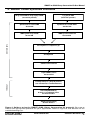
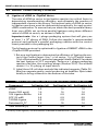
1
SMART™ cDNA Library Construction Kit User Manual Visit Visit sit our website for more details! click here… Cat. No. 634901 PT3000-1 (PR7Y2399) Published 13 November 2007 SMART TM cDNA Library Construction Kit User Manual Table of Contents I. Introduction 4 II. List of Components 9 III. Additional Materials Required 12 IV. General Considerations 15 V. SMART cDNA Synthesis by LD PCR 18 A. First-Strand cDNA Synthesis 18 B. cDNA Amplification by LD PCR 19 C. Proteinase K Digestion 21 D. SfiI Digestion 21 E. cDNA Size Fractionation 22 VI. SMART cDNA Synthesis by Primer Extension 24 A. First-Strand cDNA Synthesis 24 B. ds cDNA Synthesis by Primer Extension 25 C. Proteinase K Digestion 26 D. SfiI Digestion 27 E. cDNA Size Fractionation 27 VII. SMART cDNA Library Protocols 30 A. Ligation of cDNA to Vector 30 B. Bacterial Culture Plating 31 C. Titering the Unamplified Library 32 D. Determining the Percentage of Recombinant Clones 33 E. Library Amplification 34 F. Titering the Amplified Library 35 VIII. Troubleshooting Guide 37 IX. References 42 Appendix A: Typical Results of PCR & cDNA Synthesis 43 Appendix B: Converting λTriplEx2 to pTriplEx2 45 Appendix C: Restriction Map of λTriplEx2 48 Clontech Laboratories, Inc. www.clontech.com 2 Protocol No. PT3000-1 Version No. PR7Y2399 SMART TM cDNA Library Construction Kit User Manual List of Tables Bacterial host strain genotypes 11 Table II. Host strain applications & media additives 11 Table III. Relationship between amount of RNA starting material & optimal number of thermal cycles 20 Table IV. Ligations using three different ratios of cDNA to vector 30 Table V. Plating dilutions for titering an amplified library 36 Table I. List of Figures Figure 1. Flowchart of the SMART cDNA Library Construction Kit protocols 5 Figure 2. Sequence of SfiI A & B recognition sites 7 Figure 3. Generation of polypeptides from all three reading frames in a single recombinant λTriplEx2 clone 8 Figure 4. Guide to using the SMART cDNA Library Construction Kit protocols 17 Figure 5. Guide to troubleshooting SMART cDNA synthesis 37 Figure 6. Typical Results: ds cDNA synthesized using the SMART control reagents & protocols 43 Figure 7. Conversion of recombinant λTriplEx2 to the corresponding pTriplEx2 47 Figure 8. Restriction map of λTriplEx2 48 Protocol No. PT3000-1 www.clontech.com Version No. PR7Y2399 Clontech Laboratories, Inc. 3 SMART TM cDNA Library Construction Kit User Manual I. Introduction The SMART TM cDNA Library Construction Kit provides a method for producing high-quality, full-length cDNA libraries from nanograms of total or poly A+ RNA. This kit contains two separate protocols, allowing you to choose a method based on your starting material. The first protocol employs a novel, PCR-based method for researchers limited by their starting material (i.e., 50 ng of total RNA). The second protocol provides a more traditional protocol for researchers with abundant amounts of starting material (i.e., 1 µg or more poly A+ RNA). Both protocols utilize the patented SMART IVTM (Switching Mechanism At 5' end of RNA Transcript) Oligonucleotide in the first-strand synthesis to generate high yields of full-length, double-stranded (ds) cDNA (Figure 1). Full-length cDNA with complete 5' ends All commonly used cDNA synthesis methods rely on the ability of reverse transcriptase (RT) to transcribe mRNA into single-stranded (ss) DNA in the first-strand reaction. In some cases, RT terminates before transcribing the complete mRNA sequence. This is particularly true for long mRNAs, especially if the first-strand synthesis is primed with oligo(dT) primers only or if the mRNA contains abundant secondary structures. In addition, conventional cDNA cloning procedures use the T4 DNA polymerase to generate blunt cDNA ends after second-strand synthesis. As a result, under-represented 5' ends of genes in cDNA populations tend to be 5–30 nucleotides shorter than the original mRNA (D’Allessio, 1988). The SMART protocols are designed to preferentially enrich for full-length cDNAs, while eliminating T4 DNA polymerase and adaptor ligation. SMART libraries are proven to contain a higher percentage of full-length clones than libraries constructed by conventional methods (October 1998 Clontechniques) or other full-length cDNA synthesis protocols (Okayama & Berg, 1982; Kato et al., 1994). Thus, clones isolated from SMART cDNA libraries contain sequences corresponding to the complete 5' untranslated region of the mRNA (ibid.). SMARTTM preserves the complete 5' mRNA sequence Libraries made with SMART cDNA can facilitate preliminary mapping of transcription start sites (Fromont-Racine, 1993) due to the high percentage of full-length clones. However, SMART cDNA libraries may not be suitable for immunoscreening for certain proteins. In some cases, 5' untranslated regions (UTRs) may contain stop codons in frame with the initiating translation start site in the expression vector. Clontech Laboratories, Inc. www.clontech.com 4 Protocol No. PT3000-1 Version No. PR7Y2399 SMART TM cDNA Library Construction Kit User Manual I. Introduction continued Poly A+ RNA 5' polyA 3' GGG 5' CDS III Primer (SfiIB) SMART IV Oligonucleotide (SfiIA) First-strand synthesis coupled with (dC) tailing by RT polyA 5' GGG CC C 5' Template switching & extension by RT SMART cDNA Synthesis 5' polyA GGG CCC LD PCR (from 50 ng of total RNA) primer extension (from 1 g of poly A+ RNA) Incomplete transcripts, or premature termination of RT 5' X polyA Truncated cDNA (Lacks 5' primer binding site) Cannot be converted to ds DNA or cloned Enriched full-length ds cDNA SfiI digestion SMART λTriplEx2 Ligation SfiIB SfiIA Fractionation & Ligation into λTriplex2 arms right arm left arm SfiIA SfiIB Packaging cDNA library in λ TriplEx2 Figure 1. Flow chart of the SMART cDNA Library Construction Kit protocols. The right side of the flow chart shows the fate of incomplete transcripts caused by RNA degradation or premature termination of reverse transcription. Protocol No. PT3000-1 www.clontech.com Version No. PR7Y2399 Clontech Laboratories, Inc. 5 SMART TM cDNA Library Construction Kit User Manual I. Introduction continued cDNA synthesis by Long-Distance PCR In the first protocol (Section V), cDNA synthesis employs long-distance PCR (LD PCR; Barnes et al., 1994, Cheng et al., 1994) for generating full-length cDNA. This protocol is ideal for researchers who are limited by the amount of their available RNA starting material (i.e., 50 ng of total RNA). A modified oligo(dT) primer (CDS III/3' PCR Primer) primes the first-strand synthesis reaction, and the SMART IV Oligo serves as a short, extended template at the 5' end of the mRNA (Figure 1). When the RT reaches the 5' end, the enzyme’s terminal transferase activity adds a few additional nucleotides, primarily deoxycytidine, to the 3' end of the cDNA. The SMART IV Oligo, which has an oligo (G) sequence at its 3' end, base-pairs with the deoxycytidine stretch, creating an extended template. RT then switches templates and continues replicating to the end of the oligonucleotide. The resulting full-length ss cDNA contains the complete 5' end of the mRNA, as well as the sequence complementary to the SMART IV Oligo, which then serves as a universal priming site (SMART anchor) in the subsequent amplification by LD PCR (Chenchik et al., 1998). Only those ss cDNAs having a SMART anchor sequence at the 5' end can serve as a template and can be exponentially amplified. Incomplete cDNAs and cDNA transcribed from polyA– RNA will lack the SMART anchor and will not be amplified. Thus, contamination by genomic DNA and poly A– RNA is eliminated. This selective amplification allows you to construct a cDNA library using nanogram amounts of either total RNA or poly A+ RNA with a high percentage of full-length clones. cDNA synthesis by Primer Extension In the second protocol (Section VI), cDNA synthesis employs primer extension (Sambrook et al., 1989), which replaces the LD-PCR step in ds cDNA synthesis (Figure 1). Similar to the first-strand cDNA synthesis described above, the CDS III/3' Primer is used to prime the first-strand reaction, and the SMART IV Oligo serves as a short, extended template at the 5' end of the mRNA. After first-strand cDNA synthesis, the primer-extension step generates full-length, ds cDNA. This protocol is ideal for researchers who are not limited by their starting material (i.e., 1 µg or more poly A+ RNA). Optimized reagents and conditions The SMART kit incorporates carefully optimized reagents and conditions that improve the efficiency of all library construction steps. A specially formulated first-strand buffer and modified lock-docking oligo(dT) primers (Borson et al., 1992; Chenchik et al., 1994) optimize the efficiency of first-strand synthesis. Lock-docking primers are designed to anneal to the junction of the poly-A tail and the encoded transcript. This increases the efficiency of cDNA synthesis by eliminating unnecessary reverse transcription of long stretches of poly-A. Clontech Laboratories, Inc. www.clontech.com 6 Protocol No. PT3000-1 Version No. PR7Y2399 SMART TM cDNA Library Construction Kit User Manual I. Introduction continued We strongly recommend the use of the Advantage® 2 PCR Kits (Cat. Nos. 639206 & 639207) for PCR amplification. The specially formulated Advantage 2 Polymerase Mix is optimized for efficient, accurate, and convenient amplification of cDNA templates by LD PCR. The mix contains TITANIUM™ Taq DNA Polymerase—a nuclease-deficient N-terminal deletion of Taq DNA polymerase plus TaqStart® Antibody to provide automatic hot-start PCR (Kellogg et al., 1994)—and a minor amount of a proofreading polymerase. This polymerase system allows you to efficiently amplify full-length cDNAs with a fidelity rate significantly higher than that of conventional PCR (Barnes, 1994). SfiIA site SfiIB site 5' - GGCCATTACGGCC - 3' 5' - GGCCGCCTCGGCC - 3' 3' - CCGGTAATGCCGG - 5' 3' - CCGGCGGAGCCGG - 5' Figure 2. Comparison of the SfiI (A & B) recognition sequences. Another feature of the SMART Kit is the incorporation of asymmetrical SfiI restriction enzyme sites (A & B; Figure 2) at the 5' and 3' cDNA ends, respectively (Figure 1). After digestion with SfiI and size fractionation using CHROMA SPINTM Columns, SMART cDNA is ready for ligation into the SfiI-digested λTriplEx2TM Vector. λTriplEx2, which contains the asymmetrical SfiI sites (A & B) in the MCS, eliminates adaptor ligation and facilitates directional cloning. Furthermore, SfiI sites are extremely rare in mammalian DNA; therefore, all SMART cDNAs remain intact after SfiI digestion. Methylation steps are eliminated; valuable internal restriction sites are preserved. Typical results of cDNA synthesis, using the Human Placenta Poly A+ RNA provided, are shown in Appendix A. Protocol No. PT3000-1 www.clontech.com Version No. PR7Y2399 Clontech Laboratories, Inc. 7 SMART TM cDNA Library Construction Kit User Manual I. Introduction continued cDNA insert vector Plac 5'UTRompA RBS–ATGompA RBS–ATGlacZ (dT)13 MCS T7 vector lacZ' RNA Translation in all three reading frames: 1 Normal translation from ompA start codon f-Met N C 2 Peptide resulting from slippage of RNA polymerase &/or ribosome C 3 Normal translation from lacZ start codon f-Met N C Figure 3. Generation of polypeptides from all three reading frames in a single recombinant λTriplEx clone. λTriplEx2 contains two translation start sites (i.e., two sets of ribosome-binding sites and ATG start codons) in different reading frames and a slip site (a stretch of dTs) that DR10034pr.bw/LTrip.ORFs can cause ribosomes to shift frames between the regulated lac promoter and the MCS. RNA polymerase may also slip during transcription of the (dT)13 region. By the time the ribosomes begin translating the insert, roughly one-third will be in each of the three reading frames. Benefits of λ TriplEx2TM Vector Clontech’s λTriplEx2 Vector provides all of the following advantages for cloning in a phagemid vector: high titer libraries, blue/white screening for recombinants, regulated expression of cloned inserts, and ease of converting clones from phage to a plasmid vector via Cre-lox-mediated subcloning. In addition, every cDNA inserted into the MCS of λTriplEx2 is expressed in all three reading frames (Figure 3). SMART IVTM maximizes library utility The design of the SMART IV oligonucleotide ensures that libraries made with this kit are also compatible with the CreatorTM Cloning and Expression System. The Creator System unites our wide array of expression products, providing access to many strategies for gene function analysis. SMART cDNA libraries can be cloned into our Creator library vector, pDNR-LIB (Cat. No. 631611), via the same SfiI cloning strategy as is used with λTriplEx2 . Clontech Laboratories, Inc. www.clontech.com 8 Protocol No. PT3000-1 Version No. PR7Y2399 SMART TM cDNA Library Construction Kit User Manual II. List of Components Store CHROMA SPIN Columns, 1X Column Buffer, and Deionized H2O at room temperature. Store BM25.8 and XL1-Blue E. coli host strains, Control RNA, and SMART IV Oligonucleotide at –70°C. Store all other reagents at –20°C. First-strand synthesis • 10 µl SMART IVTM Oligonucleotide (12 µM) 5'-AAGCAGTGGTATCAACGCAGAGTGGCCATTACGGCCGGG-3' • 25 µl CDS III/3' PCR Primer (12 µM) 5'-ATTCTAGAGGCCGAGGCGGCCGACATG-d(T)30N–1N-3' (N = A, G, C, or T; N–1 = A, G, or C) • 30 µl 5X First-Strand Buffer 250 mM Tris (pH 8.3) 30 mM MgCl2 375 mM KCl • 20 µl DTT (dithiothreitol; 20 mM) • 5 µl Control Poly A+ RNA (Human Placenta; 1.0 µg/µl) cDNA amplification • 20 µl 5' PCR Primer (12 µM) 5'-AAGCAGTGGTATCAACGCAGAGT-3' Digestion of PCR enzymes • 20 µl Proteinase K (20 µg/µl) SfiI digestion • 80 µl SfiI Enzyme (20 units/µl) • 80 µl 10X SfiI Buffer • 10 µl 100X BSA cDNA purification • 10 CHROMA SPIN-400 Columns • 30 ml1X Fractionation Column Buffer Protocol No. PT3000-1 www.clontech.com Version No. PR7Y2399 Clontech Laboratories, Inc. 9 SMART TM cDNA Library Construction Kit User Manual II. List of Components continued Vector ligation • 30 µl λTriplEx2 (SfiI A & B-digested arms) (0.5 µg/µl) • 20 µl T4 DNA Ligase (400 units/µl) • 30 µl 10X DNA Ligation Buffer 500 mM Tris-HCl (pH 7.8) 100 mM MgCl2 100 mM DTT 0.5 mg/ml BSA • 20 µl ATP (10 mM) • 5 µl Control Insert (SfiI A & B) (50 ng/μl) Host cells • 0.5 mlE. coli BM25.8 (in 25% glycerol, genotype in Table I) • 0.5 mlE. coli XL1-Blue (in 25% glycerol, genotype in Table I) • 60 µl 5'-Sequencing Primer (20 μM) • 60 µl 3'-Sequencing Primer (20 μM) General reagents • 20 µl dNTP mix (dATP, dCTP, dGTP, dTTP, 10 mM each) • 200 µl Sodium Acetate (3 M; pH 4.8) • 20 µl Sodium Hydroxide (25 mM) • 60 µl Glycogen (20 µg/µl) • 5 mlDeionized H2O (Milli-Q-filtered, not DEPC-treated) Clontech Laboratories, Inc. www.clontech.com 10 Protocol No. PT3000-1 Version No. PR7Y2399 SMART TM cDNA Library Construction Kit User Manual II. List of Components continued table i: bacterial host strain genotypes Strain Genotype XL1-Blue endA1, gyrA96, hsdR17, lac –, recA1, relA1, supE44, thi-1, [F' lacI qZ ΔM15, proAB, Tn10] Note: Tn10 confers resistance to tetracycline. Reference: Wood et al., 1985 BM25.8 supE44, thi Δ(lac–proAB) [F’ traD36, proAB+, lacIqZ ΔM15] λimm434 (kanR)P1 (camR) hsdR (rk12– mk12–) Note: BM25.8 is lysogenic for phages λ and P1 and is used for automatic subcloning. Reference: Palazzolo et al., 1990 table ii: host strain applications & media additives Host strain Stock Application(s) plate XL1-Blue LB/tet (15 µg/ml) BM25.8 LB/kan (50 µg/ml)/ • Cre-lox-mediated excision of pTriplEx2 cam (34 µg/ml) from λTriplEx2 • Library plating & screening • Blue/white (β-galactosidase) screening • Regulated expression of cloned genes Note: For λ phage transductions, including plaque titering, library plating, and screening, use the following media additives for optimal adsorption of phage to bacteria: • 10 mM MgSO4 in LB agar and LB top agar • 10 mM MgSO4 and 0.2% maltose in LB broth when growing overnight bacterial cultures for λ phage transductions. • Before using XL1-Blue overnight cultures in phage transductions, centrifuge the cells, pour off the supernatant, and resuspend the pellet in 10 mM MgSO4 (in H2O). • Before using BM25.8 overnight cultures in phage transductions, add MgCl2 to a final concentration of 10 mM. • If the bacterial strains are always maintained on stock plates containing the appropriate antibiotic, there is no need to add antibiotics to the LB broth when growing overnight cultures. Protocol No. PT3000-1 www.clontech.com Version No. PR7Y2399 Clontech Laboratories, Inc. 11 SMART TM cDNA Library Construction Kit User Manual III. Additional Materials Required The following reagents are required but not supplied: Store all reagents and solutions at room temperature (20–22°C) unless specified otherwise. First-strand cDNA synthesis and SMART PCR cDNA amplification • MMLV Reverse Transcriptase (Please see Addendum PT3980-4 for details on the choice of RT enzyme.) • Advantage® 2 PCR Kit (Cat. Nos. 639206 & 639207) • Sterile, 0.5-ml microcentrifuge tubes • Poly A+ or total RNA • Mineral oil (We recommend Sigma #M-3516.) • Perkin-Elmer DNA Thermal Cyclers Note: The cycling parameters in this protocol have been optimized using Applied Biosystems GeneAmp® DNA Thermal Cyclers 480, 2400, and 9600 along with the reagents and control RNA provided in the SMART Kit. Optimal cycling parameters may vary with different templates and thermal cyclers. • DNA size markers (1-kb DNA ladder; Gibco/BRL #15615-016) • 1.1% Agarose/EtBr gel (containing 0.1 µg/ml ethidium bromide) Proteinase K digestion • Sterile, 0.5-ml microcentrifuge tubes • 95% ethanol • 80% ethanol cDNA size fractionation • 1.5-ml sterile microcentrifuge tubes • Ring-stand with small clamp (for holding column) • 1.1% agarose/EtBr gel (0.1 µg/ml ethidium bromide) • 95% ethanol (–20°) • 1% xylene cyanol λ phage packaging • λ phage packaging extract Several are commercially available. Choose a packaging system that will give you at least 1 x 10⁹ pfu/µg of DNA. PCR Insert Screening [optional] • λTriplEx LD-Insert Screening Amplimer Sets Routine plating and culture of E. coli • Kanamycin stock solution (25 mg/ml in H2O; 500X) Store at –20°C • Tetracycline stock solution (15 mg/ml in H2O; 1000X) Clontech Laboratories, Inc. www.clontech.com 12 Protocol No. PT3000-1 Version No. PR7Y2399 SMART TM cDNA Library Construction Kit User Manual III. Additional Materials Required continued Store at –20°C • Chloramphenicol stock solution (34 mg/ml in 100% ethanol; 1000X). Store at –20°C. • LB broth 10 g/LBacto-tryptone 5 g/LBacto-yeast extract 5 g/LNaCl Adjust pH to 7.0 with 5 N NaOH. Autoclave. • LB agar plates Prepare LB broth as described above. Add agar (15 g/L) and autoclave. Pour plates and store at 4°C. • LB/tet agar plates To one liter of LB broth (recipe above), add 15 g agar. Autoclave. Cool to 50°C before adding tetracycline (15 µg/ml final concentration). Pour plates and store at 4°C. • LB/kan/cam agar plates To one liter of LB broth (recipe above), add 15 g agar. Autoclave. Cool to 50°C before adding kanamycin (50 µg/ml final concentration) and chloramphenicol (34 µg/ml final concentration). Pour plates and store at 4°C. Transduction and titering of λ phage in E. coli When preparing media for phage transductions or plaque titering, use recipes containing 10 mM MgSO4 for optimal adsorption of phage to bacteria. For the same reason, add 0.2% maltose to the LB broth when growing overnight bacterial cultures for transduction/titering. • MgSO4 (1 M stock solution) Dissolve 24.65 g of MgSO4•7H2O in 100 ml of deionized H2O. Filter sterilize. • 20% Maltose stock solution Dissolve 20 g of maltose in 80 ml of deionized H2O; bring volume up to 100 ml. Filter sterilize and store at 4°C. • LB/MgSO4 agar plates To one liter of LB broth, add 10 ml 1 MMgSO4 (10 mM final concentration) 15 g agar Autoclave. Pour plates and store at 4°C. • LB/MgSO4 broth To one liter of LB broth, add 10 ml 1 MMgSO4 (10 mM final concentration). Autoclave. Protocol No. PT3000-1 www.clontech.com Version No. PR7Y2399 Clontech Laboratories, Inc. 13 SMART TM cDNA Library Construction Kit User Manual III. Additional Materials Required continued • LB/MgSO4/maltose broth Prepare 1 L LB/MgSO4 broth as described above. After autoclaving, cool to 50°C before adding maltose to a final concentration of 0.2%. (10 ml of 20% maltose stock solution.) • LB/MgSO4 soft top agar To one liter of LB broth, add 10 ml 1 M MgSO4 (10 mM final concentration) 7.2 g agar Autoclave and store at 4°C. • 10X Lambda dilution buffer Final Conc. To prepare 1 L of solution NaCl 1.0 M 58.3g MgSO4•7H2O 0.1 M 24.65 g Tris-HCI (pH 7.5) 0.35 M 350.0 ml of 1 M Add H2O to a final volume of 1 L. Autoclave and store at 4°C. • 1X Lambda dilution buffer 100 ml 10X Lambda dilution buffer 5 ml 2% Gelatin (0.01% final concentration) Add H2O to a final volume of 1 L. Autoclave and store at 4°C. Note: The 0.01% gelatin in the 1X lambda dilution buffer stabilizes the library titer for long-term storage. Gelatin addition is optional when diluting the phage for immediate titering. Amplifying a λ library • LB/MgSO4 plates • LB/MgSO4 soft top agar • 50-ml polypropylene, sterile screw-cap tubes • Chloroform Blue/white screening in E. coli (i.e., β-galactosidase) • IPTG (100 mM in H2O) Isopropyl β-D-thiogalactopyranoside. Filter sterilize. Store at 4°C. • X-Gal (100 mM) Dissolve in dimethyformamide (DMF). Store at –20°C. Long-term library storage • 100% Dimethylsulfoxide (DMSO) • 100% Glycerol Clontech Laboratories, Inc. www.clontech.com 14 Protocol No. PT3000-1 Version No. PR7Y2399 SMART TM cDNA Library Construction Kit User Manual IV.General Considerations A. Good Laboratory Practices 1.Wear gloves throughout the procedure to protect your RNA and cDNA samples from degradation by nucleases. 2.When resuspending pellets or mixing reactions, gently pipet the solution up and down or tap the bottom of the tube. Spin tube briefly to bring contents to the bottom of the tube. Do not vortex samples when resuspending pellets; vortexing may cause shearing of your cDNA. 3.Perform all reactions on ice, unless otherwise indicated. 4.Add enzymes to reaction mixtures last. Make sure that the enzyme is thoroughly blended into the reaction mixture by gently pipetting the mixture up and down. 5.Do not increase the size (volume) of any of the reactions. All components have been optimized for the volumes specified. 6.Ethidium bromide is a carcinogen. Use appropriate precautions in handling and disposing this reagent. For more information, see Sambrook et al. (1989). Several EtBr disposable cartridges are also available, such as Clontech’s BondEx Ethidium Bromide Detoxification Cartridge (see Section X, Related Products). 7.Phenol is a corrosive. Always wear gloves and protective clothing when handling solutions containing this reagent. If possible, handle solutions containing phenol and/or chloroform under a chemical fume hood. B. Preparation and Handling of Total and Poly A+ RNA 1.To avoid contamination and degradation of RNA (and to minimize the presence of RNases), use the following precautions: a. Wear gloves. b. Use freshly deionized (e.g., Milli-Q-grade) H2O, without treatment with DEPC (diethyl pyrocarbonate). c. Rinse all glassware with 0.5 N NaOH, followed by deionized H2O. Then bake the glassware at 160–180°C for 4–9 hr. d. Use only single-use plastic pipettes and pipette tips with RNA. e. Avoid using autoclaved H2O because recycled steam in some autoclaves can introduce contaminants that may interfere with PCR. Protocol No. PT3000-1 www.clontech.com Version No. PR7Y2399 Clontech Laboratories, Inc. 15 SMART TM cDNA Library Construction Kit User Manual IV. General Considerations continued 2.RNA isolation Many procedures are available for the isolation of total RNA and poly A+ RNA (Chomczynski & Sacchi, 1987; Farrell, 1993; Sambrook et al., 1989). Clontech offers several kits for the isolation of total RNA and subsequent isolation of poly A+ RNA (see Section X, Related Products). 3.RNA analysis The sequence complexity of the ds cDNA synthesized, and ultimately of the cDNA library constructed, depend on the quality of the experimental RNA starting material. Therefore, before you use it in a first-strand synthesis, we recommend that you estimate the integrity of the RNA by examining a sample on a denaturing formaldehyde/agarose gel. Total RNA from mammalian sources should appear as two distinct bands (28S and 18S ribosomal RNA) at approximately 4.5 and 1.9 kb. The ratio of intensities of the 28S and 18S rRNA bands should be 1.5–2.5:1. Intact mammalian poly A+ RNA should appear as a smear (usually 0.5–12 kb) with faint 28S and 18S rRNA bands. The size distribution may be considerably smaller (0.5–3 kb) for nonmammalian species (e.g., plants, insects, yeast, amphibians, etc.). If your experimental poly A+ RNA appears significantly smaller than expected (e.g., no larger than 1–5 kb), we suggest you prepare fresh RNA after checking your RNA purification reagents for RNase and other impurities. If problems persist, you may need to find another source of tissue/cells. Alternatively, you may wish to use one of Clontech’s Poly A+ RNAs. Clontech Laboratories, Inc. www.clontech.com 16 Protocol No. PT3000-1 Version No. PR7Y2399 SMART TM cDNA Library Construction Kit User Manual V. SMART cDNA Synthesis Protocols ONE-TWO DAYS 50 ng of total RNA or 25 ng of poly A+ RNA (researcher provides) 1 µg poly A+ RNA (researcher provides) Synthesize first-strand cDNA Section V.A Synthesize first-strand cDNA Section VI.A cDNA Synthesis by LD PCR Section V.B cDNA by Primer Extension Section VI.B Electrophorese a sample on a gel Sfi I digestion cDNA size fractionation Ligate cDNA to vector Sections VII.A THIRD DAY Package library in λ phage & plate out (see manufacturer's recommendations) Titer unamplified library & check % recombinant clones Sections VII.C & D Amplify and titer library Sections VII.E & F Figure 4. Guide to using the SMART cDNA Library Construction Kit protocols. Be sure to choose the appropriate protocol for your application. Refer to Figure 5 and Section VIII for troubleshooting tips. Protocol No. PT3000-1 www.clontech.com Version No. PR7Y2399 Clontech Laboratories, Inc. 17 SMART TM cDNA Library Construction Kit User Manual V. SMART cDNA Synthesis by LD PCR please read entire protocol before starting. Important: This protocol is designed for library construction of SMART cDNA libraries using a PCR-based protocol. The minimum amount of starting material for cDNA synthesis is 50 ng of total RNA or 25 ng of poly A+ RNA. In general, the more RNA you start with, the fewer PCR cycles will be required for the second-strand synthesis (see Table III). Fewer thermal cycles are less likely to generate nonspecific PCR products, and therefore are best for optimal cDNA and library quality. Thus, if your RNA sample is not limiting, use the higher starting amounts of RNA shown in the table. To construct SMART cDNA libraries from 1 µg of poly A+ RNA using primer extension, consult the protocol in Section VI. We strongly recommend that you perform a positive control cDNA synthesis with the human placenta poly A+ RNA provided, in parallel with your experimental cDNA synthesis. Performing a control at least once will verify that all the components are working properly. This is especially important because it may not be possible to visualize your first-strand reaction product on a gel. Furthermore, performing the control reaction through the PCR step in parallel with your experiment will allow you to evaluate the yield and size distribution of the ds cDNA synthesized from your RNA sample. Performing the control through the library construction steps will also help you troubleshoot any problems, as they arise. Notes: • If necessary, the procedure can be stopped after first-strand synthesis by placing the reaction mixture at –20°C, and also at any ethanol precipitation step after the precipitated DNA has been centrifuged or when the supernatant has been removed.The dry or resuspended DNA pellet can be stored at –20°C for up to 3 months. • For the first-strand synthesis and the PCR, all components and reaction vessels should be pre-chilled on ice. A. First-Strand cDNA Synthesis 1.Combine the following reagents in a sterile 0.5-ml microcentrifuge tube: 1–3 µl RNA sample (0.025–0.5 µg poly A+ or 0.05–1.0 µg total RNA)Forthecontrolreaction,use1µl[1µg]ofthecontrol RNA.) 1 µl SMART IV Oligonucleotide 1 µl CDS III/3' PCR Primer If total volume is <5 µl, add deionized H2O to bring volume up to 5 µl. Clontech Laboratories, Inc. www.clontech.com 18 Protocol No. PT3000-1 Version No. PR7Y2399 SMART TM cDNA Library Construction Kit User Manual V. SMART cDNA Synthesis by LD PCR continued 2.Mix contents and spin the tube briefly in a microcentrifuge. 3.Incubate the tube at 72°C for 2 min. 4.Cool the tube on ice for 2 min. 5.Spin the tube briefly to collect the contents at the bottom. 6.Add the following to each reaction tube: 2.0 µl 5X First-Strand Buffer 1.0 µl DTT (20 mM) 1.0 µl dNTP Mix (10 mM ) 1.0 µl MMLV Reverse Transcriptase* 10.0 µl Total volume *Please see Addendum PT3980-4 for details on the choice of RT enzyme. 7.Mix the contents of the tube by gently pipetting and briefly spinning the tube. 8.Incubate the tube at 42°C for 1 hr in an air incubator or a hot lid thermal cycler. Note: If you use a water bath or thermal cycler for this incubation, cover the reaction mixture with one drop of mineral oil before you close the tube. This will prevent loss of volume due to evaporation. 9.Place the tube on ice to terminate first-strand synthesis. 10.If you plan to proceed directly to the PCR step, take a 2-µl aliquot from the first-strand synthesis and place it in a clean, prechilled, 0.5-ml tube. Place the tube on ice, and proceed to Step V.B. If you used mineral oil in your first-strand reaction tube, be careful to take the 2-µl sample from the bottom of the tube to avoid the oil. 11.Any first-strand reaction mixture that is not used right away should be placed at –20°C. First-strand cDNA can be stored at –20°C for up to three months. B. cDNA Amplification by LD PCR Table III shows the relationship between the optimal number of thermal cycles to use for a given amount of RNA starting material used in the first-strand synthesis. Use the least number of cycles shown for that amount of RNA. Fewer cycles generally mean fewer nonspecific PCR products. Furthermore, undercycling can be easily rectified by placing the PCR reaction back in the thermal cycler for a few more cycles (see the Troubleshooting Guide, Section VIII.C). On the other hand, overcycling requires repeating the PCR de novo using a fresh aliquot of the first-strand reaction product. The optimal cycling parameters in Table III were determined using the Control Poly A+ Human Placenta RNA; these parameters may vary with different templates and thermal cyclers. Protocol No. PT3000-1 www.clontech.com Version No. PR7Y2399 Clontech Laboratories, Inc. 19 SMART TM cDNA Library Construction Kit User Manual V. SMART cDNA Synthesis by LD PCR continued 1.Preheat the PCR thermal cycler to 95°C. table iii: relationship between amount of rna starting material and optimal number of thermal cycles Total RNA Poly A+ RNA (µg) (µg) Number of Cycles 1.0–2.0 0.5–1.0 18–20 0.5–1.0 0.25–0.5 20–22 0.25–0.5 0.125–0.25 22–24 0.05–0.25 0.025–0.125 24–26 2.Combine the following components in the reaction tube: 2 µl First-Strand cDNA (from Step A.10) 80 µl Deionized H2O 10 µl 10X Advantage 2 PCR Buffer 2 µl 50X dNTP Mix 2 µl 5' PCR Primer 2 µl CDS III/3' PCR Primer 2 µl 50X Advantage 2 Polymerase Mix 100 µl Total volume 3.Mix contents by gently flicking the tube. Centrifuge briefly to collect the contents at the bottom of the tube. 4.Overlay the reaction mixture with 2 drops of mineral oil if necessary. Cap the tube and place it in a preheated (95°C) thermal cycler. 5.Commence thermal cycling using one of the following programs: GeneAmp 480 GeneAmp 2400/9600 •95°C 1 min •95°C 20 sec •x cycles*: •x cycles*: 95°C 15 sec 95°C 5 sec 68°C 6 min 68°C 6 min *Refer to Table III to determine the optimal number of cycles to use. 6.When the cycling is complete, analyze a 5-µl sample of the PCR product, alongside 0.1 µg of 1-kb DNA size markers, on a 1.1% agarose/EtBr gel. The ds cDNA should appear as a 0.1–4 kb smear on the gel, with some distinct bands corresponding to the abundant mRNAs for that tissue or cell source. (cDNA prepared from some tissues may not have distinct distinct bands, especially if the mRNA is highly complex.) If your PCR product does not appear as expected, refer to the Troubleshooting Guide (Section VIII.A–E). Typical results obtained with Human Placenta Poly A+ RNA are Clontech Laboratories, Inc. www.clontech.com 20 Protocol No. PT3000-1 Version No. PR7Y2399 SMART TM cDNA Library Construction Kit User Manual V. SMART cDNA Synthesis by LD PCR continued shown in Appendix A. 7.Proceed to the next step, or store ds cDNA at –20°C until use. C. Proteinase K digestion 1.In a sterile 0.5-ml tube, pipet 50 µl of amplified ds cDNA (2–3 µg), making sure you pipet below the top oil layer of the PCR tube and add 2 µl of proteinase K (20 µg/µl). Store the remaining ds cDNA at –20°C (up to 3 months). Note: Proteinase K treatment is necessary to inactivate the DNA polymerase activity. This system was optimized for 2–3 µg (~50 µl/vol) of PCR cDNA product for subsequent cloning and library construction procedures. Too much ds cDNA (>3–4 µg) will yield a low titer library. Refer to the Troubleshooting Guide (Section VIII.H) for more information. 2.Mix contents and spin the tube briefly. 3.Incubate at 45°C for 20 min. Spin the tube briefly. 4.Add 50 µl of Deionized H2O to the tube. 5.Add 100 µl of phenol:chloroform:isoamyl alcohol and mix by continuous gentle inversion for 1–2 min. 6.Centrifuge at 14,000 rpm for 5 min to separate the phases. 7.Move the top (aqueous) layer to a clean 0.5-ml tube. Discard the interface and lower layers. 8.Add 100 µl of chloroform:isoamyl alcohol to the aqueous layer. Mix by continuous gentle inversion for 1–2 min. 9.Centrifuge at 14,000 rpm for 5 min to separate the phases. 10.Move the top (aqueous) layer to a clean 0.5-ml tube. Discard the interface and lower layers. 11.Add 10 µl of 3 M Sodium Acetate, 1.3 µl of Glycogen (20 µg/µl) and 260 µl of room-temperature 95% ethanol. Immediately centrifuge at 14,000 rpm for 20 min at room temperature. Note: Do not chill the tube at –20°C or on ice before centrifuging. Chilling the sample will result in coprecipitation of impurities. 12.Carefully remove the supernatant with a pipette. Do not disturb the pellet. 13.Wash pellet with 100 µl of 80% ethanol. 14.Air dry the pellet (~10 min) to evaporate off residual ethanol. 15.Add 79 µl of Deionized H2O to resuspend the pellet. D. SfiI digestion 1. Combine the following components in a fresh 0.5-ml tube: 79 µl cDNA (Section V.C.15, above) 10 µl 10X Sfi Buffer 10 µl SfiI Enzyme 1 µl 100X BSA 100 µl Total volume Protocol No. PT3000-1 www.clontech.com Version No. PR7Y2399 Clontech Laboratories, Inc. 21 SMART TM cDNA Library Construction Kit User Manual V. SMART cDNA Synthesis by LD PCR continued 2. Mix well. Incubate the tube at 50°C for 2 hr. 3. Add 2 µl of 1% xylene cyanol dye to the tube above. Mix well. E. cDNA size fractionation by CHROMA SPIN-400 1.Label sixteen 1.5-ml tubes and arrange them in a rack in order. 2.Prepare the CHROMA SPIN-400 Column for drip procedure: a. Invert the column several times to completely resuspend the gel matrix. b. Remove air bubbles from the column. Use a 1000-µl pipettor to resuspend the matrix gently; avoid generating air bubbles. Then remove the bottom cap and let the column drip naturally. (If the column does not drain after 3 min, recap the top cap. This pressure should cause the column to drain). c. Attach the column to a ring stand. d. Let the storage buffer drain through the column by gravity flow until you can see the surface of the gel beads in the column matrix. The top of the column matrix should be at the 1.0-ml mark on the wall of the column. If your column contains significantly less matrix, adjust the volume of the matrix to the 1.0-ml mark using matrix from another column (an extra column is provided for this purpose). e. The flow rate should be approximately 1 drop/40–60 sec. The volume of 1 drop should be approximately 40 µl. If the flow rate is too slow (i.e., more than 1 drop/100 sec) and the volume of one drop is too small (i.e., less than 25 µl), you should resuspend the matrix completely and repeat the drip procedure until it reaches the above parameters. 3.When the storage buffer stops dripping out, carefully and gently (along the column inner wall) add 700 µl of column buffer to the top of the column and allow it to drain out. 4.When this buffer stops dripping (~15–20 min), carefully and evenly apply ~100 µl mixture of SfiI-digested cDNA and xylene cyanol dye (Section V.D.3 above) to the top-center surface of the matrix. An unsmooth matrix surface does not hurt the following fractionation process. 5.Before proceeding to the next step, allow the sample to be fully absorbed into the surface of the matrix (i.e., there should be no liquid remaining above the surface). 6.With 100 µl of column buffer, wash the tube that contained the cDNA, and gently apply this material to the surface of the matrix. 7.Allow the buffer to drain out of the column until there is no liquid left above the resin. When the dripping has ceased, proceed to the Clontech Laboratories, Inc. www.clontech.com 22 Protocol No. PT3000-1 Version No. PR7Y2399 SMART TM cDNA Library Construction Kit User Manual V. SMART cDNA Synthesis by LD PCR continued next step. At this point, the dye layer should be several mm into the column. 8.Place the rack containing the collection tubes (Section V.E.1) under the column, so that the first tube is directly under the column outlet. 9.Add 600 μl of column buffer and immediately begin collecting single-drop fractions (approximately 35 μl per tube) in tubes #1–16. Cap each tube after each fraction is collected. Recap the column after fraction #16 has been collected. 10.Check the profile of the fractions before proceeding with the experiment. On a 1.1% agarose/EtBr gel, electrophorese 3 μl of each fraction (separately) in adjacent wells, alongside 0.1 μg of a 1-kb DNA size marker. Run the gel at 150 V for 10 min. (Running the gel longer will make it difficult to see the cDNA bands). Determine the peak fractions by visualizing the intensity of the bands under UV. Collect the first three fractions containing cDNA (in most cases, the fourth fraction containing cDNA is usable. Make sure the fourth fraction matches your desired size distribution). Pool the above fractions in a clean 1.5-ml tube. 11.Add the following reagents to the tube with 3–4 pooled fractions containing the cDNA: (105–140 µl, respectively) 1/10 vol. Sodium Acetate (3 M; pH 4.8) 1.3 µl Glycogen (20 mg/ml) 2.5 vol. 95% ethanol (–20°C) 12.Mix by gently rocking the tube back and forth. 13.Place the tube in –20°C or a dry-ice/ethanol bath for 1 hr. (Optional: you may incubate at –20°C overnight, which may result in better recovery). 14.Centrifuge the tube at 14,000 rpm for 20 min at room temperature. 15.Carefully remove the supernatant with a pipette. Do not disturb the pellet. 16.Briefly centrifuge the tube to bring all remaining liquid to the bottom. 17.Carefully remove all liquid and allow the pellet to air dry for ~10 min. 18.Resuspend the pellet in 7 μl of Deionized H2O and mix gently. The SfiI-digested cDNA is now ready to be ligated to the SfiI-digested, dephosphorylated λTriplEx2 Vector provided. Proceed to Section VII, or store cDNA at –20°C until the ligation step. Protocol No. PT3000-1 www.clontech.com Version No. PR7Y2399 Clontech Laboratories, Inc. 23 SMART TM cDNA Library Construction Kit User Manual VI. SMART cDNA Synthesis by Primer Extension please read entire protocol before starting. Important: This protocol is designed for standard library construction of SMART cDNA libraries using primer extension. The optimal amount of starting material for cDNA synthesis is about 1 µg of poly A+ RNA (0.5 µg–2.0 µg). Reduced cloning efficiency will occur if you use less than 0.5 µg or greater than 2.0 µg of poly A+ RNA. To construct SMART cDNA libraries using a PCR-based method, consult the protocol in Section V. We strongly recommend that you perform a positive control cDNA synthesis with the Human Placenta Poly A+ RNA provided, in parallel with your experimental cDNA synthesis. Performing a control at least once will verify that all the components are working properly. This is especially important if you have difficulty visualizing your first-strand reaction product on a gel. Furthermore, performing the control reaction in parallel with your experiment will allow you to evaluate the yield and size distribution of the ds cDNA synthesized from your RNA sample. Performing the control through the library construction steps will also help you troubleshoot any problems, as they arise. Notes: • If necessary, the procedure can be stopped after the first-strand synthesis by placing the reaction mixture at –20°C, and at any ethanol precipitation step after the precipitated DNA has been centrifuged or when the supernatant has been removed.The dry or resuspended DNA pellet can be stored at –20°C for up to 3 months. • For the first-strand synthesis and the primer extension steps, all components and reaction vessels should be pre-chilled on ice. A. First-Strand cDNA Synthesis 1.Combine the following reagents in a sterile 0.5-ml microcentrifuge tube: 1 µl poly A+ RNA [1.0 µg] (For the control reaction, use 1 µl [1 µg] of the control RNA.) 1 µl SMART IV Oligonucleotide 1 µl CDS III/3' PCR Primer 2 µl Deionized H2O 5 µl Total volume 2.Mix contents and spin the tube briefly in a microcentrifuge. 3.Incubate the tube at 72°C for 2 min. 4.Cool the tube on ice for 2 min. 5.Spin the tube briefly to collect the contents at the bottom. Clontech Laboratories, Inc. www.clontech.com 24 Protocol No. PT3000-1 Version No. PR7Y2399 SMART TM cDNA Library Construction Kit User Manual VI. SMART cDNA Synthesis by Primer Extension continued 6.Add the following to each reaction tube: 2.0 µl 5X First-Strand Buffer 1.0 µl DTT (20 mM) 1.0 µl dNTP Mix (10 mM ) 1.0 µl MMLV Reverse Transcriptase* 10.0 µl Total volume *Please see Addendum PT3980-4 for details on the choice of RT enzyme. 7.Mix the contents of the tube by gently pipetting and briefly spin the tube. 8.Incubate the tube at 42°C for 1 hr in an air incubator or hot lid thermal cycler. Note: If you use a water bath or thermal cycler for this incubation, cover the reaction mixture with one drop of mineral oil before you close the tube. This will prevent loss of volume due to evaporation. 9.Place the tube on ice to terminate first-strand synthesis. 10.Add 1 µl of Sodium Hydroxide to the tube. 11.Incubate the tube at 68° C for 30 min. 12.Place the tube on ice, and proceed to Step VI.B. Any first-strand reaction mixture that is not used right away should be placed at –20°C. First-strand cDNA can be stored at –20°C for up to three months. B. ds cDNA synthesis by primer extension 1.Preheat the PCR thermal cycler to 95°C. 2.Combine the following components: 11 µl First-Strand cDNA (from Step VIA.12) 71 µl Deionized H2O 10 µl 10X Advantage 2 PCR Buffer 2 µl 50X dNTP Mix 2 µl 5' PCR Primer 2 µl CDS III/3' PCR Primer 2 µl 50X Advantage 2 Polymerase Mix 100 µl Total volume 3.Mix contents by gently flicking the tube. Centrifuge briefly to collect the contents at the bottom of the tube. 4.Overlay the reaction mixture with 2 drops of mineral oil if necessary. Cap the tube and place it in a preheated (95°C) thermal cycler. Protocol No. PT3000-1 www.clontech.com Version No. PR7Y2399 Clontech Laboratories, Inc. 25 SMART TM cDNA Library Construction Kit User Manual VI. SMART cDNA Synthesis by Primer Extension continued 5.Commence primer extension using one of the following programs: GeneAmp 480 GeneAmp 2400/9600 •72°C 10 min •72°C 10 min •95°C 1 min •95°C 20 sec •3 cycles: •3 cycles: 95°C 15 sec 95°C 5 sec 68°C 8 min 68°C 8 min 6.When the reaction is complete, analyze a 5-µl sample of the product on a 1.1% agarose/EtBr gel, alongside a 1-kb DNA size marker (0.1 µg). The ds cDNA should appear as a 0.1–9 kb smear on the gel, with some distinct bands corresponding to the abundant mRNAs for that tissue or cell source. (cDNA prepared from some tissues may not have distinct distinct bands, especially if the mRNA is highly complex.) If your product does not appear as expected, refer to the Troubleshooting Guide (Section VIII.A-B, F).Typical results obtained with Human Placenta Poly A+ RNA are shown in Appendix A. 7.Proceed to the next step, or store ds cDNA at –20°C until use. C. Proteinase K digestion 1.In a sterile 0.5-ml tube, pipet 50 µl of amplified ds cDNA (2–3 µg), making sure you pipet below the top oil layer of the PCR tube and add 2 µl of proteinase K (20 µg/µl). Store the remaining ds cDNA at –20°C (up to 3 months). Note: Proteinase K treatment is necessary to inactivate the DNA polymerase activity. This system was optimized for 2–3 µg (~50 µl/vol) of ds cDNA for subsequent cloning and library construction procedures. Too much ds cDNA (>3–4 µg) will yield a low-titer library. Refer to theTroubleshooting Guide (Section VIII.H) for more information. 2.Mix contents and spin the tube briefly. 3.Incubate at 45°C for 20 min. Spin the tube briefly. 4.Add 50 µl of Deionized H2O to the tube. 5.Add 100 µl of phenol:chloroform:isoamyl alcohol and mix by continuous gentle inversion for 1–2 min. 6.Centrifuge at 14,000 rpm for 5 min to separate the phases. 7.Move the top (aqueous) layer to a clean 0.5-ml tube. Discard the interface and lower layers. 8.Add 100 µl of chloroform:isoamyl alcohol to the aqueous layer. Mix by continuous gentle inversion for 1–2 min. 9.Centrifuge at 14,000 rpm for 5 min to separate the phases. Clontech Laboratories, Inc. www.clontech.com 26 Protocol No. PT3000-1 Version No. PR7Y2399 SMART TM cDNA Library Construction Kit User Manual VI. SMART cDNA Synthesis by Primer Extension continued 10.Move the top (aqueous) layer to a clean 0.5-ml tube. Discard the interface and lower layers. 11.Add 10 µl of 3 M Sodium Acetate, 1.3 µl of Glycogen (20 µg/µl) and 260 µl of room-temperature 95% ethanol. Immediately centrifuge at 14,000 rpm for 20 min at room temperature. Note: Do not chill the tube at –20°C or on ice before centrifuging. Chilling the sample will result in coprecipitation of impurities. 12.Carefully remove the supernatant with a pipette. Do not disturb the pellet. 13.Wash pellet with 100 µl of 80% ethanol. 14.Air dry the pellet (~10 min) to evaporate off residual ethanol. 15.Add 79 µl of deionized H2O to resuspend the pellet. D. SfiI digestion 1. Combine the following components in a fresh 0.5-ml tube: 79 µl cDNA (Section VI.C.15, above) 10 µl 10X Sfi Buffer 10 µl SfiI Enzyme 1 µl 100X BSA 100 µl Total volume 2. Mix well. Incubate the tube at 50°C for 2 hr. 3. Add 2 µl of 1% xylene cyanol dye to the tube above. Mix well. E. cDNA size fractionation by CHROMA SPIN-400 1.Label sixteen 1.5-ml tubes and arrange them in a rack in order. 2.Prepare the CHROMA SPIN-400 column for drip procedure: a. Remove the CHROMA SPIN column from the refrigerator and warm at room temperature for about 1 hr. Invert the column several times to completely resuspend the gel matrix. b. Remove air bubbles from the column. Use a 1000-μl pipettor to resuspend the matrix gently; avoid generating air bubbles. Then remove the bottom cap and let the column drip naturally. (If the column does not drain after 3 min, recap the top cap. This pressure should cause the column to drain). c. Attach the column to a ring stand. d. Let the storage buffer drain through the column by gravity flow until you can see the surface of the gel beads in the column matrix. The top of the column matrix should be at the 1.0-ml mark on the wall of the column. If your column contains significantly less matrix, adjust the volume of the matrix to the 1.0-ml mark Protocol No. PT3000-1 www.clontech.com Version No. PR7Y2399 Clontech Laboratories, Inc. 27 SMART TM cDNA Library Construction Kit User Manual VI. SMART cDNA Synthesis by Primer Extension continued using matrix from another column (an extra column is provided for this purpose). e.The flow rate should be approximately 1 drop/40–60 sec.The volume of 1 drop should be approximately 40 μl. If the flow rate is too slow (i.e., more than 1 drop/100 sec) and the volume of one drop is too small (i.e., less than 25 μl), you should resuspend the matrix completely and repeat the drip procedure until it reaches the above parameters. 3.When the storage buffer stops dripping out, carefully and gently (along the column inner wall) add 700 μl of column buffer to the top of the column and allow it to drain out. 4.When this buffer stops dripping (~15–20 min), carefully and evenly apply ~100 μl mixture of SfiI-digested cDNA and xylene cyanol dye (Section VI.D.3 above) to the top-center surface of the matrix. An unsmooth matrix surface does not hurt the following fractionation process. 5.Before proceeding to the next step, allow the sample to be fully absorbed into the surface of the matrix (i.e., there should be no liquid remaining above the surface). 6.With 100 μl of column buffer, wash the tube that contained the cDNA and gently apply this material to the surface of the matrix. 7.Allow the buffer to drain out of the column until there is no liquid left above the resin. When the dripping has ceased, proceed to the next step. At this point, the dye layer should be several mm into the column. 8.Place the rack containing the collection tubes (Step VI.E.1) under the column, so that the first tube is directly under the column outlet. 9.Add 600 μl of column buffer and immediately begin collecting single-drop fractions in tubes #1–16 (approximately 35 μl per tube). Cap each tube after each fraction is collected. Recap the column after fraction #16 has been collected. 10.Check the profile of the fractions before proceeding with the experiment. On a 1.1% agarose/EtBr gel, electrophorese 3 μl of each fraction (separately) in adjacent wells, alongside a 1-kb DNA size marker (0.1 μg). Run the gel at 150 V for 10 min (Running the gel longer will make it difficult to see the cDNA bands). Determine the peak fractions by visualizing the intensity of the bands under UV. Collect the first three fractions containing cDNA (in most cases, the fourth fraction containing cDNA is usable. Make sure the fourth fraction matches your desired size distribution). Pool the above fractions in a clean 1.5-ml tube. Clontech Laboratories, Inc. www.clontech.com 28 Protocol No. PT3000-1 Version No. PR7Y2399 SMART TM cDNA Library Construction Kit User Manual VI. SMART cDNA Synthesis by Primer Extension continued 11.Add the following reagents to the tube with 3–4 pooled fractions containing the cDNA: (105–140 µl, respectively) 1/10 vol. Sodium Acetate (3 M; pH 4.8) 1.3 µl Glycogen (20 mg/ml) 2.5 vol. 95% ethanol (–20°C) 12.Mix by gently rocking the tube back and forth. 13.Place the tube in –20°C or a dry-ice/ethanol bath for 1 hr. (Optional: you may incubate at –20°C overnight, which may result in better recovery). 14.Centrifuge the tube at 14,000 rpm for 20 min at room temperature. 15.Carefully remove the supernatant with a pipette. Do not disturb the pellet. 16.Briefly centrifuge the tube to bring all remaining liquid to the bottom. 17.Carefully remove all liquid and allow the pellet to air dry for ~10 min. 18.Resuspend the pellet in 7 μl of Deionized H2O and mix gently. The SfiI-digested cDNA is now ready to be ligated to the SfiI-digested, dephosphorylated λTriplEx2 Vector provided. Proceed to Section VII, or store cDNA at –20°C until the ligation step. Protocol No. PT3000-1 www.clontech.com Version No. PR7Y2399 Clontech Laboratories, Inc. 29 SMART TM cDNA Library Construction Kit User Manual VII. SMART cDNA Library Protocols A. Ligation of cDNA to λTriplEx2 Vector The ratio of cDNA to vector in the ligation reaction is a critical factor in determining transformation efficiency, and ultimately the number of independent clones in the library. The optimal ratio of cDNA to vector in ligation reactions must be determined empirically for each vector/ cDNA combination. To ensure that you obtain the best possible library from your cDNA, set up three parallel ligations using three different ratios of cDNA to vector, as shown in Table IV. Important note: Use a λ phage packaging system that will give you at least 1 x 10⁹ pfu/µg of DNA. Follow the supplier’s recommended protocol and perform a parallel packaging reaction with the control insert provided in the packaging kit. The following protocol is optimized for ligation of SMART cDNA to the λTriplEx2 DNA provided. 1.Set up a test ligation to determine the efficiency of ligating the vector to the Control Insert. Use 1 µl of vector, 1 µl of Control Insert, 1.5 µl of Deionized H2O, and other reagents listed inTable IV. Incubate the test ligation at 16°C overnight. Perform a λ phage packaging reaction and titer the resulting phage (Section VII.C). You should obtain ≥1 x 10⁷ pfu/µg of input vector. 2.Label three 0.5-ml tubes and add the indicated reagents (Table IV). Mix the reagents gently; avoid producing air bubbles. Spin tubes briefly to bring contents to the bottom of the tube. table iv: ligations using three different ratios of cdna to phage vector Component 1st ligation (µl) cDNA* Vector (500 ng/µl) 10X Ligation Buffer ATP (10 mM) T4 DNA Ligase Deionized H2O* Total volume (µl) 2nd ligation (µl) 3rd ligation (µl) 0.5 1.0 0.5 0.5 0.5 2.0 5.0 1.0 1.0 0.5 0.5 0.5 1.5 5.0 1.5 1.0 0.5 0.5 0.5 1.0 5.0 * To set up a test ligation, use the volumes indicated in Section VII.A.1, above. Clontech Laboratories, Inc. www.clontech.com 30 Protocol No. PT3000-1 Version No. PR7Y2399 SMART TM cDNA Library Construction Kit User Manual VII. SMART cDNA Library Protocols continued 3.Incubate tubes at 16°C overnight. 4.Perform a separate, λ-phage packaging reaction for each of the ligations. 5.Titer each of the resulting libraries (Section VII.C). From the three ligations combined, you should obtain 1–2 x 10⁶ independent clones. The unamplified libraries can be stored at 4°C for 2 weeks. 6.[Optional] If you obtained <1–2 x 10⁶ clones, you may wish to perform another ligation with the remaining cDNA. Repeat the ligation using the ratio of cDNA to vector (of the initial three ligations) that gave the best results. Scale up the volumes of all reagents according to the amount of cDNA used. Then package and titer this scaled-up ligation. If the titer is still low, see Section VIII.H for troubleshooting tips. 7.To increase the stability of your library, first combine the packaging reactions from Section VII.A.5 (you should have >1 x 10⁶ independent clones), then amplify the library as described in Section VII. E. The amplified library can be stored at 4°C for 6–7 months or at –70°C (in 7% DMSO) for at least one year. B. Bacterial Culture Plating E. coli XL1-Blue and BM25.8 are provided as a stock in LB medium with 25% glycerol and can be stored indefinitely at –70°C. 1.To recover the frozen cells, streak a small portion (~5 µl) of the frozen stock onto an LB agar plate containing the appropriate antibiotic. This is the primary streak plate. • Use LB/tet for XL1-Blue stock plates. • Use LB/kan/cam for BM25.8 stock plates. Note: When streaking bacterial cultures for use as stock plates, use LB agar medium without MgSO4. 2.Incubate at 37°C overnight. Wrap plate in Parafilm and store at 4°C for up to 2 weeks. 3.To prepare a working stock plate, pick a single isolated colony from the primary streak plate and streak it onto another LB/MgSO4 agar plate (with antibiotics). 4.Incubate at 37°C overnight. Wrap plate in Parafilm and store at 4°C for up to 2 weeks. This plate is used as a source of fresh colonies for inoculating liquid cultures and for preparing the next fresh working stock plate. 5.At 2-week intervals, prepare a fresh working stock plate from the previous working stock plate so you will always have a source of fresh colonies. Protocol No. PT3000-1 www.clontech.com Version No. PR7Y2399 Clontech Laboratories, Inc. 31 SMART TM cDNA Library Construction Kit User Manual VII. SMART cDNA Library Protocols continued 6.If you suspect contamination on your current working stock plate, prepare a new primary streak plate from the frozen culture. C. Titering the Unamplified Library Determining the titer (e.g., pfu/µl) of the unamplified library will give you an estimate of the number of independent phage and independent clones in the library. A library having at least 1 x 10⁶ independent clones, in most cases, is representative of the mRNA complexity. The following plating/titering protocol can be used to determine (1) the efficiency of the ligation of vector to positive control insert and (2) the background titer of the vector alone. Notes: • When plating bacteria + phage mixtures using melted top agar, the melted top agar should be at 45°C; higher temperatures will kill the bacteria. • Before plating with top agar, the agar plates should be prewarmed to 37°C and the agar surface free of excess moisture droplets. To dry the plates, remove the lids and shake off excess droplets from the inside of the lids. Just prior to use, place the agar plates—inverted and partially uncovered—in a 37°C incubator to warm. (Freshly prepared plates at room temperature will be warmed to 37°C in 10–15 min; plates that have been stored at 4°C will require about 1 hr to warm up. Do not over dry the plates.) • When preparing media for phage transductions or plaque titering, use recipes containing 10 mM MgSO4 for optimal adsorption of phage to bacteria. For the same reason, add 0.2% maltose to the LB broth when growing overnight bacterial cultures for transduction/titering. 1.Pick a single, isolated colony from the working stock plate (Section VII.B.5) and use it to inoculate 15 ml of LB/MgSO4/maltose broth in a 50-ml test tube or Erlenmeyer flask. Incubate at 37°C overnight while shaking (at 140 rpm) until the OD600 of the culture reaches 2.0. Centrifuge the cells at 5,000 rpm for 5 min. Pour off the supernatant, and resuspend the pellet in 7.5 ml of 10 mM MgSO4. 2.Plan the number of 90-mm LB/MgSO4 plates you will need. Warm and dry them as explained in the note above. 3.Make appropriate dilutions of each of the packaging extracts (from Section VII.A.4) in 1X lambda dilution buffer. As a general guideline, an appropriate dilution for an unamplified λ lysate is 1:5 to 1:20. Perform the following steps for each dilution of each extract you wish to plate. 4.Add 1 µl of the diluted phage to 200 µl of the XL1-Blue overnight culture, and allow the phage to adsorb at 37°C for 10–15 min. 5.Add 2 ml of melted LB/MgSO4 top agar. Mix by quickly inverting and immediately pour onto 90-mm LB/MgSO4 plates prewarmed to 37°C. Swirl the plates quickly after pouring to allow even distribution of the top agar. 6.Cool the plates at room temperature for 10 min to allow the top Clontech Laboratories, Inc. www.clontech.com 32 Protocol No. PT3000-1 Version No. PR7Y2399 SMART TM cDNA Library Construction Kit User Manual VII. SMART cDNA Library Protocols continued agar to harden. Invert the plates and incubate them at 37°C for 6–18 hr. Periodically check the plates to ensure that plaques are developing. 7.Count the plaques and calculate the titer of the phage (pfu/ml): pfu/ml = number of plaques x dilution factor x 103 µl/ml µl of diluted phage plated 8.If you are titering the three test ligations from Section VII.A, compare the titers to determine the optimal ratio of vector arms to cDNA insert. If you obtained <1–2 x 10⁶ plaques (clones) altogether, you may wish to repeat the ligation, using the optimal ratio of vector to insert. (See Section VII.A.6 and VIII.G.) D. Determining the Percentage of Recombinant Clones In λTriplEx2, as in all other λ expression vectors, the cloning site is embedded in the coding sequence for the α-polypeptide of β-galactosidase (lacZ '). This makes it possible to use lacZ α-complementation (Sambrook et al., 1989) to easily identify insert-containing phage by transducing an appropriate host strain (such as E. coli XL1-Blue) and screening for blue plaques on medium containing IPTG and X-gal. Do not attempt blue/white screening with strain BM25.8. To perform blue/white screening in E. coli XL1-Blue, follow the procedure for titering an unamplified library on LB/MgSO4 plates (Section VII.C), except add IPTG and X-gal to the melted top agar before plating the phage + bacteria mixtures. For every 2 ml of melted top agar, use 50 µl each of the IPTG and X-gal stock solutions (Section III). Aim for 500–1,000 plaques/ 90-mm plate. Incubate plates at 37°C for 6–18 hr, or until plaques and blue color develop. The ratio of white (recombinant) to blue (nonrecombinant) plaques will give you a quick estimate of recombination efficiency. A successful ligation using cDNA synthesized from the Control Poly A+ RNA provided will result in at least 80% recombinants. If your recombination efficiency for the control is lower than this, see Section VIII.I for troubleshooting tips. [Optional] PCR insert screening of SMART PCR cDNA Library To test for ligation efficiency, we recommend screening your cDNA insert using Clontech’s Advantage 2 PCR Kit and λTriplEx LD-Insert Screening Amplimers (Section X, Related Products). Efficient ligation of the cDNA to the λTriplEx2 Vector should result in more than 80% recombinants. Protocol No. PT3000-1 www.clontech.com Version No. PR7Y2399 Clontech Laboratories, Inc. 33 SMART TM cDNA Library Construction Kit User Manual VII. SMART cDNA Library Protocols continued E. Library Amplification The number of plates required depends on how many independent clones are in the library to be amplified. A general guideline is to aim for 6–7 x 10⁴ clones (or plaques) per 150-mm plate when working with λTriplEx2; thus, a library of 1 x 10⁶ clones will require 20 plates. (If you are using another vector with a larger or smaller plaque size, the optimal number of clones per plate may be different.) 1.Pick a single, isolated colony from the primary working plate of XL1-Blue (Section VII.B.5 above), and use it to inoculate 15 ml of LB/MgSO4 maltose broth. Incubate at 37°C overnight with shaking (140 rpm) until the OD600 of the culture reaches 2.0. Centrifuge the cells for 5 min at 5,000 rpm, pour off the supernatant, and resuspend the pellet in 7.5 ml of 10 mM MgSO4. 2.Plan the number of LB/MgSO4 agar plates you will need. Warm and dry them as explained in the note to Section VII.C above. 3.Set up the required number of 4-ml tubes with 500 µl of overnight bacterial culture (from Section VII.E.1, above) and enough diluted lysate to yield 6–7 x 10⁴ plaques per 150-mm plate. 4.Incubate in a 37°C water bath for 15 min. 5.Add 4.5 ml of melted LB/MgSO4 soft top agar to each tube. 6.Quickly mix and pour the bacteria + phage mixture onto LB/MgSO4 agar plates. Swirl the plate quickly, while pouring, to promote even distribution of the melted agar over the plate. 7.Cool the plates at room temperature for 10 min to allow the top agar to harden. Invert the plates and incubate at 37°C for 6–18 hr, or until the plaques become confluent (i.e., are touching each other). 8.Add 12 ml of 1X lambda dilution buffer to each plate. Store plates at 4°C overnight. The plaques are now ready to be pooled in 1X lambda dilution buffer to form an amplified library lysate. 9.On a platform shaker (~50 rpm), incubate the plates at room temperature for 1 hr. 10.Pour the λ-phage lysates into a sterile beaker. This is the pooled, amplified-library lysate. 11.To clear the phage lysate of cell debris and to lyse any remaining intact cells: a. Mix the phage lysate well and pour it into a sterile, 50-ml polypropylene, screw-cap tube. b. Add 10 ml of chloroform to the lysate. c. Screw on the cap and vortex for 2 min. Clontech Laboratories, Inc. www.clontech.com 34 Protocol No. PT3000-1 Version No. PR7Y2399 SMART TM cDNA Library Construction Kit User Manual VII. SMART cDNA Library Protocols continued d. Centrifuge in a Beckman J2-21 centrifuge at 7,000 rpm (5000 x g) for 10 min. Collect the supernatant into another sterile 50-ml tube, fasten cap tightly and place at 4°C. 12.Determine the titer of the amplified library (Section VII.F below). 13.The amplified library can be stored at 4°C for up to 6 months. For long-term storage (up to one year), make 1-ml aliquots, add DMSO to a final concentration of 7% and place at –70°C. Avoid repeated freeze/thaw cycles. F. Titering the Amplified Library 1.Pick a single, isolated colony from the working stock plate of XL1-Blue (StepVII.B.5), and use it to inoculate 20 ml of LB/MgSO4/maltose broth (without antibiotics). Incubate at 37°C overnight with shaking (140 rpm) until the OD600 of the culture reaches 2.0. Centrifuge the cells at 5,000 rpm for 5 min, pour off the supernatant, and resuspend the pellet in 7.5 ml of 10mM MgSO4. 2.Warm and dry four LB/MgSO4 agar plates (90-mm size), as explained in the note to Section VII.C. 3.Prepare dilutions of phage lysate (library): a. Pipet 10 µl of the library lysate into 1 ml of 1X lambda dilution buffer (Dilution 1 = 1:100). b. Transfer 10 µl of Dilution 1 into a second tube containing 1 ml of 1X lambda dilution buffer (Dilution 2 = 1:10,000). 4.Prepare four tubes as described inTable V using XL1-Blue overnight culture obtained from Section VII.F.1 and phage Dilution 2 from Section VII.F.3 above. Protocol No. PT3000-1 www.clontech.com Version No. PR7Y2399 Clontech Laboratories, Inc. 35 SMART TM cDNA Library Construction Kit User Manual VII. SMART cDNA Library Protocols continued table v: plating dilutions for titering an amplified library 1X Lambda Tube 1X Lambda Dilution Buffer Bacterial Overnight Culture Phage Dilution 2 1 100 µl 200 µl 5 µl 2 100 µl 200 µl 10 µl 3 100 µl 200 µl 20 µl 4 (Control) 100 µl 200 µl 0 µl 5.Incubate tubes in a 37°C water bath for 15 min. 6.Add 3 ml of melted (45°C) LB/MgSO4 top agar to each of the four tubes. 7.Quickly mix and pour the contents from each tube onto separate LB/MgSO4 agar plates. Swirl the plates quickly after pouring to promote an even distribution of the melted agar. 8.Cool plates at room temperature for 10 min to allow the soft agar to harden. 9.Incubate plates (inverted position) at 37°C for at least 6–7 hr. 10.Count the plaques and calculate the titer (pfu/ml) as follows: pfu/ml = number of plaques x dilution factor* x 103 µl/ml µl of diluted phage plated * In this case, the dilution factor = 1 x 10⁴ 11. A successfully amplified library will have a very high titer (~1010 pfu/ml). Clontech Laboratories, Inc. www.clontech.com 36 Protocol No. PT3000-1 Version No. PR7Y2399 SMART TM cDNA Library Construction Kit User Manual VIII.Troubleshooting Guide Obtain or isolate total or poly A+ RNA (Section IV.B) Recommended: Check quality of RNA on a gel (Section IV.B) Synthesize first-strand cDNA (Section V.A & VI.A) Troubleshooting: see Section VIII.A & B ds cDNA synthesis by LD PCR or by Primer Extension Check quality of ds cDNA product on a gel Proceed to next step if results normal Repeat Section IV.B if necessary Repeat Sections IV.B, V.A, or VI.A if necessary; check stability of RNA Repeat Sections IV.B, V.A, or V.B if necessary; check stability of RNA If undercycled, incubate for 3–5 more cycles and recheck product If overcycled, repeat Section V.B with a fresh aliquot of first-strand DNA using fewer cycles For Primer extension troubleshooting, see Section VIII.F Sfi I digestion cDNA size fractionation Check fractions on an agarose gel Pool fractions containing cDNA > 400 bp Ligate ds cDNA to λ arms and package (Section VII.A) PCR Screening of cDNA insert (Optional) Check titer and % recombinants of library (Sections VII.C & D) Troubleshooting: see Sections VIII.H & I Figure 5. Guide to troubleshooting SMART cDNA synthesis. Please refer to the text for further information on troubleshooting procedures. Protocol No. PT3000-1 www.clontech.com Version No. PR7Y2399 Clontech Laboratories, Inc. 37 SMART TM cDNA Library Construction Kit User Manual VIII.Troubleshooting Guide continued A. No ds cDNA product One or more essential reagents may have been inadvertently omitted from the first-strand synthesis or the ds cDNA synthesis. Repeat both of these steps, being careful to check off every item as you add it to the reaction. B. Size distribution of ds cDNA product is less than expected ds-cDNA size distribution should be similar to your mRNA, which typically appears within the range of 0.5–10 kb on an agarose/EtBr gel. If the size distributions are unmatchable, your RNA starting material may be degraded, very impure, or too dilute. Check the quality and quantity of your RNA by running a sample on a gel, as recommended in Section IV.B.3. If the RNA seems too dilute, but otherwise of good quality, restart the experiment using more RNA. If the RNA seems degraded, restart the experiment using a fresh lot or preparation of RNA. Also, check the stability of your RNA by incubating a small sample at 37°C for 2 hr. Run it on a gel, parallel to a fresh (unincubated) sample. If the RNA appears to be unstable, it will yield poor results. If this is the case, reisolate the RNA using a different method (see Section X for Related Products). Please note that problems with your RNA are easily diagnosed if you perform parallel reactions using the control RNA provided in this kit. C. Low yield of ds cDNA product using LD-PCR (Step V.B.6) 1. Too few thermal cycles used in the PCR step (i.e., PCR undercycling). Another indication of PCR undercycling is a cDNA size distribution <4 kb—if the mRNA source was mammalian. (For some sources, such as many insect species, the normal mRNA size distribution may be <2–3 kb.) If you suspect undercycling, incubate the PCR reaction mixture for two more cycles and recheck the product. If you already used the maximum recommended number of cycles indicated in Table III, increase by three more cycles. If increasing the number of cycles does not improve the yield of PCR product, repeat the PCR de novo using a fresh 2-µl aliquot of the first-strand product. 2.If you still obtain a low yield of PCR product, it may be due to a low yield of first-strand cDNA. Possible problems with the firststrand reaction include a mistake in the procedure (such as using a suboptimal incubation temperature or omitting a component) or insufficient RNA in the reaction. It is also possible that the RNA has been partially degraded (by contaminating RNases) before or during the first-strand synthesis. Reminder: problems with the first-strand cDNA synthesis can be more easily diagnosed if you perform parallel reactions using the control RNA provided in the kit. If good results were obtained with the control RNA but not with your experimental RNA, then there may be a problem with your RNA. (See Section VIII. Clontech Laboratories, Inc. www.clontech.com 38 Protocol No. PT3000-1 Version No. PR7Y2399 SMART TM cDNA Library Construction Kit User Manual VIII.Troubleshooting Guide continued B above.) The easiest way to check the quality of the first-strand cDNA is by using a small sample of it as a PCR template with 3' and 5' genespecific primers, such as Human β-Actin Control Amplimers (Cat. Nos. 639001 & 639002). If the first-strand synthesis has been successful, a PCR product of the expected size will be generated. D. No distinct bands distinguishable in the ds cDNA product using LD PCR (Section V.B.6) As explained in Appendix A, for most mammalian RNA sources, there should be several bands distinguishable against the background smear when a sample of the PCR product is run on a gel. If bands are expected but are not visible, and the background smear is very intense, this is indicative of PCR overcycling. If you suspect overcycling, then the PCR step (Section V.B) must be repeated de novo with a fresh 2-µl sample of first-strand cDNA, using 2–3 fewer cycles. In addition, gel running parameters can alter bright band visibility. Be sure to use the following conditions for optimal visibility of your bands: a 1X TAE buffer instead of 1X TBE, a gel concentration of 1.1%–1.2% agarose, and a running voltage in the range of 60–90 V. E. Presence of low-molecular-weight (<0.1 kb) material in the ds cDNA product using LD PCR (Section V.B.6) The raw cDNA (e.g., before size fractionation) is expected to contain some low-molecular-weight DNA contaminants, including unincorporated primers, SMART oligonucleotides and very short PCR products (see Figure 6, Appendix A). However, these small fragments are generally removed from the ds cDNA preparation in the size fractionation step using the columns provided. Note that a preponderance of lowmolecular-weight (<0.1 kb) material in the raw PCR product may be indicative of overcycling. If you suspect overcycling, then the PCR step (Section V.B) must be repeated de novo with a fresh 2-µl sample of first-strand cDNA, using 2–3 fewer cycles. F. No distinct bands distinguishable in the ds cDNA product using Primer Extension (Section VI.B.6) Compare the ds cDNA band profile to your starting RNA; ds cDNA generated using primer extension should reflect a similar profile. If you observe few or no bands, you will need to repeat the primer extension step with the original tube for 2–3 additional cycles. For some mammalian RNA sources (e.g., human brain, spleen, and thymus), distinct bands may be absent due to high complexity of the poly A+ RNA. In addition, gel running parameters can alter distinct band visibility. Be sure to use the following conditions for optimal visibility of your bands: a 1X TAE buffer instead of 1X TBE, a gel concentration of 1.1%–1.2% agarose, and a running voltage in the range of 60–90 V. Protocol No. PT3000-1 www.clontech.com Version No. PR7Y2399 Clontech Laboratories, Inc. 39 SMART TM cDNA Library Construction Kit User Manual VIII.Troubleshooting Guide continued G. Presence of low-molecular weight (<0.1 kb) material in the size-fractionated ds cDNA The size-fractionation columns are optimized to efficiently remove low-molecular-weight cDNA fragments, small DNA contaminants, and unincorporated nucleotides from the cDNA. Failure to remove low-molecular-weight contaminants will result in a library having a preponderance of very small inserts and/or apparently nonrecombinant clones. (Very small inserts do not necessarily knock out lacZ function.) To avoid this, be sure to check your column fractions on an agarose gel and pool only the first three fractions containing cDNA signal. If you must repeat the ds cDNA synthesis and the size-fractionation steps, keep these points in mind: 1. The resolving function of the column will be diminished if the protocol is not followed exactly. In particular, using too much or too little column buffer in washing and elution steps, or omitting a step, is likely to cause suboptimal results. 2. Do not allow the matrix to dry out between washes or steps. A dry matrix body may shrink away from the inner wall of the column casing. The ds-cDNA mixture can then flow down the sides of the column, allowing small contaminants to enter the body of the matrix too quickly and elute in earlier fractions. 3. The column should be stored and used at room temperature. If it is chilled at 4°C, and then warmed to room temperature for use, bubbles may form, which interfere with the proper functioning of the column. 4. Extreme, uneven deposition of the ds cDNA mixture on the surface of the column can cause inefficient separation of ds cDNA from low-molecular-weight contaminants. However, a slight uneven surface using the drip method is common and does not effect the resolution of the column. H. Low titer of unamplified library Check to make sure you are using a λ-phage packaging system, according to the manufacturer’s specifications, which is expected to yield at least 1 x 10⁹ pfu/µg of control λ DNA. If the unamplified library has a titer <106 pfu/µg DNA (all three ligations combined), but the control packaging reaction (using ligated, nonrecombinant λ DNA) yields at least 5 x 10⁸ pfu/µg, there may be a problem with the ligation of vector to insert (Section VII.A).The ligation reaction cannot be checked retroactively because the entire ligation mixture is generally used in the packaging reaction. However, before you repeat the ligations, check the concentration of the size-fractionClontech Laboratories, Inc. www.clontech.com 40 Protocol No. PT3000-1 Version No. PR7Y2399 SMART TM cDNA Library Construction Kit User Manual VIII.Troubleshooting Guide continued ated cDNA by electrophoresing 1 µl of the resuspended cDNA on an agarose EtBr gel next to a known amount of control DNA. View the gel under UV light. Alternatively, spot 1 µl of the cDNA on an agarose EtBr plate next to small spots of known amounts (10–1,000 ng) of control DNA. The concentration of the resuspended cDNA should be in the range of 100–200 ng/µl. Check to make sure the concentration of your ds cDNA product is within 2–3 µg, before proceeding to the subsequent cloning procedure. Too much starting cDNA (>3–4 µg) often yields poor cloning efficiencies or low-titer libraries. If the cDNA concentration is too high, apply 5 µl of ds cDNA product to a DNA fluorometer and determine the DNA concentration. Alternatively, the control RNA can provide a rough estimate of your target cDNA concentration. On a 1.1% agarose gel, you can compare the intensity of your ds cDNA with the cDNA synthesized from the control RNA, provided with this kit.Typically, the control RNA generates 4–6 µg of ds cDNA by this protocol. I. Low (<80%) recombination efficiency The SfiI (A & B)-digested λTriplEx2 arms, provided in the SMART Kit, have been dephosphorylated; background due to re-ligation of nonrecombinant vector molecules should be minimal. This can be checked by performing a control ligation using vector alone (no cDNA) in Step VII.A.1. A high titer, combined with an apparently low recombination efficiency, may indicate that your cDNA population contains a lot of small DNA fragments, which are preferentially ligated into the vector. If you suspect this may be the case, repeat the cDNA synthesis, and check the size distribution of the fractionated cDNA. Also, see Section VIII.G. J. Small insert sizes If more than 50% of your clones appear to have small insert sizes (i.e., <0.4 kb), it is likely that your cDNA preparation was not successfully size fractionated. Small cDNA fragments, unincorporated primers, and primer dimers are preferentially ligated to the vector, and should be completely removed before the vector ligation step. Because this problem cannot be retroactively solved, you will have to repeat the cDNA synthesis procedure. However, before you do so, read SectionVIII. G above for tips on troubleshooting the size-fractionation procedure. Protocol No. PT3000-1 www.clontech.com Version No. PR7Y2399 Clontech Laboratories, Inc. 41 SMART TM cDNA Library Construction Kit User Manual IX. References Barnes, W. M. (1994) PCR amplification of up to 35-kb DNA with high fidelity and high yield from λ bacteriophage templates. Proc. Natl. Acad. Sci. USA 91:2216–2220. Birnboim, H. C. & Doly, J. (1979) A rapid alkaline extraction procedure for screening recombinant plasmid DNA. Nucleic Acids Res. 7:1513. Borson, N. D., Sato, W. L. & Drewes, L. R. (1992) A lock-docking oligo(dT) primer for 5' and 3' RACE PCR. PCR Methods & Appl. 2:144–148. Chenchik, A., Diatchenko, L., Chang, C. & Kuchibhatla, S. (January 1994) Great Lengths cDNA Synthesis Kit for high yields of full-length cDNA. Clontechniques IX(1):9–12. Chenchik, A., Zhu, Y. Y., Diatchenko, L., Li, R., Hill, J. & Siebert, P. D. (1998) Generation and use of high-quality cDNA from small amounts of total RNA by SMART PCR. In Gene Cloning and Analysis by RT-PCR (BioTechniques Books, MA), pp. 305–319. Chomczynski, P. & Sacchi, N. (1987) Single-step method of RNA isolation by acid guanidinium thiocyanate-phenol-chloroform extraction. Anal. Biochem. 162:156–159. D’Alessio, J. M. & Gerard, G. F. (1988) Second-strand cDNA synthesis with E. coli DNA polymerase I and RNase H: the fate of information at the mRNA 5' terminus and the effect of E. coli DNA ligase. Nucleic Acids Res. 16:1999–2014. Farrell, Jr., R. E. (1993) RNA Methodologies—A Lab Guide for Isolation and Characterization (Academic Press, San Diego, CA). Fromomt-Racine, M., Bertrand, E., Pictet, R. & Grange, T. (1993) A highly sensitive method for mapping the 5' termini of mRNAs. Nucleic Acids Res. 21:1683–1684. Furuichi,Y. & Miura, K. (1975) A blocked structure at the 5' terminus of mRNA from cytoplasmic polyhedrosis virus. Nature 253:374–375. Gubler, U. & Hoffman, B. J. (1983) A simple and very efficient method for generating complementary DNA libraries. Gene 25:263–269. Huynh, T. V., Young, R. A. & Davis, R. W. (1985) DNA Cloning: A Practical Approach, ed. Glover, D. M. (IRL Press, Oxford). Kato, S., Sekine, S., Oh, S.-W., Kim, N.-S., Umezawa, Y., Abe, N., Yokoyama-Kobayashi, M. & Aoki, T. (1994) Construction of a human full-length cDNA bank. Gene 150:243–250. Kellogg, D. E., Rybalkin, I., Chen, S., Mukhamedova, N., Vlasik,T., Siebert, P. & Chenchik, A. (1994) TaqStart Antibody: Hot start PCR facilitated by a neutralizing monoclonal antibody directed against Taq DNA polymerase. BioTechniques 16:1134–1137. Okayama, H. & Berg, P. (1982) High efficiency cloning of full-length cDNA. Mol. Cell. Biol. 2:161–170. Sambrook, J., Fritsch, E. F. & Maniatis,T. (1989) Molecular Cloning: A Laboratory Manual, Second Edition (Cold Spring Harbor Laboratory Press, Cold Spring Harbor, NY). SMART cDNA Library Construction Kit (October1998) Clontechniques XIII(4):12–13. Note: SMART technology was originally described as Capfinder. Clontech Laboratories, Inc. www.clontech.com 42 Protocol No. PT3000-1 Version No. PR7Y2399 SMART TM cDNA Library Construction Kit User Manual Appendix A. Typical Results of ds cDNA Synthesis A. kb M B. ds cDNA M 12 6 5 4 ds cDNA 12 6 5 4 3 3 2 1.6 1 kb 2 1.6 ← 0.5 1 ← 0.5 Figure 6.Typical results: ds cDNA synthesized using the SMART control reagents and protocol. 1 µl (1.0 µg) of the Control Human Placenta poly A+ RNA provided in the kit was used as starting material in a first-strand cDNA synthesis. Panel A. 2 µl of the ss cDNA served as template for LD-PCR based, second-strand synthesis using 15 thermal cycles. Panel B. 10 ul of ss cDNA served as a template for primer-extension based, second-strand synthesis using 3 cycles. A 5-µl sample of the ds cDNA product was electrophoresed on a 1.1% agarose/EtBr gel. Lane M: 1-kb DNA size marker (0.1 µg loaded). The arrow indicates a strong band at 900 bp typically seen for human placenta poly A+ RNA. Figure 6A & 6B shows typical gel profiles of ds cDNA synthesized using the control human placenta poly A+ RNA and the SMART protocols. Typical results for ds cDNA synthesis using LD PCR or Primer Extension, should have the following characteristics: A. Results for LD PCR (Figure 6A) 1. A moderately strong smear of cDNA from 0.1 to 4 kb Compare the intensity of the banding pattern of your PCR product to the 1-kb DNA size marker (0.1 µg run on the same gel). For cDNA made from all mammalian RNA sources, the overall signal intensity (relative to the marker DNA) should be roughly similar to that shown for the control experiment in Figure 6A. If the intensity of the cDNA smear is much stronger than that shown for the control (relative to 0.1 µg of size markers), especially if no bands are distinguishable, this may indicate that too many thermal cycles were used, i.e., PCR overcycling has occurred (see Section VIII.D). If the smear is much fainter and the size distribution is less than 4 kb, then too few thermal cycles (i.e., PCR undercycling) may be the problem (see Section VIII.C). Protocol No. PT3000-1 www.clontech.com Version No. PR7Y2399 Clontech Laboratories, Inc. 43 SMART TM cDNA Library Construction Kit User Manual Appendix A. Typical Results continued 2. Several distinct bands corresponding to abundant transcripts The pattern of distinct bands shown in Figure 6A is characteristic of the PCR product made using the control human placenta poly A+ RNA. A very strong smear of cDNA in the control reaction without the characteristic distinct bands may be indicative of PCR overcycling (see Section VIII.D). If the characteristic bands are present but weak, this may be indicative of PCR undercycling (see Section VIII.C). The number and position of the bands you obtain with your experimental RNA may differ from those shown for the control reaction. Furthermore, cDNA prepared from some mammalian tissue sources (e.g., human brain, spleen, and thymus) may not display distinct bands due to the very high complexity of the poly A+ RNA. 3. Some low-molecular-weight material Most raw PCR reaction products will contain some small cDNA fragments (<0.4 kb) and some very small (i.e., <0.1 kb) non-cDNA contaminants, as seen in Figure 6A. The non-cDNA contaminants include unincorporated primers, and SMART oligonucleotides, as well as primer dimers. A preponderance of material in the lower part of the gel (i.e., <0.1 kb) may indicate that PCR overcycling has occurred (see Section VIII.D). B. Results for Primer Extension (Figure 6B) 1. A moderately strong smear of cDNA from 0.1 to 9 kb (or more) Compare the intensity of the banding pattern of your ds cDNA product to the 1-kb DNA size marker (0.1 µg run on the same gel). Primer extension generates a longer smear than the ds cDNA synthesized using LD PCR. If your cDNA is less than 3 kb, it is possible that your RNA is degraded (see Section VIII.F). 2. Weak bands correspond to abundant transcripts Primer extension requires more poly A+ RNA for first-strand synthesis than required by LD PCR; therefore, the bands in Figure 6B appear weaker than the bands in Figure 6A. Weaker bands correspond to a higher complexity of the poly A+ starting material. The number and position of the bands you obtain with your experimental RNA may differ from those shown for the control reaction. 3. Some low-molecular-weight material The ds cDNA generated using primer extension will contain some degraded RNA fragments (<0.1 kb); a strong signal can be seen at position of about 100 bp in Figure 6B. These fragments are removed by the size-fractionation columns (see Section VI.E & Section VIII.G). Clontech Laboratories, Inc. www.clontech.com 44 Protocol No. PT3000-1 Version No. PR7Y2399 SMART TM cDNA Library Construction Kit User Manual Appendix B. Converting λTriplEx2 to pTriplEx2 A. Background The conversion of a λTriplEx2 clone to a pTriplEx2 plasmid involves in vivo excision and circularization of a complete plasmid from the recombinant phage. The plasmid is released as a result of Cre recombinase-mediated site-specific recombination at the loxP sites flanking the embedded plasmid (Figure 7). Release of the plasmid occurs automatically when the recombinant phage is transduced into a bacterial host in which Cre recombinase is being expressed. In this system, E. coli BM25.8 (growing at 31°C) provides the necessary Cre recombinase activity. Conversion may be performed on individual positive plaques picked from the secondary or tertiary screening plates. The released version of the plasmid differs from pTriplEx2 by a 100-bp loxP insert at the Cla I site (please refer to the Vector Information packet for a vector map and mcs). The excised plasmid is propagated stably in E. coli. Do not attempt blue/white screening with strain BM25.8. B. Conversion Protocol 1.Pick a single, isolated colony from the working stock plate of BM25.8 host cells (Step VII.B.4) and use it to inoculate 10 ml of LB broth in a 50-ml test tube or Erlenmeyer flask. Incubate at 31°C overnight with shaking (at 150 rpm) until the OD600 of the culture reaches 1.1–1.4. 2.Add 100 µl of 1 M MgCl2 to the 10-ml overnight culture of BM25.8 (10 mM final concentration of MgCl2). 3.Pick a well-isolated positive plaque from secondary- or tertiaryscreening plates, and place it in 350 µl of 1X lambda dilution buffer. Vortex the plaque and incubate at 37°C for 3–4 hr without shaking (200–250 rpm). (Alternatively, allow phage to elute at 4°C overnight.) 4.In a 20-ml test tube, combine 200 µl of overnight cell culture with 150 µl of the eluted positive plaque. (Reserve the remainder of the eluted plaque in case you need to repeat the conversion.) 5.Incubate at 31°C for 30 min without shaking. 6.Add 400 µl of LB broth. 7.Incubate at 31°C for an additional 1 hr with shaking (225 rpm). 8.Using a sterile glass spreader, spread 1–10 µl of infected cell suspension on an LB/carbenicillin plate to obtain isolated colonies and grow at 31°C. Note: Ampicillin can be used instead of carbenicillin, but may result in more satellite colonies. 9.Pick several well-isolated colonies from each clone and prepare plasmid DNA separately from each one. For a quick analysis of Protocol No. PT3000-1 www.clontech.com Version No. PR7Y2399 Clontech Laboratories, Inc. 45 SMART TM cDNA Library Construction Kit User Manual Appendix B. Converting λTriplEx2 to pTriplEx2 continued insert size, the alkaline lysis method of Birnboim & Doly (1979) is recommended for isolation of bacterial plasmids. The isolated plasmid DNA should be pure enough for direct sequencing. The pTriplEx2 sequencing primers provided may be used with standard ds-DNA-sequencing protocols. Clontech Laboratories, Inc. www.clontech.com 46 Protocol No. PT3000-1 Version No. PR7Y2399 SMART TM cDNA Library Construction Kit User Manual Appendix B. Converting λTriplEx2 to pTriplEx2 continued λTriplEx2 (42.3 kb) MCS loxP loxP λ Left Arm (25.5 kb) λ Right Arm (16.8 kb) pTriplEx2 Directionally Clone cDNA into Sfi I (A & B) site of λTriplEx loxP loxP insert Sfi IA Sfi IB High-efficiency, single-step conversion via site-specific recombination at loxP sites (in E. coli BM25.8) Plac pUC ori MCS with insert lacZ' pTriplEx2 3589 bp Ampr f1 ori loxP Figure 7. Conversion of a recombinant λTriplEx2 to the corresponding pTriplEx2. The λTriplEx2 MCS is located within an embedded plasmid, which is flanked by loxP sites at the λ junctions. Transduction of a λTriplEx2 lysate into E. coli strain BM25.8 promotes Cre recombinase-mediated release and circularization of pTriplEx2 at the loxP sites. pTriplEx2 carries the bla gene for ampicillin resistance and the pUC ori for autonomous replication in E. coli. The MCS provides several unique restriction sites flanking the SfiI A & B sites to facilitate the subcloning and analysis of inserts. See Appendix C for a restriction map of λTriplEx2. Protocol No. PT3000-1 www.clontech.com Version No. PR7Y2399 Clontech Laboratories, Inc. 47 SMART TM cDNA Library Construction Kit User Manual Appendix C. Restriction Map of λTriplEx2 Multiple cutters Unique sites BamHI (36.0) Bgl II (33.0 & 33.1) Right arm (16.8 kb) 42.3 kb BamHI (29.8) KpnI Bgl II SfiIA SfiIB BamHI MCS (26.1) EcoRI XbaI HindIII NotI SacI (26.0) BsiWI (19.3) KpnI (18.6) Left arm (25.5 kb) KpnI (17.1) SnaBI (12.2) BamHI (5.5) Bgl II (0.42) 0 kb Figure 8. Restriction map of λTriplEx2. Clontech Laboratories, Inc. www.clontech.com 48 Protocol No. PT3000-1 Version No. PR7Y2399 SMART TM cDNA Library Construction Kit User Manual Notes Notice to Purchaser Clontech products are to be used for research purposes only.They may not be used for any other purpose, including, but not limited to, use in drugs, in vitro diagnostic purposes, therapeutics, or in humans. Clontech products may not be transferred to third parties, resold, modified for resale, or used to manufacture commercial products or to provide a service to third parties without written approval of Clontech Laboratories, Inc. SMART™ Technology is covered by U.S. Patent Nos. 5,962,271 and 5,962,272. For-Profit and Not-For-Profit purchasers of SMART™ Products are entitled to use the reagents for internal research. However, the following uses are expressly prohibited: (1) performing services for third parties; (2) identifying nucleic acid sequences to be included on nucleic acid arrays, blots, or in libraries or other cDNA collections which are then sold to third parties. Reproduction, modification, reformulation, or resale of the reagents provided in SMART™ Products is not permitted. For information on licensing SMART™ Technology for commercial purposes, please contact a licensing representative by phone at 650.919.7320 or by e-mail at licensing@ clontech.com. Use of the SfiI cloning strategy is licensed under U.S. Patent No. 5,595,895. U.S. Patent No. 5,436,149 for LA Technology is owned by TAKARA BIO INC. Milli-Q is a trademark of Millipore. NucleoTrap® and NucleoSpin® are registered trademarks of Macherey-Nagel GmbH and Co., KG Parafilm is a registered trademark of American National Can Company. GeneAmp® is a registered trademark of Applera Corporation or its subsidiaries in the US and/or certain other countries. Clontech, the Clontech logo and all other trademarks are the property of Clontech Laboratories, Inc., unless noted otherwise. Clontech is aTakara Bio Company. ©2007 Clontech Laboratories, Inc. Protocol No. PT3000-1 www.clontech.com Version No. PR7Y2399 Clontech Laboratories, Inc. 49